1
Protective effects of beta-cyclodextrins vs. zearalenone-induced toxicity in
1
HeLa cells and Tg(vtg1:mCherry) zebrafish embryos
2
3
Zelma Faisal a,b,#, Edina Garai c,#, Rita Csepregi b,d, Katalin Bakos c, Eszter Fliszár-Nyúl a,b, 4
Lajos Szente e, Adrienn Balázs f, Mátyás Cserháti f, Tamás Kőszegi b,d, Béla Urbányi c, Zsolt 5
Csenki c,*, Miklós Poór a,b,*
6 7
aDepartment of Pharmacology, Faculty of Pharmacy, University of Pécs, Szigeti út 12, H- 8
7624 Pécs, Hungary 9
bJános Szentágothai Research Center, University of Pécs, Ifjúság útja 20, H-7624 Pécs, 10
Hungary 11
cDepartment of Aquaculture, Institute of Aquaculture and Environmental Safety, Faculty of 12
Agricultural and Environmental Sciences, Szent István University, Páter Károly u. 1, H-2100 13
Gödöllő, Hungary 14
dDepartment of Laboratory Medicine, Medical School, University of Pécs, Ifjúság út 13, H- 15
7624 Pécs, Hungary 16
eCycloLab Cyclodextrin Research & Development Laboratory, Ltd., Illatos út 7, H-1097 17
Budapest, Hungary 18
fDepartment of Environmental Safety and Ecotoxicology, Institute of Aquaculture and 19
Environmental Safety, Faculty of Agricultural and Environmental Sciences, Szent István 20
University, Páter Károly u. 1, H-2100 Gödöllő, Hungary 21
22
#These authors contributed equally to this work.
23 24
*Corresponding authors:
25
2
Miklós Poór, PharmD, PhD; Department of Pharmacology, Faculty of Pharmacy, University 26
of Pécs, Szigeti út 12, H-7624 Pécs, Hungary; Phone: +36-72-536-000 ext: 35052; E-mail:
27
poor.miklos@pte.hu 28
Zsolt Csenki, PhD; Department of Aquaculture, Institute of Aquaculture and Environmental 29
Safety, Faculty of Agricultural and Environmental Sciences, Szent István University, Páter 30
Károly u. 1, H-2100 Gödöllő, Hungary; Phone: +36-28-522-000 ext: 2316; E-mail:
31
csenki.zsolt@mkk.szie.hu 32
33
E-mail addresses: faisal.zelma@gytk.pte.hu (Z.F.), edina.garai@phd.uni-szie.hu (E.G.), 34
ritacsepregi93@gmail.com (R.C.), bakos.katalin@mkk.szie.hu (K.B.), 35
eszter.nyul@aok.pte.hu (E. F-N.), szente@cyclolab.hu (L.S.), balazs.adrienn@mkk.szie.hu 36
(A.B.), cserhati.matyas@mkk.szie.hu (M.C.), koszegi.tamas@pte.hu (T.K.), 37
urbanyi.bela@mkk.szie.hu (B.U.) 38
3 Abstract
39
Zearalenone is a xenoestrogenic mycotoxin produced by Fusarium species. High exposure 40
with zearalenone induces reproductive disorders worldwide. Cyclodextrins are ring-shaped 41
host molecules built up from glucose units. The apolar cavity of cyclodextrins can entrap so- 42
called guest molecules. The formation of highly stable host-guest type complexes with 43
cyclodextrins can decrease the biological effect of the guest molecule. Therefore, 44
cyclodextrins may be suitable to decrease the toxicity of some xenobiotics even after the 45
exposure. In this study, the protective effect of beta-cyclodextrins against zearalenone- 46
induced toxicity was investigated in HeLa cells and zebrafish embryos. Fluorescence 47
spectroscopic studies demonstrated the formation of stable complexes of zearalenone with 48
sulfobutyl-, methyl-, and succinyl-methyl-substituted beta-cyclodextrins at pH 7.4 (K = 1.4- 49
4.7 × 10⁴ L/mol). These chemically modified cyclodextrins considerably decreased or even 50
abolished the zearalenone-induced loss of cell viability in HeLa cells and mortality in 51
zebrafish embryos. Furthermore, the sublethal effects of zearalenone were also significantly 52
alleviated by the co-treatment with beta-cyclodextrins. To test the estrogenic effect of the 53
mycotoxin, a transgenic bioindicator zebrafish model (Tg(vtg1:mCherry)) was also applied.
54
Our results suggest that the zearalenone-induced vitellogenin production is partly suppressed 55
by the hepatotoxicity of zearalenone in zebrafish. This study demonstrates that the formation 56
of stable zearalenone-cyclodextrin complexes can strongly decrease or even abolish the 57
zearalenone-induced toxicity, both in vitro and in vivo. Therefore, cyclodextrins appear as 58
promising new mycotoxin binders.
59 60
Keywords: zearalenone; beta-cyclodextrins; mycotoxin binders; transgenic; bioindicator;
61
vitellogenin 62
63
4 1. Introduction
64
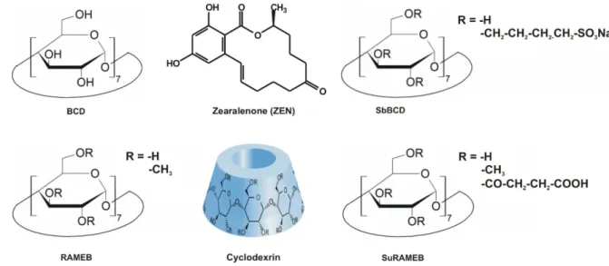
Zearalenone (ZEN; Fig. 1) is a xenoestrogenic mycotoxin produced by Fusarium species, 65
which is a contaminant in cereals (e.g., maize and wheat), spices, and in different beverages, 66
e.g., milk and beer (Maragos, 2010; EFSA, 2017). Because of the high thermal stability and 67
wide occurrence of ZEN, its removal from the food chain is difficult (Ryu et al., 1999). Based 68
on cell and animal experiments, several adverse effects are attributed to ZEN, e.g., 69
hepatotoxicity and genotoxicity (Zinedine et al., 2007; Cheraghi et al., 2015). Furthermore, 70
ZEN can activate estrogen receptors in humans and animals, therefore, ZEN is an endocrine 71
disruptor molecule which induces reproductive disorders (EFSA, 2017; Shier et al., 2001).
72
ZEN is extensively metabolized in the body, during which reduced derivatives (zearalenols, 73
zearalanone, and zearalanols) and glucuronic acid conjugates of ZEN and its reduced 74
metabolites are produced (EFSA, 2017). Some of these metabolites (e.g., α-zearalenol and α- 75
zearalanol) bind with significantly higher affinity to the estrogen receptors (and consequently 76
exert higher toxicity) than ZEN (Shier et al., 2001; Filannino et al., 2011).
77
Cyclodextrins (CDs) are ring-shaped host molecules with a hydrophilic external part, which 78
ensures excellent aqueous solubility, and an apolar internal cavity, which can accommodate 79
lipophilic guest molecules (Szente and Szejtli, 1999; Szente et al., 2018). Therefore, they are 80
frequently utilized by food, cosmetic, and pharmaceutical industries. The pharmaceutical 81
application of beta-CDs is most common, due to their favorable cavity size for drugs (Challa 82
et al., 2005). The native beta-CD (BCD) is often contained by orally administered drugs, 83
however, its parenteral use is limited due to its nephrotoxicity and relatively low aqueous 84
solubility of BCD (Jambhekar and Breen, 2016a). Methylated beta-CDs are absorbed from the 85
gastrointestinal tract and cause nephrotoxic effects, therefore, they are not used neither orally 86
nor parenterally (Jambhekar and Breen, 2016a). The sulfobutylated beta-CD is an excellent 87
solubilizer without nephrotoxic adverse effect, thus, it is even suitable for parenteral 88
5
application (Jambhekar and Breen, 2016b). Generally, the pharmaceutical industry applies 89
drug-CD complexes with low binding constants to increase the aqueous solubility, 90
gastrointestinal absorption, and/or cellular uptake of drugs (Jambhekar and Breen, 2016a).
91
However, formation of highly stable CD complexes can strongly decrease the 92
pharmacological effect and tissue uptake of drugs and other xenobiotics (Schaller and Lewald, 93
2016; Weiss-Errico et al., 2017).
94
Native and chemically modified beta-CDs can form stable complexes with mycotoxins, 95
including aflatoxins (Dall’asta et al., 2003), citrinin (Poór et al., 2016), ochratoxin A (Poór et 96
al., 2015a), and ZEN/zearalenols (Poór et al., 2017). The interaction of ZEN with beta-CDs 97
has been reported in previous studies, demonstrating that native and chemically modified 98
beta-CDs form highly stable complexes with ZEN (K is in the 104-105 L/mol range) 99
(Dall’Asta et al., 2008; Dall’Asta et al., 2009; Poór et al., 2015b). Among beta-CDs tested, 100
ZEN formed the most stable complexes with methyl and sulfobutyl derivatives (Poór et al., 101
2015b).
102
A beta-CD bead polymer has been shown recently to effectively remove ZEN and zearalenols 103
added to aqueous solutions and corn beer samples (Poór et al., 2018). Furthermore, BCD 104
strongly alleviated the toxic effect of ZEN in HepG2 cells, probably by limiting toxin uptake 105
by the cells, as a result of the formation of highly stable mycotoxin-CD complexes (Poór et 106
al., 2015b). Based on these observations, we hypothesize that CDs may also be effective as in 107
vivo binders of ZEN.
108
There are numerous of endocrine disruptors in the environment, especially estrogenic 109
xenobiotics. Sensitive biomonitor/bioindicator organisms are commonly applied to test 110
xenoestrogenic effects. Among these biomonitoring organisms, several fish models, including 111
zebrafish, exist (Chen et al., 2010; Fetter et al., 2014; Bakos et al., 2019). The main advantage 112
of zebrafish as a biosensor is the transparent body of embryos and larvae; therefore, the 113
6
fluorescence signal of a reporter protein can be easily studied in vivo in the living animal 114
(Strähle et al., 2012). Zebrafish embryo is widely used as a model in developmental 115
toxicology tests (Braunbeck et al., 2005; Scholz et al., 2008) because the developing and 116
transparent zebrafish can be assessed conveniently for lethality and developmental 117
abnormalities from fertilization through larval stages. Furthermore, the development of 118
zebrafish embryos is very similar to the embryogenesis in higher vertebrates (including 119
humans); therefore, this species is highly suitable for the investigation of the fundamental 120
processes underlying embryonic development (Nagel, 2002; Weight et al., 2011). In addition 121
to animal protection, it is also favorable that the same individual fish can be studied 122
throughout the treatment (Segner, 2009). In our experiments, we used a vitellogenin reporter 123
transgenic zebrafish line, the Tg(vtg1:mCherry) (Bakos et al., 2019).
124
In this study, we examined the hypothesis that beta-CDs can limit the toxic effects of ZEN, 125
employing BCD and its chemically modified derivatives, namely sulfobutylated beta- 126
cyclodextrin (SbBCD), randomly methylated beta-cyclodextrin (RAMEB), succinyl-beta- 127
cyclodextrin (SucBCD), and succinyl-methyl-beta-cyclodextrin (SuRAMEB) (Fig. 1). The 128
stability of ZEN-CD complexes was tested in a physiological buffer by fluorescence 129
spectroscopy. In our previous study, the cytotoxic effects of ZEN in the absence and presence 130
of CDs were examined on HepG2 cell line (Poór et al., 2015b). Because HepG2 liver cells 131
may significantly biotransform ZEN. Therefore, in this study, the toxic actions of ZEN were 132
examined in HeLa (cervical cancer) cell line, in the absence and presence of CDs. The 133
cytotoxicity of ZEN and CDs were evaluated based on ATP levels/well. Furthermore, the 134
acute toxicity of ZEN was also examined on zebrafishembryos, in the absence and presence 135
of CDs. Our results demonstrate that CDs can strongly alleviate the ZEN-induced toxicity 136
both in vitro and in vivo.
137 138
7 139
Fig. 1: Chemical structures of zearalenone and beta-cyclodextrins tested.
140 141
2. Materials and Methods 142
2.1. Reagents 143
Zearalenone (ZEN), Dulbecco’s Modified Eagle Medium (DMEM), and fluorescamine 144
(Fluram) were purchased from Sigma-Aldrich (St. Louis, MO, US). Cyclodextrins, including 145
beta-cyclodextrin (BCD), sulfobutylated beta-cyclodextrin (SbBCD), randomly methylated 146
beta-cyclodextrin (RAMEB), succinyl-beta-cyclodextrin (SucBCD), and succinyl-methyl- 147
beta-cyclodextrin (SuRAMEB) were provided by CycloLab Cyclodextrin Research and 148
Development Laboratory, Ltd (Budapest, Hungary). Bioluminescent ATP Assay Kit CLSII 149
(Roche; Basel, Switzerland), fetal bovine serum (Pan-Biotech; Aidenbach, Germany), and 150
bovine serum albumin (Biosera;Nuaille, France) were used as received.
151 152
2.2. Steady-state fluorescence spectroscopic studies 153
Fluorescence spectroscopic measurements were performed using a Hitachi F-4500 fluorimeter 154
(Tokyo, Japan). Increasing amounts of CDs (final concentrations: 0, 25, 50, 100, 250, and 500 155
μM) were added to ZEN (2 μM), after which fluorescence emission spectra of ZEN and ZEN- 156
CD complexes were recorded (λex = 315 nm). To approximate extracellular physiological 157
8
conditions, experiments were carried out in phosphate-buffered saline (PBS, pH 7.4;
158
containing 8.00 g/L NaCl, 0.20 g/L KCl, 1.81 g/L Na2HPO4 × 2H2O, and 0.24 g/L KH2PO4).
159
Stock solution of ZEN (5000 μM) was prepared in 96 v/v(%) ethanol (Reanal; Budapest, 160
Hungary). In fluorescence spectroscopic studies, the concentration of ethanol did not exceed 161
0.04 v/v (%). Binding constants (K, unit: L/mol) of ZEN-CD complexes were determined 162
employing the graphical application of the Benesi-Hildebrand equation, assuming 1:1 163
stoichiometry of complex formation (Poór et al., 2015b):
164
𝐼0 (𝐼−𝐼0) = 1
𝐴+ 1
𝐴×𝐾×[𝐶𝐷]𝑛 (1) 165
where I0 and I are the fluorescence emission intensity of ZEN without and with CDs, 166
respectively (λex = 315 nm, λem = 455 nm). [CD] denotes the molar concentration of CDs 167
(unit: mol/L), A is a constant, and n is the number of binding sites.
168 169
2.3. Cell experiments 170
2.3.1. Cell culturing and treatment 171
Cell experiments were performed on HeLa cervical cancer cell line (ATCC: CCL-2). The 172
adherent cells were cultured in DMEM with high glucose (4500 mg/L) containing 10% fetal 173
bovine serum, penicillin (100 U/mL), and streptomycin (100 μg/mL) in 75 cm2 sterile cell 174
culture flasks in humidified atmosphere with 5% CO2 and at 37 °C. Cells were trypsinized 175
and plated onto 96-well sterile plastic plates. Stock solution of ZEN (5000 μM) were prepared 176
in 96 v/v(%) ethanol. In cell experiments, solvent controls were also applied; however, the 177
final concentrations of ethanol did not exceed 1 v/v(%), which did not influence significantly 178
the viability of HeLa cells. During the treatments, the culture medium was replaced with fresh 179
one, containing the appropriate concentrations of ZEN (50 μM) and/or CDs (0.0-1.0 mM).
180
Then the cells were incubated for 48 h before analysis.
181 182
9
2.3.2. Measurements of cellular ATP and total protein levels 183
To test the effects of ZEN and CDs alone and in combinations on the viability of HeLa cells, 184
intracellular ATP and total protein levels were quantified (based on luciferin-luciferase ATP 185
and fluram protein assays, respectively) as described previously (Csepregi et al., 2018).
186 187
2.3.3. Statistical analyses in cell experiments 188
Means and standard error (± SEM) values were derived from at least three independent 189
experiments. The data showed normal distribution based on the Shapiro-Wilk normality test 190
(IBM SPSS Statistics, V21). Statistical evaluation was performed using one-way ANOVA 191
test (IBM SPSS Statistics, V21). The level of significance was set at p < 0.05 and p < 0.01.
192 193
2.4. Experiments on zebrafish embryos 194
2.4.1. Characterization of the Tg(vtg1:mCherry) biomarker zebrafish line 195
The zebrafish line used in these experiments is a vitellogenin reporter transgenic zebrafish 196
line. Vitellogenin is a glycoprotein that is inducible by environmental estrogens. The 197
transgene construct used for the development of Tg(vtg1:mCherry) carried a long (3.4 kbp) 198
natural vitellogenin-1 promoter sequence with a high number of ERE (estrogen responsive 199
element) sites. The mCherry reporter is only produced in the liver, similarly to endogenous 200
vitellogenin. The sensitivity and usability of the embryos of the line have been tested on 201
several estrogenic compounds (including ZEN) as well as on environmental samples (Bakos 202
et al., 2019).
203 204
2.4.2. Zebrafish maintenance and egg collection 205
Laboratory-bred Tg(vtg1:mCherry) zebrafish strain was held in breeding groups of 30 females 206
and 30 males at the Department of Aquaculture (Szent István University, Hungary) in a 207
10
Tecniplast ZebTEC recirculation system (Tecniplast S.p.a., Italy) at 25.5 ± 0.5 °C (system 208
water: pH 7.0 ± 0.2, conductivity 550 ± 50 µS) and on a 14h:10 h light:dark cycle. The fish 209
were fed twice a day with dry granulate food (Zebrafeed 400-600 µm, Sparos Lda., Portugal) 210
supplemented with freshly hatched live Artemia salina once a day. The fish were placed in 211
breeding tanks (Tecniplast S.p.a.) late in the afternoon before the day of the experiment and 212
allowed to spawn by removing the dividing walls next morning. The collected eggs were 213
incubated in system water with methylene blue (2 mL 0.1% methylene blue in 1 L system 214
water) (25 ± 2 °C) in Petri dishes (diameter: 10 cm). After 24 h, coagulated and/or non- 215
fertilized eggs were assorted, and a part of the embryos were disinfected with bleaching 216
method to keep the experiment sterile.
217 218
2.4.3. Embryo bleaching 219
Bleaching of embryos was necessary because some microorganisms can break down the CD 220
ring to glucose units during long-term experiments in aqueous solution. System water was 221
removed with a plastic pipette and embryos were bathed in a bleach solution (0.0035%
222
sodium hypochlorite) for 5 min. Then, the bleach solution was removed, and Petri dishes were 223
filled with sterilized E3 medium (5.0 mM NaCl, 0.17 mM KCl, 0.33 mM CaCl2, and 0.33 224
mM MgSO4 in 1 L sterilized deionized water) for 5 min. E3 medium was removed and dishes 225
were filled with new E3 solutions under a sterile box.
226 227
2.4.4. Determination of lethal concentration (LC) values of ZEN 228
96 hpf (hours post-fertilization) Tg(vtg1:mCherry) embryos were placed in groups of five in 229
24-well plates (JET Biofil; Guangzhou, China). E3 medium were removed then zebrafish 230
embryos were treated (2 mL/well) with 0, 1, 2, 3, 4, 5, 6, and 7 mg/L (equal to 0-22 μM) 231
ZEN, each treatment was performed in four replicates. ZEN was dissolved in methanol, the 232
11
final concentrations of the solvent did not exceed 0.4 v/v (%) during the treatments. Solvent 233
controls were also tested: At the applied concentrations, methanol alone did not affect the 234
viability of zebrafish embryos. The mortality was evaluated after 24 h exposure.
235 236
2.4.5. Testing the effects of CDs on zebrafish embryos in the absence and presence of ZEN 237
Three concentrations (0.25 mM, 0.5 mM, and 1 mM) of beta-CDs (BCD, SbBCD, RAMEB, 238
and SuRAMEB) with and without ZEN (final concentration: 4.0 mg/L or 12.6 μM) were 239
diluted in sterilized E3 medium. Mixtures were filtered with 0.2 µm syringe filters (VWR 240
International Ltd., Hungary) to gain bacteriologically sterile solutions. ZEN control (with 241
methanol solvent) was diluted in E3 medium to 4.0 mg/L (12.6 μM) final concentration. Each 242
treatment were prepared with bleached and non-bleached larvae to test disinfection procedure.
243
96 hpf transgenic larvae were transferred in groups of ten in sterile 6-well plates (JET Biofil, 244
China), the experiment was performed in three replicates. Thereafter, E3 medium was 245
removed, each well were filled with 10 mL of treatment solution and larvae were incubated at 246
26°C (± 1°C) on 14 h:10 h light:dark cycle for 24 h in each treatment.
247 248
2.4.6. Imaging and analysis 249
Five-day old embryos were placed to petri dishes (diameter: 6 cm; JET Biofil; Guangzhou, 250
China) from each group. Overplus solutions were removed with a plastic pipette and were 251
filled with 2 mL of 0.02% MS-222 (Tricane-methane-sulfonate; from Sigma-Aldrich; St.
252
Louis, MO, US) anesthetic solution. Special designed petri dishes (with two cube-shaped 253
tape, diameter: 10 cm) were filled with 4% methyl-cellulose solution. Anaesthetized embryos 254
were placed to methyl-cellulose, oriented to the left side, and pushed to the bottom of the 255
cellulose solution with a cut ended Microloader pipette tip (Eppendorf; Hamburg, Germany).
256
Bright field (exposure time: 6 msec, magnification: 30x and 60x), and fluorescent (mCherry 257
12
filter, exposure time: 2 sec, magnification: 60x) images of larvae were taken under a 258
fluorescent stereomicroscope (Leica M205 FA fluorescent stereomicroscope, Leica DFC 259
7000T camera, Leica Application Suite X, Leica Microsystems GmbH; Wetzlar, Germany).
260
Signals in the red range of the RGB (Red, Green, Blue) color rangewas evaluated by ImageJ 261
software (Schneider et al., 2012) based on the prepared fluorescent images. An ellipticalarea 262
of the same size was selected on each image and moved to the areaof the liver, then the signal 263
strength and the size of the affected areawere determined. The integrated density values were 264
determined for each treatment. The results of ZEN treatments (ZEN and ZEN+CDs) were 265
corrected with the integrated density values of test solutions without the mycotoxin.
266 267
2.4.7. Statistical analyses in zebrafish experiments 268
The concentration-lethality curve was fitted and LC values were calculated by non-linear 269
regression. Integrated density data were checked for normality with Shapiro-Wilk normality 270
test and non-compliance with the requirements of parametric methods was established.
271
Statistical significance was evaluated employing Kruskal-Wallis analysis with Dunn's 272
multiple comparisons test. Results were analyzed and plotted by GraphPad Prism 6.01 273
(GraphPad Software; San Diego, CA, US).
274 275
3. Results and Discussion 276
3.1. Interaction of ZEN with beta-CDs in physiological buffer 277
Whereas the complex formation of ZEN with some beta-CDs has been reported, the 278
interaction of SucBCD and SuRAMEB with ZEN has not been tested. Furthermore, previous 279
experiments did not try to approximate extracellular physiological conditions; therefore, our 280
spectroscopic experiments were performed in PBS buffer (pH 7.4). Each tested CD induced a 281
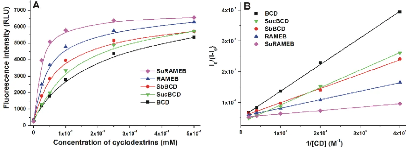
strong increase in the fluorescence of ZEN (which is the sign of complex formation), showing 282
13
the following order in the fluorescence enhancement: SuRAMEB > RAMEB > SbBCD >
283
SucBCD > BCD (Fig. 2A). Our results are in agreement with the previously published 284
studies, which also suggest that the chemical modifications of BCD strongly increase the 285
fluorescence signal of ZEN (Dall’Asta et al., 2009; Poór et al., 2015b). Then, the binding 286
constants of ZEN-CD complexes were determined employing the Benesi-Hildebrand equation 287
(Eq. 1). As it is demonstrated in Fig. 2B, Benesi-Hildebrand plots showed excellent linearity 288
with the 1:1 stoichiometry model, and suggesting the formation of stable mycotoxin-CD 289
complexes. ZEN forms similarly stable complexes with SucBCD (K = 5.5 × 10³ L/mol) than 290
with BCD (K = 6.5 × 10³ L/mol), while other chemically modified beta-CDs bound to ZEN 291
with higher affinity. The most stable mycotoxin-CD complexes were formed with SuRAMEB 292
(K = 4.7 × 10⁴ L/mol) followed by RAMEB (K = 2.0 × 10⁴ L/mol) and SbBCD (K = 1.4 × 10⁴ 293
L/mol). Since succinyl substitution of BCD resulted in slightly less stable ZEN-CD 294
complexes than BCD, we did not use SucBCD in the following experiments.
295
Our results demonstrate that each beta-CDs tested form stable complexes with ZEN in PBS 296
(pH 7.4). The binding constants of BCD, SbBCD, and RAMEB complexes were similar but 297
slightly lower than those previously found in ammonium acetate buffer (0.05 M) at pH 5.0 298
(Poór et al., 2015b). These findings indicate that methyl and sulfobutyl substitutions of BCD 299
strongly increase the stability of ZEN-CD complexes (Dall’Asta et al., 2009; Poór et al., 300
2015b). Despite succinyl derivative of BCD slightly decreased the stability of the complexes 301
formed, the simultaneous presence of succinyl and methyl substituents in SuRAMEB resulted 302
in higher binding constants compared to both BCD and RAMEB. Succinyl-methyl, methyl, 303
and sulfobutyl substitutions of BCD led to the approximately 7.2-, 3.1-, and 2.2-fold increase 304
in binding constants of ZEN-CD complexes, respectively.
305 306
14 307
Fig. 2: (A) Fluorescence emission intensity of ZEN (2 μM) in the absence and presence of 308
increasing concentrations of CDs (0-500 μM) in PBS (pH 7.4). (B) Benesi-Hildebrand plots 309
of ZEN-CD complexes (λex = 315 nm, λem = 455 nm).
310 311
3.2. Effects of ZEN on HeLa cells in the absence and presence of beta-CDs 312
To test the effects of CDs on the ZEN-induced cytotoxicity, HeLa cells were treated with 313
ZEN and/or CDs. After 48 h incubation, cell viability was mainly evaluated based on the 314
cellular ATP content/well. Quantitation of cellular ATP levels is a widely accepted method to 315
determine cell viability. However, previous studies indicated that the ATP level alone may be 316
a misleading parameter (Sali et al., 2016; Kőszegi et al., 2007; Hochachka and Mcclelland, 317
1997; Andreoli and Mallett, 1997). Therefore, to confirm the results from ATP assay, total 318
protein levels were also quantified. Changes of cellular ATP and total protein levels showed 319
good correlation (Fig. 3). To produce a strong decrease in cell viability, ZEN was applied at 320
50 μM concentration in these experiments. Our data are in good agreement with a previous 321
study on HeLa cells which reported that the IC50 value of ZEN is approximately 60 µM (Ayed 322
et al., 2011). However, a wide cytotoxic concentration range for ZEN has been found in other 323
cell lines: 5-40 µM in Caco-2 (colorectal adenocarcinoma) cells and 31-157 µM in HL-60 324
(human leukemia) cells (Rai et al., 2019). As Fig. 3 demonstrates, BCD failed to significantly 325
alleviate the ZEN-induced toxicity (it caused only slight increases in ATP and total protein 326
15
levels); however, other CDs considerably decreased or even abolished the toxic effects of 327
ZEN. In a concentration-dependent fashion, the co-treatment of ZEN-exposed cells with 328
SbBCD, RAMEB, or SuRAMEB increased both ATP and total protein levels compared to the 329
cells exposed to ZEN alone. Low CD concentrations (0.25 mM) were minimally effective 330
(only the total protein level was increased significantly by SuRAMEB), while 0.5 mM 331
concentrations of CDs induced spectacular elevation of cell viability. In addition, SbBCD, 332
RAMEB, and SuRAMEB completely abolished the ZEN-induced loss of cell viability at the 333
highest concentration (1 mM).
334
Considering the high stability of ZEN-CD complexes as well as the previously reported 335
protective effect of BCD against ZEN in HepG2 cells (Poór et al., 2015b), it was reasonable 336
to hypothesize that some of these CDs may also effectively alleviate the ZEN-induced 337
cytotoxicity in vivo. BCD failed to significantly affect cell viability even at 1 mM 338
concentration in HeLa cells, although it strongly alleviated the toxic effects of ZEN in HepG2 339
cells in a previous study (Poór et al., 2015b). However, chemically modified beta-CDs 340
(SuRAMEB, RAMEB, and SbBCD) caused the significant decrease of ZEN-induced loss of 341
cell viability. This can be explained by the higher binding affinity of the mycotoxin towards 342
these CDs. In the cell medium, ZEN can form stable complexes with bovine serum albumin 343
contained by the fetal bovine serum. In previous fluorescence spectroscopic studies, similar K 344
values (6.0 × 10⁴ and 2.6 × 10⁴ L/mol) of ZEN-BSA complex have been reported (Faisal et 345
al., 2018; Ma et al., 2018). Therefore, ZEN is likely present in the cell medium mainly in 346
albumin-bound form. Because CDs can form similarly stable complexes with ZEN than with 347
albumin (see in 3.1), CDs can further decrease the free fraction of ZEN in the cell medium, 348
thus further decreasing the cellular uptake of the mycotoxin.
349
Under the applied conditions, even the highest concentration (1 mM) of CDs (BCD, SbBCD, 350
RAMEB, and SuRAMEB) did not affect significantly ATP and total protein levels (Fig. S1).
351
16
Based on previous studies, SbBCD is a less toxic while the methylated derivatives are less 352
tolerable compared to the native BCD (Kiss et al., 2010; Jambhekar and Breen, 2016b).
353
Furthermore, in vitro studies suggests that SuRAMEB is a less toxic derivative compared to 354
RAMEB (Kiss et al., 2010). In previous cell experiments, alpha-, beta-, and gamma-CDs as 355
well as their hydroxypropyl, methyl, and carboxymethyl derivatives did not induce significant 356
toxicity at 1 mM or lower concentrations in HEK293T (human embryonic kidney), HeLa, and 357
TZM-bl (endocervical adenocarcinoma) cells (Szente et al., 2018).
358 359
360
17
Fig. 3: Effects of ZEN (50 μM) on the cellular ATP (A) and total protein (B) levels in HeLa 361
cells, in the absence and presence of CDs (0-1 mM) after 48 h incubation (compared to the 362
control: *p < 0.05, **p < 0.01; compared to ZEN alone: #p < 0.05, ##p < 0.01).
363 364
3.3. Effects of ZEN on zebrafish embryos in the absence and presence of beta-CDs 365
To confirm our results indicating the protective effects of CDs in HeLa cell in vitro, their 366
influence on the ZEN-induced toxicity was further examined in zebrafish embryos. As the 367
first step, the toxicity indicators in the selected exposure window were determined. Therefore, 368
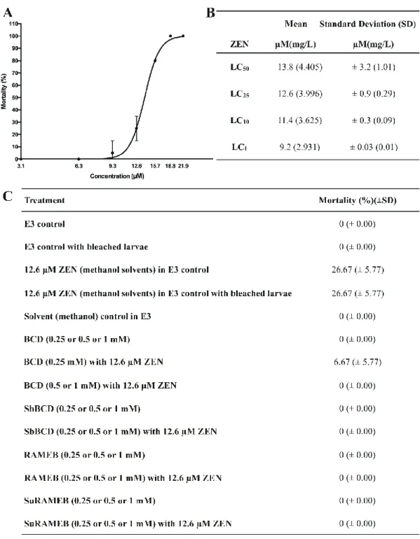
the effect of ZEN on Tg(vtg1:mCherry) embryos was determined between 96-120 hpf. Fig.
369
4A demonstrates the concentration-mortality curve of ZEN. LC values (Fig. 4B) were higher 370
than in an earlier study using the same strain, in which 0.893 mg/L (or 2.81 µM; in this study:
371
4.405 mg/L or 13.84 µM) and 0.335 mg/L (or 1.05 µM; in this study: 3.625 mg/L or 11.39 372
µM) LC50 and LC10 values of ZEN were reported, respectively (Bakos et al., 2013). Since 373
earlier studies suggest that the survival of fish embryos decreases with their age (Gellert and 374
Heinrichsdorff, 2001), these differences likely resulted from the different length of exposure 375
(96-120 vs. 1-120 h period). The LC25 concentration of ZEN (4.0 mg/L or 12.6 μM) was 376
selected for the following experiments (Fig. 4A and B) because it did not induce marked 377
mortality while its sublethal effects were significant. Mortality data observed in the presence 378
of ZEN and/or CDs are listed in Fig. 4C. ZEN-induced mortality (26.67%) was consistent 379
with the previous treatments (see in Fig. 4A and B), and the bleaching method did not affect 380
the viability of larvae. The OECD guideline criteria for fish embryo test accepts a maximum 381
of 10% lethality in the control groups (OECD236, 2013). Since the mortality of the control 382
groups was 0%, it obviously fulfills this criteria. Under the applied circumstances, CDs (0.25- 383
1.0 mM) alone did not increase the mortality. Furthermore, the co-exposure of ZEN with beta- 384
CDs completely abolished the lethal effects of ZEN (except 0.25 mM BCD) (Fig. 4C), 385
18
suggesting the considerable protective effects of CDs vs. ZEN-induced toxicity. The weaker 386
protective effect of BCD on ZEN-induced mortality is in agreement with the previous 387
observation that BCD forms less stable complexes with ZEN compared to SuRAMEB, 388
RAMEB, and SbBCD (see in 3.1), as well as it is also in accordance with the results of cell 389
experiments (Fig. 3). Some previous studies also pointed out that CDs can decrease the toxic 390
actions of several compounds, due to the formation of stable host-guest type complexes. BCD 391
strongly decreased the LC50 values of1-dodecyl-3-methylimidazolium tetrafluoroborate 392
(Hodyna et al., 2016), 20(S)-Protopanaxadiol-20-O-D-glucopyranoside (Nam et al., 2017), 393
and perfluorooctanoic acid (Weiss-Errico et al., 2017) in zebrafish.
394 395
19 396
Fig. 4: LC values and mortality data of Tg(vtg1:mCherry) zebrafish embryos (120 hpf).
397
Concentration-mortality curve of ZEN (A); lethal concentration and the corresponding 398
standard deviation (SD) values of ZEN with (B); and mortality data of ZEN and CDs alone as 399
well as in combination (C). All experiments were performed in 96-120 hpf exposure window.
400 401
20
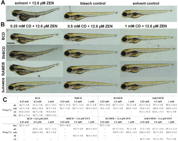
Besides the lethal outcome, sublethal effects of beta-CDs were also studied on 120 hpf 402
zebrafish embryos. In general, beta-CDs caused mild phenotypic lesions on the treated 403
embryos, such as uninflated swim bladder and mild pericardial edema (Fig. 5). Furthermore, a 404
slight upward curvature of the body axis can be observed as a result of BCD and SuRAMEB 405
treatments (Fig. 5A), whereas moderately irregular edges of the dorsal and ventral fins were 406
noticed only on BCD-treated embryos (Fig. 5B). Quantitative values of sublethal effects are 407
demonstrated in Fig. 6C. In previous studies, the presence of 1% or lower concentrations of 408
hydroxypropyl-beta-CD did not affect the development in zebrafish embryos and larvae 409
(Maes et al., 2012), and even 3 mM concentration of methyl-beta-CD did not induce abnormal 410
cytokinesis of zebrafish embryos (Feng et al., 2002). Our results also suggest that BCD, 411
SbBCD, RAMEB, and SuRAMEB do not cause strong malformations up to 1 mM 412
concentrations.
413 414
415
21
Fig. 5: Representative developmental defects in 120 hpf zebrafish embryos after 24 h 416
treatment with beta-CDs. (A) An untreated control and embryos treated with 1 mM of BCD, 417
SbBCD, RAMEB, and SuRAMEB. Pericardial edema and uninflated swim bladder appeared 418
as a result of beta-CD treatments. Slight upward curvature of the body axis can be observed 419
after BCD and SuRAMEB treatments. (B) Moderately irregular edges (marked with asterisks) 420
of the dorsal fin are apparent in the BCD-treated embryo. Scale bar: 500 µm.
421 422
Sublethal effects were also studied on 120 hpf embryos treated with ZEN in the absence and 423
presence of beta-CDs (Fig. 6). The effects of ZEN on the development of zebrafish embryos 424
have been reported. During the 72-h treatment of the embryos, ZEN caused pericardial 425
edema, eye deformity, and concentration-dependent dorsal curvature of the body axis (heart 426
and soul (has) phenotype), which is also characterized by other estrogenic substances (Bakos 427
et al., 2013). The has phenotype can be observed in ZEN-treated (12.6 μM) embryos as well 428
as after the co-exposure of ZEN with 0.25 mM BCD, SbBCD, or RAMEB (Fig. 6). However, 429
this morphological alternation was no longer observed during the co-treatment of ZEN- 430
exposed embryos with higher concentrations (0.5 and 1.0 mM) of beta-CDs (Fig. 6B).
431
Interestingly, the ZEN-induced formation of has phenotype was also eliminated by the co- 432
treatment of 0.25 mM SuRAMEB, which is in good agreement with our previous 433
observations: (1) SuRAMEB forms the most stable complex with ZEN among the beta-CDs 434
tested (see in section 3.1) and (2) SuRAMEB was the only CD which significantly increased 435
the total protein levels in ZEN-treated HeLa cells even at 0.25 mM concentration (Fig. 3B).
436
Another developmental effect of ZEN is the lack of the gap in the melanophore streak along 437
the ventral side at the base of the caudal fin (Bakos et al., 2013). This phenotype is also 438
typical for endocrine disruptors in zebrafish embryos treated between 0-72 hpf (Yang et al., 439
2010; Georgescu et al., 2011). Less pigmentation disorder was observed only after ZEN 440
22
treatment (without CDs), and there was no complete closure of melanophores streak. This 441
may be explained by the fact that experiments were started with 96 hpf embryos when the 442
process of pigmentation was slowed down compared to the previous developmental stages, 443
and the duration of the treatment was too short for complete closure.
444
The co-treatment of ZEN with 0.5 and 1 mM BCD typically resulted in uninflated swim- 445
bladder, and some individuals exhibited mild pericardial edema. Fin disorder, which was 446
specific to BCD-treated embryos, was not observed. The co-exposure of ZEN with 0.5 and 1 447
mM SbBCD caused inflated swim bladder in most of the individuals (which appeared 448
regularly only with this CD) and a slight pericardial edema was also noticed. Typical 449
sublethal symptoms, as a result of the simultaneous treatment with ZEN and RAMEB (0.5 450
and 1 mM), were uninflated swim bladder and pericardial edema. There was no similar 451
abnormality with the has phenotype regarding ZEN-SuRAMEB co-treatments; however, a 452
slight pericardial edema was observed in all treated embryos. Furthermore, the 0.25 and 0.5 453
mM concentrations of SuRAMEB (in the presence of ZEN) led to a slight downward 454
curvature of the body axis. During the ZEN-SuRAMEB co-exposures, the swim bladders of 455
the embryos were not inflated until the end of the experiment. Based on the above-listed 456
observations, beta-CDs reduced the sublethal effects of ZEN (Fig. 6C).
457 458
23 459
Fig. 6: Representative developmental defects in 120 hpf zebrafish embryos after 24 h 460
treatment with ZEN (12.6 μM) in the absence and presence of beta-CDs. ZEN-exposed 461
embryos as well as bleach and solvent controls are demonstrated in panel A, while embryos 462
co-treated with ZEN and CDs are represented in panel B. CDs reduced the sublethal effects of 463
ZEN, as it can be observed on the bright field images. Scale bar: 500 µm. (C) The mean 464
appearance of representative developmental defects after ZEN and ZEN+CD exposure (%) 465
(has: heart and soul phenotype; pe: pericardial edema; sb: inflected swim bladder; usb:
466
uninflected swim bladder; cb: curvature of the body axis; fin: irregular edges of dorsal fin).
467
As a result of ZEN treatment (without CDs), the has phenotype appeared in each zebrafish 468
embryo (100%).
469 470
24
Transgenic bioindicator models for estrogenic effects are increasingly used in toxicological 471
studies. In vivo models allow the investigation of complex processes in the organism. Several 472
transgenic zebrafish lines are suitable for the investigation of estrogenic effects of test 473
compounds, of which Tg(vtg1:mCherry) was used in our studies (Bakos et al., 2019). In these 474
experiments, the effects of ZEN in the absence and presence of beta-CDs were examined on 475
96 hpf-120 hpf zebrafish embryos. We investigated the potential appearance of fluorescence 476
signal in the liver of fish (at the end of the exposure time), indicating the xenoestrogenic 477
effect of ZEN. ZEN treatment induced the transgene to function, which is indicated by 478
fluorescence signal in the liver (Fig. 7A and B). The weakest fluorescence signal was 479
observed in the liver of ZEN-treated embryos (in the absence of CDs; Fig. 7A). In ZEN-BCD 480
co-treated fish, the intensity of the fluorescence signal was almost the same than in the 481
presence of ZEN alone (without CDs). Surprisingly, ZEN-SbBCD co-exposure caused a 482
concentration-dependent increase in the fluorescent signal; while SuRAMEB induced the 483
strongest elevation at its lowest concentration (0.25 mM), above which the fluorescence 484
signal gradually decreased (Fig. 7B). Simultaneous treatment of ZEN-exposed cells with 485
RAMEB led to the strong increase in the fluorescence (0.5 mM RAMEB produced the highest 486
effect in this whole experiment), however, no clear concentration-dependence can be 487
observed. In untreated control embryos, no fluorescent signal was visible (Fig. 7A). The 488
integrated density values were in agreement with the differences in the fluorescence 489
intensities (Fig. 7C). When the integrated density values of ZEN-CD co-treatments were 490
compared to ZEN, no significant differences were observed in the presence of BCD, however, 491
statistically significant changes were noticed in the presence of 0.5 and 1.0 mM 492
concentrations of SbBCD and RAMEB, and each concentration (0.25, 0.5, and 1.0 mM) of 493
SuRAMEB.
494
25
The germ layers from which the liver of the zebrafish is formed start to develop 4 to 6 h after 495
fertilization, hepatic budding starts at 24 h after fertilization, and the liver starts working after 496
50 h (Villenueve et al., 2014). First, the left lobe of the liver is formed, where the endogenous 497
vitellogenin (and the fluorescent reporter) is produced; then, after 96 h (96 hpf), the right lobe 498
of the liver also appears (Ober et al., 2003; Tao and Peng, 2009). The final shape of the liver 499
appears around day 5 (120 hpf), and becomes well-defined in a relatively large area, where 500
the fluorescence signal can be easily detected with a stereomicroscope (Bakos et al., 2019).
501
Therefore, the liver works during the 96-120 h exposure window for short-term treatments 502
when Tg(vtg1:mCherry) embryo model is used to test estrogen effects (as it is also confirmed 503
in the current study). There are large variations in the fluorescence intensities and the 504
integrated density values within the treatments. Interestingly, ZEN-CD co-treatments induced 505
stronger fluorescence signals compared to ZEN alone. The reason is likely the high individual 506
sensitivity of the embryos to the treatment. The cells of the embryos (including their liver 507
cells) can be damaged by higher concentrations of toxic substances (Bakos et al., 2013, 2019).
508
In that case, the induction of vitellogenin production can be strongly decreased, thus lowering 509
the fluorescence signal in ZEN-treated fish as compared to ZEN-CD co-treatment. This 510
hypothesis is also supported by our observations that the stronger fluorescence signal of the 511
reporter and the higher integrated density values (Fig. 7) are accompanied with the 512
considerably lower mortality (Fig. 4) and the substantially weaker sublethal symptoms (Fig.
513
6). Furthermore, Fig. 7D shows that both ZEN and ZEN+BCD treatments significantly altered 514
the size, shape and color of the liver compared to the liver of untreated control embryos, 515
suggesting the significant hepatotoxic effect of ZEN. In contrast, simultaneous treatment of 516
embryos with ZEN and other CDs (SbBCD, RAMEB, SuRAMEB) resulted in much smaller 517
hepatic lesions, confirming their hepatoprotective effects against ZEN. These observations are 518
26
in agreement with the integrated density values, where stronger fluorescent signal and higher 519
mCherry protein affected area were observed in the less damaged liver.
520 521
27 522
28
Fig. 7: Fluorescence signals of the vitellogenin reporter and integrated density values in 523
Tg(vtg1: mCherry) embryos (n = 30/treatment) as well as the changes in liver size and shape 524
as a result of ZEN and ZEN+CD treatments. ZEN-treated (12.6 μM) embryos as well as 525
bleach (bl.cont.) and solvent controls (s.cont.) are demonstrated in panel A, while embryos 526
co-treated with ZEN and CDs are represented in panel B. Livers of the treated embryos are 527
demonstrated in Bright Field (B.F.) and in fluorescent (Fluo.) images. (C) Integrated density 528
values of ZEN-CD co-treatments were compared to ZEN (**p < 0.01, *** p < 0.001, **** p 529
< 0.0001). Data represent that co-treatments of embryos with ZEN and beta-CDs cause higher 530
fluorescence signal than ZEN alone. (D) The changes in the liver size and shape (marked with 531
white line) are shown as the results of ZEN and ZEN+CD treatments (ZEN: 12.6 μM; CDs: 1 532
mM). Integrated density values which were equal to or less than the untreated controls were 533
excluded from the evaluation.
534 535
In addition to the solubilizing effect of CDs, the low stability of CD complexes (K ≈ 102-103 536
L/mol) may support the cellular uptake of guest molecules, whereas CD complexes with 537
significantly higher stability may impair uptake (Redenti et al., 2001; Irie and Uekama, 1999;
538
Poór et al., 2015b). Therefore, the stability of CD complexes strongly affect the field of their 539
application. Furthermore, some CD derivatives have proved to be suitable in the treatment of 540
endotoxin shock in animal studies, likely due to their interactions with lipopolysaccharides 541
(Arima et al., 2005). Moreover, CDs are also applied in the human therapy: hydroxypropyl- 542
beta-CD is applied for the treatment of Niemann-Pick disease (Davidson et al., 2019) and 543
Sugammadex (a chemically-modified gamma-CD derivative) terminates the muscle relaxant 544
effect of rocuronium (Cada et al., 2016). These effects result from formation of highly stable 545
complexes of hydroxypropyl-beta-CD and Sugammadex with cholesterol and rocuronium, 546
respectively. In addition to these pharmaceutical applications, it is reasonable to hypothesize 547
29
that CD technology may suitable for development mycotoxin binders, which may counteract 548
the toxic effects of mycotoxins even after exposure. Our results demonstrate that some beta- 549
CDs are promising as binders of ZEN.
550 551
4. Conclusions 552
In summary, the protective effects of native and chemically modified beta-CDs on ZEN- 553
induced toxicity were investigated in HeLa cells and in zebrafish embryos. The chemically 554
modified beta-CDs that formed more stable complexes with ZEN had considerably stronger 555
protective effect on HeLa cells and zebrafish embryos against the toxic consequences of ZEN- 556
exposure. Since beta-CDs strongly decreased or even abolished the ZEN-induced toxicity 557
both in our in vitro and in vivo models, it is reasonable to hypothesize that CD technology 558
may be suitable for the development of new ZEN binders. However, further in vivo studies are 559
needed to confirm the suitability of CDs as protective agents against ZEN exposure.
560 561
Acknowledgements 562
Supported by the ÚNKP-18-3 New National Excellence Program of the Ministry of Human 563
Capacities (Zelma Faisal). Miklós Poór and Zelma Faisal are thankful for support of the 564
Hungarian National Research, Development and Innovation Office (FK125166). This project 565
was supported by the János Bolyai Research Scholarship of the Hungarian Academy of 566
Sciences (Miklós Poór and Mátyás Cserháti). This work was supported by the National 567
Research, Development and Innovation Office (NKFIH) from the National Research, 568
Development and Innovation Fund (NKFIA) (NVKP_16-1-2016-0003), by the EFOP-3.6.3- 569
VEKOP-16-2017-00008 project cofinanced by the European Union, and by the Thematic 570
Excellence Programme (TUDFO/51757/2019-ITM) of Szent István University 2019, awarded 571
30
by Ministry for Innovation and Technology. Tamás Kőszegi and Rita Csepregi are grateful for 572
the support of the University of Pécs, Medical School, Hungary (KA-2018-17).
573 574
Declarations of interest: The authors declare no conflict of interest. We have full control of 575
all primary data and we agree to allow the journal to review our data if requested.
576 577
References 578
Andreoli, S.P., Mallett, C.P., 1997. Disassociation of oxidant-induced ATP depletion and 579
DNA damage from early cytotoxicity in LLC-PK1 cells. Am. J. Physiol. Renal Physiol. 272, 580
F729–735. DOI: https://doi.org/10.1152/ajprenal.1997.272.6.F729 581
582
Arima, H., Motoyama, K., Matsukawa, A., Nishimoto, Y., Hirayama, F., Uekama, K. 2005.
583
Inhibitory effects of dimethylacetyl-beta-cyclodextrin on lipopolysaccharide-induced 584
macrophage activation and endotoxin shock in mice. Biochem. Pharmacol.70, 1506-17. DOI:
585
10.1016/j.bcp.2005.08.021 586
587
Ayed, Y., Ayed-Boussema, I., Ouanes, Z., Bacha, H., 2011. In vitro and in vivo induction of 588
chromosome aberrations by alpha- and beta-zearalenols: comparison with zearalenone. Mut.
589
Res. 726, 42–46. DOI: https://doi.org/10.1016/j.mrgentox.2011.08.003 590
591
Bakos, K., Kovács, R., Staszny, Á., Kánainé Sipos, D., Urbányi, B., Müller, F., Csenki, Z., 592
Kovács, B., 2013. Developmental toxicity and estrogenic potency of zearalenone in zebrafish 593
(Danio rerio). Aquat. Toxicol. 136–137. DOI: https://doi.org/10.1016/j.aquatox.2013.03.004 594
595
31
Bakos, K., Kovacs, R., Balogh, E., Kanaine Sipos, D., Reining, M., Gyomorei-Neuberger, O., 596
Balazs, A., Kriszt, B., Bencsik, D., Csepeli, A., Gazsi, G., Hadzhiev, Y., Urbanyi, B., 597
Mueller, F., Kovacs, B., Csenki, Z., 2019. Estrogen sensitive liver transgenic zebrafish (Danio 598
rerio) line (Tg(vtg1:mCherry)) suitable for the direct detection of estrogenicity in 599
environmental samples. Aquat. Toxicol. 208, 157–167. DOI:
600
https://doi.org/10.1016/j.aquatox.2019.01.008 601
602
Braunbeck, T., Boettcher, M., Hollert, H., Kosmehl, T., Lammer, E., Leist, E., Rudolf, M., 603
Seitz, N., 2005. Towards an alternative for the acute fish LC(50) test in chemical assessment:
604
the fish embryo toxicity test goes multi-species - an update. ALTEX-Altern. Anim. Exp. 22, 605
87–102.
606 607
Cada, D.J., Levien, T.L., Baker, D.E., 2016. Sugammadex. Hosp. Pharm. 51, 585–596. DOI:
608
10.1310/hpj5107-585 609
610
Challa, R., Ahuja, A., Ali, J., Khar. R.K., 2005. Cyclodextrins in Drug Delivery: An Updated 611
Review. AAPS PharmSciTech 6, E329. DOI: https://doi.org/10.1208/pt060243 612
613
Chen, H., Hu, J., Yang, J., Wang, Y., Xu, H., Jiang, Q., Gong, Y., Gu, Y., Song, H., 2010.
614
Generation of a fluorescent transgenic zebrafish for detection of environmental estrogens.
615
Aquat. Toxicol. 96, 53–61. DOI: https://doi.org/10.1016/j.aquatox.2009.09.015 616
617
Cheraghi, S., Razi, M., Malekinejad, H., 2015. Involvement of cyclin D1 and E2f1 in 618
zearalenone-induced DNA damage in testis of rats. Toxicon 106, 108–116. DOI:
619
https://doi.org/10.1016/j.toxicon.2015.09.018 620
32 621
Csepregi, R., Temesfői, V., Poór, M., Faust, Z., Kőszegi, T., 2018. Green Fluorescent Protein- 622
Based Viability Assay in a Multiparametric Configuration. Molecules 23, E1575. DOI:
623
https://doi.org/10.3390/molecules23071575 624
625
Dall’asta, C., Ingletto, G., Corradini, R., Galaverna, G., Marchelli, R., 2003. Fluorescence 626
enhancement of aflatoxins using native and substituted cyclodextrins. J. Incl. Phenom.
627
Macrocycl. Chem. 45, 257–263. DOI: https://doi.org/10.1023/A:1024572426577 628
629
Dall’Asta, C., Faccini, A., Galaverna, G., Corradini, R., Dossena, A., Marchelli, R., 2008.
630
Complexation of the mycotoxin zearalenone with β-cyclodextrin: Study of the interaction and 631
first promising applications. Mycotoxin Res., 24, 14–18. DOI:
632
https://doi.org/10.1007/BF02985265 633
634
Dall’Asta, C., Faccini, A., Galaverna, G., Corradini, R., Dossena, A., Marchelli, R., 2009.
635
Complexation of zearalenone and zearalenols with native and modified β-cyclodextrins. J.
636
Incl. Phenom. Macrocycl. Chem. 64, 331–340. DOI: https://doi.org/10.1007/s10847-009- 637
9572-3 638
639
Davidson, J., Molitor, E., Moores, S., Gale, S.E., Subramanian, K., Jiang, X., Sidhu, R., Kell, 640
P., Zhang, J., Fujiwara, H., Davidson, C., Helquist, P., Melancon, B.J., Grigalunas, M., Liu, 641
G., Salahi, F., Wiest, O., Xu, X., Porter, F.D., Pipalia, N.H., Cruz, D.L., Holson, E.B., 642
Schaffer, J.E., Walkley, S.U., Maxfield, F.R., Ory, D.S. 2019. 2-Hydroxypropyl-β- 643
cyclodextrin is the active component in a triple combination formulation for treatment of 644
33
Niemann-Pick C1 disease. Biochim. Biophys. Acta Mol. Cell Biol. Lipids 1864, 1545–1561.
645
DOI: 10.1016/j.bbalip.2019.04.011 646
647
European Food Safety Authority (EFSA), 2017. Risks for animal health related to the 648
presence of zearalenone and its modified forms in feed. EFSA J. 15, 4851. DOI:
649
https://doi.org/10.2903/j.efsa.2017.4851 650
651
Faisal, Z., Lemli, B., Szerencsés, D., Kunsági-Máté, S., Bálint, M., Hetényi, C., Kuzma, M., 652
Mayer, M., Poór, M., 2018. Interactions of zearalenone and its reduced metabolites α- 653
zearalenol and β-zearalenol with serum albumins: species differences, binding sites, and 654
thermodynamics. Mycotoxin Res. 34, 269-278. DOI: https://doi.org/10.1007/s12550-018- 655
0321-6 656
657
Feng, B., Schwarz, H., Jesuthasan, S., 2002. Furrow-specific endocytosis during cytokinesis 658
of zebrafish blastomeres. Exp Cell Res. 279, 14–20. DOI:
659
https://doi.org/10.1006/excr.2002.5579 660
661
Fetter, E., Krauss, M., Brion, F., Kah, O., Scholz, S., Brack, W. 2014. Effect-directed analysis 662
for estrogenic compounds in a fluvial sediment sample using transgenic cyp19a1b -GFP 663
zebrafish embryos. Aquat. Toxicol. 154, 221–229. DOI:
664
https://doi.org/10.1016/j.aquatox.2014.05.016 665
666
Filannino, A., Stout, T.A., Gadella, B.M., Sostaric, E., Pizzi, F., Colenbrander, B., 667
Dell'Aquila, M.E., Minervini, F., 2011. Dose-response effects of estrogenic mycotoxins 668
(zearalenone, alpha- and beta-zearalenol) on motility, hyperactivation and the acrosome 669
34
reaction of stallion sperm. Reprod. Biol. Endocrinol. 9, 134. DOI:
670
https://doi.org/10.1186/1477-7827-9-134 671
672
Gellert, G., Heinrichsdorff, J., 2001. Effect of age on the susceptibility of Zebrafish eggs to 673
industrial wastewater. Water Res. 35, 3752-3757. DOI: https://doi.org/10.1016/s0043- 674
1354(01)00084-7 675
676
Georgescu, B., Georgescu, C., Dǎrǎban, S., Bouaru, A., Pas¸ cal˘au, S., 2011. Heavy metals 677
acting as endocrine disrupters. J. Anim. Sci. Biotechnol. 44, 89–93.
678 679
Irie, T., and Uekama, K., 1999. Cyclodextrins in peptide and protein delivery.Adv. Drug 680
Deliv. Rev. 36, 101–123.
681 682
Hochachka, P.W., Mcclelland, G.B., 1997. Cellular metabolic homeostasis during large-scale 683
change in ATP turnover rates in muscles. J. Exp. Biol. 200, 381–386.
684 685
Hodyna, D., Bardeau, J-F., Metelytsia, L., Riabov, S., Kobrina, L., Laptiy, S., Kalashnikova, 686
L., Parkhomenko, V., Tarasyuk, O., Rogalsky, S., 2016. Efficient antimicrobial activity and 687
reduced toxicity of 1-dodecyl-3-methylimidazolium tetrafluoroborate ionic liquid/β- 688
cyclodextrin complex. Chem. Eng. J. 284, 1136–1145. DOI:
689
https://doi.org/10.1016/j.cej.2015.09.041 690
691
Jambhekar, S.S., Breen. P., 2016a. Cyclodextrins in pharmaceutical formulations I: structure 692
and physicochemical properties, formation of complexes, and types of complex. Drug Discov.
693
Today 21, 356. DOI: http://dx.doi.org/10.1016/j.drudis.2015.11.017 694