R E S E A R C H A R T I C L E Open Access
Effect of airway acidosis and alkalosis on airway vascular smooth muscle responsiveness to
albuterol
Jose E Cancado1†, Eliana S Mendes1*†, Johana Arana1, Gabor Horvath2, Maria E Monzon1, Matthias Salathe1† and Adam Wanner1†
Abstract
Background:In vitroand animal experiments have shown that the transport and signaling ofβ2-adrenergic agonists are pH-sensitive. Inhaled albuterol, a hydrophilicβ2-adrenergic agonist, is widely used for the treatment of obstructive airway diseases. Acute exacerbations of obstructive airway diseases can be associated with changes in ventilation leading to either respiratory acidosis or alkalosis thereby affecting albuterol responsiveness in the airway.
The purpose of this study was to determine if airway pH has an effect on albuterol-induced vasodilation in the airway.
Methods:Ten healthy volunteers performed the following respiratory maneuvers: quiet breathing, hypocapnic hyperventilation, hypercapnic hyperventilation, and eucapnic hyperventilation (to dissociate the effect of pH from the effect of ventilation). During these breathing maneuvers, exhaled breath condensate (EBC) pH and airway blood flow response to inhaled albuterol (ΔQ̇aw) were assessed.
Results:Mean ± SE EBC pH (units) andΔQ̇aw(μl.min-1.mL-1) were 6.4 ± 0.1 and 16.8 ± 1.9 during quiet breathing, 6.3 ± 0.1 and 14.5 ± 2.4 during eucapnic hyperventilation, 6.6 ± 0.2 and -0.2 ± 1.8 during hypocapnic hyperventilation (p = 0.02 and <0.01 vs. quiet breathing), and 5.9 ± 0.1 and 2.0 ± 1.5 during hypercapnic hyperventilation (p = 0.02 and <0.02 vs quiet breathing).
Conclusions:Albuterol responsiveness in the airway as assessed byΔQ̇awis pH sensitive. The breathing maneuver associated with decreased and increased EBC pH both resulted in a decreased responsiveness independent of the level of ventilation. These findings suggest an attenuated response to hydrophilicβ2-adrenergic agonists during airway disease exacerbations associated with changes in pH.
Trial registration:Registered at clinicaltrials.gov: NCT01216748.
Keywords:Airway surface liquid pH, Airway blood flow, Respiratory alkalosis, Respiratory acidosis, Albuterol
Background
In vitro and animal experiments have shown that trans- port of and signaling by β2-adrenergic agonist are pH- sensitive. At acidic pH, the transport of β2-adrenergic agonists across the airway epithelium is decreased [1],β2- adrenergic receptor function is impaired [2,3], endothelial function is diminished [4-6], and systemic vascular
smooth muscle tone is increased [7]. Conversely, epithelial β2-adrenergic agonist transport is increased at alkaline pH [1]. The effects of alkalosis on β2-adrenergic receptor function, endothelial function and systemic vascular smooth muscle tone are less clear, with studies showing minimal or no changes inβ2-adrenergic signaling [5], but an increase in vascular smooth muscle tone [7].
Inhaled albuterol, a hydrophilic β2-adrenergic agonist, is widely used for the treatment of obstructive airway disease. Acute exacerbations of obstructive airway dis- eases can be associated with changes in ventilation lead- ing to either respiratory acidosis or alkalosis. The
* Correspondence:emendes@med.miami.edu
†Equal contributors
1Division of Pulmonary, Allergy, Critical Care and Sleep Medicine, University of Miami School of Medicine, Miami, FL 33136, USA
Full list of author information is available at the end of the article
© 2015 Cancado et al.; licensee BioMed Central. This is an Open Access article distributed under the terms of the Creative Commons Attribution License (http://creativecommons.org/licenses/by/2.0), which permits unrestricted use, distribution, and reproduction in any medium, provided the original work is properly credited. The Creative Commons Public Domain Dedication waiver (http://creativecommons.org/publicdomain/zero/1.0/) applies to the data made available in this article, unless otherwise stated.
resulting changes in airway pH could have an effect on albuterol responsiveness. We therefore sought to test the hypothesis that the magnitude of vasodilation in the airway caused by inhaled albuterol could be altered by changes in airway pH. To investigate this possibility, we determined the effect of airway surface pH on airway blood flow (Q̇aw) responsiveness to inhaled albuterol in healthy subjects by manipulating airway pH through ventilatory maneuvers. Healthy subjects were chosen be- cause the required respiratory maneuvers would be diffi- cult to impose on patients with airflow obstruction. Q̇aw was chosen as a“biomarker” of albuterol responsiveness because airflow responses would only be marginally sensitive to albuterol in healthy subjects.
Methods Subjects
Ten healthy lifetime non-smokers participated in the study. The exclusion criteria were as follows: 1) a phys- ician diagnosis of cardiovascular or pulmonary disease;2) the use of cardiovascular or airway medication;3) a body mass index >30; and 4) a forced expiratory volume in 1 second (FEV1) < 80% of predicted and FEV1-to-forced vital capacity ratio < 0.7. All subjects had been free of an acute respiratory infection for at least 4 weeks before be- ginning the study, and no subject had an acute respiratory infection during the study. The study was approved by the Western Institutional Review Board and by the Human Subjects Research Office at the University of Miami. A signed informed consent was obtained from the subjects.
The study is registered at clinicaltrials.gov: NCT01216748.
Measurements
Airway blood flow (Q̇aw)
A previously validated soluble inert gas uptake method was used to measure Q̇aw[8,9]. The subjects first inhaled room air to total lung capacity. After exhaling 500 mL, they rapidly re-inhaled the same volume of a pre-mixed gas consisting of 10% dimethylether (DME), balance ni- trogen. After a predetermined breathhold time, the subjects then exhaled through a critical flow orifice to standardize the expiratory flow. During the entire man- euver, the instantaneous concentrations of DME and ni- trogen were measured at the airway opening with a mass spectrometer (Perkin-Elmer; Pomona, CA). The maneu- ver was performed with two breathhold times each of 5 and 15 sec in random order. The DME concentration (FDME) at the end of phase 1 of the nitrogen wash-in curve (defining a virtual anatomical dead space, VD) was obtained. The difference in FDME between the two breathhold times (ΔFDME) multiplied by VDwas used to calculate DME uptake (V̇DME) over the intervening 10 sec. From V̇DME, the mean DME concentration be- tween the two breathholds (FmDME) and the solubility
coefficient for DME in blood and tissue (α), was calculated using the Fick principle (Q̇aw= V̇DME/(α•FmDME). Q̇awwas normalized for VD; therefore, VD cancels out and wasn’t measured. Q̇awwas expressed asμl.min-1.mL-1, whereμl.
min-1reflects blood flow and mL reflects the virtual ana- tomical deadspace. At each Q̇awdetermination, data from two 5 sec and two 15 sec breathholds were analyzed.
A Q̇awdetermination took less than 5 min.
Blood pressure and arterial oxygen saturation (SaO2) by pulse-oximetry were monitored at each measurement point. Mean systemic arterial pressure (perfusion pres- sure for airway blood flow) was calculated as diastolic pressure plus 1/3 pulse pressure.
Spirometry
For spirometry (Forced Expired Volume in one sec- ond/FEV1, Forced Vital Capacity/FVC, FEV1/FVC), a Koko spirometer was used (Ferraris Respiratory, Louis- ville, CO). The tracing with the highest FVC of three forced vital capacity maneuvers was analyzed. Pre- dicted normal values were taken from Crapo et al [10].
The values were expressed in absolute values and per- cent of predicted.
Exhaled Breath Condensate (EBC) pH was obtained as recommended by an American Thoracic Society/Euro- pean Respiratory Society task force [11]. The EBC samples were collected with the condenser temperatures close to 0°C. We determined EBC pH immediately following sam- ple collection without argon purging [12], using a Thermo Orion 3 Star pH Meter and Micro pH Electrode (Thermo Scientific Orion Inc., Carlsbad, CA). During the different breathing maneuvers, EBC samples were collected by directing the subject’s exhaled breath into a pre-cooled (-10°C) tube for 5 min, using the disposable R-tubes® from Respiratory Research System (Charlottesville, VA). Over this period of time, approximately 0.5-1 mL of condensate was collected. For further standardization, the subjects were not allowed to drink or eat for at least one hour be- fore the EBC samples were collected [13,14].
Ventilation
Compressed air was lead through a calibrated airflow regulator (Dakota Instruments, Orangeburg, NY) and an anesthesia bag to a one-way valve at the mouthpiece. Dur- ing the ventilatory maneuvers, the airflow was adjusted to keep the anesthesia bag from collapsing or overinflating until a steady state was reached [15]. The airflow was read at that point and expressed as l.min-1. The system had a deadspace of 100 mL between the mouthpiece and the valve separating inspiration from expiration. Subjects wore a nose clip for all measurements.
Respiratory maneuvers
Different respiratory maneuvers were used to change airway pH as reflected by EBC pH.
The same measurements were made in all subjects dur- ing quiet breathing, hypercapnic hyperventilation, hypo- capnic hyperventilation and eucapnic hyperventilation. To induce hypercapnic hyperventilation, we employed a modi- fication of a previously described procedure [15]. While monitoring SaO2using pulse oximetry and end-tidal CO2
by mass-spectrometry (Perkin-Elmer, Pomona, CA) on a breath by breath basis, CO2was bled into the inspired air to achieve an end-tidal pCO2 of at least 55 mmHg, expected to result in a decrease in systemic pH of about 0.1 pH units. For hypocapnic hyperventilation, the subjects were instructed to breathe fast and deep until their end- tidal pCO2fell to 30 mmHg, corresponding to a systemic pH increase of about 0.1 pH units. For eucapnic hyperven- tilation, the subjects were instructed to increase their venti- lation to the highest level of ventilation recorded in the previous two hyperventilation maneuvers, while CO2 was bled into the inspired air to maintain end-tidal pCO2 at 40 mmHg. This maneuver was used to separate the effect of ventilation from the effect of pH on albuterol respon- siveness. The same mouthpiece set-up was used for the measurement of Q̇aw, EBC pH, and ventilation.
Protocol
The subjects were instructed to abstain from ingesting alcoholic beverages the night before each study day and not to ingest caffeinated drinks for at least 12 hours be- fore the study. The subjects were also instructed not to use phosphodiesterase type 5 inhibitors for 12 hours be- fore coming to the laboratory.
There were 6 visit days. On day 1, informed consent was obtained and the subjects underwent a physical examination to ensure good general health. In females, a urine pregnancy test was performed to rule out current pregnancy. Then, spirometry was performed to ensure normal lung function. For technical reasons, EBC pH, Q̇aw responses to albuterol and the level of ventilation could not be assessed simultaneously during the breathing maneuvers. Therefore, these pa- rameters were measured during different breathing maneuvers on different days in random order (quiet breathing, hypercapnic hyperventilation, hypocapnic hyperventilation and eucapnic hyperventilation.
Exhaled breath condensate collection
For each respiratory maneuver, the subjects breathed at the respective ventilatory level for 2 minutes followed by a 5 minutes EBC collection while maintaining the same breathing pattern.
Determination of ventilation
This was done during the different respiratory maneu- vers as described for the EBC collection. Ventilation was measured during the 5 min steady state period.
Q̇awresponse to albuterol
This was done during the four breathing protocols as described above. During the 5 min steady state breathing period, Q̇aw was first measured with a short break in the breathing maneuver. After resuming the designated breath- ing maneuver, the subjects inhaled albuterol (180μg) deliv- ered by a metered dose inhaler using a holding chamber during a brief interruption of the breathing maneuver. The subjects then continued to perform the prescribed respira- tory maneuver for another 5 min. Q̇awwas again measured 15 min after drug administration during quiet breathing.
Albuterol responsiveness was expressed as the difference between pre-and post albuterol Q̇aw(ΔQ̇aw).
Statistical analysis
Values are presented as mean ± standard error (SE). Dif- ferences between the groups were analyzed by a non- parametric Kruskal-Wallis ANOVA test followed, when significant, by the Mann-Whitney U test for compari- sons between groups. Values were expressed as mean ± SE and a p value less than 0.05 was accepted as a statis- tically significant difference. All statistics were analyzed with SPSS software (Statistical Product and Services So- lutions, version 18.0; SPSS Inc., Chicago, IL).
Results
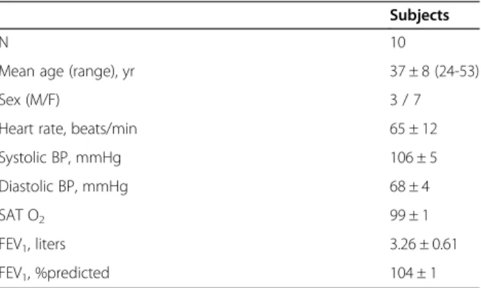
The demographics and baseline characteristics of study participants are shown in Table 1, consistent with good cardiovascular and respiratory health. All subjects com- pleted the protocol.
Table 1 Demographics and baseline characteristics of study participants (visit 1)
Subjects
N 10
Mean age (range), yr 37 ± 8 (24-53)
Sex (M/F) 3 / 7
Heart rate, beats/min 65 ± 12
Systolic BP, mmHg 106 ± 5
Diastolic BP, mmHg 68 ± 4
SAT O2 99 ± 1
FEV1, liters 3.26 ± 0.61
FEV1, %predicted 104 ± 1
Values are mean ± SE.
N = number of subjects; M, male; F, female; BP, blood pressure; SAT O2, arterial oxygen saturation measured by pulse oximetry; FEV1, forced expiratory volume in 1 second.
Ventilation and EBC pH
The levels of ventilation at the time of albuterol admin- istration during the four respiratory maneuvers are shown in Table 2. Hypercapnia and hypocapnia changed EBC pH, while eucapnic hyperventilation had no effect on EBC pH. Thus, it was possible to unlink the level of ventilation from the changes in EBC pH, which presum- ably is a reflection of airway surface liquid pH.
Airway blood flow response to albuterol
Mean systemic blood pressure and oxygen saturation were not different at the Q̇aw measurement points (baseline, pre-albuterol and post albuterol). The lack of changes in mean systemic blood pressure obviated the need to ex- press the airway blood flow responses as airway blood flow conductance. Vasodilator responses therefore were re- ported asΔQ̇aw.
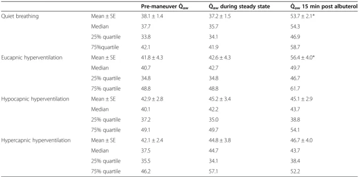
Baseline mean Q̇awvalues were similar before the four breathing maneuvers and remained unchanged during the subsequent breathing maneuvers as reflected by the pre-albuterol values (Table 3). All subjects had similar albuterol response to the different breathing maneuvers. Albuterol increased mean Q̇awsignificantly, by 46.2 and 33.8% 15 min post drug inhalation during quiet breath- ing and eucapnic hyperventilation, respectively (Table 3, Figure 1). In contrast, albuterol had no effect on mean Q̇awduring hypercapnic hyperventilation (4.9%) or hypo- capnic hyperventilation (-1.3%) maneuvers, which were associated with a decrease or increase in EBC pH.
Discussion
The purpose of this study was to determine if respiratory acidosis and alkalosis have an effect on the physiological response to inhaled albuterol in airway tissue and if the effect is related to the ventilation-associated changes in airway pH as reflected by exhaled breath condensate (EBC) pH. In order to demonstrate the role of pH in the observed changes in albuterol responsiveness associated with re- spiratory acidosis and alkalosis, it was necessary to unlink the changes in EBC pH from the changes in ventilation.
This was done by comparing quiet breathing with eucapnic hyperventilation, where ventilation changes while pH is
kept constant. Albuterol responsiveness was the same dur- ing the two maneuvers, suggesting that hyperventilation per se did not alter albuterol responsiveness. Likewise, the preserved albuterol responsiveness during eucapnic hyper- ventilation ruled out the possibility that cooling and drying of the airway could have been the cause of the blunted albuterol responsiveness during respiratory alkalosis and acidosis. Eucapnic hyperventilation was investigated last in order to be able to reproduce the highest level of ventila- tion achieved in any of the other maneuvers. We therefore are confident that ventilation per se had no effect on albu- terol responsiveness. The level of ventilation during quiet breathing was higher than one would have expected in healthy subjects at rest (mean 14.4 L.min-1). It has previ- ously been reported that wearing a nose clip and breathing through a mouthpiece increases tidal volume and minute ventilation [16]. In addition, the breathing setup we used for our study included a 100 mL deadspace, another stimu- lus for increasing tidal volume and respiratory rate.
In our study, the intended target of albuterol was airway vascular smooth muscle contained in the airway wall. Since the different respiratory maneuvers by them- selves had no effect on Q̇aw we were able to assess the effect of respiratory acidosis and alkalosis on albuterol responsiveness. In some systemic vascular beds, hyper- capnic acidosis causes relaxation and hypocapnic alkal- osis causes constriction, resulting in corresponding blood flow changes [5]. The airway circulation appears not to be subject to this regulation at least in the range of pCO2 changes seen in the present study in which changes in pH had no effect on Q̇aw; however, they affected albuterol responsiveness. We allowed 5 min for albuterol absorption during the four breathing maneu- vers, and measured Q̇aw 15 min after drug inhalation.
This was done because in previous studies we found that the maximum response typically occurs after 15 min while a vasodilator response to inhaled albuterol is already seen after 5 min [17].
We found that both airway alkalosis and acidosis attenuated albuterol responsiveness. The pH-sensitivity of albuterol responsiveness could have been related to a combination of several factors, including absorption and transport of albuterol from the airway surface to the airway vascular smooth muscle, β2-adrenergic re- ceptor function, vascular endothelial function or vascu- lar smooth muscle responsiveness. In vitro and animal experiments suggest that all of these functions can be pH-dependent.
Acidosis
The majority of the currently used β2-adrenergic bron- chodilators, including albuterol, cannot freely diffuse across the epithelial cell membrane because they are hydrophilic and carry a transient or permanent positive Table 2 Ventilation and exhaled breath condensate pH
during respiratory maneuvers
Challenges V̇(L.min-1) EBC pH (units)
Quiet breathing 14.4 ± 4.2 6.39 ± 0.14
Eucapnic hyperventilation 35.5 ± 3.4* 6.31 ± 0.08 Hypocapnic hyperventilation 35.2 ± 3.3* 6.59 ± 0.15**
Hypercapnic hyperventilation 24.4 ± 2.9* 5.88 ± 0.14**
V̇, ventilation.
EBC, exhaled breath condensate.
*p < 0.05 vs. quiet breathing.
**p < 0.02 vs. quiet breathing.
charge at physiological pH. Thus, the epithelium of the airway becomes a barrier to these agents, requiring cel- lular or paracellular transport across the epithelial lining of the airway to reach their intended target tissues in- cluding airway vascular smooth muscle. We have previ- ously demonstrated the existence of an organic cation transport machinery in the human airway epithelium
and showed that this process is largely mediated by the organic cation/carnitine transporter OCTN2, which is likely involved in the delivery of inhaled hydrophilic cationic bronchodilators to the airway tissue [1]. We showed that cationic drug uptake is pH dependent, with about 3-fold lower rates at an acidic pH (5.7) than alka- line pH (8.2). This mechanism could have been fully or Table 3 Effects of respiratory maneuvers on airway blood flow (Q̇aw)
Pre-maneuver Q̇aw Q̇awduring steady state Q̇aw15 min post albuterol
Quiet breathing Mean ± SE 38.1 ± 1.4 37.2 ± 1.5 53.7 ± 2.1*
Median 37.7 35.7 54.3
25% quartile 33.8 34.1 46.9
75%quartile 42.1 41.9 58.7
Eucapnic hyperventilation Mean ± SE 41.8 ± 4.3 42.6 ± 4.3 56.4 ± 4.0*
Median 40.7 42.7 49.7
25% quartile 34.8 34.8 46.7
75% quartile 48.8 48.8 61.7
Hypocapnic hyperventilation Mean ± SE 42.9 ± 2.8 45.2 ± 3.4 45.1 ± 2.9
Median 40.1 42.2 43.7
25% quartile 37.2 35.0 38.8
75% quartile 49.1 49.7 54.1
Hypercapnic hyperventilation Mean ± SE 42.1 ± 2.4 44.8 ± 3.8 46.7 ± 4.0
Median 37.5 44.7 43.7
25% quartile 35.5 34.1 38.4
75% quartile 46.2 57.1 52.2
Q̇awis expressed inμl.min-1.mL-1. * p < 0.05 vs. steady state.
Figure 1Relative albuterol-induced changes in airway blood flow (ΔQ̇aw) during four breathing maneuvers.Values are mean ± SE. *p <
0.01 and ** p < 0.02 vs quiet breathing and eucapnic hyperventilation.
partially responsible for the blunted albuterol respon- siveness during respiratory acidosis associated with a de- creased airway surface liquid pH. We have also shown that albuterol crosses the airway epithelium via the para- cellular route [18]. The paracellular pathway can also mediate pH-dependent permeability to pH-dependent changes in negative charges.
It has also been reported that acidosis can cause rapid desensitization and uncoupling of β2-adrenergic recep- tors [2], possibly leading to albuterol unresponsiveness as seen in the present investigation.
Albuterol-induced vasodilation is endothelium-dependent, involving endothelial relaxant factors including nitric oxide [19]. Although the observations on the effects of intracellu- lar and extracellular acidosis and pCO2on endothelial func- tion have not been consistent, the majority of studies have shown that acidosis can impair endothelial function [4-6]. It is likely that airway surface liquid pH is a reflection of extra- cellular pH, but changes in both extracellular and intracellu- lar pH have been implicated in the effect of acidosis on endothelial function. Thus, endothelial dysfunction could have had a role in the blunted albuterol responsiveness in our study. Finally, airway vascular smooth muscle function could be directly affected by acidosis. In particular, acidosis can lead to smooth muscle cell hyperpolarization, which in turn could attenuate albuterol-induced vasodilation [5].
Alkalosis
Respiratory alkalosis also attenuated albuterol-induced vasodilation in our study.In vitro, alkalosis increases the transport of organic cations such as albuterol across the airway epithelium via the transcellular and paracellular routes [18]. Alkalosis may also increaseβ2-adrenergic re- ceptor ligand binding [3]. Finally, alkalosis has been shown to cause endothelium-dependent vasodilation without altering endothelial nitric oxide synthase function [5]. All of these actions would be expected to potentiate inhaled albuterol-induced vasodilation. The mechanistic explan- ation for our observation that respiratory alkalosis has the same attenuating effect on albuterol responsiveness as acidosis remains unclear at this time.
Conclusions
Patients with airway disease are likely to have highly vari- able airway surface liquid pH and adrenergic airway smooth muscle responsiveness [20-23]. Therefore, we de- cided to investigate the pH dependence of β2-adrenergic responsiveness as a marker of albuterol responsiveness in healthy subjects with normalβ2-adrenergic smooth muscle responsiveness in whom the airway surface liquid pH can be artificially manipulated. From a clinical perspective, airway smooth muscle would have been a more meaningful airway wall target to assess responsiveness to inhaled albu- terol. However, healthy subjects do not have an increased
airway smooth muscle tone and responses to albuterol would have been too small to study the effects of respira- tory acidosis and alkalosis. We chose not to include patients with asthma or COPD in the investigation because they have a blunted airway blood flow response to albute- rol due to endothelial dysfunction [18], and because meas- uring airflow responses by pulmonary function testing would have been technically difficult under the experimen- tal conditions of the study.
Our in vivo observation showed that both respiratory acidosis and alkalosis blunt albuterol responsiveness in the airway wall, although it is not known whether the effect is driven by intracellular or extracellular pH or pCO2 and which of the above-mentioned mechanisms may be involved. In this study we found that albuterol responsiveness as assessed by Q̇aw in the airway is blunted by acidosis and alkalosis, using Q̇aw as a bio- assay. It remains to be shown whether the clinical bene- fits of inhaled albuterol, i.e., bronchodilation may be less than expected during acute respiratory acidosis and alkalosis, which can be associated with exacerbations of asthma and COPD.
Competing interests
The authors declare that they have no competing interests.
Authors’contributions
AW: Conception and design, analysis and interpretation of data, drafting of the manuscript, critical revision of the manuscript for important intellectual content, supervision and final approval of the version to be published. MS:
Conception and design and manuscript editing. ESM: Acquisition of data, statistical analysis, and interpretation of data. JEC: Acquisition and analysis of data, GH: Conception and design of the study. MEM: Sample analysis and interpretation. JA: Acquisition of data. All authors read and approved the final manuscript.
Authors’information
Matthias Salathe and Adam Wanner are senior authors contributed equally to this paper.
Funding
The study was supported by NIH 1R01HL060644.
Author details
1Division of Pulmonary, Allergy, Critical Care and Sleep Medicine, University of Miami School of Medicine, Miami, FL 33136, USA.2Department of Pulmonology, Semmelweis University School of Medicine, Budapest, Hungary.
Received: 15 October 2013 Accepted: 16 March 2015
References
1. Horvath G, Schmid N, Fragoso MA, Schmid A, Conner GE, Salathe M, et al.
Epithelial organic cation transporters ensure pH dependent drug absorption in the airway. Am J Respir Cell Mol Biol. 2007;36:53–60.
2. Davies AO. Rapid desensitization and uncoupling of human beta-adrenergic receptors in anin vitromodel of lactic acidosis. J Clin Endocrinol Metab.
1984;59:398–405.
3. Modest VE, Butterworth JF. Effect of pH and lidocaine on beta-adrenergic receptor binding: interaction during rescuscitation. Chest. 1995;108:1373–9.
4. Crimi E, Taccone FS, Infante T, Scoletta S, Crudele V, Napoli C. Effects if intracelular acidosis on endotelial function: an overview. J Crit Care.
2012;27:108–18.
5. Celotto AC, Capellini CF, Baldo CF, Dalio MB, Rodriguez AJ, Evora PRB.
Effects of acid-base imbalance on vascular reactivity. Braz J Med Biol Res.
2008;41:439–45.
6. Nagy S, Harris MB, Ju H, Bhatia J, Venema RC. pH and nitric synthase activity and expression in bovine endothelial cells. Acta Pediatr. 2006;95:814–7.
7. Kontos HA, Wei EP, Raper AJ, Patterson JL. Local mechanism of CO2action of cat pial arterioles. Stroke. 1977;8:226–9.
8. Wanner A, Mendes ES, Atkins ND. A simplified noninvasive method to measure airway blood flow in humans. J Appl Physiol. 2006;100:1674–8.
9. Scuri M, McCascill V, Chediak AD, Abraham WM, Wanner A. Measurement of airway blood flow with dimethylether: validation with microspheres. J Appl Physiol. 1995;79:1386–90.
10. Crapo RO, Morris AH, Gardner RM. Reference spirometric values using techniques and equipment that meet ATS recommendations. Am Rev Respir Dis. 1981;123:659–64.
11. Horvath G, Hunt J, Barnes PJ. On behalf of the ATS/ERS Task Force on Exhaled Breath Condensate. Eur Respir J. 2005;26:523–48.
12. Paget-Brown AO, Ngamtrakulpanit L, Smith A, Bunyan D, Hom S, Nguyen A, et al. Normative data for pH of exhaled breath condensate. Chest.
2006;129:426–30.
13. Wells K, Vaughan J, Pajewski TN, Hom S, Ngamtrakulpanit L, Smith A, et al.
Exhaled breath condensate pH assays are not influenced by oral ammonia.
Thorax. 2005;60:27–31.
14. Effros RM, Casaburi R, Su J, Dunning M, Torday J, Biller J, et al. The effects of volatile salivary acids and bases on exhaled breath condensate pH. Am J Respir Crit Care Med. 2006;173:386–92.
15. Gilbert IA, McFadden ER. Airway cooling and rewarming. J Clin Invest.
1992;90:699–704.
16. Bloch KE, Barandun J, Sackner MA. Effect of mouthpiece breathing on cardiorespiratory response to intense exercise. Am J Respir Crit Care Med.
1995;151:1087–92.
17. Onorato DJ, Demirozu MC, Breitenbucher A, Atkins ND, Chediak AD, Wanner A. Airway mucosal blood flow in man: response to adrenergic agonists. Am J Respir Crit Care Med. 1994;149:1132–7.
18. Unwalla HJ, Horvath G, Roth FD, Conner GE, Salathe M. Albuterol modulates its own transepithelial flux via changes in paracellular permeability. Am J Respir Cell Mol Biol. 2012;46:551–8.
19. Wanner A, Mendes ES. Airway endothelial dysfunction in asthma and COPD: a challenge for future research. Am J Respir Crit Care Med. 2010;182:1344–51.
20. Hunt JF, Fang K, Malik R, Snyder A, Malhotra N, Platts-Mills TA, et al. En- dogenous airway acidification. Implications for asthma pathophysiology. Am J Respir Crit Care Med. 2000;161:694–9.
21. Kodric M, Shah A, Fabbri L, Confalonieri M. An investigation of airway acidification in asthma using induced sputum. Am J Respir Crit Care Med.
2007;175:905–10.
22. McShane D, Davies JC, Davies MG, Bush A, Geddes DM, Alton EW. Airway surface pH in subjects with cystic fibrosis. Eur Respir J. 2003;21:37–42.
23. Brieva J, Wanner A. Adrenergic airway vascular smooth muscle responsiveness in healthy and asthmatic subjects. J Appl Physiol.
2001;90:665–9.
Submit your next manuscript to BioMed Central and take full advantage of:
• Convenient online submission
• Thorough peer review
• No space constraints or color figure charges
• Immediate publication on acceptance
• Inclusion in PubMed, CAS, Scopus and Google Scholar
• Research which is freely available for redistribution
Submit your manuscript at www.biomedcentral.com/submit