ESTABLISHMENT AND CHARACTERIZATION OF EPITHELIAL CELL CULTURES FROM TUMORS OF
THE HUMAN URINARY TRACT
A. Y. ELLIOTT D. L, BRONSON J, CERVENKA P. H. CLEVELAND
Å. E. FRALEY
Department of Urologie Surgery
University of Minnesota Health Sciences Center Minneapolis, Minnesota
SUMMARY
Fifty-four surgical specimens of transitional cell cancer (TCC) of the human urinary tract were placed in culture. Epithe- lium-like cells grew out from expiants of 38 (70%) of the tumors cultured, and 11 (22%) have been maintained in culture for more than 18 months. Characterization studies performed on six of these lines show the cells to (1) be small, (2) have a rapid dou- bling time, (3) exhibit multilayering, (4) produce tumors in the cheek pouches of immune-depressed hamsters, (5) form colonies in soft agar, C6I possess a hyperdiploid karyotype, and C7) contain
711
712 Á. Õ. ELLIOTT et al.
ultrastructural features common to epithelial cells. The estab- lished cultures grow rapidly in roller bottles and therefore can be produced in large quantities. These cells also remain viable after storage for 4 years in liquid nitrogen (LN2).
I. INTRODUCTION
Since the initial work of Burrows, et al. (1917), many labora- tories have attempted to establish long-term cell lines from transitional cell cancers (TCC) of the human urinary tract. Al- though there are presently available to investigators three long- term lines: R T4, established by Rigby and Franks (1970); T2 4, established by Bubenik et al. (1973); and 253J established by El- liott et al. (1974); no single laboratory has been successful in establishing more than one long-term epithelial cell line. Re- ports by Walker et al. (1965), Jones (1967), Yajima (1968), and Bubenik et al. (1970) emphasize the problems involved in estab- lishing epithelial-cell cultures from human tumors.
Previously, Elliott et al. (1974) reported the establishment and characterization of a cell line (253J) derived from a metast- ic lymph node from a patient with multiple TCC of the urinary tract. Utilizing the technique employed to establish the 253J cell line, as well as another method described by Elliott et al.
(1976), we have established ten additional long-term TCC cell lines.
EPITHELIAL CELL CULTURES 713 II. MATERIALS AND METHODS
A. Tissue Culture Procedures
1. Establishment of Cell Cultures
Tumor tissues obtained at surgery were brought directly to the tissue culture laboratory in RPMI-1640 medium (Gibco, Grand Island, New York) containing 20 percent heat-inactivated fetal bovine serum (Armour Co., Kankakee, Illinois), 10 percent tryp- tose phosphate broth (Difco Laboratories Inc., Detroit, Michigan), and 100 U of penicillin and 100 ug of streptomycin (Gibco) per milliliter.
In most cases, processing of the tumors began within 15 min- utes of the time they were removed from the patient. The tissue was minced very fine (1 mm) with sharp scissors in a sterile Petri dish. The minced tissue was floated in an appropriate amount of RPMI outgrowth medium in a Petri dish, and the small fragments and the medium were drawn up into a 10-ml plastic sy- ringe without a needle. Approximately 1 ml of outgrowth medium containing tumor fragments was placed in each of several 25-cm plastic flasks (Falcon Plastics, Oxnard, California), and the
flasks were rotated to distribute the fragments evenly over the surface. The flasks were then incubated at 37°C without C 02 for 24 hours. It is very important to use as little medium as pos- sible, or many of the tissue fragments will not attach to the surface of the flask. After growth began, 2 to 3 ml of outgrowth medium were added carefully so as not to disturb the attached pieces.
2. Subculture
When the primary cultures reached a monolayer, the cells were removed from the flask by the technique previously described by Elliott et al. C1974I.
714 Á. Õ. ELLIOTT et al.
3, Freezing and Storage
Cells were removed from the flasks by trypsinization, washed several times in outgrowth medium, counted, and diluted in storage medium (RPMI-1640 with 30 percent fetal bovine serum, 10 percent dimethyl sulfoxide, penicillin, and streptomycin) to a concentra- tion of 5.0 x 10^ cells/ml. Three ml of the cell suspension were placed in a 5-ml blue-line break ampoule (Kimble Glass Products, Vineland, N. J.) and heat-sealed. The ampoules were placed at 4°C for one hour and then put into the vapor phase compartment of a liquid nitrogen refrigerator (Minnesota Valley Engineering Co., New Prague, Minnesota).
To revive the stored cells, the ampoules were removed from the refrigerator and thawed at 40°C. The cells were washed sev- eral times in outgrowth medium and seeded at a density of 5.0 x
10 per ml of outgrowth medium in 75-cm Falcon plastic flasks. 6 2
4. Large Volume Production
Glass roller bottles (#7000 Bellco disposable bottles, Bellco Glass Co., Vineland, N. J.) were used to produce large volumes of tumor cells. Before the cells were planted, the surface of each bottle was conditioned by adding 100 ml of outgrowth medium and rotating the bottles at 37°C for 30 minutes. The medium was then removed from the bottles and replaced with 100 ml of fresh out- growth medium containing 1 x 10^ tumor cells. The bottles were placed in a Bellco roller bottle apparatus, incubated at 37°C, and rotated at 0.5 rpm.
B, Malignancy Studies
1. Hamster Inoculation
The procedure employed for inoculation of TCC into the cheek pouches of immune-depressed hamsters have been previously de- scribed by Elliott et al. C1974).
EPITHELIAL CELL CULTURES 715
2. Cell Culture in Agar
Cultures were prepared according to the procedure described by McAllister et al, Q-967I, The cell concentrations employed, as well as the medium and cultural requirements, were previously described by Elliott et al, ( 1 9 7 4 } ,
3. Electron Microscopy
A portion of the original tumor tissue from each TCC was fixed in 2 percent glutaraldehyde (Electron Microscopy Sciences, Fort Washington, Pa.I as previous described by Elliott et al. ( 1 9 7 4 ) . Tissue culture cells were fixed, dehydrated, and embedded in Epon 812 (Electron Microscopy Sciences) as monolayers while still in the culture flasks by a method described by Elliott et al. ( 1 9 7 3 ) ,
4 . Chromosome Studies
Near-monolayer cultures of TCC cells were harvested by tryp- sinization after incubation with 0 . 0 5 mg colcemid/ml (Ciba Phar- maceutical Products Inc., Summit, N. J.) for 5 to 10 hours. The remainder of the procedure was previously described by Elliott et al. ( 1 9 7 4 ) .
III. RESULTS
A, Cells in Culture
1. Cell Outgrowth
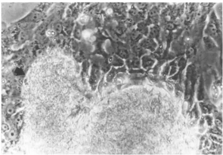
Cultures were observed daily for evidence of cell growth from the tumor expiants. In 48 of 54 tumors (89%), epithelial-like cells grew out from the tumor within 24 to 48 hours after planting
(Fig. 1 1 . In 12 tumors, the epithelial cells continued to grow for 1 to 4 weeks, then began to round up and detach from the sur-
716 Á. Õ. ELLIOTT et ai
FIGURE 1 Phase contrast photograph of epithelial cells grow- ing out from tumor (292W) piece (at arrow). X 400.
face of the flask. Three cultures exhibiting this spontaneous degeneration have been described in detail by Elliott et al.
(1973) .
Each of these cultures showed complete destruction of the cells within 7 days, and attempts to subculture the detached, floating cells proved futile. Extensive culture and ultrastruc- ture studies failed to detect bacteria, mold, yeast, mycoplasma, or ubiquitous viruses.
Cells from 38 tumors (70%) grew rapidly and were subcultured.
However, 27 of the epithelium-cell cultures could not be kept in culture longer than 12 months. At various times between 2 and 12 months after planting, the cells would either slow their growth rate or cease to grow altogether. The cells that remained on the flask usually were much larger than the characteristically small TCC cells and were frequently multinucleated. All attempts to keep these cells in culture by increasing the serum concentration.
EPITHELIAL CELL CULTURES 717
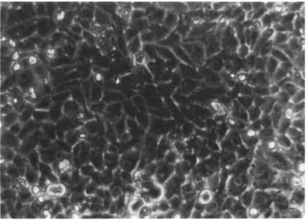
FIGURE 2 Phase contrast photograph of monolayer culture of 292VT cells in passage #30. X 400,
by adding L-glutamine, nonessential amino acids, insulin, corti- sone, or testosterone, or by using Eagle's MEM, Medium 199, or Earle1s MEM instead of our usual medium, did not prevent the death of these cells. Even when earlier passage levels of the same cells were revived from storage in L N2 and placed in culture, the cells stopped growing after a short time.
Eleven long-term cell lines have been in culture for more than a year. Six of these lines (253J, 292W, 192B, 647V, 639V and 486P) have been subcultured more than 50 times and continue to grow at a constant rate. The cells are small (averaging 10 μ in diameter), epitheloid, and, usually, uninucleate. They grow rap- idly in culture, exhibit multilayering, and can be subcultured at a l-to-3 split at weekly intervals (Fig. 2 ) . For practical rea- sons, the other 5 lines, after more than 20 passages, have been stored in LN2 for future study,
718 Á. Õ. ELLIOTT et al.
Cells retrieved from LN2 storage settled onto the surface of the flasks and grew; within 1 hour after planting, often forming a monolayer in 4 to 7 days. Cells have been revived from LN2 stor- age of early passages of each of the 11 long-term cultures with no apparent damage or change in growth pattern.
After the tumor cells had undergone approximately 12 passages in the RPMI outgrowth medium, it was possible to switch to Eagle's MEM (GIBCO) containing 10 percent heat-inactivated fetal bovine serum, penicillin, and streptomycin as both outgrowth and mainte- nance medium.
B. Large Volume Cell Production
TCC cells grew better on plastic flasks than on disposable glass roller bottles, but after several passages of the cells on plastic flasks, tumor cells seeded in roller bottles at a high cell density (1 x 10? cells/bottle) would grow and form a heavy multilayered cell sheet. Good cell attachment and outgrowth were not obtained if the surface of the bottles was not exposed to out- growth medium before the cells were seeded. The cells grew out more slowly on glass, requiring 10 to 14 days to form a monolayer.
When confluent, the cells continued to grow in multilayers; and after 3 to 4 weeks in culture, 1 x 1 09 cells could be recovered from a single bottle. This procedure has allowed us to produce larger numbers of tumor cells in early passages and to store them for future studies.
Culture and ultrastructure studies in our laboratory and by HEM Corporation (Rockville, Maryland) have failed to detect bac- teria, yeast, molds, or mycoplasma in these cell lines.
1. Cell Growth xn Agar
Passage-10 cells from each of the TCC lines listed in Table I produced multilayered colonies in soft agar. Microscopic examina- tion of the agar plates 24 hours after the cells were planted re-
Ό Ο U ft U · Q
Φ
•H Φ
fi
O QrH ft Φ U
MH O -P Φ
•H
ù
O CO M1 H CO CM
ó\ vo m in m
+ + + + + +
+ + + + + +
+ + + + + +
r- m ο ο o vo m m vo oo t é i i t i
O CN O VO VO VO vo L O m m vo
cn w w w ω co
co o rH C M m
^ C M C N C N ^ co
4J + J AJ Jp +) 4J
•H -H -H -H -H -H 04 ft ft ft ft
w w w ù ù w
õ õ _
Å-« Tl I ϋ Ñ* Φ S « Ê D CQ CQ
pq ^ > > pu CO CN CN CT> VO LT) <Τß (Tï CO <tf CO CN rH CN VO VO
φ ϋ
ϋ rd φ ÎH Ο UH Φ
φ Â • 0 CQ ω U
0 φ
 ϋ 0 Φ u rd ϋ
¼ rH
rH ΓΗ rtf φ ΤΙ ϋ 0 å rH å rd
Φ 0 -ñ -Η -Η -Ρ -ñ CO φ rd Μ Φ •μ
ft Φ
Φ rH U -H ft
U •Ρ · φ rH U
G II
3 Ö rH U
å ¼ (d Ö Tl C -Ρ
ô é cd Λ
rd Φ Φ II II II CQ Δ D ϋ Ό Φ
^ „ I
a
719
720 Á. Õ. ELLIOTT et al.
FIGURE 3 Electron micrograph of 192B cells in passage §34, Numerous microvilli are shown, X 6000,
vealed single cells well dispersed throughout the top agar layer;
areas could be marked and the colonies from these single cells
EPITHELIAL CELL CULTURES 721
observed. Except in the ca.se of 639V and 486P, colonies consist- ing of a few cells were visible several days after inoculation onto soft agar. At the end of 21 days in culture, the colonies consisted of tightly packed, multilayered cell sheets. The size of the colonies again depended on the growth rate of the cells in culture, with the largest colonies seen in 192B cells and the smallest in 486P cells. Approximately 65 percent of the 192B cells and 10 percent of the 486P cells planted in agar formed colonies. At this time, clones were established by transplanting individual colonies to Petri dishes under liquid medium. These colonies attached to the surface of the dish and, except for the 192B cells, grew slowly; the cells were usually a monolayer within 3 weeks (see Fig. 3).
C. Electron Microscopy
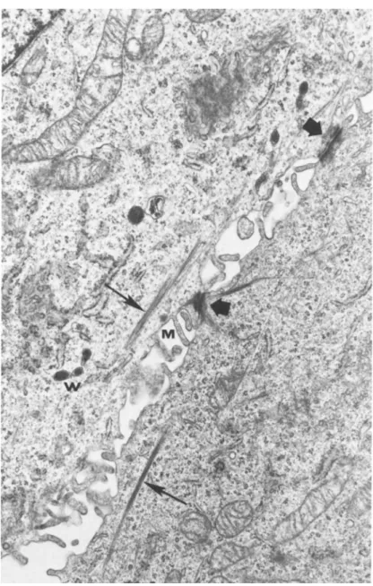
Sections from both the original surgical specimen and from passaged TCC cells revealed structures which are considered to be consistently found in epithelial cells. Most of the cells, re- gardless of the passage level, contained numerous microvilli (Fig.
4). Mitochondria were abundant, as were the ribosomes and poly- somes. Golgi bodies were found in the cells, although they were not well developed. Residual bodies, vacuoles, and, to lesser extent, lysosomes and phagosomes were also observed. Multivesicu- lar bodies were found in abundance, especially in the 253J and 192B cell lines. Desmosomes and bundles of tonofibrils were scarce in most of the cell lines but readily seen in the 647V and 486P cell lines (Fig. 5). No remarkable ultrastructural differ- ences were noted between the early and the late passages of any of the cell lines.
722 Á. Õ. ELLIOTT et al.
FIGURE 4 Electron micrograph of 647V cells In passage §19.
Desmosomes (large arrows!, bundles of tonofibrils (small arrows), microvilli (M), and mibel Palade bodies (Jiïl are shown. X 18,000.
EPITHELIAL CELL CULTURES 723
I I H * l ι»
eil 1 4* I i l l β I I I I I Ê η
FIGURE 5 Karyotype of 253J cells in passage §76. X 1500.
D. Chromosome Studies
Chromosome analysis was performed on each cell line at pas- sage 10 and at every tenth passage thereafter. Although there was some shifting and chromosomal rearrangement the basic karyo- type of each cell line did not change drastically with time. The karyotype of passage 76 253J cells is shown in Fig. 5. This cell line has been in culture since April 1972 and has undergone more than 90 subcultures. The basic karyotype has not changed since passage 10.
The karyotype of passage-1 192B cells is shown in Fig. 6.
This cell line is the fastest growing and has the smallest number of chromosomes, the modal number for this line being 52 to 54.
In general, all of the cell lines possessed hyperdiploid karyo- types with numerous minutes and breaks. Although no definite chromosome abnormality or rearrangement seems consistent with TCC cells, all of the lines: CH are definitely of human origin, C2) possess karyotypes consistent with malignant cells, (31 can be distinguished from one another on the basis of distinctive chro-
724 Á. Õ. ELLIOTT et al.
FIGURE 6 Karyotype of 192B cells in passage #1, X 1500.
mosome patterns, and C4) do not have karyotypes in common with either HeLa or any other characterized human cell line,
IV. DISCUSSION
Using the procedure with which we established the 253J cell line, and a second method, we have been able to establish 10 ad- ditional long-term TCC cell lines. Six of these have been studied in detail, and characterization studies have been performed.
Properties common to all six cell lines, as listed in Table I, are (1) epithelial cell morphology, (2) multilayering of cells in culture, (3) hyperdiploid chromosome profiles, (4} production of tumors in immune-depressed hamsters, C5Î growth in soft agar, C6) rapid doubling times, and C7] presence of ultrastructural features common to epithelial cells.
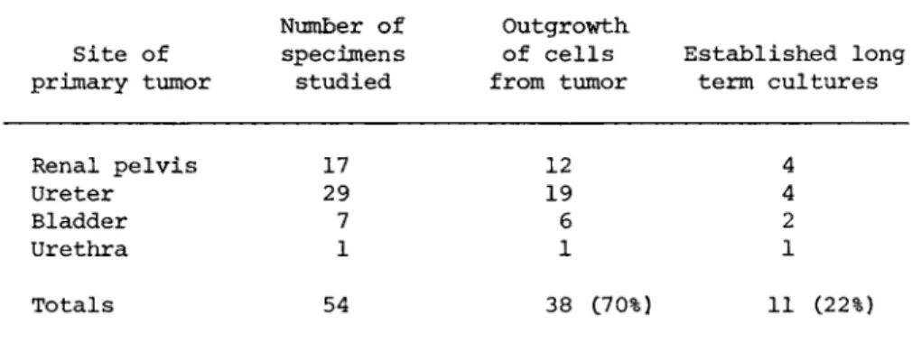
EPITHELIAL CELL CULTURES 725 The difficulties encountered by other investigators in their attempts to establish epithelial cell lines from TCC are also re- flected in our work. As shown in Table II, there was initial cell outgrowth from 38 C70%) of the 54 tumors placed in culture, but cells from 27 of these tumors could be maintained in culture for no longer than six months or four subcultures, and, as pre- viously mentioned, all attempts to keep these cells in culture by the addition of growth-promoting hormones or other nutrients proved futile. No similar "crisis period" was noted in the eleven cell lines which have been carried for at least 20 passages.
Employing the roller bottle technique described in this re- port, it was possible to produce large volumes of cells from each of the 11 TCC cell lines. This technique provided large numbers of cells from both early and late tissue culture passages so that we could compare the results of cytogenetic, immunologic, bio- chemical, ultrastructural, oncogenicity, and growth studies. It was also possible to store large numbers of these cells in LN2 and later revive them for tissue culture passage and study.
Not only has it been difficult to establish long-term cell cultures from human transitional cancers of the urinary tract, but similar problems have been encountered by investigators at- tempting to culture epithelium-like cells from other solid human tumors. For example, Giard et al. (1973) summarized the results of their attempts to establish cell lines from 200 human tumors.
Having obtained only a 6 percent success rate, the authors con- cluded that, in contrast to human fibroblasts, epithelium-like cell cultures from human tumors were indeed difficult to develop into long-term lines.
However, the studies reported herein suggest that a reasonable success rate can be achieved with TCC if a certain protocol is followed. First, tumors should be placed in culture as soon as possible. We believe that it is important to process the tumors and place them in culture within an hour of their removal from the patient. Second, although TCC may be obtained by many surgi-
726 Á. Õ. ELLIOTT et al.
Site of primary tumor
Number of specimens studied
Outgrowth of cells from tumor
Established long term cultures
Renal pelvis 17 29 7 1
12 19 6 1
4 4 2 1 Ureter
Bladder Urethra
Totals 54 38 C70%) 11 (22%)
aOnly epithelial-like cells are counted in the 70% figure listed under outgrowth of cells from tumor. Fibroblasts were disregarded. Each of the 11 cell lines listed in the last column has been in culture for over 18 months and has undergone at least 50 subcultures.
cal techniques, we have had our best results with specimens re- moved mechanically through the resectoscope with biopsy forceps.
Specimens obtained by transurethral resection with an electrical- ly activated cutting loop often are so badly damaged by the elec- trical current that they are not satisfactory for cultivation.
Third, it is imperative that the outgrowth medium provides the proper nutrients for rapid outgrowth of cells from tumor explants.
The medium described herein appears to meet this requirement for TCC cells.
Cell lines established from human TCC have been used in vari- ous immunologic and biochemical studies. It was previously shown by Hakala et al. (1974a) that humoral complement-dependent cyto- toxic to this cell line were obtained more frequently from pa- tients. Hakala et al. (1974b) also showed that lymphocytes cyto- toxic to this cell line were obtained more frequently from pa- tients with TCC than from patients without TCC,
Transitional cell cancer of the urinary tract has been studied in the laboratory as comprehensively as any other solid
TABLE II Growth, of Tumor Cells from Tissue Culture Expiantsa
EPITHELIAL CELL CULTURES 727
human tumor. There has been special interest in both the irnmuno- biology and virology of these tumors. Almost all of these studies have utilized an established TCC cell line, usually either T2 4 or RT4. If studies on the basic biology of TCC are to continue and to be expanded, additional cell lines will be required in order to establish or refute the accuracy of data gathered on TCC in the above assays.
ACKNOWLEDGMENTS
We thank H. Asmussen, S. Dombrovskis, and N. Stein for excel- lent technical assistance.
Supported in part by Public Health Service Training Grant AM 05514-10 from the National Institute of Arthritis and Metabolic Diseases, and by grants CA 13095-03 and CA 15551-02 from the National Cancer Institute.
REFERENCES
1. Bubenik, J., Baresova, M., Viklick, V., Jakoubkova, J., Sainerova, H. and Donner, J. (1973). M T . J. Cancer 11, 765- 773.
2. Bubenik, J., Perlmann, P., Helmstein, Ê. and Moberger, G.
(1970). Int. J. Cancer 5, 39-46.
3. Burrows, M. T., Burns, J. E. and Suzuki, Y. (1917). Johns Hopkins Hosp. Bull. 28, 178-179.
4. Elliott, A. Y., Bronson, D. L., Stein, Ν., and Fraley, Å. E.
(1976). Cancer Res. 36, 365-369.
5. Elliott, A. Y., Cleveland, P., Cervenka, J., Castro, Á. Å., Stein, N., Hakala, T. R. and Fraley, E. E, C1974). J. Natl.
Cancer Inst, 53, 1341^1349.
6. Elliott, A. Y,t Fraley, Å. Å., Cleveland, P., Castro, Á. Å., and Stein, N. (1973). Science 179, 393-395.
728 Á. Õ. ELLIOTT et al.
7. Giard, D, J,, Aaronsonf S, A., Todaro, G, J., Arnstein, P., Kersey, J. H., Dosik, H,, and Parks, W. P. 0.973}. J. Natl.
Cancer Inst, 51, 1417-1423,
8. Hakala, T. R., Castro, A, E., Elliott, A, Y. and Fraley, E.
E. C1974a). J. Urol. 111, 3 8 2 - 3 8 5 ,
9. Hakala, T, R., Lange, P, H,, Castro, Á. Å., Elliott, A, Y.
and Fraley, E. E. C1974bl. Cancer 34, 1 9 2 9 - 1 9 3 4 , 10. Jones, G. W. 0-9671. Cancer 20, 1 8 9 3 - 1 8 9 8 .
11. McAllister, R., Reed, G., and Huebner, R. 0- 9 6 7 ) . j, Natl.
Cancer Inst. 39, 43-53.
12. Rigby, C., and Franks, L. M. ( 1 9 7 0 ) . Brit. J, Cancer 24, 7 4 6 - 7 5 4 .
13. Walker, D, G., Lyons, M. M., and Wright, J. C. ( 1 9 6 5 ) . Eur.
J. Cancer ! , 2 6 5 - 2 7 3 .
14. Yajima, T. 0- 9 6 8 ) . Jap. J. Urol. 59, 1 2 8 - 1 3 4 ,