Received 1 March 2018; revised 13 June 2018 and 18 August 2018; accepted 3 September 2018.
Date of publication 1 November 2018; date of current version 15 November 2018.
Digital Object Identifier 10.1109/JTEHM.2018.2878724
Virtual Neonatal Echocardiographic Training System (VNETS): An Echocardiographic Simulator for
Training Basic Transthoracic Echocardiography Skills in Neonates and Infants
BIJAN SIASSI 1, MAHMOOD EBRAHIMI1, SHAHAB NOORI2, SHUYANG SHENG1, DEBJIT GHOSH1, AND ISTVAN SERI3
1Los Angeles County University of Southern California Medical Center, Los Angeles, CA 90033, USA
2Division of Neonatology, Fetal and Neonatal Institute, Children’s Hospital Los Angeles, Department of Pediatrics, Keck School of Medicine, University of Southern California, Los Angeles, CA 90027, USA
3First Department of Pediatrics, Faculty of Medicine, Semmelweis University, Budapest H-1083, Hungary
CORRESPONDING AUTHOR: B. SIASSI (bijans@msn.com)
This work was supported from an Educational Fund in part by the Fetal and Neonatal Institute, Division of Neonatology, Children’s Hospital Los Angeles, in part by the Department of Pediatrics, Keck School of Medicine, University of Southern California, Los Angeles, CA, USA, and in part by the Division of Neonatology, Department of Pediatrics, LAC+USC Medical Center. B. Siassi, M. Ebrahimi, S. Noori and I. Seri have
financial interest in Virtual Echo Training System Inc., which has developed VNETS.
ABSTRACT There is a great need for training in pediatric echocardiography. In addition to physicians being trained in pediatric cardiology and echocardiography technologists, neonatologist, pediatric intensivists, and other health care professionals may be interested in such training. Since, there is limited opportunity of training on live patients, echocardiographic simulators may be of help. No simulator with complete range of echocardiographic modalities is available for neonates and infants. The aim of this project was to develop a mannequin-based echocardiographic simulator capable of simulating full range of pediatric 2D, color flow Doppler, spectral Doppler, and M-mode echocardiograms. A mannequin, a laptop computer, a magnetic tracking device, and a six-degree freedom (6DOF) sensor incorporated in a dummy transducer serve as the hardware platform of the simulator. We obtained six to seven 4D echocardiographic datasets in DICOM format through five acoustic windows from each infant along with a complete set of 2D video clips of color flow, Doppler, and M-mode. The 4D datasets are sliced into 3D slices using the visualization toolkit and are displayed as 2D echocardiograms through the information obtained by the 6DOF sensor. The coordinates from specific 3D slices triggers display of video clips of color flow, M-mode, and Doppler echocardiogram.
Software written in C++ programming language controls the basic function of the program. The main simulator screen displays the full range of 2D echocardiograms including color flow Doppler, spectral Doppler, and M-mode from each acoustic window, whereas the side screen display the position and motion of the cutting planes through a 3D heart model. The system includes a software module to perform hemodynamic measurements from specific video clips images. Our hybrid, mannequin-based pediatric echocardiography simulator provides full range of pediatric echocardiography training experience. This simulator may help training in pediatric echocardiography for which there is a growing demand in clinical medicine.
INDEX TERMS Echocardiography simulation, infants, mannequin-based simulation, neonates, point of care echocardiography.
I. INTRODUCTION
Echocardiography is the predominant non-invasive method of assessing cardiac structure and function. Although originally developed as a diagnostic tool for cardiologists, recently it has been increasingly used across different specialties for acute patient care [1].
The utility of point-of-care echocardiography has been demonstrated by neonatologists in neonatal intensive care [2], anesthesiologists during the perioperative period [3], inten- sivists in the critical care setting [4]. Development of more advanced and portable systems has facilitated this change. However, the widespread use of point-of-care
2168-23722018 IEEE. Translations and content mining are permitted for academic research only.
echocardiography, especially in neonatal intensive care, has been hampered by the lack of adequately trained non-cardiology specialists. Training in echocardiography is a complex process which, in addition to thorough theoretical knowledge, requires extensive hands-on training to acquire the all-so-important technical skills. To overcome this obsta- cle, echocardiography simulators have been developed for transthoracic and transesophageal echocardiography [6]–[9].
These simulators utilize images from adult patients which are usually acquired by other imaging platforms such as magnetic resonance imaging and are converted to echocar- diographic images after acquisition. These systems provide sharp and clear images delineating various cardiac structures and include full range of echocardiographic modalities such as color flow Doppler, spectral Doppler and M-mode [10].
There is even less opportunity in hands-on training in pedi- atric echocardiography due to limited access especially to small and sick infants and a greater need for such training.
In addition to physicians being trained in pediatric cardi- ology, the need extends to echocardiography technologists with limited pediatric training (who are often called upon to obtain echocardiogram in neonates and infants), neona- tologists, pediatric intensivists, emergency room physicians and anesthesiologists. The most difficult and time-consuming part of learning neonatal and pediatric echocardiography is the ability of obtaining multiple sector cuts, each involv- ing roll, pitch and yaw manipulation of the transducer on the chest of neonates through the specific cardiac windows.
This requires manual dexterity and hand-eye coordination, which can be comfortably practiced and reinforced using Mannequin-based echocardiographic simulators (Figure 1).
It is not feasible to obtain 3D images from other imaging platforms on large scale in sick infants; therefore presently available simulators rely on one to three real 4D images obtained from subcostal or apical acoustic windows, which are combined to produce a relatively complete 3D image of the heart. These simulators are limited to two-dimensional echocardiography and images obtained from parasternal and suprasternal windows are suboptimal. The purpose of the present project was to develop a mannequin-based Virtual Neonatal Echocardiographic Training Simulator (VNETS) with exclusive emphasis on neonates and young infants, with the capability of real-time scanning from all acoustic win- dows and color flow Doppler, spectral Doppler and M-mode modalities from standard sector scans.
II. MATERIAL AND METHODS A. IMAGE ACQUISITION
Images were obtained using a Philips IE33 echocardiography system. Up to eight 4D DICOM volumes (3D+time) were acquired from each neonate and appropriate video clips of color flow, spectral Doppler and M-mode recorded from 27 sector cuts using five transthoracic echocardiographic acoustic windows (Right and Left Parasternal, Apical, Right Apical, Subcostal and Suprasternal). The image volumes
were obtained using a Philips Matrix X 7-2 transducer. The 2D images were obtained using a Philips S12-7 sector trans- ducer. All images were recorded in DICOM format (Digital Imaging and Communication in Medicine). All the echocar- diograms were obtained for clinical indications according to treating neonatologists.
B. HARDWARE
We employed a modified neonatal mannequin, a laptop com- puter and an integrated electromagnetic tracking device (trak- STAR; Ascension Technologies, Shelburne, VT). The elec- tromagnetic tracking device includes a magnetic transmitter and 6 degrees of freedom (6DOF) sensor incorporated into a dummy transducer (Figure 2).
C. VNETS CONFIGURATION
Since ultrasound images of the heart are obtained as sector scans through specific access points on the chest known as acoustic windows, one 4D image volume of the heart is limited to portion of the heart enclosed in that particular sector scan and is not sufficient for complete imaging of the heart which are obtained from 5 separate acoustic windows in complete echocardiography. For each subject from whom data were obtained for use in the simulator, six to seven 4D volumes were recorded in DICOM format and were oriented and virtually placed in their designated echocardiographic acoustic windows. The location of cardiac windows and the orientation of 4D volumes are specific to each infant dupli- cating the actual clinical settings.
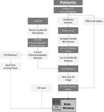
‘‘Visualization Tool Box’’ (VTK), a software system for 3D computer graphic in C++language, was programmed for slicing of the 4D volumes. Proprietary DICOM 4D volumes obtained are first converted to standard DICOM 4D volumes using Philips Q-Lab software and subsequently to sets of DICOM 3D volumes using Matlab software. 3D volumes go through VTK pipeline1 and are sliced and shown on the Main Window of the display. Simultaneously, sensor location and orientation are tracked through VTK pipeline2 which control the motion of cutting planes that are displayed in the side window of the display (Figure 1and 3).
The DICOM volumes were sliced through Roll, Pitch and Yaw orientations of the 6DOF sensor incorporated into the dummy transducer resulting in continuous real time echocar- diographic images displayed on the main window of the com- puter screen. When the sector scans of interest are displayed on the monitor, by activating the appropriate short cuts, their orientation parameters are used to initiate display of video clips of associated color flow Doppler, spectral Doppler or M-mode (Figure 4 and 5).
III. RESULTS
The simulator consists of the following three main compo- nents: a 2D echocardiography display, a 3D heart model with cutting planes and a sector-based color and spectral Doppler and M-mode display.
FIGURE 1. Virtual Neonatal Echocardiographic Training Simulator.
FIGURE 2. Hardware components of VNETS.
A. TWO-DIMENSIONAL ECHOCARDIOGRAPHY DISPLAY For each infant whose echocardiographic data were utilized, six or seven 4D DICOM echocardiographic volumes are virtually placed and oriented in their respective five echocar- diographic acoustic windows specific to each patient. In addi- tion, an image volume can be placed in the right apical position if patients with dextrocardia were imaged. The 4D volumes are arranged so that one 4D volume is placed at the Apical, Left and Right Parasternal windows and two 4D vol- umes are placed at the Subcostal and Suprasternal windows.
The use of multiple volumes allows for the complete range of 2D imaging from each infant.
B. 3D HEART MODEL WITH CUTTING PLANES
For visualization of how sector cuts are generated, a 3D heart model on the side screen of the computer duplicates the motion of the fan shaped cutting planes from each acoustic window. The motion of the cutting plane is controlled by 6DOF sensor incorporated in the dummy probe. The orien- tation of the cutting planes parallels sector cuts displayed on the main screen of the simulator. A small arrow indicates the direction of the motion of the cutting planes which is also indicated on the marker of the probe (Figure 6).
FIGURE 3. Software structure of VNETS.
FIGURE 4. Slicing of 4D dataset and display of video loops of color Doppler, spectral Doppler and M-mode from coordinates of 26 specific cuts of the 4D dataset.
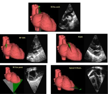
FIGURE 5. Approximate locations of the acoustic windows are displayed.
Typical two dimensional and color flow images are demonstrated.
C. SECTOR-BASED COLOR FLOW AND SPECTRAL DOPPLER AND M-MODE DISPLAY
Color flow Doppler can be displayed from each of the 27 spe- cific cuts obtained through the 5 echocardiographic windows.
Spectral Doppler display may be obtained from flow through
FIGURE 6. The cutting planes of the three-dimensional heart displayed in the side screen demonstrate the typical images obtained from the five acoustic windows.
aortic, pulmonary, tricuspid and mitral valves, superior vena cava (SVC), patent foramen ovale and patent ductus arterio- sus and from any other abnormal flows detected on color flow study. M-mode measurements may be obtained from Parasternal long and short axes.
D. VNETS AS A TRAINING ECHOCARDIOGRAPHIC SIMULATOR
As the simulator was designed for hands-on training pur- poses, it has the capability to simulate actual echocardiogra- phy including image acquisition via the use of the probe in the standard acoustic windows. All essential echocardiographic modalities can be simulated, including 2D, color flow, spec- tral Doppler and M-mode imaging along with hemodynamic measurements and report generation.
1) COMPLETE 2D ECHOCARDIOGRAPHY
A complete range of echocardiographic images is obtained from the following windows on the mannequin: Paraster- nal window, Apical window, Subcostal window, Right parasternal window and Suprasternal window. Continuous two-dimensional images can be obtained from all windows and optimized for up to 27 standard echocardiographic views.
Using 2D echocardiography, the dimension of cardiac struc- tures can be measured (Figure 7) and cardiac defects present in congenital heart disease can be detected. In addition, 2D echocardiography can be used for evaluation of left and right ventricular function. By tracing endocardial surface of left ventricle at end diastole and end systole from apical 4 cham- ber view and by use of modified Simpson’s algorithm, ejec- tion fraction of the left ventricle can be measured. Similarly by measuring the area of right ventricle in end diastole and end systole, the right ventricular function can be estimated.
Measurement of the area of left atrium at end systole in apical
FIGURE 7. M-mode echocardiography using VNETS. Panel A: M-mode of left ventricle for anatomical and functional measurements.
Panel B: Aortic blood flow measured from aortic spectral Doppler tracing and the aortic diameter.
4 chamber view allows for a better evaluation of left atrial dimension.
2) COLOR FLOW ECHOCARDIOGRAPHY
Normal and abnormal color flow Doppler from all standard echocardiographic views can be visualized and investigated.
Particular attention is paid to any left to right shunt though the atrial/ventricular septa, or through a patent ductus arterio- sus. Regurgitant flows across the mitral, tricuspid, aortic and pulmonary valves, as well as turbulent flow through stenotic aortic and pulmonary valves may be evaluated. Careful eval- uation of color flow Doppler in addition to the twelve to seventeen 2D echocardiographic images will be sufficient to suspect presence of most congenital cardiac defects for trainees in point of care echocardiography. Mastery and care- ful evaluation of 26 2D cuts with color flow Doppler will be sufficient to lead to a detailed diagnosis of most congenital cardiac defects for trainees in pediatric cardiology and pedi- atric cardiology technologists provided that appropriate 2D and color flow images were recorded from the index patient for the simulator.
3) SPECTRAL DOPPLER ECHOCARDIOGRAPHY
Spectral Doppler can be used to display normal flow profile through normal vasculature as well as abnormal flow profiles through stenotic or incompetent heart valves, stenotic ves- sels or defects in interatrial or interventricular septa. Doppler velocity profiles can be accessed from Apical 5 chamber view for aortic flow measurement (Figure 7), from the main pulmonary artery for pulmonary flow, from Subcostal coronal or sagittal bicaval for superior vena cava flow measurement.
Similarly, Doppler tracings of the tricuspid flow profiles can be assessed from Apical 4 chamber view and from Paraster- nal long axis right ventricular inflow view for inflow and regurgitant flow profile for assessment of pulmonary arterial pressure. Flow through the ductus arteriosus can be assessed
from suprasternal ductal cut or from parasternal short axis base or parasternal long axis outflow for direction of flow and duration of right to left and left to right shunt through a patent ductus arteriosus. Flow through the foramen ovale may be evaluated from the subcostal coronal posterior or subcostal sagittal bicaval view for its direction and velocity. Finally, other abnormal stenotic or regurgitant flows detected by color Doppler can be further investigated by pulse or continuous wave Doppler.
4) M-MODE ECHOCARDIOGRAPHY
M-mode tracing are obtained from parasternal long axis or parasternal short axis at the level of the mitral valve leaflets for right and left ventricular sizes, function and wall thickness, and from parasternal long and short axis at the base of the heart for measurement of aortic root and left atrial diameters (Figure 7). M-mode from other areas of the heart may be obtained as desired.
5) CASE STUDIES AND REPORT GENERATION
Although new cases are added to the simulator in an ongo- ing basis, presently we have stored complete echocardio- grams of 30 cases for training purposes. They include normal term and preterm infants as well as neonates with func- tional or congenital cardiac defects. Each simulator case takes up 2.1-2.2 gigabytes of hard drive memory. Therefore, 100 to 400 cases can be stored on computers with a hard drive memory of 250 gigabytes to 1 terabytes. The trainee is able to record 2D echocardiography images for evaluation by the instructor. The integrated calculation package allows the trainee to obtain a full range of echocardiographic mea- surements that are automatically transcribed to the report.
For each case, the trainee is able to generate a neonatal echocardiography report describing his/her impression of the case.
E. EVALUATION OF THE SIMULATOR
We have been using the simulator to teach the echocardiog- raphy skills to neonatologists and neonatology fellows in our annual echocardiography course. In 2017, eleven participants (3 neonatologists and 8 neonatology fellows) in the course with no experience in echocardiography were tested on the simulator after 4 hours of didactic session on topographical anatomy of the heart and standard echocardiographic views.
Each participant was given 20 minutes to obtain and save all the 26 standard views. After the hands-on training on the simulator for 6 hours over 3 days, they were tested again.
All the pre- and post-training obtained images were blindly reviewed by one of the instructors. Each image was scored from 0 to 3 based on the quality. The number of echocardio- graphic views obtained, the quality of acquired images, and the duration of time to complete the task before and after the hands-on training was compared using Wilcoxon signed-rank test. After hands-on training, the number of echocar- diographic views obtained (from median [interquartile]
22 [15, 25] to 27 [26, 27], p=0.0033) and the score of the
quality of acquired image (23.5 [14.5, 29] to 37.5 [33, 50.5], p=0.0033) increased and the time spent in completing the task decreased (18 [17, 20] to 12 [12, 18], p = 0.0074).
We are currently in the process of formally studying the effectiveness of the simulator in teaching echocardiography.
IV. DISCUSSION
The use of simulators to aid in acquiring specific skills dates back to 1922 when Edward Link presented his home-made flight simulator, which has been subsequently widely used both in military and civilian aviation [11]. Medical appli- cation of simulation training was introduced in the early 1960s with the invention of Resusci-Anne for resuscitation training [12]. The appearance of minimally invasive surgi- cal procedures in the early 1990s gave further impetus for the development of simulators. In order to reduce the steep learning curve required for performing complex procedures, simulators were used in multiple surgical and medical spe- cialties [13], [14]. Among others, simulation has been used for training in endoscopic [15] and interventional cardiol- ogy procedures [16], to train and evaluate anesthesiologists’
responses to critical incidents [17], in trauma training [18]
and craniomaxillofacial surgery [19].
Acquiring the skills to perform echocardiography requires both medical knowledge and manual dexterity [20]. Tradi- tionally training has been done on patients or volunteers under supervision. The training is arduous, the access to patients is limited and, until recently, it was restricted mainly to cardiac sonographers and cardiologists. However, the need to obtain noninvasive information about cardiac function in anesthesia, emergency medicine and intensive care has resulted in the development of point-of-care echocardiography training pro- grams, which have been struggling to meet the demand [21].
Specific training guidelines have been published for adult and pediatric cardiologists [22]–[24], anesthesiologists [25], [26], emergency care physicians [27]–[29] and neonatologists [30].
However, as the training programs are resource intensive and logistical barriers, including limited access to appropriate subjects for training purposes exist, they have not yet been widely adopted.
The recent emergence of high fidelity, mannequin based echocardiography simulators are having a major effect on the training echocardiography for older child and adults.
Use of real 4D echocardiographic volume datasets, i.e. 4D echocardiography volumes obtained directly from patients, was first introduced by Weidenbach and colleagues [31].
With this approach, either a single or multiple sub-volume are incorporated into a single 3D volume, which when sliced, generates real, 2D images. However, since these volumes are recorded from the Apical or Subcostal acoustic windows, imaging from Parasternal and Suprasternal acoustic windows is rendered suboptimal.
Indeed, the use of real 4D volumes for transthoracic echocardiography (TTE) in older children and adults has been hampered by poor image quality. Therefore, almost all com- mercially available TTE simulators rely on rendered images
obtained from other imaging platforms such as computed tomography or magnetic resonance scans and are modified to simulate echocardiograms. The images obtained this way are sharp and well delineated with full range of echocardio- graphic modalities. These simulators are being extensively used for training in point-of-care echocardiography in older children and adults, however are not available for neonates and infants.
There is increasing evidence in neonatal medicine, that bedside echocardiography is valuable in helping the physi- cian to make the correct diagnosis earlier and its use influ- ences treatment decisions for critically ill neonates [32].
Although there is great interest in training neonatologists in point-of-care echocardiography [33], [34], organizing an effective training program has been hampered by the signifi- cant logistical difficulties and ethical considerations in gain- ing access to neonates for training purposes. In addition, there is a limited number of qualified trainers interested in train- ing point-of-care echocardiography to neonatologists. Under these circumstances, an effective simulator for transthoracic echocardiography developed for the neonate can significantly contribute to providing efficient training and skill mainte- nance. For this, a mannequin-based simulator is required as it has the potential to provide the haptic experience of appropriately manipulating the transducer on the chest of the mannequin to obtain the desired images. The only other TTE simulator based on real 4D image volumes available for neonates and young infants is restricted to 2 dimensional echocardiography [35]. Furthermore, since it only uses 4D volumes recorded from Apical or Subcostal acoustic win- dows, imaging from parasternal and surprasternal windows become suboptimal. In addition, it lacks color flow Doppler, Spectral Doppler and M-mode modalities which are essential for hemodynamic measurements and thus for the provision of comprehensive training in point-of-care echocardiography in neonates.
Our hybrid transthoracic echocardiography simulator is specifically designed for neonates and young infants. It uses echocardiography images obtained in neonates from five transthoracic acoustic windows. Multiple real 4D volume datasets are arrayed in a fashion to cover all acoustic windows specific for each neonate. Real time 2D imaging may be per- formed from all acoustic windows. In addition, from specific 3D slices, 2D video clips of color flow and spectral Doppler and M-mode can be accessed providing a complete range of echocardiographic experience. The integrated hemodynamic measurement and reporting module allows for a complete range of anatomical and functional measurements along with report generation capability.
REFERENCES
[1] Y. Beaulieu, ‘‘Specific skill set and goals of focused echocardiography for critical care clinicians,’’Crit. Care, vol. 35, no. 5, pp. S144–S149, 2007.
[2] N. Evans, ‘‘Echocardiography on neonatal intensive care units in Aus- tralia and New Zealand,’’J. Paediatrics Child Health, vol. 36, no. 2, pp. 169–171, 2000.
[3] B. Cowie, ‘‘Focused cardiovascular ultrasound performed by anesthesi- ologists in the perioperative period: Feasible and alters patient manage- ment,’’J. Cardiothoracic Vascular Anesthesia, vol. 23, no. 4, pp. 450–456, 2009.
[4] B. P. Cholley, A. Vieillard-Baron, and A. Mebazaa, ‘‘Echocardiography in the ICU: Time for widespread use!’’Intensive Care Med., vol. 32, no. 1, pp. 9–10, 2006.
[5] R. Mayron, F. E. Gaudio, D. Plummer, R. Asinger, and J. Elsperger,
‘‘Echocardiography performed by emergency physicians: Impact on diag- nosis and therapy,’’ Ann. Emerg. Med., vol. 17, no. 2, pp. 150–154, 1988.
[6] M. Weidenbachet al., ‘‘Simulation of congenital heart defects—A novel way of training echocardiography,’’Heart, vol. 95, no. 8, pp. 636–641, 2009.
[7] R. Bose et al., ‘‘Transesophageal echocardiography simulator: A new learning tool,’’ J. Cardiothoracic Vascular Anesthesia, vol. 23, no. 4, pp. 544–548, 2009.
[8] R. Matyalet al., ‘‘Transthoracic echocardiographic simulator: Normal and the abnormal,’’J. Cardiothoracic Vascular Anesthesia, vol. 25, no. 1, pp. 177–181, 20ll.
[9] M. Weidenbach et al., ‘‘Computer-based training in two-dimensional echocardiography using an echocardiography simulator,’’J. Amer. Soc.
Echocardiogr., vol. 18, no. 4, pp. 362–366, 2005.
[10] O. Shakil, F. Mahmood, and R. Matyal, ‘‘Simulation in Echocardiogra- phy: An ever-expanding Frontier,’’J. Cardiothoracic Vascular Anesthesia, vol. 23, no. 3, pp. 476–485, 2012.
[11] K. R. Rosen, ‘‘The history of medical simulation,’’J. Crit. Care, vol. 23, no. 2, pp. 157–166, 2008.
[12] A. Grenvik and J. Schaefer, ‘‘From Resusci-Anne to Sim-Man: The evolution of simulators in medicine,’’Crit. Care Med., vol. 32, no. 2, pp. S56–S57, 2004.
[13] A. Cushieri, ‘‘Technology for minimal access surgery,’’Brit. Med. J., vol. 319, no. 7220, pp. 1304–1305, 1999.
[14] R. M. Satava, ‘‘Accomplishments and challenges of surgical simulation:
Dawning of the next-generation surgical education,’’Surgical Endoscopy, vol. 15, no. 3, pp. 232–241, 2001.
[15] C. B. Williams, J. Baillie, D. F. Gillies, D. Borislow, and P. B. Cotton,
‘‘Teaching gastrointestinal endoscopy by computer simulation: A proto- type for colonoscopy and ERCP,’’Gastrointestinal Endoscopy, vol. 36, no. 1, pp. 49–54, 1990.
[16] S. Cotinet al., ‘‘ICTS, an interventional cardiology training system,’’Stud Health Technol. Inf., vol. 70, pp. 59–65, 2000.
[17] H. A. Schwid and D. O’Donnell, ‘‘The anesthesia simulator-recorder:
A device to train and evaluate anesthesiologists’ responses to critical incidents,’’Anesthesia, vol. 72, no. 1, pp. 191–197, 1990.
[18] C. Kaufmann and A. Liu, ‘‘Trauma training: Virtual reality applications,’’
Stud. Health Technol. Inf., vol. 81, pp. 236–241, 2001.
[19] T. Liebregts, T. Xi, R. Schreurs, B. van Loon, S. Bergé, and T. Maal,
‘‘Three-dimensional virtual simulation of alar width changes following bimaxillary osteotomies,’’Int. J. Oral Maxillofacial Surg., vol. 45, no. 10, pp. 1315–1321, 2016.
[20] D. Ehleret al., ‘‘Guidelines for cardiac sonographer education: Recom- mendations of the American society of echocardiography sonographer training and education committee,’’J. Amer. Soc. Echocardiogr., vol. 14, no. 1, pp. 77–84, 2001.
[21] Y. Beaulieu, ‘‘Bedside echocardiography in the assessment of the critically ill,’’Crit. Care Med., vol. 35, no. 5, pp. S235–S249, 2007.
[22] S. P. Sanderset al., ‘‘ACCF/AHA/AAP recommendations for training in pediatric cardiology. Task force 2: Pediatric training guidelines for noninvasive cardiac imaging endorsed by the American society of echocar- diography and the society of pediatric echocardiography,’’J. Amer. College Cardiol., vol. 46, no. 7, pp. 1384–1388, 2005.
[23] P. H. Mayoet al., ‘‘American college of chest physicians/La Société de Réanimation de Langue Française statement on competence in critical care ultrasonography,’’Chest, vol. 135, no. 4, pp. 1050–1060, 2009.
[24] S. Scrivastavaet al., ‘‘Task force 2: Pediatric cardiology fellowship training in noninvasive cardiac imaging. SPCTPD/ACC/AAP/AHA,’’Circulation, vol. 132, no. 6, pp. e57–e67, Aug. 2015
[25] J. S. Shanewiseet al., ‘‘ASE/SCA guidelines for performing a comprehen- sive Intraoperative multiplane transesophageal echocardiography exami- nation: Recommendations of the American society of echocardiography council for intraoperative echocardiography,’’Anesthesia Anal., vol. 89, no. 4, pp. 870–884, Oct. 1999.
[26] R. T. Hahnet al., ‘‘Guidelines for performing a comprehensive trans- esophageal echocardiographic examination: Recommendations from the American society of echocardiography and the society of cardiovas- cular anesthesiologists,’’ J. Amer. Soc. Echocardiogr., vol. 26, no. 9, pp. 921–964, 2013.
[27] W. J. Stewartet al., ‘‘Echocardiography in emergency medicine: A policy statement by the American society of echocardiography and the Ameri- can college of cardiology. Task force on echocardiography in emergency medicine of the American society of echocardiography and the echocardio- graphy and technology and practice executive committees of the American college of cardiology,’’J. Amer. Collage Cardiol., vol. 33, no. 2, pp. 586–
588, Feb. 1999.
[28] S. Priceet al., ‘‘Peri-resuscitation echocardiography: Training the novice practitioner,’’Resuscitation, vol. 81, no. 11, pp. 1534–1539, 2010.
[29] A. N. Neskovic et al., ‘‘Emergency echocardiography: The European association of cardiovascular imaging recommendations,’’Eur. Heart J.
Cardiovascular Imag., vol. 14, no. 4, pp. 1–11, 2013.
[30] M. Weidenbachet al., ‘‘Augmented reality simulator for training in two- dimensional echocardiography,’’Comput. Biomed. Res., vol. 33, no. 1, pp. 11–22, 2000.
[31] M. Weidenbachet al., ‘‘EchoComTEE—A simulator for transoesophageal echocardiography,’’Anaesthesia, vol. 62, no. 4, pp. 347–357, 2007.
[32] A. Sehgal and P. J. McNamara, ‘‘Does point-of-care functional echocar- diography enhance cardiovascular care in the NICU?’’J. Perinatol, vol. 28, pp. 729–735, Jul. 2008.
[33] R. Wagneret al., ‘‘Effectiveness of simulator-based echocardiography training of noncardiologists in congenital heart diseases,’’Echocardiog- raphy, vol. 30, no. 6, pp. 693–698, 2013.
[34] J. Nguyen, R. Amirnovin, R. Ramanathan, and S. Noori, ‘‘The state of point-of-care ultrasonography use and training in neonatal-perinatal medicine and pediatric critical care medicine fellowship programs,’’J.
Perinatol., vol. 36, no. 11, pp. 972–976, Nov. 2016.
[35] ECHOCOM—Training Simulator for Echocardiography in Neonates.
[Online]. Available: https://www.echocom.de