UNCORRECTED
PROOF
1 23
4
Intestinal Autotransplantation Using PACAP-38-Containing
5
Preservation Solution
6 Klara Nedvig&Gyorgy Weber&Jozsef Nemeth&
7 Krisztina Kovacs&Dora Reglodi&Agnes Kemeny&
8 Andrea Ferencz
9 Received: 23 March 2012 / Accepted: 31 July 2012 10 #Springer Science+Business Media, LLC 2012 11
12 Abstract Small bowel is one of the most sensitive organs to 13 ischemia–reperfusion injury, which is a significant pro- 14 blem during transplantation. Pituitary adenylate cyclase- 15 activating polypeptide (PACAP) has cytoprotective effect 16 in ischemic injuries of various tissues. The aim of our study 17 was to measure changes of PACAP-38 and PACAP-27 18 immunoreactivities and cytokine levels in intestinal grafts 19 stored in PACAP-38-containing preservation solution.
20 Small bowel autotransplantation was performed on male 21 Wistar rats. Grafts were stored in University of Wisconsin 22 (UW) solution at 4 °C for 1 h (group (G)I), for 3 h (GII), and
23 for 6 h (GIII) and in PACAP-38-containing UW solution for
24 1 h (GIV), for 3 h (GV), and for 6 h (GVI). After preserva-
25 tion, performing vessel anastomosis reperfusion began,
26 which lasted 3 h in each group. Tissue biopsies were col-
27 lected after laparotomy (control) and at the end of the
28 reperfusion periods. Intestinal PACAP-38 and PACAP-27
29 immunoreactivities were measured by radioimmunoassay.
30 To measure cytokine array from tissue homogenates, we
31 used rat cytokine array and Luminex Multiplex Immunoas-
32 say. Levels of PACAP-38 and PACAP-27 immunoreactivity
33 decreased after 1 and 3 h preservation compared to control
34 levels. This decrease was significant following 6 h cold storage (p<0.05). Values remained significantly higher in 35
36 grafts stored in PACAP-38-containing UW. Cytokine array
37 revealed that expression of the soluble intercellular adhesion
38 molecule-1 (CD54) and L-selectin (CD62L/LECAM-1) was
39 increased in GIII. Both 6 h cold storage in PACAP-38-
40 containing UW solution and 3 h reperfusion caused strong
41 reduction in these cytokines activation in GVI. RANTES
42 (CCL5) levels were increased in all groups. Strong activa-
43 tion of the tissue inhibitor of metalloproteinase-1 was in
44 GIII. However, PACAP-38-containing cold storage could
45 decrease its activation in GVI. Furthermore, strong activa-
46 tion of the tissue inhibitor of metalloproteinase-1 was
47 detected in 6 h preserved grafts without PACAP-38 (GIII).
48 PACAP-38-containing cold storage could decrease its acti-
49 vation in GVI. Our present study showed that PACAP-38
50 and PACAP-27 immunoreactivities decreased in a time-
51 dependent manner during intestinal cold preservation,
52 which could be ameliorated by administration of exogenous
53 PACAP-38 to the preservation solution. Moreover, PACAP-
54 38 could attenuate tissue cold ischemic injury-induced
55 changes in cytokine expression.
56 Keywords Small bowel . Transplantation . PACAP-38 .
57 PACAP-27 . Cytokine
K. Nedvig
:
G. WeberDepartment of Surgical Research and Techniques, University of Pecs, Pecs, Hungary
Q1
G. Weber
Department of Surgical Research and Techniques, Semmelweis University, Budapest, Hungary J. Nemeth
Department of Pharmacology and Pharmacotherapy, University of Debrecen, Debrecen, Hungary K. Kovacs
Department of Biochemistry and Medical Chemistry, University of Pecs, Pecs, Hungary
D. Reglodi
Department of Anatomy,
“PTE-MTA Lendulet PACAP Research Group”, University of Pecs, Pecs, Hungary
A. Kemeny
Department of Pharmacology and Pharmacotherapy, University of Pecs, Pecs, Hungary
A. Ferencz (*)
Department of Surgical Research and Techniques,
Faculty of Medicine, Semmelweis University, Nagyvarad sqr. 4, 1089 Budapest, Hungary
e-mail: andrea.ferencz@gmail.com
UNCORRECTED
PROOF
58 Introduction
59 Small bowel is a highly sensitive tissue to ischemia/reperfu- 60 sion (I/R) injury in the body. Intestinal I/R injury is caused 61 by many clinical conditions, including small bowel trans- 62 plantation. Both clinical and experimental data demonstrate 63 that transplant I/R injury has deleterious short- and long- 64 term effects, manifesting as increased episodes of acute 65 rejection and chronic allograft dysfunction (Ferencz et al.
66 2002,2010a,b; Linfert et al.2009; Yuan et al.2011). Graft 67 viability prior to implantation is a key factor in the outcome 68 after organ transplantation. Along with surgical manipula- 69 tion, I/R injury and preservation damage are some of the 70 many essential factors that affect the quality of intestinal 71 graft and its multiple functions. The current standard in 72 organ preservation with University of Wisconsin (UW) so- 73 lution was developed for kidney/liver preservation and it is 74 suboptimal for the intestinal graft despite good results for 75 other organs (Maathuis et al.2007; Roskott et al.2011).
Q2 The
76 benefit of the UW solution for the preservation of other 77 intraabdominal organs remains unclear and the maximum 78 storage time for small bowel remains relatively brief 79 (6–8 h). Thus, no general agreement exists about optimal 80 preservation solution for intestinal grafts so far (Kokotilo 81 et al.2010; Roskott et al.2011). Recently, there are contin- 82 uous research efforts to modify the commercially available 83 solutions (adding more components, high-energy intermedi- 84 ates, and nutrients) or to develop new preservation solutions 85 (Inuzuka et al.2007; Wei et al.2007; Ferencz et al. 2009;
86 Yandza et al.2011).
87 Pituitary adenylate cyclase-activating polypeptide 88 (PACAP) is a widespread neuropeptide with diverse effects 89 not only in the nervous system but also in the cardiovascular 90 system and peripheral organs including endocrine glands, 91 respiratory organs, and gastrointestinal tract. The polypep- 92 tide exists in two forms, with 38 and 27 amino acid residues, 93 named PACAP-38 and PACAP-27 (Vaudry et al. 2009).
94 Endogenous PACAP-38 and PACAP-27 were demonstrated 95 in all parts of the gastrointestinal tract with high levels 96 detected in the jejunum and ileum. PACAP-38 and 97 PACAP-27-immunoreactivities have been shown in the cell 98 bodies and nerve fibers throughout the gastrointestinal tract 99 (Hannibal et al. 1998). PACAP-38 and PACAP-27 act 100 through the specific PAC1 receptor and VPAC1 and VPAC2 101 receptors that bind VIP and PACAP with equal affinity 102 (Koves et al.1993; Vaudry et al.2009). All three types of 103 PACAP receptors have been shown in the intestinal system:
104 in the mucosa and myenteric neurons, in neuroendocrine 105 cells, blood vessels, and smooth muscle (Schulz et al.2004;
106 Pirone et al.2011).
107 Endogenous PACAP-38 has been implicated in protec- 108 tion against harmful stimuli; the peptide has anti-apoptotic, 109 anti-inflammatory, and anti-oxidant actions in numerous
110 different in vivo and in vitro models (Ferencz et al.2010a;
111 Horvath et al. 2010a; Gasz et al. 2006; Racz et al. 2006,
112 Q3
2010; Reglodi et al.2011,2012; Roth et al.2009). Recently, 113 in warm and cold I/R small bowel models, the in vivo
114 protective effects of PACAP-38 have been shown. These
115 experiments confirmed an important protective effect of
116 endogenous PACAP-38 against warm I/R tissue damage.
117 Moreover, it has been shown that adding exogenous
118 PACAP-38 to UW solution prevented the oxidative stress
119 and tissue structure injury in rat small intestine (Ferencz
120 et al.2010a,b). Although the exact mechanisms, by which
121 intestinal I/R and gut injury contribute to the systemic
122 inflammatory response, are not completely known, cyto-
123 kines play a key role in these actions. There is also evidence
124 that intestinal injury resulting from I/R can lead to the bowel
125 becoming a cytokine-generating organ (Grotz et al. 1999).
126 The anti-inflammatory actions of PACAP in several inflam-
127 matory models are partially mediated through its suppress-
128 ing effect on cytokine/chemokine production (Delgado and
129 Ganea 2001; Horvath et al. 2010b; Ohtaki et al. 2010).
130 However, there are no data in the literature about changes
131 of intestinal PACAP-38 or PACAP-27 and tissue cytokine
132 levels using PACAP-38-containing preservation solution in
133 stored and transplanted small bowel grafts. The aim of our
134 study was to measure changes of PACAP-38 and PACAP-
135 27 immunoreactivities and cytokine values using PACAP-
136 38-containing UW preservation solution during intestinal
137 autotransplantation.
138 Materials and Methods
139 Animals
Adult male Wistar rats (250–300 g,n035) were purchased 140 141 from the Laboratory Animal Center of University of Pecs,
142 housed under pathogen-free conditions, and were fasted for
143 24 h preoperatively but had free access to water. Rats were
144 anesthetized with intramuscular ketamine hydrochloride
145 (0.075 mg/g of body weight) and diazepam (0.075 mg/g of
146 body weight; Richter Gedeon, Budapest, Hungary). All
147 procedures were performed in accordance with the ethical
148 guidelines of National Institute of Health and guidelines
149 approved by the University of Pecs (BA02/2000-9/2008)
150 to minimize pain and suffering of the animals.
151 Intestinal Autotransplantation Model
152 Rats were randomly divided into groups (G). In group S,
153 animals underwent only median laparotomy (sham operated, n05). In GI–GVI after heparin administration (0.2 U/g), 154
155 small bowel was resected to descending colon, and the
156 lumen was flushed with normal saline. Grafts were perfused
UNCORRECTED
PROOF
157 by the superior mesenteric artery and were preserved in 158 100 ml 4 °C University of Wisconsin solution (ViaSpan, 159 DuPont Pharma, Bad Homburg, Germany) for 1 h in GI (n0 160 5), for 3 h in GII (n05), and for 6 h in GIII (n05). Grafts 161 were preserved in 100 ml UW solution containing 100μg 162 PACAP-38 dissolved in 2 ml of physiological saline (Sigma, 163 Hungary) for 1 h in GIV (n05), for 3 h in GV (n05), and for 164 6 h in GVI (n05). After preservation, end-to-end anastomo- 165 sis was performed between the stumps of mesenteric vessels 166 with microvascular technique. Reperfusion lasted 3 h in 167 each group. Small bowel biopsies were collected after lap- 168 arotomy (control, C) and at the end of the reperfusion 169 periods.
170 Radioimmunoassay
171 Intestinal tissue samples (600 mg) were homogenized in ice- 172 cold distilled water. After centrifugation at 12,000 rpm/4 °C 173 for 30 min, the supernate was further processed for RIA 174 analysis of PACAP-38- and PACAP-27-like immunoreac- 175 tivity, as previously described (Brubel et al.2011). Briefly, 176 the antiserum for PACAP-38 was “88111-3” and for 177 PACAP-27 was“88123”. The tracer was mono-125I-labeled 178 ovine PACAP 24-38 and mono-125I-labeled ovine PACAP- 179 27 (5,000 cpm/tube). The standard was ovine PACAP-38 180 and PACAP-27 (0–1,000 fmol/ml). Assays were prepared in 181 1 ml phosphate buffer (0.05 mol/l, pH 7.4) containing 0.1 M 182 NaCl, 0.05 % NaN3, and 0.25 % bovine serum albumin. The 183 assay procedures include 100μl antiserums (working dilu- 184 tions—PACAP-38“88111-3”antiserum, 1:10,000; PACAP- 185 27“88123”antiserum, 1:45,000), 100μl RIA tracers, and 186 100μl standards or unknown samples measured into poly- 187 propylene tubes with assay buffer. After 48–72 h incubation 188 at 4 °C, the antibody-bound peptides were separated from 189 the free ones by addition of 100μl separation solution (10 g 190 charcoal, 1 g dextran, and 0.2 g commercial fat-free milk 191 powder in 100 ml distilled water). Following centrifugation 192 (3,000 rpm for 20 min at 4 °C), the tubes were gently 193 decanted, and the radioactivity of the precipitates was mea- 194 sured in a gamma counter. PACAP-38 and PACAP-27 con- 195 centrations of the unknown samples were read from the 196 appropriate calibration curves. Results of PACAP-38- and 197 PACAP-27-like immunoreactivities are given as femto- 198 moles per milligram tissue.
199 Cytokine Array After Small Bowel Autotransplantation 200 Intestinal tissues from control bowel sample (A), from tissue 201 exposed to 6 h cold storage in UW (B), from 6 h cold preser- 202 vation in PACAP-38-containing UW solution (C), and subse- 203 quent 3 h reperfusion period (D) were measured as previously 204 described (
Q4 Horvath et al. 2011). Briefly, cytokine array from 205 tissue homogenates was performed using rat cytokine array
206 (Panel A Array kit from R&D Systems, Biomedica Hung.,
207 Budapest, Hungary). Small bowel samples were excised then
208 homogenized in PBS with protease inhibitors. Triton X-100
209 was added to the final concentrations of 1 %. The samples were
210 stored at−80 °C prior to use. After blocking the array mem-
211 branes for 1 h and adding the reconstituted Detection Antibody
212 Cocktail for another 1 h at room temperature, the membranes were incubated with 1 ml of tissue homogenates at 2–8 °C 213
214 overnight on a rocking platform. After washing with buffer for
215 three times and addition of horseradish peroxidase-conjugated
216 streptavidin to each membrane, we exposed them to a chemi-
217 luminescent detection reagent (Amersham Biosciences,
218 Hungary) then side up to an X-ray film cassette.
219 Luminex Multiplex Immunoassay
220 The levels of three host markers (soluble intercellular adhesion
221 molecule-1 (sICAM1), L-selectin, and metalloproteinase-1
222 (TIMP-1)) were determined in the selected bowel samples
223 (see in cytokine array) using customized Flurokine MAP Rat
224 Base Kit (R&D Systems). This was done according to the
225 manufacturer's instructions (R&D Systems). Following previ-
226 ous optimizations, all samples were tested undiluted, in a
227 blinded manner. All analyte levels in the quality control
228 reagents of the kits were within the expected ranges. Standard curve for sICAM-1 is 17–12,500 pg/ml, for L-selectin is 229 100–73,000 pg/ml, and for TIMP-1 is 55–40,600 pg/ml. Meas- 230
231 urements were done with Luminex100 instrument, and Lumi-
232 nex 100 IS software was used for the analysis of bead median
233 fluorescence intensity. The R&D Systems Rat Base kit assay
234 was carried out according to the manufacturer's instructions,
235 with a few exceptions as stipulated below. Briefly, an eight-
236 point standard curve was generated by performing serial dilu-
237 tions of the reconstituted normalized standard (lot # 1279612).
238 This was done in order to ensure that the matrix used in the
239 generation of the standard curve resembled that of the samples
240 as closely as possible as preliminary test showed that this
241 method was superior to dilution of standards in standard dilu-
242 ent (data not shown). Bowel samples were homogenized with
243 RPMI-1640 (GIBCO) containing 1 % protease inhibitor cock-
244 tail. In order to assess recovery, bowel samples were used in
245 20 mg/ml concentrations. The assays were run in duplicate,
246 which produced in total of six concentration replicates. A 50-μl volume of each sample, control, or standard was added 247
248 to a 96-well plate (provided with the kit) containing 50μl of
249 antibody-coated fluorescent beads. Biotinylated secondary and
250 streptavidin-PE antibodies were added to the plate with alter-
251 nate incubation and washing steps. After the last wash step,
252 100μl of wash buffer was added to the wells; the plate was
253 incubated and read on the Luminex100 array reader, using a
254 four-PL regression curve to plot the standard curve. Data were
255 subsequently analyzed using the Luminex100 manager
256 software.
UNCORRECTED
PROOF
257 Statistics
258 Results are expressed as mean values ± SEM. Data were 259 analyzed with one-way analysis of variance. The level of 260 significance was set atp<0.05. The MicroCal Origin (ver.
261 6.0) program (Microcal Software Inc, Northampton, USA) 262 was used for data evaluation.
263 Results
264 Radioimmunoassay
265 Level of intestinal PACAP-38-like immunoreactivity (LI) was 266 55.1±2.5 fmol/mg in sham-operated group and it was 57.32±
267 3.5 fmol/mg in control samples. After 1 h cold storage, intes- 268 tinal PACAP-38-LI was 50.4±3.5 fmol/mg (GI), and after 3 h 269 preservation it decreased to 40.1±5.5 fmol/mg (GII). These 270 changes were significant following 6 h cold storage (GIII, 271 32.6±3.0 fmol/mg; p<0.05) compared to control or sham 272 values. Levels remained significantly higher in grafts stored 273 in PACAP-38-containing UW solution (GIV–GVI). In GIV, 274 levels (65.2±3.4 fmol/mg) increased above the control values, 275 which was statistically significant. After 3 and 6 h cold storage 276 in PACAP-38-containing preservation solution, the PACAP- 277 38-LI levels were 55.6 ± 4.2 fmol/mg (GV) and 48.9 ± 278 3.2 fmol/mg (GVI). These resulted significantly higher com- 279 pared to preservation only in UW without PACAP-38 (vs. GII 280 and GIII;p<0.05) (Fig.1).
281 Tissue PACAP-27 level also decreased during cold stor-
282 age and autotransplantation procedure compared to the con-
283 trol value (4.2±0.2 fmol/mg). This decrease was significant
284 in the 1 h (GI, 2±0.2 fmol/mg; p<0.05), 3 h (GII, 1.6±
285 0.3 fmol/mg; p<0.05), and 6 h (GIII, 0.9±0.2 fmol/mg;
p<0.01) groups. Levels of PACAP-27-LI remained signifi- 286 287 cantly higher in grafts stored in PACAP-38-containing UW solution (GIV–GVI) at the end of the reperfusion periods. In 288
289 GIV, its concentration was 3.5±0.3 fmol/mg, which was
290 significantly higher than in GI. Three and 6 h cold storage
291 in PACAP-38-containing preservation solution resulted in
292 elevated PACAP-27 levels in GV (3.0±0.2 fmol/mg) and in
293 GVI (2.6±0.15 fmol/mg). These values were significantly
294 higher compared to tissues preserved in UW without
295 PACAP-38 (vs. GII and GIII;p<0.05) (Fig.2).
296 Cytokine Measurements
297 Among several cytokines, according to cytokine array, the
298 expression of the sICAM-1 (CD54) (1) and L-selectin
299 (CD62L/LECAM-1) (2) regulated upon activation was
300 detectable in control bowel samples. The expression did
301 not changed after 6 h cold preservation in UW and subse-
302 quent reperfusion period in GIII. Both 6 h cold storage in
303 PACAP-38-containing UW solution and 3 h reperfusion
304 caused a strong reduction in the activation of these cyto-
305 kines in GVI. The RANTES (CCL5) (3) levels were high in
306 all groups and did not change, as could be observed in the
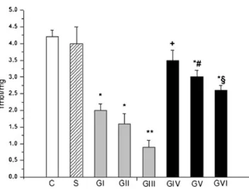
Fig. 1 Level of immunoreactive PACAP-38 in small bowel tissue after intestinal autotransplantation. Small intestinal grafts were perfused and preserved in cold UW solution for 1 h (GI), for 3 h (GII), and for 6 h (GIII) and in UW containing PACAP-38 for 1 h (GIV), for 3 h (GV), and for 6 h (GVI). In sham-operated group (S), animals underwent only median laparotomy. Small bowel biopsies were collected after laparot- omy (control, C) and at the end of the reperfusion periods. Final value of PACAP-38 was given as femtomoles per milligram wet weight. Data are presented as mean ± SEM.*p<0.05 vs. control;+p<0.05 vs. GI;
#p<0.05 vs. GII;§p<0.05 vs. GIII
Fig. 2 Level of immunoreactive PACAP-27 in small bowel tissue during autotransplantation. Small intestinal grafts were perfused and preserved in cold UW solution for 1 h (GI), for 3 h (GII), and for 6 h (GIII) and in UW containing PACAP-38 for 1 h (GIV), for 3 h (GV), and for 6 h (GVI). In sham-operated group (S), animals underwent only median laparotomy. Small bowel biopsies were collected after laparot- omy (control, C) and at the end of the reperfusion periods. Final value of PACAP-27 was given as femtomoles per milligram wet weight. Data are presented as mean ± SEM. *p<0.05 vs. control; **p< 0.01 vs.
control;+p<0.05 vs. GI;#p<0.05 vs. GII;§p<0.05 vs. GIII
UNCORRECTED
PROOF
307 PACAP-treated groups. We found no activation of the tissue 308 inhibitor of TIMP-1 (4) in the control samples, but strong 309 activation was detected in 6 h preserved grafts without 310 PACAP-38 (GIII). PACAP-38-containing cold storage 311 could decrease its activation in GVI (Fig.3).
312 Measurement of cytokines levels by Luminex Immuno- 313 assay confirmed these results (Fig. 4a–c). sICAM and 314 L-selectin were expressed at similar levels in the control 315 (A in Fig.4a, b) and ischemic groups (B in Fig.4a, b), while 316 both were significantly reduced in the PACAP-treated 317 groups (C and D in Fig.4a, b). TIMP, on the other hand, 318 was expressed at detection limit in the control group 319 (A in Fig.4c), and it was markedly increased upon ischemia 320 (B in Fig.4c). The elevated TIMP levels were significantly 321 attenuated by PACAP treatment (C and D in Fig.4c).
322 Discussion
323 This study examined the intestinal levels of PACAP-38 and 324 PACAP-27 and tissue cytokine expression using PACAP-
325 38-containing UW preservation solution during small bowel
326 autotransplantation.
327 Small bowel transplantation is increasingly performed in
328 recent years, yet, clinically, there are still many obstacles to
329 improve patient and graft survival. For most grafts, the
330 preservation solution plays a fundamental role in minimiz-
331 ing the detrimental effects of ischemia during cold storage
332 and subsequent reperfusion periods. The current clinical
333 standard for small bowel consists of a vascular flush with
334 cold UW solution. This solution has many advantages in the
335 preservation of liver and kidney; however, for small bowel
336 storage it still is unclear whether UW is optimal (Kokotilo et
337 al. 2010; Roskott et al. 2011; Salehi et al. 2004). Several
338 research efforts have been directed towards methods to
339 protect against I/R injury, using modified composition of
340 the commercially available solutions or adding new compo-
341 nents appropriate for intestinal storage.
Fig. 3 Cytokine array showing the appearance on various cytokines in control intestine (a) and in small bowel tissue exposed 6 h cold storage in UW (b) or 6 h cold preservation in PACAP-38-containing UW solution (c) and subsequent 3 h reperfusion period (d). Marked changes are observed in sICAM-1 (1), L-selectin (2), and TIMP-1 (4) expres- sions.+Crepresents positive control. Other spots, where no changes were observed are (fromupper left corner,without numbers): CINC-1, CINC-2alpha/beta, CINC-3, GM-CSF, IFN-gamma, IL-1alpha, IL- 1beta, IL-1ra, IL-2, IL-3, IL-4, IL-6, IL-10, IL-13, IL-17, IP-10, LIX, MIG, MIP-1alpha, TNF-alpha, and VEGF
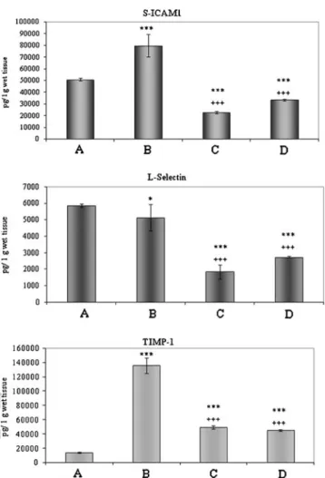
Fig. 4 Cytokine measurement by Luminex immunoassay showing the appearance on various cytokines in control intestine (A) and in small bowel tissue exposed to 6 h cold storage in UW (B) or 6 h cold preservation in PACAP-38-containing UW solution (C) and subse- quent 3 h reperfusion period (D). Marked changes are observed in sICAM-1 (a), L-selectin (b), and TIMP-1 (c) expressions. Final values were given as picogram per gram wet tissue weight. Data are presented as mean ± SEM.*p<0.05 vs. A,***p<0.001 vs. A,+++p<0.001 vs. B
UNCORRECTED
PROOF
342 In the present study, we demonstrated that intestinal 343 tissue PACAP-38 and PACAP-27 levels decreased in a 344 time-dependent manner after 1 and 3 h cold preservation 345 procedure. These changes were significant following 6 h 346 cold storage. Our previous study showed similar tendency 347 in the results of endogenous PACAP-38 concentration 348 changes in warm I/R intestinal model (Ferencz et al.
349 2009). The reason for the decreased PACAP-38 levels after 350 cold ischemia may be due to either excessive uptake by 351 ischemic cells or decreased synthesis/increased degradation 352 paralleling tissue degeneration. Similar observations have 353 been made by others in an experimental ulcer model, where 354 an acute decrease in PACAP immunoreactivity was 355 observed (Mei and Sundler1998). Values remained signif- 356 icantly higher in grafts stored in PACAP-38-containing UW 357 solution. Interestingly, PACAP-38 levels increased above 358 control values following 1 h preservation. Three and 6 h 359 cold storage in PACAP-38-containing preservation solution 360 resulted in significantly higher PACAP-38 and PACAP-27 361 levels in bowel tissue homogenates compared to only in 362 UW-preserved grafts without PACAP-38. There are no data 363 indicating the exact mechanism of the elevated values at the 364 end of the reperfusion periods. It could be due to the 365 decrease in intracellular cyclic adenosine monophosphate 366 (cAMP) through the reduction of adenylate cyclase activity 367 induced by hypoxia in endothelial cells in vitro (Yan et al.
368 1997). These changes were confirmed in an in vivo small 369 intestine preservation study. Among these mechanisms, the 370 cellular cAMP signal may represent a major determinant of 371 the intestinal integrity after global ischemic preservation 372 (Minor and Isselhard1998). Studies confirmed that admin- 373 istration of PACAP-38 enhancing the cAMP level exer- 374 ted tissue protection against I/R injury (Riera et al.
375 2001). Moreover, after extrinsic denervation, which is an 376 indispensable procedure during intestinal transplantation, 377 PACAP-38 concentration decreased in the stomach, but 378 not in the small intestine. These findings suggested a dual 379 intrinsic and extrinsic origin of the PACAP-containing nerve 380 fibers in the small intestine (Hannibal et al.1998). Another 381 explanation of the present result is that PACAP-38 intake 382 from the preservation solution and attached to the specific 383 receptors could result in the anti-oxidant and protective 384 effect to the bowel structure as described in our previous 385 studies (Ferencz et al.2009,2010a,b).
386 I/R injury is one of the main factors affecting the function 387 and structure of the small intestine, by generation of pro- 388 inflammatory mediators including cytokines. The generated 389 inflammatory cascade may activate leukocytes and endothe- 390 lial cells, which ultimately lead to tissue inflammation, 391 multiple organ dysfunction, and death. Following I/R in 392 small bowel transplantation, the gut turns into a cytokine- 393 producing organ, threatening graft and patient survival 394 (Kostopanagiotou et al.2011).
395 In the present study, we found that the expression of the
396 sICAM-1 (CD54) and L-selectin (CD62L/LECAM-1) regu-
397 lated upon activation was detectable in control bowel sam-
398 ples, and those after 6 h cold preservation in UW and
399 subsequent reperfusion period. In contrast, 6 h cold storage
400 in PACAP-38-containing UW solution caused strong reduc-
401 tion in the activation of these cytokines. Increased expres-
402 sion of sICAM-1 and L-selectin was also observed after
403 renal I/R, and it was decreased in PACAP-treated groups
404 in renal model (Horvath et al.2010b). In fact, these adhesion
405 molecules, involved in the distinct cellular crosstalk
406 between leukocytes, platelets, T cells, and endothelial cells,
407 can cause microvascular dysfunction and reperfusion dam-
408 age (Vollmar and Menger 2011). The RANTES (CCL5)
409 chemokine is not constitutively expressed; it is released
410 during inflammation. In our model, the RANTES (CCL5)
411 levels were increased in all groups, but slight reduction was
412 observed in PACAP-treated groups. During inflammatory
413 events, the transcription of matrix metalloproteinase-9 and
414 its endogenous inhibitor TIMP-1 is induced by pro-
415 inflammatory mediators. In our experiment, TIMP-1
416 showed a strong activation in 6 h preserved grafts without
417 PACAP-38. PACAP-38-containing cold storage could
418 decrease its activation. The anti-inflammatory actions of
419 PACAP in several inflammatory models are partially medi-
420 ated through its suppressing effect on cytokine/chemokine
421 production (Delgado and Ganea 2001; Horvath et al.
422 2010b). In summary, our present results support the protec-
423 tive role of PACAP-38 in cold UW solution-stored and
424 autotransplanted small intestine, which may have clinical
425 relevance in bowel transplantation in the future.
426427 Acknowledgments This study was supported by the Hungarian Sci-
428 entific Research Fund (Grant OTKA PD77474, K72592, CNK78480),
429 TAMOP (4.2.1.B-10/2KONV-2010-002, 4.2.2.B-10/1-2010-0029),
430 and“Lendulet”program of the Hungarian Academy of Sciences.
431
References 432
433 Brubel R, Horvath G, Reglodi D et al (2011) Presence of pituitary
434 adenylate cyclase activating polypeptide and its type I receptor in
435 the rat kidney. Transplant Proc 43:1297–1299
436 Delgado M, Ganea D (2001) Inhibition of endotoxin-induced macro-
437 phage chemokine production by vasoactive intestinal peptide and
438 pituitary adenylate cyclase-activating polypeptide in vitro and in
439 vivo. J Immunol 167:966–975
440 Ferencz A, Szanto Z, Borsiczky B et al (2002) The effects of precon-
441 ditioning on the oxidative stress in small bowel autotransplanta-
442 tion. Surgery 132:877–884
443 Ferencz A, Racz B, Tamas A et al (2009) Influence of PACAP on
444 oxidative stress and tissue injury following small bowel autotrans-
445 plantation. J Mol Neurosci 37:168–176
446 Ferencz A, Nedvig K, Fekecs T et al (2010a) Comparison of intestinal
447 cold preservation injury on pituitary adenylate cyclase-activating
448 polypeptide in knockout and wild-type mice. Transplant Proc
449 42:2290–2292
UNCORRECTED
PROOF
450 Ferencz A, Kiss P, Weber G et al (2010b) Comparison of intestinal 451 warm ischemic injury in PACAP knockout and wild-type mice. J 452 Mol Neurosci 42:435–442
453 Gasz B, Rácz B, Roth E et al (2006) Pituitary adenylate cyclase 454 activating polypeptide protects cardiomyocytes against oxidative 455 stress-induced apoptosis. Peptides 27:87–94
456 Grotz M, Deitch EA, Ding J, Xu D, Huang Q, Regel G (1999) 457 Intestinal cytokine response after gut ischemia. Role of gut barrier 458 failure. Ann Surg 229:478–486
459 Hannibal J, Ekblad E, Mulder H, Sundler F, Fahrenkrug J (1998) 460 Pituitary adenylate cyclase activating polypeptide (PACAP) in 461 the gastrointestinal tract of the rat: distribution and effects of 462 capsaicin or denervation. Cell Tissue Res 291:65–79
463 Horvath G, Mark L, Brubel R et al (2010a) Mice deficient in pituitary 464 adenylate cyclase activating polypeptide display increased sensi- 465 tivity to renal oxidative stress in vitro. Neurosci Lett 469:70–74 466 Horvath G, Racz B, Reglodi D et al (2010b) Effects of PACAP on 467 mitochondrial apoptotic pathways and cytokine expression in rats 468 subjected to renal ischemia/reperfusion. J Mol Neurosci 42:411–
469 418
470 Inuzuka K, Unno N, Yamamoto N et al (2007) Effect of hyperbarically 471 oxygenated-perfluorochemical with University of Wisconsin so- 472 lution on preservation of rat small intestine using an original 473 pressure-resistant portable apparatus. Surgery 142:57–66 474 Kokotilo MS, Carter J, Thiesen A et al (2010) Optimizing the concen- 475 tration of hydroxyethylstarch in a novel intestinal-specific preser- 476 vation solution. Cryobiology 61:236–242
477 Kostopanagiotou G, Avgerinos ED, Markidou E et al (2011) Protective 478 effect of NAC preconditioning against ischemia–reperfusion in- 479 jury in piglet small bowel transplantation: effects on plasma TNF, 480 IL-8, hyaluronic acid, and NO. J Surg Res 168:301–305 481 Koves K, Arimura A, Vigh S, Somogyvari-Vigh A, Miller J (1993) 482 immunohistochemical localization of PACAP in the ovine diges- 483 tive system. Peptides 14:449–455
484 Linfert D, Chowdhry T, Rabb H (2009) Lymphocytes and ischemia– 485 reperfusion injury. Transplant Rev 23:1–10
486 Maathuis MH, Leuvenink HG, Ploeg RJ (2007) Perspectives in organ 487 preservation. Transplantation 83:1289–1298
488 Mei Q, Sundler F (1998) Changes in pituitary adenylate cyclase acti- 489 vating polypeptide and vasoactive intestinal peptide innervation 490 of rat oxyntic mucosa during ulcer healing. Neuropeptides
491 32:527–535
492 Minor T, Isselhard W (1998) Cellular signal level of cyclic AMP and 493 functional integrity of the small bowel after ischemic preserva- 494 tion: an experimental pilot study in the rat. Eur Surg Res 30:144–
495 148
496 Ohtaki H, Satoh A, Nakamachi T et al (2010) Regulation of oxidative 497 stress by pituitary adenylate cyclase-activating polypeptide 498 (PACAP) mediated by PACAP receptor. J Mol Neurosci 499 42:397–403
500 Pirone A, Baoan D, Piano I, Santina LD, Baglini A, Lenzi C 501 (2011) Pituitary adenylate cyclase-activating peptide (PACAP)
502 immunoreactivity distribution in the small intestine of the adult
503 New Hampshire chicken. Acta Histochem 113:477–483
504 Racz B, Gallyas F Jr, Kiss P et al (2006) The neuroprotective effects of
505 PACAP in monosodium glutamate-induced retinal lesion involves
506 inhibition of proapoptotic signaling pathways. Regul Pept
507 137:20–26
508 Reglodi D, Kiss P, Horvath G et al (2011) Effects of pituitary adenylate
509 cyclase activating polypeptide in the urinary system, with special
510 emphasis on its protective effects in the kidney. Neuropeptides
511 46:61–70
512 Reglodi D, Kiss P, Szabadfi K et al (2012) PACAP is an endogenous
513 protective factor—insights from PACAP-deficient mice. J Mol
514 Neurosci. doi:10.1007/s12031-012-9762-0
515 Riera M, Torras J, Cruzado JM et al (2001) The enhancement of
516 endogenous cAMP with pituitary adenylate cyclase-activating
517 polypeptide protects rat kidney against ischemia through the
518 modulation of inflammatory response. Transplantation 72:1217–
519
1223 520
Roskott AMC, Nieuwenhuijs VB, Dijkstra G et al (2011) Small bowel 521 preservation for intestinal transplantation: a review. Transplant Int
522 24:107–131
523 Roth E, Weber G, Kiss P et al (2009) Effects of PACAP and precondi-
524 tioning against ischemia/reperfusion-induced cardiomyocyte apo-
525 ptosis. Ann N Y Acad Sci 1163:512–516
526 Salehi P, Spratlin J, Chong T-F, Churchill TA (2004) Beneficial effects
527 of supplemental buffer and substrate on energy metabolism during
528 small bowel storage. Cryobiology 48:245–253
529 Schulz S, Rocken C, Mawrin C, Weise W, Hollt V, Schulz S (2004)
530 Immunocytochemical identification of VPAC1, VPAC2, and
531 PAC1 receptors in normal and neoplastic human tissues with
532 subtype-specific antibodies. Clin Cancer Res 10:8235–
533
8242 534
Vaudry D, Falluel-Morel A, Bourgault S et al (2009) Pituitary adeny- 535 late cyclase-activating polypeptide and its receptors: 20 years
536 after the discovery. Pharmacol Rev 61:283–357
537 Vollmar B, Menger MD (2011) Intestinal ischemia/reperfusion: micro-
538 circulation pathology and functional consequences. Langenbecks
539 Arch Surg 396:13–29
540 Wei L, Hata K, Doorschodt BM, Büttner R, Minor T, Tolba RH (2007)
541 Experimental small bowel preservation using Polysol: a new
542 alternative to University of Wisconsin solution, Celsior and histi-
543 dine–tryptophan–ketoglutarate solution? World J Gastroenterol
544 13:3684–3691
545 Yan SF, Ogawa S, Stern DM, Pinsky DJ (1997) Hypoxia-induced
546 modulation of endothelial cell properties: regulation of barrier
547 function and expression on interleukin-6. Kidney Int 51:419–425
548 Yandza T, Tauc M, Canioni D et al (2011) Effect of polyethylene
549 glycol in pig intestinal allotransplantation without immunosup-
550 pression. J Surg Res. doi:10.1016/j.jss.2011.10.012
551 Yuan Y, Guo H, Zhang Y et al (2011) Protective effects ofL-carnitine
552 on intestinal ischemia/reperfusion injury in a rat model. J Clin
553 Med Res 3:78–84
554