ASYMPTOMATIC CARRIAGE OF ANTIBIOTIC- RESISTANT GRAM-POSITIVE COCCI AMONG DIFFERENT BACKGROUND POPULATIONS IN
EAST TIMOR, SOUTHEAST ASIA
RAQUEL SANTOS1, MIGUEL GRILO1, MONICA´ ARAÚJO1, JOSÉLUÍS MONTEIRO2
and MANUELA OLIVEIRA1*
1CIISA–Centre for Interdisciplinary Research in Animal Health, Faculty of Veterinary Medicine, University of Lisbon, Lisbon, Portugal
2OIKOS–Cooperação e Desenvolvimento, Oeiras, Portugal
(Received: 1 May 2018; accepted: 28 May 2018)
Dissemination of pathogenic multiresistant bacteria is of public health concern.
Reliable data can be difficult to obtain, especially in developing countries. This work aimed to characterize the skin and oropharyngeal microbiota, as well as their antimicrobial resistance profiles, of East-Timor populations to identify potentially pathogenic Gram-positive cocci. In order to assess the prevalence of pathogenic bacteria in East-Timor, the oropharyngeal and skin microbiota of 74 individuals was characterized. Gram-positive cocci were evaluated and their antimicrobial resistance profiles were determined. A total of 228 oropharyngeal and 278 skin samples were obtained. The population consisted of 36.5% of asymptomatic carriers of Gram- positive cocci.Kocuria rosea(n=7, 19.4%),Staphylococcusspp. (n=6, 16.7%), and Micrococcus luteus (n=6, 16.7%) were isolated, among others. Antimicrobial resistance levels ranged between 0% and 36.1%, and a multiresistance profile was observed in one third of the isolates. Gram-positive cocci colonization was associated with age group. Prevalence of multiresistant isolates was higher in males who were sampled at the refugee camp. Results show that the prevalence of antimicrobial resistance on East Timor may be underestimated. This study represents thefirst step toward the full characterization of potential pathogenic Gram-positive cocci present in the populations from East Timor.
Keywords: antimicrobial resistance, East Timor, Gram-positive cocci, oropharynx, skin
*Corresponding author; E-mail:moliveira@fmv.ulisboa.pt
Acta Microbiologica et Immunologica Hungarica 65 (4), pp. 501–513 (2018) DOI: 10.1556/030.65.2018.032 First published online July 14, 2018
Introduction
The oropharynx and skin are epithelial surfaces constantly exposed to micro- organisms, being important sites for pathogen colonization. Gram-positive cocci include several commensal genera and also important human pathogens. Streptococci and staphylococci, in particular, are a major threat to human health, since they can cause severe invasive infections. It is estimated that they are responsible for at least one third of all bacterial infections of humans, including tonsillitis, pneumonia, otitis media, meningitis, food poisoning, skin diseases, and septic shock [1].
These potentially pathogenic bacterial groups can live as commensals in individuals with a competent immune system, which may allow horizontal spread- ing within the community, or be responsible for transient or persistent opportunistic infections [2,3]. For that reason, independently of the occurrence of an infection, it is extremely important to monitor the prevalence and distribution of this potentially pathogenic bacterial group and to determine its antimicrobial resistance profile [4].
In fact, Gram-positive bacteria often present high levels of antimicrobial resistance [5,6]. Emergence of resistant bacteria is generally attributed to selective pressure due to the excessive use or misuse of antimicrobial compounds [6]. Resistant pathogen dissemination routes must be considered for the establishment of adequate measures for treatment and prevention of infectious diseases in humans [7].
A comprehensive knowledge of the human microbiota begins with in-depth surveys of the bacterial community present in each individual. Identification and characterization of the antimicrobial resistance profile of this microbiota provides fundamental information for the monitorization of possible pathogens present in asymptomatic carriers. These data may provide relevant information about the epidemi- ological situation and the occurrence of resistance determinants that spread in developing countries, where the use and access to antimicrobial compounds are scarce.
This work aimed to characterize the skin and oropharyngeal microbiota of East Timor populations, to identify potentially pathogenic Gram-positive cocci and to determine their antimicrobial resistance profiles. Samples were obtained from diversified population groups, including both genders, a large range of ages, and different socioeconomic status.
Methods Participants and data collection
A total of 148 swab samples were collected from 74 healthy individuals in East Timor, Southeast Asia, between July and August 2006, including skin (n=74) and oropharyngeal swabs (n=74). Samples were collected from three
502 SANTOS ET AL.
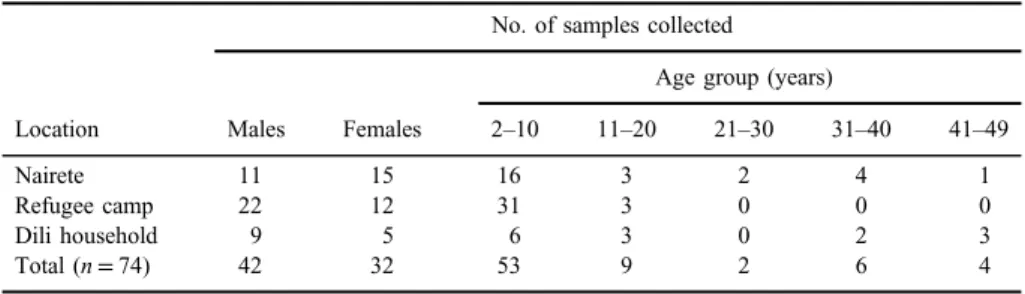
distinct locations: a household and a refugee camp in Dili and in Nairete, a small rural village. Sampled population was composed by males (n=42) and females (n=32) who were aged between 2 and 49 years, as discriminated in TableI.
After collection, Amies swabs (Oxoid Ltd., Hampshire, UK) were then kept refrigerated until transported to our laboratory, where they were further processed.
Bacterial isolation and phenotypic identification
Skin and oropharyngeal swab samples were plated onto Columbia agar plates supplemented with 5% sheep blood (COS, bioMérieux, Marcy l’Etoile, France) and incubated aerobically at 37 °C for 24–48 h. A total of 506 isolates were obtained through conventional microbiological methods and characterized through their macro- and microscopic morphologies and staining and biochemical characteristics (catalase and oxidase). Gram-positive cocci (n=36) were further identified using biochemical identification galleries: API ID 32 Staph®and API 20 Strep®(bioMérieux), according to the manufacturer’s instructions. Isolates were maintained at−80 °C until further characterization.
Antimicrobial resistance profiling
Antimicrobial resistance profile of the isolates was determined using the following nine compounds that are commonly used in human medicine and directed toward different bacterial targets: vancomycin (VA; 30 μg), amikacin (AK; 30 μg), penicillin G (P; 10 units), tetracycline (TE; 30 μg), erythromycin (ERY; 15μg), gentamicin (CN; 10μg), clindamycin (DA; 2μg), sulfamethoxazole/
trimethoprim 19:1 (SXT; 25 μg), and norfloxacin (NOR; 10 μg) (Table II).
Antimicrobial disks were purchased from Oxoid Ltd. Susceptibility to the com- pounds was tested by the disk diffusion method, as recommended by the Clinical and Laboratory Standards Institute guidelines [8].
Table I.Characteristics of the sampled populations: location, gender, and age group
Location
No. of samples collected
Males Females
Age group (years)
2–10 11–20 21–30 31–40 41–49
Nairete 11 15 16 3 2 4 1
Refugee camp 22 12 31 3 0 0 0
Dili household 9 5 6 3 0 2 3
Total (n=74) 42 32 53 9 2 6 4
ANTIMICROBIAL RESISTANCE IN EAST-TIMOR POPULATIONS 503
TableII.Identificationandantimicrobialresistanceprofileofthe36Gram-positivecocci OriginGenderAgegroup (years)IsolateIDAPIIDVAAKTESXTERYPCNDANORDoubledisk NaireteM31–4010S1StaphylococcuscapitisSISSIRSSSSusc. M2–1013O4MicrococcuslytaeSSSSSSSSSSusc. F2–1014O1MicrococcusluteusSSSSSSSSSSusc. 14O4Staphylococcusspp.SSSRSSSSSSusc. M2–1015O1Aerococcusspp.RRSSRRSRSLSA 15O3M.lytaeSSSSSSRSSSusc. M2–1018O3KocuriaroseaRSSIIRSRScMLSB F31–4019O4K.roseaSSSSISSSSM F2–1020O2StaphylococcussaprophyticusRSSSRSSSSM Refugee campF2–1028O1M.luteusRSSSRRSRSLSA M2–1030O2Staphylococcusspp.SSISISSRIcMLSB M11–2031S1K.roseaSSSRSSSRILSA M2–1034O1EnterococcusfaeciumRRISSRRRIcMLSB 34O2E.faeciumRRISISRRIcMLSB M2–1037S2S.capitisRIISRRSRSLSA 37S3StaphylococcusschleiferiRIIIRRSRScMLSB M2–1038O2M.luteusSISSSSSSISusc. F2–1039S6StaphylococcusxylosusSRSSSSIRRLSA M2–1042O1Staphylococcusspp.SSSRRSSRSiMLSB 42O3K.roseaSSSRSSSRSLSA M2–1043S4K.roseaSSSSSSIIScMLSB M2–1045S1M.luteusSRIISRSIILSA M2–1046S2StaphylococcuslentusSSIISRSISLSA 46S3K.roseaSSSSRRSRSLSA M11–2047O1K.roseaRSSSSRSSSSusc. 47O3Aerococcusspp.SISRSSSISLSA F2–1048S3M.luteusSSSSRSSSIM M2–1049O2S.xylosusSSISRRSRScMLSB M2–1057O2Staphylococcusspp.SSSSRSSRSLSA 57O4Staphylococcusspp.SSSRRSSIILSA F2-1058O1Streptococcusspp.RRSIISISSSusc. 58O3Streptococcusspp.SRSSSSSSSLSA
HouseholdM2–1065S4Micrococcusspp.SRSSSRRRSLSA M2–1072S4M.luteusSSSSSSSRSLSA F2–1073S2Staphylococcusspp.SSSSRSSIScMLSB M21–3074S2StaphylococcuscohniiurealticusSSSSRSSIScMLSB Note:M:male;F:female;S:susceptible;R:resistant;I:intermediate;S:skin;O:oropharynx;Susc.:susceptibilitytomacrolidesandlincosamide;M:resistancetomacrolidesand susceptibilitytolincosamides;LSA:susceptibilitytomacrolidesandresistancetolincosamides;cMLSB:constitutiveresistancetomacrolidesandlincosamides;iMLSB:resistance tomacrolidesandinducibleresistancetolincosamides.
In addition, to determine the macrolide resistance profile, a double-disk test was performed using the antimicrobial compounds ERY and DN. Mueller–Hinton agar (bioMérieux) plates were inoculated and both disks were placed in the center with a 15-mm distance. Plates were then incubated aerobically at 37 °C for 24 h.
Formation of inhibition halos allows the isolates classification intofive possible macrolide resistance phenotypes as follows: Susc., susceptibility to macrolides (ERY) and lincosamides (DA); M, resistance to macrolides and susceptibility to lincosamides; LSA, susceptibility to macrolides and resistance to lincosamides;
cMLSB, constitutive resistance to macrolides and lincosamides; and iMLSB, resistance to macrolides and inducible resistance to lincosamides. The cMLSB phenotype can be identified when an inhibition halo does not occur around the ERY nor the DA disks. The iMLSBphenotype can be identified when an inhibition halo does not appear around the ERY, but a distorted (D-shaped) inhibition halo appears around the DA disk.
Statistical analysis
In order to test the independence between the prevalence of Gram-positive cocci and multiresistant isolates with sociobiological data (age group, gender, and origin), aχ2test was performed. A log-linear model was further applied to identify dependencies between the prevalence of multiresistant isolates, gender, age group, and individual origin. The analysis of standardized residuals was used to identify cells with bad adjustments. Standardized residuals which were greater than 1.96 (in absolute value) were considered to be significantly different from 0 for a probability of type 1 error of 0.05. Statistical significance was considered at p<0.05. All statistical analysis were performed using IBM SPSS Statistics®, version 24 (IBM Analytics, New York, USA).
Statement of human and animal rights
In this study, no experimental research was conducted on vertebrates or any regulated invertebrates. This research was reviewed and approved by the Faculty of Veterinary Medicine, University of Lisbon.
Results Bacterial isolation and phenotypic identification
From the oropharyngeal samples, 228 isolates were obtained, including Gram-positive cocci (16 catalase-positive and 6 catalase-negative isolates),
506 SANTOS ET AL.
Gram-positive bacilli (76 catalase-positive and 28 catalase-negative isolates), and Gram-negative bacilli (55 oxidase-positive and 47 oxidase-negative isolates). From the skin samples, it was possible to obtain 278 isolates, including Gram-positive cocci (14 catalase-positive and 1 catalase-negative isolates), Gram-positive bacilli (172 catalase-positive and 14 catalase-negative isolates), and Gram-negative bacilli (61 oxidase-positive and 16 oxidase-negative isolates).
Gram-positive cocci were selected for further analysis, due to their relevance as potentially pathogenic microorganisms and their role in antimicro- bial resistance dissemination. Evaluation of results concerning age, gender, and socioeconomic status revealed that 36.5% (n=27) of the population were asymptomatic carriers of Gram-positive cocci. This prevalence was higher in children aged between 2 and 10 years, reaching 29.7% (n=22). On the other hand, only 6.7% (n=5) of the population older than 11 years were carriers of Gram-positive cocci. Individuals from the refugee camp presented the higher prevalence of Gram-positive cocci (n=16, 21.6%), followed by the ones from Nairete and the Dili household with a prevalence of 9.5% (n= 7) and 5.4%
(n= 4), respectively. Almost half of the male population carried Gram-positive cocci (n=19, 25.7%), whereas in females, only 10.8% (n=8) were found to be carriers.
The results of the biochemical identification of Gram-positive cocci by API ID 32 Staph® and API 20 Strep® (bioMérieux) are shown in Table II.
Most prevalent genera wereStaphylococcus(n=14, 38.9%) [Staphylococcusspp.
(n=6, 16.7%),S. capitis(n=2, 5.6%),S. xylosus(n=2, 5.6%),S. cohniiurealticus (n=1, 2.8%),S. lentus(n=1, 2.8%),S. saprophyticus(n=1, 2.8%),S. schleiferi (n=1, 2.8%)],Micrococcus(n=9, 25%) [M. luteus(n=6, 16.7%),Micrococcus lylae(n=2, 5.6%),Micrococcusspp. (n=1, 2.8%)], andKocuria (n=7, 19.4%) [K. rosea (n=7, 19.4%)]. Isolates belonging to Streptococcus (n=2, 5.6%) [Streptococcus spp. (n=2, 5.6%)], Enterococcus (n=2, 5.6%) [E. faecium (n=2, 5.6%)], and Aerococcus (n=2, 5.6%) [Aerococcus spp. (n=2, 5.6%)]
genera were also obtained.
Antimicrobial resistance testing
The antimicrobial resistance profile of the Gram-positive cocci (n=36) is presented in TableII. None of the Gram-positive isolates was resistant to TE. Low levels of resistance were observed for NOR (n=1, 2.8%), CN (n=2, 5.6%), and SXT (n=6, 16.7%), whereas intermediate levels of resistance were observed for other tested antimicrobials, such as AK (n=8, 22.2%), VA (n=10, 27.8%), ERY (n=13, 36.1%), and P (n=13, 36.1%). The highest level of resistance was observed regarding DA (n= 17, 47.2%).
ANTIMICROBIAL RESISTANCE IN EAST-TIMOR POPULATIONS 507
It was observed that one third of the Gram-positive cocci (n=10, 27.7%) presented a multiresistance profile, which is characterized by resistance to more than three classes of antimicrobial compounds.
The double-disk test for macrolide resistance characterization revealed that only 22.2% (n=8) of the isolates presented the S phenotype. Remaining isolates presented the iMLSB(n=1, 2.7%), M (n=3, 8.3%), cMLSB(n=9, 25%), and LSA (n=15, 41.7%) phenotypes (TableII).
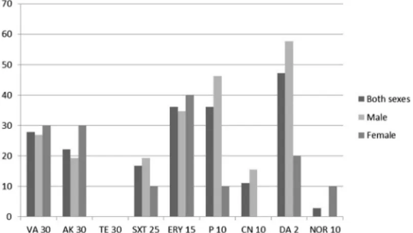
Evaluation regarding age, gender, and socioeconomic status showed that males presented higher resistance levels for DA and P, whereas females presented higher resistance for ERY, VA, and AK (Figure1). Children from the 2–10 years age group presented the highest multidrug resistance levels (Table II). Nairete population presented higher levels of resistance for VA and P, the refugee camp population for DA, and the Dili Household population for DA and ERY (Figure2).
Statistical analysis
Analysis revealed that colonization with Gram-positive cocci in children in the age group of 2–10 years [χ2(1)=4.331; p=0.037], as well as the prevalence of multiresistant isolates in individuals who were located to the refugee camp [χ2(1)= 7.608; p=0.006], were statistically significant. Furthermore, log-linear analysis showed a dependency between variables regarding prevalence of multiresistant isolates, gender, and origin of individuals [χ2L(7)=13.784;p=0.037]. Standardized residual analysis showed that multiresistant isolate prevalence was significantly associated with male individuals who were sampled in the refugee camp.
Figure 1.Antimicrobial resistance distribution according to gender
508 SANTOS ET AL.
Discussion
In recent years, there has been a growing concern about antimicrobial resistance dissemination. Epidemiological studies on the microbiota of asymp- tomatic populations are especially relevant, since they allow the detection of potentially pathogenic bacteria. However, such reports on developing countries are extremely scarce.
This study aimed to characterize the skin and oropharynx microbiota and its antimicrobial resistance profile of East Timor individuals. Three distinct popula- tions were sampled: two located in Dili, the city capital of East-Timor, and one in Nairete. Thefirst sampled population in Dili was composed by internally displaced people from a refugee camp, with a population of more than 400 individuals who were living in extremely close quarters on tends, within the boundaries of an empty pharmaceutical warehouse. Habitants ran away or lost their homes and belongings due to violence occurring between different groups in the capital. Food supply was scarce and irregular, complemented by the support of international organizations. The camp had only two water supply sources, and the waste disposal system was severely overwhelmed by the large number of people present.
Health care was almost inexistent.
The other population sampled in Dili was composed by individuals belong- ing to a single household with access to running water, energy, stable supply of food and health care, including antimicrobial compounds, when necessary.
Nairete is a small rural village, being the mostly Eastern province of East Timor. It mainly depends on agriculture. The village has a relatively stable food supply and is known as one of the most promising villages in the area, benefiting
Figure 2.Antimicrobial resistance distribution according to geographical location
ANTIMICROBIAL RESISTANCE IN EAST-TIMOR POPULATIONS 509
from being close to Lospalos, the region capital. It has no running water and uses traditional means of waste disposal. Water is abundant, with the village sitting on top of a small river. It has a very small health station, but access to pharmaceuticals is scarce.
Based on conventional microbiological procedures, bacterial isolation from the sampled populations originated a total of 506 isolates. Gram-positive bacteria were the most prevalent (64.6%), although a high percentage of Gram-negative (35.4%) was also observed. Since this bacterial group is very susceptible to environmental conditions, the high percentage of Gram-negative isolates seems to indicate that sample collection and transport were performed in adequate conditions [9].
For subsequent analysis, a group of 36 Gram-positive cocci was selected, including Micrococcus (n=9), Kocuria (n=7), Streptococcus (n=2), Entero- coccus(n=2), andAerococcus(n=2), and due to their relevance as potentially pathogenic microorganisms and their role in the transmission of antimicrobial resistance determinants [5]. However, since sampled individuals were apparently healthy, showing no gross signs of disease, they were classified as asymptomatic carriers.
Regarding isolates distribution among different genders, age groups, and geographic location, it was observed that children up to 10 years old presented a higher relative frequency (41.5%) of Gram-positive cocci, whereas on the remaining population only 23.8% of the individuals were asymptomatic carriers of this bacterial group. The statistical association was evident in the age group of 2–10 years. Young children present specific characteristics, including a develop- ing immune system and an increased contact with environmental microbiota, rendering them more susceptible to bacterial colonization [3]. The higher contact with potentially contaminated environments can also explain the higher preva- lence of Gram-positive cocci in males (45.2%) than in females (25%).
Socioeconomic constraints may have an important role in the dissemina- tion of potentially pathogenic bacteria, as suggested by the higher prevalence (47.1%) in the refugee camp population. This population lived in extremely poor conditions with scarce food and water supplies. Moreover, the waste disposal system was almost inexistent and people did not have adequate hygiene conditions. Considering their extremely difficult situation, these prevalence results were not surprising. In this study, this hypothesis is further corroborated by the association of multiresistant isolates with males who were sampled in the refugee camp.
Antimicrobial-resistant bacteria are a major concern. In this study, TE was the only antimicrobial to which all isolates were susceptible, while the highest resistance levels were obtained for DA (47.2%) and ERY and P (36.1%).
510 SANTOS ET AL.
Resistance in Gram-positive cocci has increased dramatically worldwide, partic- ularly to macrolides [10,11], as corroborated by the fact that 77.8% of the isolates studied presented a resistance profile to these drugs. This is in accordance with the earlier studies that demonstrated that the resistance to macrolides (such as ERY) and lincosamides (such as DA) is prevalent among Gram-positive cocci [12–15].
Emergence, selection, and dissemination of resistant bacteria are mainly attributed to the selective pressure of antimicrobials misuse and abuse [16, 17].
Thus, it was expected that the individuals from the Dili household, who have improved socioeconomic conditions, access to health care system and pharma- ceuticals, would present a higher prevalence of antimicrobial-resistant bacteria.
However, our results showed that the prevalence among all three sampled populations was similar. As antimicrobial drugs administration was extremely rare in the refugee camp and in the Nairete village, multiresistance levels observed might be originated from environmental contamination. In fact, the refugee camp was located within the boundaries of an empty pharmaceutical warehouse, while in the Nairete region, antimicrobial resistance could have disseminated due to the presence of international soldiers during the 30 years of East Timor occupation, which had access to antimicrobial drugs. Salyers and Shoemaker [18] showed that resistant bacteria and antimicrobial compounds can travel through the ground water originated from human wastes, entering environments where no antibiotics are being administrated directly to the populations, supporting our theory. In addition, soldiers proceeded to perform intense fumigation, which may also contributed to the high level of antimicrobial resistance observed. In fact, references to non-antibiotic selection pressure due to heavy metals or disinfectants have already been described [7].
Identification of skin and oropharyngeal microbiota is an importantfirst step toward the whole characterization of potential pathogenic bacteria of developing populations. Our results suggest that drug resistance may be very significant in these countries and should be monitored to provide accurate information on antimicrobial resistance prevalence. Furthermore, risk factors detected in this study, such as a higher prevalence of multiresistant isolates at the refugee camp, should be considered in the development of appropriate health management practices in emergency situations.
Acknowledgements
This work would not be possible without the support of Professor Cristina Lobo Vilela, 1958–2013, and Professor Ilda Sanches, 1958–2017. Work was supported by CIISA – Centre for Interdisciplinary Research in Animal Health,
ANTIMICROBIAL RESISTANCE IN EAST-TIMOR POPULATIONS 511
Faculty of Veterinary Medicine, University of Lisbon, funded by Project UID/CVT/276/2013 (CIISA).RS, MG, and MA have contributed equally to this work.
Conflict of Interest None.
References
1. Nitsche-Schmitz, D., Rohde, M., Chhatwal, G.: Invasion mechanisms of Gram-positive pathogenic cocci. Thromb Haemost98, 488–496 (2007).
2. Bogaert, D., De Groot, R., Hermans, P. W.:Streptococcus pneumoniaecolonization: The key to pneumococcal disease. Lancet Infect Dis4, 144–154 (2004).
3. Jourdain, S., Smeesters, P. R., Denis, O., Dramaix, M., Sputael, V., Malaviolle, X., Van Melderen, L., Vergison, A.: Differences in nasopharyngeal bacterial carriage in preschool children from different socio-economic origins. Clin Microbiol Infect17, 907–914 (2011).
4. Kaszanyitzky, E. J., Jánosi, S., Egyed, Z., Agost, G., Semjén, G.: Antibiotic resistance of staphylococci from humans, food and different animal species according to data of the Hungarian resistance monitoring system in 2001. Acta Vet Hung51, 451–464 (2003).
5. Rice, L. B.: Antimicrobial resistance in Gram-positive bacteria. Am J Med119, S11–S19 (2006).
6. Weigel, L. M., Donlan, R. M., Shin, D. H., Jensen, B., Clark, N. C., McDougal, L. K., Zhu, W., Musser, K. A., Thompson, J., Kohlerschmidt, D., Dumas, N., Limberger, R. J., Patel, J. B.: High-level vancomycin-resistantStaphylococcus aureusisolates associated with a polymicrobial biofilm. Antimicrob Agents Chemother51, 231–238 (2007).
7. Harbottle, H., Thakur, S., Zhao, S., White, D. G.: Genetics of antimicrobial resistance.
Anim Biotechnol17, 111–124 (2006).
8. Clinical and Laboratory Standards Institute: Performance Standards for Antimicrobial Susceptibility Testing; Twenty-Third Informational Supplement. CLSI document M100- S23. Clinical and Laboratory Standards Institute, Wayne, 2013.
9. Oliveira, M., Monteiro, J. L., Rana, S., Vilela, C. L.: Antimicrobial resistance in Gram- positive bacteria from Timorese River Buffalo (Bubalus bubalis) skin microbiota. Trop Anim Health Prod42, 833–839 (2010).
10. Halpern, M. T., Schmier, J. K., Snyder, L. M., Asche, C., Sarocco, P. W., Lavin, B., Nieman, R., Mandell, L. A.: Meta-analysis of bacterial resistance to macrolides. J Antimicrob Chemother55, 748–757 (2005).
11. Reyes, J., Hidalgo, M., Díaz, L., Rinc´on, S., Moreno, J., Vanegas, N., Castaneda, E., Arias,˜ C. A.: Characterization of macrolide resistance in Gram-positive cocci from Colombian hospitals: A countrywide surveillance. Int J Infect Dis11, 329–336 (2007).
12. Leclercq, R.: Mechanisms of resistance to macrolides and lincosamides: Nature of the resistance elements and their clinical implications. Clin Infect Dis34, 482–492 (2002).
512 SANTOS ET AL.
13. Schmitz, F. J., Verhoef, J., Fluit, A. C., The Sentry Participants Group: Prevalence of resistance to MLS antibiotics in 20 European university hospitals participating in the European SENTRY surveillance programme. J Antimicrob Chemother 43, 783–792 (1999).
14. Schreckenberger, P., Ilendo, E., Ristow, K.: Incidence of constitutive and inducible clindamycin resistance inStaphylococcus aureus and coagulase-negative staphylococci in a community and a tertiary care hospital. J Clin Microbiol42, 2777–2779 (2004).
15. Spiliopoulou, I., Petinaki, E., Papandreou, P., Dimitracopoulos, G.: erm(C) is the predomi- nant genetic determinant for the expression of resistance to macrolides among methicillin- resistantStaphylococcus aureusclinical isolates in Greece. J Antimicrob Chemother53, 814–817 (2004).
16. Monroe, S., Polk, R.: Antimicrobial use and bacterial resistance. Curr Opin Microbiol3, 496–501 (2000).
17. Sayah, R. S., Kaneene, J. B., Johnson, Y., Miller, R.: Patterns of antimicrobial resistance observed in Escherichia coli isolates obtained from domestic- and wild-animal fecal samples, human septage and surface water. Appl Environ Microbiol71, 1394–1404 (2005).
18. Salyers, A., Shoemaker, N. B.: Reservoirs of antibiotic resistance genes. Anim Biotechnol 17, 137–146 (2006).
ANTIMICROBIAL RESISTANCE IN EAST-TIMOR POPULATIONS 513