ROLE OF POTASSIUM IONS AND ION CHANNELS IN THE MUSCLE OR WHY IS ONE PLATE MEAT SOUP SO DELI-
CIOUS?
SZUTS Viktoria1,2*, HOUSHMAND Nazanin1, OTVOS Ferenc3, KOVACS Andras S1, SZEGLE- TES Zsolt4, UGHY Bettina1, DOMONKOS Ildiko1, VÁGVÖLGYI Csaba5, TOTH Laszlo1, VEHA Antal2, SZABO P Balazs2, SZILAGYI Mihaly1,2, PUSKAS G Laszlo1, SZEGLETES Zita1, HALASY Katalin6, ROVO Laszlo7, KISS G Jozsef7, NAGY Roland7, CSANADY Miklos7, GAL Jozsef8,
CSANADI Jozsef2, JARABIN Janos A7
1 Institute of Plant Biology, Biological Research Centre, Szeged, Hungary
2 Department of Food Engineering, Faculty of Engineering, University of Szeged, Hungary
3 Institute of Biochemistry, Biological Research Centre, Szeged, Hungary
4 Institute of Biophysics, Biological Research Centre, Szeged, Hungary
5 Department of Microbiology, Faculty of Science and Informatics, University of Szeged, Hungary
6 Department of Anatomy and Histology, University of Veterinary Medicine, Budapest, Hungary
7 Department of Otorhinolaryngology, Head and Neck Surgery, University of Szeged, Szeged, Hun- gary
8 Department of Economic and Rural Development, Faculty of Engineering, University of Szeged, Sze- ged, Hungary
Abstract
The sensory pleasure of a good soup represents a complex synthesis of molecular and cellular inputs of ions. Here we are focusing on the intake of potassium ions affecting the muscles along with the sensorineural science of eating a satisfying bowl of soup. Unbalanced ionic homeostasis and modified expression of ion channels is presumably involved in the inhibition of normal physiological functions. Salts are necessary components of soup with the fla- vorful broth that the salt complements turn out to activate another compo- nents of the sense of taste, the sense of savoury. Here we show the properties, molecular composition and pharmacological effects of potassium ions for the main channels of Kv-type, Kir and connexin channels in the ear system and the muscle components under physiological concentration of K-ions. However, the high concentration of K+- and Na+-ions evokes an aversive response, which is curiously not well understood at molecular level that respond to sour or bit- ter flavors. The ionic homeostasis has the main role to keep the cell tone in the muscle cells and, within all cells, what is supported by soup containing a wide variety of ions and flavors.
Keywords: ions, meal, potassium ion, ion channel complexes
*Corresponding Author: Viktoria Szuts; szutsv@hotmail.com
Introduction
About 98 % of total potassium in the body is located in the intracellular space, and mus- cle 44 containing 80% of the intracellular po- tassium. But in the extracellular space only 2%
of the total potassium ion (K+) is present, and the remaining amount is distributed in the bone, liver and erythrocytes. K+in the extra- cellular space critically determines the resting membrane potential of the cells and, together with intracellular K-ions, connected with the cellular potassium homeostasis. The move- ment of K+in and out of the muscle plays es- sential role in extracellular potassium homeostasis mediated by plants by their transporters. The K+uptake is processed by Na+-, K+- adenosine triphosphatase (Na+, K+- ATPase) and released by inward-rectifier K+ channels.
To keep our health we intake several ions from meals which are needed to keep and re- gulate the physiological function of cells. The sensory pleasure of a good soup represents complex formulation of molecular and cellu- lar inputs of ions. Here we are focusing on the intake of potassium ions to take a look into the muscle by the sensorineural science of eating a satisfying bowl of soup.
Ions in the organs serve as structural and functional elements; classified as primary, se- condary and micronutrients. The primary ele- ments in plants are nitrogen, phosphorus, and potassium, originated from the soil and are often used in relatively large amounts by the transport system of plants. Ions involve both anions and cations: NO3
-, NH4
+, H2PO4
-, HPO4
2-and K+ions.
The animals contain the same ions but the concentration depends on the species and also on the organs. Secondary nutrients are avai- lable in adequate supply and the main com- ponents are calcium, magnesium and sulphur [1]. These are usually used in large amounts.
In contrast, the microelements are iron, zinc, molybdenum, manganese, boron, copper, co- balt, and chlorine known as micronutrients or
trace elements needed in small concentration in organs.
H I G H P O TA S S I U M C O N T E N T S I N A N I - M A L S A N D P L A N T S
Potassium in milk and soups
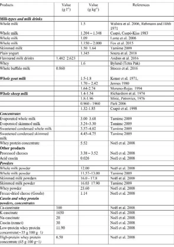
Mammalians are known to have the milk productions which are rich in all type of ions si- milarly to the case of soups regarding the ionic content (Table 1 and Table 2). The daily potassium demand of a healthy human body under normal physical activity is 2-5 g (50–125 mmol). We can provide this amount for our body through diffe- rent foodstuffs. Potassium in foods is present in the form of salts. The same amount of foodstuffs can satisfy the demand of our body in different proportions. There is no enormous difference in the potassium content of the most consumed foods, although we can discover 2-4 fold diffe- rences.
The potassium content of milk from dairy animals can play a significant role to satisfy our daily potassium demand. Comparing the milk of some milking animal species, several researchers determined that the milk of small ruminants (goats, sheep) contains more potassium than the milk of cows [2-4A].
The potassium content of milk depends on many factors, such as species, feeding (the amo- unt of potassium consumed by the animal) and others. The potassium content of dairy products is, of course, determined by the potassium con- tent of the milk, however, some separation tech- niques e.g. membrane separation and other concentration techniques can significantly affect the amount of potassium in the products.
A tight negative correlation was observed between lactose content and calcium and potas- sium content in goat and other species milk (Table 1). This suggests that osmotic pressure may also affect the amount of different elements in milk.
Table 1. Potassium content of milk-types and products of milk
References: [8-9], [12], [60-74]
Examining the effect of lactation, some authors observed a continuous decrease in mi- nerals, but in contrast to other macro ele- ments, there was no change in the observed potassium level during lactation [5-6]. Accor- ding to the results, the potassium content in- creases in the first 3 months [7],and then decreases slightly (but continuously) until the end of lactation (1.386 - 1.419 - 1.267). The al- teration can be described by quadratic equa- tion [8]. In contrast, in milk samples coming from merino ewes, a slight but continuous de- crease was observed in the first 30 days of lac- tation [9].
Examining the effect of different feeding systems, significant difference could be detec- ted not only in the main milk components and milk quantities, but also in the minerals par- tially bound to the main milk components [10]. It is emphasized that the concentration of the ingredients can be very important in case of some milk products and considering the processing plant capacity. It is especially im- portant for specialty products such as thera- peutic products and infant formulas. At the same time, they draw a"ention to significant changes in the composition of milk through feeding which can affect the thermal stability of milk during processing.
Effect of the processing of milk
Examining the potassium content of different flavoured milk drinks [11], higher potassium va- lues were determined in samples containing orange juice, but the statistical variance of the re- sults was also greater. Without orange, the po- tassium content ranged from 1.482 to 2.623, while that of the milk drink with orange juice ranged from 2.93 to 3.95 g / kg.
As a result of nanofiltration, the decrease in the potassium content of cheese whey can be ap- proximately 31% in the retentive [12]. However, examining the effect of heat treatment and ad- vanced technology (High Pressure Treat- ment)[13], it was found that the potassium content of milk samples did not change signifi- cantly (1.926-2.022 g /L).
Potassium in meat
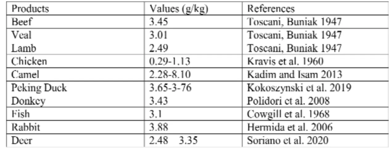
Meat and meat products usually contain more potassium than milk. In average, potassium content of meats can be characterized with a range of 2.5 – 4.00 g/kg, affected by many factors as well as by species differences (Table 2) [14].
Not only pure meats but different edible meat by- products contain remarkable potassium. These values stand close to the values of meats. In case of different species we can say that the range of potassium is similar as follows (in g/kg): beef 0.16-4.29, pork 0.55-3.58, lamb 2.38-4.28, and veal 2.43-4.33 [15-16].
Table 2. Potassium content of meat in different species
References: [16], [75-81]
It is important to note that the body weight gain of animals depends on the potas- sium content of feeds, but this fact alone does not affect the potassium level in tissues. Con- sidering the moisture content and the potas- sium salts used as additive in the production of some meat products, the end products may contain more potassium than the raw meat.
Potassium content in plants and soups
The highest concentration of K+content is in kale (savory cabbage, brussels sprout too) among plants, and also in salmon and sea- weed is the richest with potassium and anot- her macro- and micronutrients. The king is kale because it contains vitamins C, A and K1.
Vitamin B6, potassium, calcium, magnesium, copper and manganese are also in high concen- trations in kale. Furthermore, it has 2 grams of fiber, 3 grams of protein per 100g, rather high amounts, and only 50 calories [17]. Most probably kale is healthier than spinach. Both are very nut- ritious, but kale contains less amount of oxalate, which can bind minerals like calcium in your in- testine, preventing them from being absorbed.
Kale and other green leaves also contain high amount of various bioactive compounds, inclu- ding isothiocyanates and indole-3-carbinol, which have been shown to fight cancer in test- tube experiments and animal studies [18].
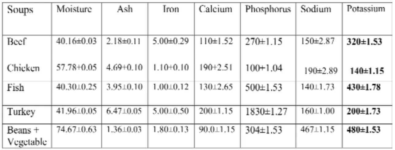
Table 3. The main minerals in meat soups. Values of minerals are in mg/100 g
1.) Data = mean ± SD; n = 3
2.) References: h"ps://d1wqtxts1xzle7.cloudfront.net/47034644/Micro_Nutrient_Content_of_Se- lected_Indig20160705-6328-1i1mm3h.pdf?1467748336=&response-contentdisposition=inline
%3B+filename%3DMicro_Nutrient_Content_of_Selected_Indig.pdf&Expires= 1593202399 &Sig- nature=AdmRUZh1GZM9hhOs
The measured pivotal macro- and micro- elements in the studies indicated that these concentration variable in different meat soups (Table 3).
We were interested in to get an answer to our question: Why are potassium ions (along with their channels) so important ion in our life? The hypothesis is that the ionic homeo- stasis changes under stress (i.e. stress or hun- griness) in cells and balanced by ions in meal to keep the tone of cells. Potassium ions and ion channels are dominant in the muscle cells.
Role of potassium ions in cells and organs, focusing on muscle
The physiological role of potassium ions with channels in the healthy muscle is widely studied [19]. At physiological ion concentrations (3–5 mM K+extracellularly, 140 mM K+intracel- lularly), the electrochemical gradient for K+(the driving force for movement of K+through a K+ channel) is outward. Potassium is mainly an in- tracellular ion. The Na+, K+-ATPase pump is pri- marily responsible for regulating the homeostasis
between sodium and potassium which pumps out sodium in exchange for potassium which moves into the cells.
Vascular smooth muscle (VSM) cells, in small arteries and arterioles that develop myo- genic tone when pressurized, are relatively depolarized, with membrane potentials on the order of -45 to -30 mV [20-21]. When the K+ channels are open, it causes K+diffusion out of the cell, leading to the loss of positive charge and membrane hyperpolarization [22].
Conversely, the closure of open K+channels will result in a decrease in this hyperpolari- zing current, and membrane depolarization.
Voltage-gated Ca2+channels contribute sub- stantially to the regulation of intracellular Ca2+
and contraction of differentiated, contractile VSM cells, particularly in resistance arteries and arterioles. Depolarization (voltage- de- pendent activation) and deactivation (hyper- polarization) by these channels importantly regulates VSM contraction.
The structure of the pore of K+channels is supposed to be similar across all of the chan- nels based on the studies of two transmem- brane (TM) domain K+channels [23].
Potassium disorders are related to cardiac arrhythmias. Hypokalemia occurs when serum potassium levels under 3.6 mmol/L.
Weakness, fatigue and muscle twitching pre- sent in hypokalemia. Hyperkalemia occur when the serum potassium levels above 5.5 mmol/L; which can result in arrhythmias.
Muscle cramps, muscle weakness, rhabdo- myolysis, myoglobinuria present the signs and symptoms in hyperkalemia [24].
Potassium channels in vascular smooth muscle
Potassium ions and another ion channels contribute to the regulation of vascular smo- oth muscle (VSM) in contraction and growth.
These ion channels are dominant in the ion conductance of the VSM cell membrane; de- termine and regulate the membrane potential of VSM cells. They are expressed in multiple
isoforms (five classes) of K+channels and contri- bute to the regulation of contraction and cell pro- liferation. The membrane potential regulates several physiological functions of voltage-gated Ca2+channels (VGCC), the open-state probabi- lity, also the Ca2+influx, intracellular Ca2+, VSM contraction and affects release of Ca2+from in- ternal stores and the Ca2+sensitivity of the con- tractile machinery [25-26, 19].
The large-conductance Ca2+ -activated K+ (BKCa) channels, intermediate-conductance Ca2+-activated K+(KCa3.1) channels, the isoforms of voltage-gated K+(KV or Kv) channels, ATP- sensitive K+(KATP) channels, and inward-recti- fier K+ (KIR) channels in both contractile and proliferating VSM cells [19]. The members of the two-pore K+(K2P) channel family have a role of K+channels. The accessory β1-subunits slow the gating kinetics, increase the Ca2+sensitivity, and affect the pharmacology of the channels [27]. Ge- nerally, K+channels participate in all aspects of regulation of VSM contraction. Furthermore, they contribute to the regulation of proliferation of VSM cells [28, 29].). The Kv3.4 channels mo- dulate proliferation of smooth muscle cells me- diated by cell cycle-dependent expression in human uterine artery.
The Ca-dependent big conductance (BKCa) K- channels are macro-complex in expression but the activity of BKCa channels is depressed in obesity [30]. In diabetic patients the impaired function reduced expression and function of the β1-subunits was measured in smooth muscle of
retinal arteria ([31]. Voltage-gated K+channels are active at the resting membrane potential of VSM cells in blood vessels displaying myogenic tone; closure of these channels leads to mem- brane depolarization and vasoconstriction [25- 26]. The function of VSM ATP dependent K- ion channels (KATP) seems to be decreased in obe- sity.
VSM cells also express one or more members of the strong inward rectifier K+channels, with Kir2.1 being the dominant isoform expressed in small resistance arteries and arterioles [32]. These channels act to amplify the hyperpolarization in- duced by opening of other K-ion channels or cel-
lular processes, i.e. the Na+/K+ATPase, and thus, may contribute to the mechanism of ac- tion of a number of vasodilators. The effects of hypertension on Kir channel function are not clear; increases, decreases, or no change in function was all observed. In diabetes it has been reported to increase.
Role of potassium channels in skeletal muscle
The role of potassium channels located on the skeletal muscle membrane is the in vivo and in vitro reduction of muscle con- tractile activity. Activation of voltage-gated potassium channels (Kv7) represented in many tissues including the excitable cells- neuronal and muscular.
The voltage gating Na+and K+channels together with Cl-channels are the main ion channels involved in muscle excitability. Se- veral mutations of the genes encoding the pore forming subunits of these channels can cause muscle hyper-excitability and stiffness or hypo- excitability and weakness or paraly- sis. However, the Ca2+channels have a pivotal function in the excitation-contraction events of skeletal muscle. From the development of action potential (AP) of the sarcolemma thro- ugh the increase in intracellular Ca2+which ac- tivates contraction anchored to transverse tubular membrane, plays an essential role of voltage-sensor: controlling the release of Ca2+
ions into the cytosol.
KCNQ channels have been identified in all type muscle tissues. However, their role in vasoregulation and chronic vascular diseases remains elusive. These data suggest that KCNQ channels play a pivotal role in vasore- gulation and forming the shape of action po- tentials along with electrocardiogram. More than 300 mutations have been detected in genes for KCNQs causing mild and severe di- seases [33].
Energy homeostasis in mitochondria is pivotal for proper muscle cell function. The proper physiological function of mitochond-
rial K-ion channels and uncoupling proteins may both regulate the generation of reactive oxygen species despite the molecular differences between these proteins [34]. The mitoKATP channel can protect cardiac tissue against ischemia; even the details of this protective mechanism are still un- known because the macromolecular composition of the mitoKATP channel is still remains unclear.
Earlier studies suggested that the inward rectify- ing K+channel subunit Kir6.1 is a pore-forming unit of the mitoKATP channel, but it is still not evaluated. However, a screen using pharmacolo- gical and genetic manipulations provided evi- dence that a splice variant of the renal outer medullary potassium channel (ROMK) is a pore- forming unit of the cardiac mitoKATP channel.
The mitochondrial Ca-dependent big-con- ductance (mitoBKCa) ion channel constitutes a unique potassium channel in the mitochondria of cardiac muscle. In contrast to the KATP channel, which is also present in the surface membrane, the mitoBKCa channel is present only in the inner mitochondrial membrane in skeletal and cardiac muscles [35].
Potassium channels in heart and cardio- myopathy
In cardiomyocytes we can detect the out- ward and inward currents linked to more than 32 ion channels and more than 70 isoforms [36-38].
They form big macromolecules with accessory subunits and regulator elements. The Kir2.x K+ channels (encoded by KCNJ genes) maintain and regulate the inward rectifier current (IK1) contri- buting to the final repolarization phase of the ac- tion potential (AP) in cardiomyocytes. Kir-type ion channels share structural similarities [39] and have a role in a wide variety of physiological functions including insulin release, vascular tone, heart rate, buffering of potassium, and renal salt flow [37-38]. These ion channels strongly modu- late cell excitability and repolarization of AP, and determine the cellular resting membrane poten- tial [40-42]. Kir2.x subunits (Kir2.1, Kir2.2, Kir2.3,
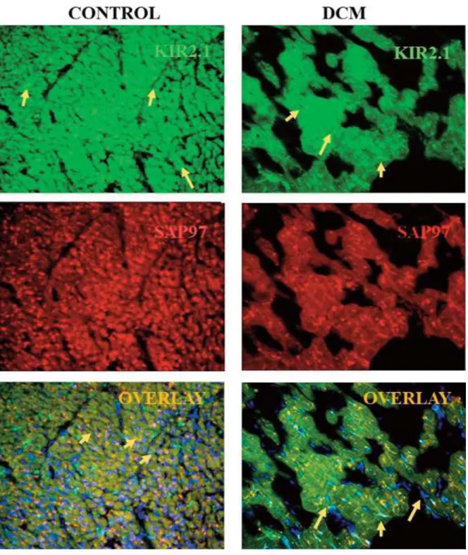
Kir2.4 and Kir2.6), assemble to form homo- or hetero-tetrameric inward rectifier potassium channels in cardiomyocytes. These channels interact with protein complexes that may be important to target and traffic ion channels, anchor and stabilize the channels into the plasma membrane [41- 43]. There are sex dif- ferences in the expressed potassium channel proteins. We revealed in earlier results that endogenous five isoforms of Kir2.x channels associate with anchoring protein of synaptic associated protein97 (SAP97) (Figure 1), for- ming specific signaling complexes [41, 44]. Kir 2.1, Kir2.2 proteins strongly bind SAP97 and they show co-localization near the T-tubules.
In cardiomyopathic tissue, studying the human dilated cardiomyopathic (DCM) sam- ples in the heart, the expressed Kir2.1, Kir2.2, Kir2.3 isoforms are drastically decreased with the anchoring and modulator protein SAP97 at gene and protein level too. The physiologi- cal studies evaluated that when the density of inward rectifier currents (IK1) decreased of Kir channel expression in cardiomyopathy, the genes and protein level of Kir ion channels decreased comparing to the healthy heart samples (Figure 1). Also the KvLQT ion chan- nels with accessory subunits and two-pore ion channels, K2P (TWIK1, TASK1) are less than 50% decreased in the heart tissues of DCM pa- tients [45-46].
The Kv –type ion channels are respon- sible for the transient outward currents (ITO1). To test the toxin effects on the outward potassium channels for Kv4.3 and anchoring protein SAP97 channels also decreased these genes and proteins using 6-epi-ophiobolin A (6EOPA) toxins in heart cell culture (Kv4.x) [46]. We measured the physico-chemical parameters which also changed, and less amo- unt of kv4.3 ion channel macro complex were stained on the surface of heart cell line
( Figure2) [42]. The elasticity of cardiomyocytes after treatment with 6EOPA suffered a mild change. The altered function found in DCM patients, however, may lead to severe heart disease, even sudden death. DCM is a myocar- dial disorder leading to left ventricular dilation and systolic dysfunction often, also to progres- sive heart failure, arrhythmias, and premature death [36-38, 41-42, 46-48]. The impaired mito- chondria were studied where KATP ion channels are damaged in the cardiovascular system [49].
Fig. 1. Alteration of Kir2.1 ion channel complex in dilated cardiomyopathy (DCM). Arrows show the Kir2.1 ion channels labeled with green dye, synaptic associated protein97 (SAP97 anchoring protein) with red and nucleus with 2,4-diamino-2-phenylindole (DAPI, blue). Tissue was a kind gift of Professor Dr. Andras Varro.
These results suggested us that the mea- sured mild effects on physiological parame- ters may be causing severe alteration in the macro-complexes of potassium channels that
can change the proper gating of these channels.
Fig. 2. Alteration of Kv4.3 ion channel complex on the heart in the presence of ophiobolin A toxin. The Kv4.3 ion channel (Cx43) protein was labeled with green, synaptic associated protein97 (SAP97) with red d nucleus with 2,4-diamino-2-phenylindole (DAPI, blue).
Potassium ions and channels in the cell- cell contact of human body
Hypercholesterolemia suppresses in- wardly rectifying K+channels in aortic en- dothelium in vitroandin vivo[50]. Connexins have a major role in cell-to-cell interactions and in the permeability of the channels car- rying diverse ions and small molecules, which maintain the physiological condition of the cells (muscle, neural, cochlea etc.) [51].
Connexin 43 (Cx43) expression decreased in myocardial tissues of DCM patients, showed positive dyeing spots in the heart tissues, and these were different in size, distribution, color and disparity, some of them were dis- tributed in the form of particles, compared to control group. Quantitative data showed that there was significant difference between the two groups in Cx43 expressive area, but there was no difference between the left and right ventricles in each group itself [41, 52- 53].
Role of potassium channels in the sen- sorineural cells focusing on ear system
Connexin26 (Cx26) and connexin43 (Cx43) have a major role in cell-to-cell inte- ractions and in the permeability of the chan- nels for diverse ions and small molecules which maintain the physiological condition of the sensorineural system as well as in the cochlea [51-56]. Different connexins may be important factors in the flawless linked to cell-to-cell interactions that maintain the fast electrogenic mechanisms between the coch- lea and neuronal system.
Connexins, Kv-type ion channels, and pannexins have a dominant role in maintai- ning the potassium ion homeostasis in the cochlea. The cellular background currents are
sustained by Kir2.1 ion channels; however, their involvement in the hearing system is less clear.
Over 50% of hearing loss or nonsyndromic deaf- ness cases in different human populations investigated in the last decades [54-56]. There are mutations in a few genes causing deafness.
Even in the early childhood, it should be very important the detection of genetic muta- tions for inner ear impairment which is crucial to provide hearing rehabilitation with an out- standing functional outcome, i.e. to maintain the development of the peripheral and central audi- tory pathway for unhindered future benefits.
Cx26 proteins were localized in the outer mem- brane of the cochlea forming hemichannels in the cochlea where the Cx26 channels are the most abundant and are involved in the potassium-re- cycling pathway. In non-syndromic hearing loss it causes mutations in connexin26 (Cx26) and connexin30 (Cx30) which have frequently been associated with hearing loss and deafness [54].
Hereditary hearing diseases are known to be as- sociated with mutations, i.e. the autosomal re- cessive non-syndromic hearing loss (ARNSHL), seizures, and sensorineural deafness [56]. Pen- dred syndrome (PDS), deafness, and the hearing loss overlap with Andersen-Tawil syndrome rarely. The Andersen-Tawil syndrome is charac- terized by malfunctioning Kir2.1 proteins, which can cause deafness with cardiomyopathy too [54-57].
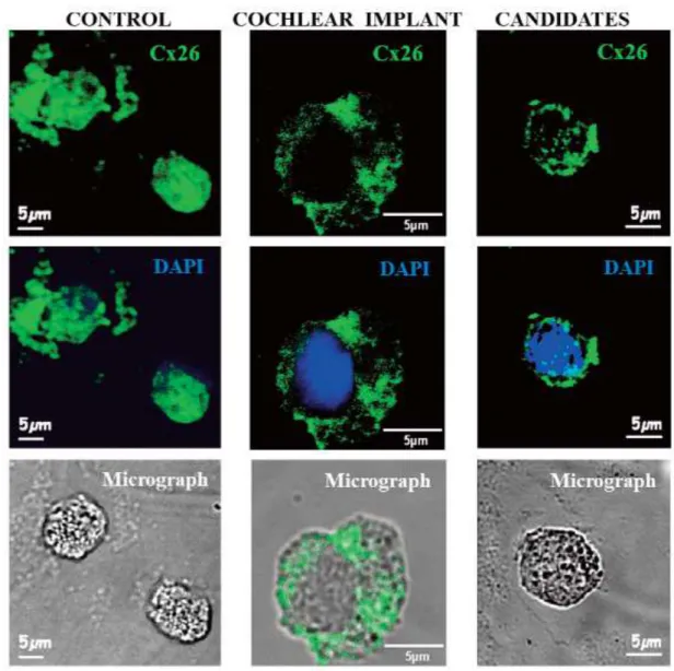
Fig. 3A. Expression of Cx26 proteins is abundant on control cells and decreased in the presence of the GJB2 gene mutation. The connexin 26 (Cx26) protein was labeled with green and nucleus with 2,4-diamino-2- phenylindole (DAPI, blue)
We investigated non-randomized, profo- undly hearing-impaired cochlear implant can- didates. All patients in the diseased group had nonsyndromic sensorineural hearing loss, altogether 80 Hungarian patients including with overlapping diseases. Prior Objective tests were used for the evaluation of hearing sensitivity on patients. Otoacoustic emissions (OAEs) indicate the functional integrity of the outer hair cells in the inner ear [54, 58]. Our representative results evaluated the levels of connexins 26 and connexins 43 proteins of non-diseased patients compared to deafness in Figure 3A and B. Earlier we investigated gene mutation analysis of connexins. The re- sults are shown that 25.0% of hearing loss pa-
tients carried a mutation in the GJB2 gene (enco- ded Cx26), and only 1.2% had a mutation in the GJB3 gene (encoded Cx30) out of 80 Hungarian patients [54].
The high intensity staining on the surface of lymphocyte cells indicated a high abundance of Cx26 in Figure 3. In the presence of the GJB2 gene mutation, the Cx26 protein level decreased and the pa"ern of the distribution was disrupted in deafness. Kir2.x isoforms colocalized with Sap97 in healthy patients but it was reduced in disease.
The regulation and modulation of these ion chan- nel complexes have a strong effect in the deve- lopment of different tissue types in hereditary diseases associated with cardiomyopathy and deafness too [54].
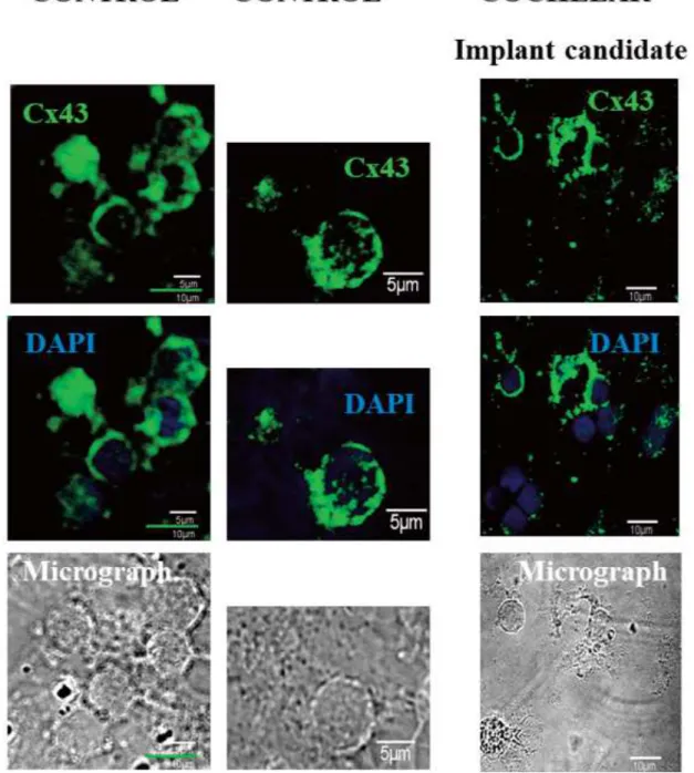
Fig. 3B. The level of Cx43 ion channel protein decreased in hearing loss patient. The connexin 26 (Cx43) pro- tein was labeled with green dye and nucleus with 2,4-diamino-2-phenylindole (DAPI, blue)
Direct and/or indirect interactions are es- sential for the normal physiological function of the Kir2.1 complex and inward rectifier currents contributing to normal potassium ion homeostasis. These genes have demon- strated gender differences and have been re- modeled in cardiomyopathy too [38].
Drugs, antibiotics and other molecules may cause fatal deafness or heart diseases i.e.
acetyls, semicillin in high dose or frequently used drugs. Not only the drugs but the low
amount of potassium intake can also cause seve- ral diseases in our life [59].
Summary
The purpose was to demonstrate that the muscle tissues and the sensorineural system in the ear affect ion homeostasis originating from potassium. We could understand when the har- monic ion balance is impaired, and the non-ba- lanced K-ion channels blocked, how it can be
restored the normal physiological functions partially or not.
The low level of SAP97 anchoring protein decreased drastically in hearing loss patients.
Our earlier studies confirmed the colocaliza- tion of Kir2.1 ion channel with SAP97 ancho- ring protein in non-diseased patients but only partial colocalization with disrupted cluste- ring occurred on the surface of blood cells in patients with DCM and deafness.
Milk and milk products are the richest foods with all kind of elements. Kale, salmon, seaweeds are rich in potassium and kale is one of the most nutrient-dense vegetables you can eat, containing large amounts of vitamins, minerals and cancer-fighting compounds.
Fish, salmons, beef and other meats are car- rying high amount of K-ions.
Even we have learnt a lot about the ex- pression and function of K-ion channels in the regulation of muscle contraction and prolife- ration in the past 40 years, there remained se- veral outstanding questions. First, why are do expressed so many different KV-type chan- nels in muscle cells? Whether this is simply a ma"er of redundancy or the pa"ern of the ex- pression of these channels tunes the particular electrophysiology of VSM cells in different vascular beds is not yet clear [19]? Second, while is it clear that, like all ion channels, the existence in multi-protein signaling domains [41-42], our knowledge about understanding of the regional heterogeneity in nature and composition of these signaling domains in different vascular beds is yet incomplete. Fi- nally, our understanding of the regulation of expression and function of K+ channels in major cardiovascular disease states also re- mains elusive, especially that they relate to different vascular beds all around the body [19, 26].
Potassium, first of all, is a “good friend”
for our healthy system [91]. Nowadays some studies evaluated that the beneficial effects of potassium intake on blood pressure and cli- nical outcomes. In a meta-analysis it was found that higher potassium intake resulted
in blood pressure lowering in the overall popu- lation studied, with more pronounced effects in patients with hypertension or consuming a high sodium diet [17, 33]. Furthermore, analysis of 11 cohort studies with a total of 127,038 participants showed that potassium intake in high range (90–
120 mmol/day) was associated with a decreased risk of stroke. Based on these studies the World Health Organization (WHO) recommends daily potassium intake of at least 90 mmol/day[1], while the Institute of Medicine recommends an intake at least 155 mmol/day.
Take home messages
Potassium channels participate in all aspects of regulation of VSM contraction.
K+channels have a dominant role in excitability in muscle cells to keep the tone of cells and re- gulate the outward and inward flow of K ions mediated by KV-, Kir-type and other ion chan- nels in human body.
K-ions contribute to the cell-cell communication with connexins (Cx 26, 30, 32, 42, 43, 45) in mus- cle, endothelial and neural tissues.
Serving our body, we have to consume eno- ugh potassium with foodstuffs. We know, that many foodstuff contain outstanding amount of potassium, but it is coming from the use of addi- tives e.g. potassium sorbate, potassium nitrate, potassium polyphosphates. Furthermore, we know that foods may also contain substances that inhibit the absorption of ions, such as spices.
We serve our health if we consider these facts and we create a well-balanced, mixed diet from animal and plant origin meals preferably with additive-free foods.
Methods and ethical statement
All the methods were described in the refe- rences of [41-42, 46 and 54]. Human heart tissues were a kind gift of Professor Dr. Andras Varro and prepared in the University of Szeged, Fa- culty of Medicine, Department of Pharmacology and Pharmacotherapy, Szeged, Hungary [41].
The samples were obtained from organ donors
whose hearts were explanted to obtain pul- monary and aortic valves for transplant sur- gery. The investigations conform to the principles of the Declaration of Helsinki. Ex- perimental protocols were approved by the University of Szeged and National Scientific and Research Ethical Review Boards (No. 51- 57/1997 OEj and 4991- 0/2010-1018EKU (339/
PI/010.)).
The investigations conformed to the De- claration of Helsinki. Experimental protocols were authorized by the University of Szeged and National Scientific Research Ethical Re- view Boards (No. 38/2014 and 2017). The blood cells were taken and kept in cold (4–
6°C) for 2–4 hours prior to investigations.The Cx23, Cx43, kir2.x and kv4.x channels were la- beling as we reported earlier both in tissue and cells using confocal microscopy after im- munofluorescence labeling [41-42, 54].
Conflict of interest
The authors declares no conflict of inte- rest.
Acknowledgements
This work was supported by the projects GINOP-2.3.2-15-2016-00012, and GINOP- 2.3.2-15-2016-00001. We thanks for J.Cs, F. O.
and J.G. intensive and critical work. Authors express their thanks for help to Professor Dr.
Győző Garab, Professor Dr. András Varró, Professor Dr. Julius G. Papp and Dr. Norbert Jost.
Resumo
La sensa plezuro de bona supo reprezentas kompleksan sintezon de molekulaj kaj ĉelaj enigoj de jonoj. Ĉi tie ni fokusiĝas pri la ingestaĵo de ka- liojonoj influantaj la muskolojn kune kun la sen- sneŭrala scienco manĝi kontentigan bovlon da supo. Malekvilibra jona homeostazo kaj modifita esprimo de kanaloj estas supozeble implikitaj en la
inhibicio de normalaj fiziologiaj funkcioj. Saloj estas necesaj eroj de supo kun la bongusta buljono, kiun la salo kompletigas por aktivigi aliajn erojn de la senca gusto, la sento de sekeco. Ĉi tie ni montras la pro- praĵojn, molekulan konsiston kaj farmakologiajn efi- kojn de kaliojonoj por la ĉefaj kanaloj de specoj Kv, Kir kaj koneksinaj kanaloj en la orela sistemo kaj la mu- skolaj komponantoj sub fiziologia koncentriĝo de K- jonoj. Tamen la alta koncentriĝo de K+kaj Na+jonoj elvokas avidan respondon, kurioze ne bone komprena- tan ĉe molekula nivelo, kiu reagas al acidaj aŭ amaraj gustoj. La jona homeostazo havas la ĉefan rolon kon- servi la ĉelan tonon en la muskolaj ĉeloj kaj, ene de ĉiuj ĉeloj, kio estas subtenata de supo enhavanta ampleksan
varion de jonoj kaj gustoj...
References
1. Wardlaw Gordon M and Smith Anne M C Contemporary Nutrition (2nd Ed.), Mc Graw Hill Companies, 2012.
2. Chamdan RC, A"aie R, Shahani KM Nutri- tional aspect of goat milk and its products. Pro- ceedings of V. Int. Conf. Goats. New Delhi, India.
1992; II. (Part II): 399.
3. Haenlein GF W and Caccese R. Goat milk ver- sus cow milk. In: Haenlein, G.F.W., Ace, D.
(Eds.), Extension Goat Handbook. Fact Sheet E-1.
United States Department of Agriculture, Wa- shington, DC 1984.
4. Park YW and Chukwu HI. Macro-mineral concentration in milk of two goat breeds at diffe- rent stage of lactation. Small. Rum, Res. 1988;
1:157.
4A. Park Young W. and Chukwu Hyginus I.
Trace mineral concentrations in goat milk from French-Alpine and Anglo-Nubian breeds during the first 5 months of lactation. Journal of Food Composition and Analysis 2, 1989; (2) 161-169.
5. Chamdan RC, A"aie R, Shahani KM. Nutritio- nal aspect of goat milk an its products. Procee-
dings of V. Int. Conf. Goats. New Delhi India.
1992; II., Part II. 399.
6. Maraval B, Vignon, B. Mineral composi- tion of goat’s milk in early lactation Milsch- wissenchaft 1982; 34, 464-466.
7. Csapó J. Composition of colostrum and milk in ca"le of different genotype. PhD dis- sertation, PANNON Agricultural University Faculty of Animal Science (Hungary) 1994.
8. Csapó J, Csapó-Kiss Zs. Examination of the macro- and microelement content of co- lostrum and milk in ca"le of different geno- types (A kolosztrum és a tej makro- és mikroelem tartalmának vizsgálata eltérő ge- notípusú szarvasmarhákon). Animal Hus- bandry and Feeding (Álla"enyésztés és Takarmányozás). 1983; 32 (2) 109-121.
9. Csapó J, Keszthelyi T, Csapó-Kiss Zs, Lengyel A, Andrássy-Baka G., Varga É. Visi:
Composition of colostrum and milk from dif- ferent genotypes of ewes. Acta Agraria De- breceniensis 2. 1998; (1) 1-21.
10. Arunima G, Galvin N, Lewis E, Hen- nessy D, O'Donovan M, McManus J J., Mark A, Timothy F, Guinee P. Outdoor grazing of dairy cows on pasture versus indoor feeding on total mixed ration: Effects on gross com- position and mineral content of milk during lactation, J. Dairy Sci. 2018; 101:2710–2723.
11. Andrés V, Tenorio D M, Villanueva J M.
Sensory profile, soluble sugars, organic acids, and mineral content in milk- and soy-juice based beverages. Food Chem. 2015; 1100- 1106.
12. Bylund Gösta. Dairy processing han- dbook. Tetra Pak Processing Systems AB edi- tion, 1995.
13. Andres V, Villanueva M-J, Tenorio M.- Dolores Influence of high pressure processing
on microbial shelf life, sensory profile, soluble su- gars, organic acids, and mineral content of milk- and soy-smoothies. Food Science and Technol.
2016 65 98-105.
14. Dickeman M., Devine C. Encyclopaedia of Meat Sciences. II. Edition Elsevier, Academic Press, 2014.
15. Pearson AM, Dutson TR. Edible Meat By- products Advances in meat research. Volume: 5.
Elsevier Sciences Publisher Ltd. 1988.
16. Soriano A, Murillo P, Peralesa M, Sánchez- Garcíac C, Murillo J A, Ruiz AG. Nutritional qua- lity of wild Iberian red deer (Cervus elaphus hispanicus) meat: Effects of sex and hunting pe- riod. Meat Sci. 2020; 168: 108-189.
17. h"ps://www.healthline.com/nutrition/11- most-nutrient-dense-foods-on-the-planet#TOC_
TITLE_HDR_4.
18. 11Trusted Source, 12: h"ps://www.ncbi.nlm.
nhi.gov/pubmed/18504070.
19. Jackson WF. Potassium Channels in Regula- tion of Vascular Smooth Muscle Contraction and Growth. Adv Pharmacol. 2017; 78: 89–14, doi:10.
1016/bs.apha.2016.07.001
20. Burns WR, Cohen KD, Jackson WF. K+-in- duced dilation of hamster cremasteric arterioles involves both the Na+/K+-ATPase and inward- rectifier K+ channels. Microcirculation. 2004;
11(3):279– 293. DOI: 10.1080/10739680490425985.
21. Welsh DG, Jackson WF, Segal SS. Oxygen in- duces electromechanical coupling in arteriolar smooth muscle cells: a role for L-type Ca2+ chan- nels. Am J Physiol. 1998; 274(6 Pt 2): H2018–2024.
22. Nelson MT, Patlak JB, Worley JF, Standen NB. Calcium channels, potassium channels, and voltage dependence of arterial smooth muscle tone. Am J Physiol. 1990; 259(1 Pt 1):C3–18.
23. Huang C, Pollock CA, Chen XM. KCa3.1:
a new player in progressive kidney disease.
Curr Opin Nephrol Hypertens. 2015; 24(1):
61–66. DOI: 10.1097/MNH.0000000000000083.
24. Chen YJ, Lam J, Gregory CR, Schrepfer S, Wulff H. The Ca(2)(+)-activated K(+) chan- nel KCa3.1 as a potential new target for the prevention of allograft vasculopathy. PLoS One. 2013; 8(11): e81006.doi: 10.1371/journal.
pone. 0081006.
25. Jackson WF. Ion channels and vascular tone. Hypertension. 2000; 35(1 Pt 2):173–178.
26. Jackson WF. Potassium channels in the peripheral microcirculation. Microcircula- tion. 2005; 12(1): 113–127. DOI: 10.1080/
10739680590896072 [PubMed: 15804979].
27. McManus OB, Helms LM, Pallanck L, Gane&ky B, Swanson R, Leonard RJ. Func- tional role of the beta subunit of high con- ductance calcium-activated potassium channels. Neuron. 1995; 14(3): 645–650.
28. Bi D, Toyama K, Lemaitre V, Takai J, Fan F, Jenkins DP, Miura H. The intermediate conductance calcium-activated potassium channel KCa3.1 regulates vascular smooth muscle cell proliferation via controlling cal- cium-dependent signaling. J Biol Chem 2013;
288(22):15843–15853. DOI: 10.1074/jbc.M112.
427187.
29. Cidad P, Jimenez-Perez L, Garcia-Arri- bas D, Miguel-Velado E, Tajada S, Ruiz- McDavi" C, PerezGarcia MT. Kv1.3 channels can modulate cell proliferation during phe- notypic switch by an ion flux independent mechanism. Arterioscler Thromb Vasc Biol.
2012; 32(5):1299–1307. DOI: 10.1161/ATV- BAHA.111.242727.
30. Borbouse L, Dick GM, Asano S, Bender SB, Dincer UD, Payne GA, Tune JD. Impaired function of coronary BK(Ca) channels in me-
tabolic syndrome. Am J Physiol Heart Circ Phy- siol. 2009; 297(5):H1629–1637. DOI: 10. 1152/aj- pheart.00466.2009.
31. McGahon MK, Dash DP, Arora A, Wall N, Dawicki J, Simpson DA, Curtis TM. Diabetes downregulates large-conductance Ca2+ -activa- ted potassium beta 1 channel subunit in retinal arteriolar smooth muscle. Circ Res. 2007;
100(5):703–711. DOI: 10.1161/01.RES. 0000260182.
36481.c9.
32. Longden TA, Nelson MT. Vascular inward rectifier K+ channels as external K+ sensors in the control of cerebral blood flow. Microcirculation.
2015; 22(3):183–196. DOI: 10.1111/micc.12190.
33. Zavaritskaya O, Zhuravleva N, Schleifen- baum J, Gloe T, Devermann L, Kluge R, Mlade- nov M, Frey M, Gagov H, Fésüs G, Gollasch M, Schubert R. Hypertension. 2013; 61(1):151-9. doi:
10.1161/HYPERTENSIONAHA. 112.197566.
34. Jarmuszkiewicz W and Szewczyk A. Energy- dissipating hub in muscle mitochondria: Potas- sium channels and uncoupling proteins.
(Warsaa) . 2019; 664: 102-109.
35. Koenig X, Ebner J, Hilber K. Voltage-De- pendent Sarcolemmal Ion Channel Abnormali- ties in the Dystrophin-Deficient Heart. Int J Mol Sci. 2018; 19(11): 3296. doi: Review.10.3390/ ijms 19113296.
36. Gaborit N, S Le Bouter , Szuts V, Varro A, Na"el S, Escande D, Demolombe S. Regional and Tissue Specific Transcript Signatures of Ion Channel Genes in the Normal Human Heart. The J. Physiol-London, March, 2007 582(Pt2): 675-93.
37. Gaborit, N, Wichter T, Varró A, Szuts V, La- mirault G, Eckardt L, Paul M, Breithardt G, Schulze-Bahr E, Escande D, Na"el S, Demolombe S. Transcriptional profiling of ion channel genes in Brugada syndrome and other right ventricu- lar arrhythmogenic diseases. Eur. Heart J. 2009;
30: 487–496. doi:10.1093/eurheartj/ehn520.
38. Gaborit N,, Varro A, Le Bouter S, Szűts V, Escande D, Na"el S, Demolombe S. Gender- related Differences in Ion-Channel and Trans- porter Subunit Expression in non-Diseased Human Hearts. J MolCell Cardiol., 2010;
49(4): 639-646.
39. Anumonwo JM and Lopatin AN Cardiac strong inward rectifier potassium channels. J Mol Cell Cardiol. 2010; 48: 45–54. doi:10. 1016/
j. yjmcc.2009.08. 013.
40. Munoz V, Vaidyanathan R, Tolkacheva EG, Dhamoon AS, Taffet SM Anumonwo JMB. Kir2.3 isoform confers pH sensitivity to heteromeric Kir2.1/Kir2.3 channels in HEK293 cells. Heart Rhythm, 2007; 4: 487–496. doi:
10.1016/ j.hrthm. 2006.12.033.
41. Szuts V, Ménesi D, Varga-Orvos Z, Zvara Á, Houshmand N, Bitay M, Bogáts G, Baczkó I, Virág L, Szalontai B, Geramipoor A, Cotella D, We"wer E, Ravens U, Deák F, Puskás LG, Papp J Gy, Kiss I , Varró A, Jost N, Altered ex- pression of genes for Kir channels in dilated cardiomyopathy. Can J Physiology Pharma- cology 2013; 91(8): 648-656. doi: 10.1139/cjpp- 2012-0413.
42. Szuts V, Otvos F, Bencsik O, Varo G, Ja- rabin AJ, Kovacs A, Rovo L, Kiss GJ, Szenasi T, Veha A, Szekeres A, Vagvolgyi Cs, Halasy K, Szegletes Z. Effects of 6-epi-ophiobolin on mechanical and physiological parameters of cardiomyocytes and on the reorganization and amounts of their kv4.x ion channels. Acta Biol Szegediensis 2016; 60:(2) pp. 157-166.
43. Vaidyanathan R, Taffet, SM, Vikstrom KL, Anumonwo MBJ. Regulation of cardiac in- ward rectifier potassium current (IK1) by sy- napse associated protein-97. J. Biol. Chem.
2010; 285: 28000–28009. doi:10.1074/jbc.M110.
110858.
44. Leonoudakis D, Mailliard WS, Wingerd
KL, Clegg DO, Vandenberg C. Inward rectifier potassium channel Kir2.2 is associated with sy- napse-associated protein SAP97. J. Cell Sci. 2000;
114: 987–998.
45. Szuts V, Otvos F, Dézsi L, Vágvölgyi Cs, Sza- lontai B, Dobrzynski H, Boye" M, Zhang H, Papp GyJ, Varró A, Benyhe S, Erdélyi L. What have we learned from two-pore potassium channels?
Their molecular configuration and function in the human heart. Minireview, Acta Biol Szegedien- sis. 2012; 56(2):93-107. h"p://www.sci.u-sze- ged.hu/ABS.
46. Bendahhou, S., Marionneau, C., Haurogne, K., Larroque, M.M., Derand, R., Szűts, V., Es- cande, D., Demolombe, S., Barhanin, J., In vitro molecular interactions and distribution of KCNE family with KCNQ1 in the hu- man heart. Car- diovasc Res. 2005; 67: 529-38.
47. Csanády M, Faragó M, Forster T, Hőgye M, Piros Gy. 1991 Study of the course of inheritance of dilated familial cardiomyopathy. Eur Heart J.
1991; 12: 191.
48. Jefferies JL and Towbin JA. Dilated cardio- myopathy. Lancet. 2010; 375: 752–762. 11.
49. Foster MN, Coe&ee WA. KATP Channels in the Cardiovascular System. Physiol Rev. 2016;
96(1): 177–252. DOI: 10.1152/physrev.00003.2015.
50. Fang Y, Mohler ER 3rd, Hsieh E, Osman H, Hashemi SM, Davies PF, Levitan I. Hyperchole- sterolemia suppresses inwardly rectifying K+
channels in aortic endothelium in vitro in vivo.
Circ Res. 2006; 98(8): 1064–1071. DOI: 10.1161/01.
RES.0000218776.87842.43.
51. Kemperman MH, Hoefsloot LH, Cremers CWRJ. Hearing loss and connexin 26. J of Royal Soc of Medic. 2002;95(4): 171-177. DOI: 10.1258/
jrsm.95.4.171.
52. Xinshan C and Zhang Y. Myocardial Cx43 Expression in the Cases of Sudden Death Due to Dilated Cardiomyopathy. (17th Triennial Mee- ting of The International Association of Forensic
Sciences 2005, Hong Kong) Forensic Sci Int.
2006; 162(1-3): 170-3. doi: 10.1016/j.forsciint.
2006;. 06.044.
53. Fontes MS C, Raaijmakers AJ A; van Doorn T; Kok B, Nieuwenhuis S; van der Nagel R, Vos MA; de Boer TP, van Rijen HVM, Bierhuizen M A. Changes in Cx43 and NaV1.5 expression precede the occurrence of substantial fibrosis in calcineurin-induced murine cardiac hypertrophy. 2014-01-01 pub- med.
54. Szuts V, Jarabin JA, Nagy N, Otvos F, Nagy R, Nagy A, Halasy K, Rovo L, Szell M, Kiss JG. Altered potassium ion homeostasis in hearing loss: Altered Expression of Connexins and Kir2.1 Potassium Ion Channels in Hearing Loss Patients. In: Ion Channel; Edited by Pro- fessor Fatima Shad Kaneez; (ISBN 978-935-51- 616666-09). InTechOpen. 2018; Chapter 5:
79-104. DOI: 10.5772/intechopen.77732.
55. Chan DK, Schrijver I, Chang KW. 2010 Connexin-26-associated deafness: Phenotypic variability and progression of hearing loss.
Genetics in Medicine.;12(3):174-181. DOI: 10.
1097/GIM.0b013e3181d0d42b.
56. Nagy AL, Csáki R, Klem J, Rovó L, Tóth F, Tálosi G, Jóri J, Kovács K, Kiss JG. Mini- mally invasive genetic screen for GJB2 related deafness using dried blood spots. Int J of Pe- diat Otorhinolaryngology. 2010; 74(1): 75-81.
DOI: 10.1016/j.ijporl. 2009.10.021.
57. Janssen T, Gehr DD, Klein A, Muller J.
Distortion product Otoacoustic emissions for hearing threshold estimation and differentia- tion between middle-ear and cochlear disor- ders in neonates. J Acoustical Soc of America.
2005; 117(5):2969-2979. DOI: 10.1121/1.
1853101.
58. Lu CW, Lin JH, Rajawat YS, Jerng H, Rami TG, Sanchez X, DeFreitas G, Carabello B, DeMayo F, Kearney DL, Miller G, Li H,
Pfaffinger PJ, Bowles NE, Khoury DS, Towbin JA.
2006 Functional and clinical characterization of a mutation in KCNJ2 associated with Andersen- Tawil syndrome. Journal of Medical Genetics.
2006; 43(8):653-659.
59. Rodan, Aylin R. 2017 Potassium: Friend or Foe? Pediatr Nephrol. July; 32(7): 1109–1121.
doi:10.1007/s00467-016-3411-8.
60. Walstra P., Wouters J.T.M., Geurts T.J. Dairy Science and Technology. CRC Taylor & Francis Group, Boca Raton, London, New York, ISBN: 0- 8247-2763-0. 2006; 782.
61. Rebman H., Höth H.J. Bestimmung von Na, K, Ca, Mg, Cu and Fe in Milch mit einem Ato- mabsorption-Spektralphotometer. Milchwissen- chaft, 1971; 26: 411-413.
62. Lante A., Lomolino G., Cagnin M, Spe"oli P. Content and characterisation of minerals in milk and in Crescenza and Squacquerone Italian fresh cheeses by ICP-OES. Food Control 17: 2006;
229–233.
63. Fox P.F., Uniacke-Lowe T., McSweeney P.L.H., O’Mahony J.A. Dairy Chemistry and Bio- chemistry. Springer International Publishing AG.
2015; 243.
64. Tamime A.Y. Dairy Powders and Concentra- ted Products. Wiley–Blackwell ISBN: 978-1-405- 15764-3. 2009; 408.
65. Souza S O, Santos VS., Santos ES, Ávila D VL., Nascimento Cristiane C.,. Costa Silvânio Silvério L, Garcia CAB., Araujo RGO. Evaluation of the mineral content in milk and yogurt types using chemometric tools, Microchemical Journal. 2018;
143: 1-8.
66. Andres V. Villanueva M-J., Tenorio M-D. In- fluence of high pressure processing on microbial shelf life, sensory profile, soluble sugars, organic acids, and mineral content of milk- and soy-smo- othies Food Science and Technology. 2016; 65: 98-
105.
67. Stocco G., Cipolat-Gotet C., Bonfa"i V., Schiavon S., Bi"ante G., Cecchinato A. Varia- tions in major mineral contents of Mediterra- nean buffalo milk and application of Fourier-transform infrared spectroscopy for their prediction, J. Dairy Sci. 2016; 99: 8680–
8686.
68. Konar A., Thomas P.C., Rook A.F. The concentrations of some water-soluble consti- tuents in the milks of cows, sows, ewes and goats. Journal of Dairy Research. 1971; 38 (3):
333-341.
69. Jenness R. Composition and Characteris- tics of Goat Milk: Review 1968-1979. Journal of Dairy Sci. 1980; 63: 1605-1630.
70. Moreno-Rojas R., Zurera-Cosano G., Amaro-Lopez M.A. Concentration and seaso- nal variation of calcium, magnesium, sodium and potassium in raw cow, ewe and goat milk. Int. J. Food Sci. Nutr. 1994; 45: 99–105.
71. Rincon F., Moreno R., Zurera G., Amaro M. Mineral composition as a characteristics for identification of animal origin of raw milk.J. Dairy Res. 1994; 61: 151-154.
72. Misic D, Petrovic D. Main compositional characteristics of ewes milk in the Rtanj pas- ture area with particular regard to minerals.
Mljekarstvo 1976; 26: 175-182.
73. Park Young W. Handbook of Non-Bovine Mammals, Blackwell Publishing ISBN 978-0- 8138-2051-4 2006; 449.
74. Noel L., Carl M., Vastel C., Guerin T. De- termination of sodium, potassium, calcium and magnesium content in milk products by flame atomic absorption spectrometry (FAAS): A joint ISO/IDF collaborative study.
Int. Dairy J. 2008; 18: 899–904.
75. Toscani V. and Buniak V. Sodium and po- tassium content of meats. J of Food Sci. 1947; 12 (4): 328-331.
76. Kravis Eugene M., Morley Kare. R. Changes with Age in Tissue Levels of Sodium and Potas- sium in the Fowl. Poultry Sci. 1960; 39(11): 13-15.
77. Kadim IT, Isam T. Camel meat and meat pro- ducts. CAB International. 2013.
78. Kokoszynski DR. Steczny Wasilewski K M, Hrncar Kotowicz C, Arpasova H. Meat traits from genetic resources ducks. Poultry Sci. 2019; 98 (71):
3029-3039.
79. Polidori PS. Cavallucci Vincenze"i C, Beg- helli D. Quality of donkey meat and carcass cha- racteristics. Meat Sci. 2008; 80: 1222–1224.
80. Cowgill Ursula M,. Hutchinson GE, Cathe- rine H, Skinner W. The elementary composition of Latimeria chalumnae. Zoology. 1968; 60: 456- 463.
81. Hermida M, Gonzale M, Miranda M, Rod- rıguez-Otero JL. Mineral analysis in rabbit meat from Galicia (NW Spain). Meat Scie. 2006; 73:
635–639.