APPLICATION OF LAMINAR MULTIELECTRODES AND BIOCOMPATIBLE COATINGS FOR
INTRACORTICAL APPLICATIONS
Thesis booklet of the Ph.D. dissertation László Bálint Grand
Scientific advisors: Prof. Dr. György Karmos Dr. István Ulbert
Interdisciplinary Technical Sciences Doctoral School Faculty of Information Technology
Pázmány Péter Catholic University Budapest, 2010
Introduction
Extracellular recordings of action potentials of single neurons as well as local field potentials of neuronal populations have basic importance in investigating neural networks of the central nervous system. For this reason, glass capillaries and wire electrodes have been used for a long while, which allow of low channel number recording. Their fabrication technology is often laborious and not adequately precise and reproducible. In contrary, by employing the fabrication technology of Micro-Electro-Mechanical-Systems (MEMS) small scale multisite probes can be developed in a highly reproducible and effective manner. It is extremely important to cause minimal damage in the brain tissue during probe implantation. The degree of trauma depends on the dimensions and shape of the probe as well as on the mode of implantation. One advantage of the MEMS technology, namely the high number of recording sites that can be placed on the surface of a probe, is a key attribute from the viewpoint of the future of extracellular neuroscience research, since investigation of neural activity on the population level is essential in order to elucidate complex brain mechanisms. Modus operandi of
at the Institute for Psychology of the Hungarian Academy of Sciences (IP-HAS), as well as in a project at the Research Institute for Technical Physics and Materials Science which aim was to design and fabricate silicon based novel multisite brain probes. In addition, I could take part in a project at the Institute of Experimental Medicine of the Hungarian Academy of Sciences which subject was biocompatibility issues of silicon brain probes. Helping to start up the Electrophysiology Lab of the Péter Pázmány Catholic University and the first animal experiments there constituted a part of my Ph.D.
work as well. The research results presented in my thesis booklet and dissertation unite an engineer’s innovations with a neurobiologist’s investigations. I have a strong belief that these two different directions not only complete each other but mutually beneficial to facilitate the successful research on both scientific fields.
One aim of my investigations was to design and develop a silicon brain probe with 24 sites, which excellent mechanical properties allow us to penetrate both the dura and pia mater without breaking or bending, and which does not damage neural structures too seriously during implantation thanks to its sharp bow-like tip and rounded edges. It was taken into consideration from the very first phase of the design process of the fabrication technology that the probe dimensions must be variable according to the requirements of a certain biologic task. The fabricated silicon probes were characterized by measuring the impedance of their electrodes and by analyzing the bioelectric activities that were recorded from the rat cerebellar cortex and caudate-putamen in acute experiments.
The motivation to the second goal of my investigations was followed from the fact that the implanted probe is a foreign body for the brain tissue. The tissue reacts to its presence with short– and long–term immunoresponse. This results in continuous degradation of the recorded signal and makes it useless sooner or later. I investigated biocompatibility of inactive NeuroProbes silicon probes that were coated with various bioactive agents. The short– and long–
term efficiency of coatings was defined by comparing the number of neurons located in the vicinity of the probe track to control. In addition analysis of synapses microbleedings and morphology of glial cells in the surroundings of the probe track provided us valuable results.
My intent, beside the above mentioned, was to present the advantageous application of multichannel recording throughout the research of an actual biologic topic. For this purpose I used a laminar array with stainless steel shank that was fabricated at the IP-HAS in the 90’s. I analyzed bioelectric signals recorded from the different layers of the cat auditory cortex during the natural sleep-wake cycle and ketamine-xylazine (KX) anesthesia. The mode of information processing of the auditory cortex in the different sleep-wake stages
Novel scientific results
I. Thesis group: Design, fabrication and test of a novel double–
sided wet etched multisite silicon neural probe [Grand et al.
(2010a)].
The idea of designing and fabricating a novel multisite silicon probe in Hungary came to my mind after spending my first Ph.D.
year at the NeuroNexus Technologies and the Neural Engineering Laboratory of the University of Michigan.
On one hand the multisite small scale but fragile silicon probes they had developed for a long while in Michigan, on the other hand the robust, reliable but handmade, therefore labouriously reproducible stainless steel shank based microwire laminar arrays that were fabricated in Hungary in the early 90’s, motivated me to give rise to a highly reproducible batch fabricated silicon multisite probe with small dimensions and less tissue damaging shape but good mechanical properties.
I.1. I designed a multisite probe with 24 sites and good mechanical properties for acute intracortical recordings.
The dimensions of the current probes extended to 12 mm in length, to 280 µm in width and to 80 µm in thickness. The probe shaft that can be inserted into the brain tissue is 7 mm in length.
The length of the tip is defined by the fabrication process, since yacht bow like and sharp tip was an elementary requirement from the very first phase of the design process. This design supports 24
square shaped (30µm x 30µm) Pt potential recording sites with 100 µm site spacing. The corresponding leads are made out of Pt as well.
Four masks are needed to form the 3 dimensional geometry as well as the Pt sites, corresponding leads and contact pads on the microprobes. I designed these 4 masks with the Mask Edit software.
A Printed Circuit Board (PCB) specifically designed for this purpose was used for packaging. Connection between the silicon probe and the PCB was established via ultrasonic wire bonding.
Since Al formed better bonding on SiO2 than on Pt, SiO2-Pt micro grids (200 µm x 200 µm) were designed to form the bonding pads. We used the technology that is introduced in thesis I.2. to fabricate the first Hungarian silicon multisite probe.
I.2. By using a CMOS compatible process, based on double–
sided anisotropic etching of silicon monocrystal, a multisite neural probe with parallel lateral walls, rounded edges and a yacht-bow like tip was fabricated, which dimensions are variable in a wide range.
Standard <100> oriented, p-type silicon wafer with a diameter of 3 inch and thickness of 200 µm polished on both sides were used at the Research Institute for Technical Physics and Materials Science for the probe fabrication (denoted by 1 on Figure 2), that proceeded in four steps. In initial thin-film deposition steps the bottom insulating layers (2,3,4), the electrodes and output leads (5), the passivation layer (6) are formed and the contact holes and bonding pads are opened (Figure 2.A). In the next step we developed a relief silicon–dioxide pattern on the backside of the wafer by using anisotropic wet etching (Figure 2.B). This relief pattern with parallel lateral walls defined the thickness of the probe. In the next step the SiO2 masking layer was removed from the back side of the probe and anisotropic etching of bulk silicon without mask was carried out until the whole thickness of silicon was removed from desired places (Figure 2.C). These two steps of different types of wet chemical etching processes resulted in a probe with sharp tip. Rounded edges and yacht–bow like less tissue damaging tip shape were achieved by using a special isotropic etching. This is important to avoid serious tissue damage (see Thesis group II.). The final phase of the probe fabrication process consisted of the removal of protecting and masking layers
(7), flipping out the probes of the holder frame and connecting the probes with the special shaped PCBs. Residual stress in the passivation layers or other mechanical stress had no influence on the probe performance (i.e. bending).
Figure 2. Schematic representation of the main steps of the probe fabrication process flow given in a cross-section through an electrode site (not to scale).
I.3. I proved that the probe presented under thesis point I.2., is capable to record good quality field potentials, multiunit and unit activities in acute rat experiments.
Before the acute animal experiments were conducted, impedance between each probe site and a counter platinum electrode had been measured in Ringer`s lactate solution at 1 kHz with 500 nA current (BAK model EASI-1, BAK Electronics). The average impedance, according to measuring on 72 sites (3 probes), was 1067.36±99.91 kΩ. I recorded neural activities from the caudate putamen (CPu) and motor cortex of ketamine- xylazine (KX) anesthetized rats (n=2) in acute experiments.
Multichannel Local Field Potentials (LFP, BP filter: 0.1Hz- 500Hz, 24 dB/decade, zero phase shift) and Multiple Unit Activities (MUA, BP filter: 500Hz-5000Hz, 24dB/oct, zero phase shift) were separated offline from the recorded raw data via band- pass filtering. I separated Single Unit Activities (SUA) from MUAs by using the built in CEM sorter of DataView 4.7 demo and Klustawin.
Good quality LFPs and MUAs were recorded on every site. In the first experiment SUAs could be separated at least from 2 channels (Figure 3.), while in the second experiment (not shown) this number increased at least to 6. This fact proves the functionality of this probe.
Figure 3. Neural signal recorded from the CPu of a KX anesthetized rat with the probe described in thesis point I.2. The figure represents 4 channels with the highest MUAs and corresponding LFPs. The vertical scale bar represents two values, one belongs to MUA (+32 µV) and the other to LFP (+300 µV) respectively. Gain: EEG (1k), MUA (50k).
II. Thesis group: Analyzing tissue reactions around inactive NeuroProbes silicon probes coated with biocompatible coatings and implanted into the rat cortex for short– and long–term [Grand (2010b)].
NeuroProbes, a project within the frame of the European Union’s FP6 program, has 14 participants. 11 research laboratories and 3 companies. Its main aim is to fabricate 3D multisite electrode arrays that, beyond recording neural activities, are capable to stimulate the tissue, have drug delivery capacities and less tissue damaging. One of the consortial projects, which I had the luck to work on and which the IP-HAS was responsible for, was biocompatibility issues related investigations. I have gathered precious experiences in this project that I can use to predict the effect of expected immunoreactions around the Hungarian probes.
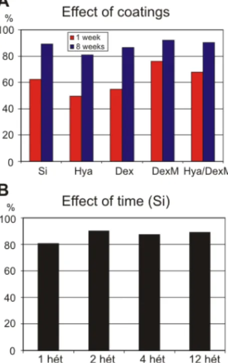
II.1. I have shown that biocompatible coatings deposited on the surface of inactive NeuroProbes silicon probes have immunosuppressing/reaction decreasing effect, especially on short–term. The efficiency of the coatings in terms of optimal neuronal survival followed the same order on short– (1 week) and long–term (8 week): 1.
dexamethasone (DexM), 2. a mixture of hyaluronic acid (Hya) and DexM, 3. uncoated Si, 4. dextrane (Dex), 5. Hya (less efficient).
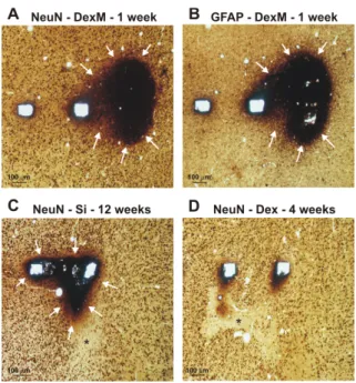
I demonstrated the changes in neuronal density around the probe tracks of implanted inactive (without output cable), four-
shank, 2 mm long NeuroProbes silicon probes on short– (1, 2 week) and long–term (4, 8, 12 week). Besides, immunoreactions evolved on short– (1 week) and long–term (4, 8, 12 week) around inactive NeuroProbes silicon probes, that were coated with Hya, Dex, DexM or the mixture of Hya/DexM and implanted into the rat (n=13) cortex, were compared to what was found in the vicinity of the uncoated silicon probe. The efficiency order of different coatings was determined by calculating the ratio of surviving neurons in the vicinity of the probe tracks to control.
After adequate time of implantation, the fixation of the brain and explantation of the probes were performed. Horizontal sections of the tissue adjacent to the probe track were cut with a Vibratome. Sections were then stained with a specific neuron (NeuN) and a glia marker (GFAP). Sections containing probe tracks were examined by light microscope, and digitized with high resolution at 10x magnification. For manual analysis of NeuN positive cells, a grid of 100 µm x 100 µm squares was placed over the probe tracks, and neurons were counted with stereological methods in squares at a distance of 100, 200, 300 and 400 µm from the side of the probe tracks on sections derived
decrease in neuronal numbers was reduced on long–term (8 week) and converged to the number that was counted at control areas located far from the probe track and the inflammation.
The efficiency of the coatings in terms of optimal neuronal survival followed the same order either on short– and long–term:
Hya<Dex<Si<Hya/DexM<DexM (Figure 4.A).
We obtained further data on how neuronal survival depends on time, by measuring neuronal densities around implantated uncoated silicon probes at both short (1, 2) and long survival (4, 12) times. The value of neuronal survival was the lowest at 1 week (77.8%), that increased to ~85% after 2 week and did not change significantly on long–term.
In summary, maximal neuronal loss occurs at 1 week after probe implantation and at distances less than 100 µm. The considerable neuron loss observed at this time point is reduced with time after implantation as neuronal densities return towards control values.
Figure 4. The efficiency order of the different coatings is the same for short– (1 week) and long–term survival (8 week) (A). The efficiency was determined by calculating the ratio of surviving neurons in the 100 µm vicinity of the probe track to control. The biocompatibility values of uncoated silicon probes, defined by using the same method, as a function of time after the implantation (B).
bleeding, decreases by going from superficial towards the deep layers. These results might explain the reason why a fraction of sites often cannot record satisfactory quality bioelectrical activities (especially from the superficial layers).
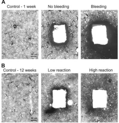
Inserted probes may puncture larger or smaller superficial blood vessels and so cause serious or minor bleeding. We examined the short– or long–term effect of bleeding on neuronal and glial cell densities with a light microscope. Signs of serious bleeding were visible at both 1 and 12 week after surgery (Figure 5.A-D.). Tissue around the probe tracks was damaged and very few, if any neurons or glial cells could be observed. In some cases, but not always, patches with severe neuron loss were detected near damaged tissue, and in these regions glial cells were larger and darker than in the control case.
Figure 5. Effects of bleeding during implantation after 1, 4 and 12 week of insertion. Dex coating facilitate lysis of clots, therefore reduced tissue reaction around Dex coated probes was seen after 4 week (D).
Next, we attempted to estimate differences in blood vessel distribution between superficial and deep layers of rat neocortex.
Both small and large blood vessels were more abundant in supragranular layers of the cortex (274 vessels/2.55 mm2, cross
that the chance of puncturing a blood vessel decreases by going down from surface to deep.
Gliosis around probe tracks was investigated in qualitative analysis at light and electron microscopic levels of sections stained with the astroglial marker GFAP.
On short–term (1 week), large, strongly stained reactive astroglial cells were visible around the probe tracks, independent of the coating (Figure 6.A). At longer delays (6 week), glial cell numbers and shape returned to control levels even close to probe tracks and a dense glial scar that was published many times by other groups was not observed visually (Figure 6.B). Light microscopic examination revealed no major influence visually, of probe size or coating on the gliotic response. Larger or smaller glial reactions were sometimes associated with different shanks of the same probe, possibly related to local bleeding. If a major bleeding occurred during probe insertion, tissue was considerably damaged with large, dark glial cells surrounding the injured area.
Figure 6. Glial reaction around the probe tracks at 1 (A) and 12 week (B) after the implantation with and without the presence of local bleeding.
Healthy neuronal cell bodies must exist sufficiently close to implanted probes for making us possible to record MUA/SUA
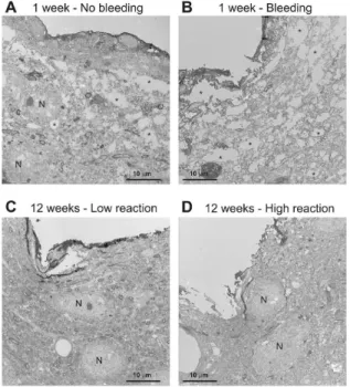
preservation within distances of 100 µm from probe tracks was very good (Figure 7.C-D.). Little structural difference was evident although the number of GFAP-positive astroglial processes seemed to be higher in a 30-50 µm thick region around the track with high glial reaction. This network of astroglial processes did not form a dense glial scar according to our light microscopic visual inspection.
Figure 7. Transmission electron micrographs of the probe tracks 1 and 12 week after implantation in the presence of bleeding (A), without bleeding (B), in the presence of low (C) or high (D) immunoreaction.
Large caverns (asterisks) and neurons in the vicinity of the probe track are indicated.
We examined intactness of synapses close to tracks of implanted silicon probes. Damaged synapses were detected, mostly at 1 or 2 week survival times and their extent was closely related to the degree of tissue damage. In general, the outer membranes of pre- and postsynaptic elements were discontinuous, whereas the synaptic cleft was unaffected as were synaptic vesicles.
III. Thesis group: Information processing in the cat auditory cortex during the natural sleep–wake cycle and anesthesia
The field of application of multi-site laminar probes, in studying the cortical mechanisms of information processing, has been widening. I investigated the alteration of acoustic evoked potentials and spontaneous cell activities during the natural sleep-wake cycle and anesthesia on freely moving cats that were implanted with a 24 sites microwire based laminar stainless steel array for chronic use.
III.1. After analyzing bioelectrical signals recorded from a chronically implantable multisite probe that was inserted into the auditory cortex of cats I have shown that local positivity, following the early components of intracortical
responsible for filtering out redundant sound stimuli. I revealed that intracortical features of down-states occurring spontaneously during the slow-wave phase (SWS) of natural sleep and activity evolving after the transient components of acoustically evoked potentials are very similar. Moreover, the acoustically evoked cortical hyperpolarization might be responsible for the periodical gating of cortical information processing.
The bioelectric activities of different layers of the acoustic cortex of cats (n=5) were recorded with a 24 sites stainless steel shank laminar multielectrode array. In addition, fine wire and wire single channel stainless steel electrodes were used to monitor the vigilance state of animal by recording activities from the surface of motor and visual cortices, the hippocampi, the neck muscles (electromiogram, EMG) and the muscles located around the eyes (Electrooculogram, EOG).
A high amplitude positive LFP component (25-100 ms) emerged from the deeper layers of the auditory cortex in response to acoustic stimuli during SWS. It was apparent after the transient components. After comparing the results of time-frequency, current source density and MUA analysis of potentials acoustically evoked during the rapid eye movement (REM) SWS and wakefulness, I revealed that the cortical power is significantly decreased in the 10-100 Hz range during the deep positive component (25-100 ms) of the Auditory Evoked Potential (AEP) (Figure 8.). The MUA was drastically depressed in every layer of the cortex, while the CSD map showed outflowing currents
(hyperpolarization) in the middle layers during the deep positive component (25-100 ms) of the AEP.
The drastic decrease of power and MUA of the cortex as well as the presence of hyperpolarizing current led me to the conclusion that disfacilitation might be responsible for generating the deep positive component of the AEP during SWS.
I revealed that the acoustically evoked down-state has similar features than that of the down-state emerging in KX narcosis.
The similarity of activity patterns emerging in EEG during KX anesthesia and natural SWS, with emphasis on the slow activity, was investigated. The auditory cortex under KX anesthesia exhibited much more rhythmic and higher amplitude slow oscillation (SO) than even in deep SWS. The recurrence frequency of the spontaneous down-states was significantly lower in SWS (0.2-0.5 1/sec) than under KX (0.5-1.5 1/sec). The average duration of the down-states in KX was significantly longer than in SWS. In deep SWS 60-100% of the random or rhythmic stimuli elicited precisely synchronized down-states, while this measure significantly dropped in deep KX anesthesia (5-10%). According to these observations, the working mechanism of auditory cortex is different in KX anesthesia than during the natural sleep. Since KX freezes cortical oscillations and prevents natural sleep related cortical dynamics to develop it is not accepted to use it as a proper model of natural SWS phase as it was suggested by other groups.
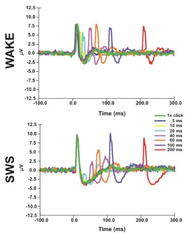
III.2. I have shown, by application of paired stimuli, that the second click stimulus is impaired and evokes significantly smaller response in the 25-100 ms timeframe during SWS compared to wakefulness, that is namely the down-state phase and which seems to have sleep protecting role in the auditory cortex.
Double sound clicks were delivered to cats (n=5) via a bone conductor. The first click was followed by a second one with
alternating interstimulus interval (ISI =5, 10, 20, 40, 60, 100, 200 ms). I assessed the potentials evoked with both members of the acoustic stimulus as well as the MUAs (Figure 9.) and CSDs.
It has been revealed that the second member of the paired acoustic stimulus presented with various ISI after the first one in SWS, but not during SWS or wakefulness, elicits almost none MUA or LFP response in the 0-15 ms timeframe. The evoked LFP and MUA were significantly smaller at 25 ms and recovered to at least 80%
of its initial value after an 80-100 ms delay (Figure 9.). The recovery period was significantly shorter, about a few tens of ms, in wakefulness and REM.
Figure 9. MUAs elicited in response to paired acoustic stimulus in SWS and wakefulness. The first member of the stimulus arrived at 0 ms, that was followed by the second one with alternating ISIs (i.e. 5, 10, 20, 40, 60, 100, 200 ms).
Based on these results I concluded that the acoustic evoked down-state (synaptic silence) in SWS, that was described under thesis point III.1., acts a sleep protecting mechanism which shuts- down the information processing of the acoustic cortex in the 25- 100 ms timeframe after the stimulus. The evoked early component
of LFP did not decrease during SWS, a fact that indicates existing cortical input during SWS. It means, the evoked activity reaches the cortex in SWS, thus the thalamus bahaves not like a switch in off mode during SWS, because information coming from the periphery passes through the thalamus. During SWS, the cortex has a sleep protecting function that prevents the subject awakening and continues sleep at least in case of irrelevant incoming acoustic information.
Application of the results
The three thesis groups of my dissertation focus on the topic of application of intracortical multisite probes and recording of bioelectric signals. The presented Hungarian silicon probe is a specific example for application, however, with its fabrication technology and with the new Deep Reactive Ion Etcher (DRIE) of the Research Institute for Technical Physics and Materials Science of the Hungarian Academy of Sciences, the research for a novel, 5-8 cm long silicon based deep brain probe with 2 hidden drug delivery channels has been started. The dimensions and geometry of the existing silicon probe, thus its mechanical properties, as well as the
the application of ion selective membranes. Suchlike of research directions are expected to come in the near future.
In order to reliably employ multisite probes in chronic recordings and to prevent the recorded signal from degradation it would be important to efficiently reduce or suppress short– and long–term immunoreactions around the probe track. It is visible, after reading the results section of the biocompatibility topic related chapter of my thesis, that different coatings did have impact on the degree of emerging immunoreaction around the probe track, especially on short–term. Significantly reducing the immunoreaction would be probably possible by administrating dexamethasone slowly in a controlled manner on long–term. Dexamethasone filled nanoparticles embedded into the coating or immobilized on the surface of the probe might be a solution of the future. According to our results the degree of neuronal loss and immunoreaction increase, and very likely the quality of recorded signal decreases, thanks to the considerable amount of cytokines entering from blood vessels to extracellular space at the time of microbleedings by probe implantation occurs. It was proven that more and larger blood vessels are present in superficial layers, than in the deep layers of the neocortex. Therefore, the chance of puncturing a vessel during implantation is higher in the supragranular layers. The chance of hitting a vessel might be lowered by using modern imaging processes before the surgery. Employing biocompatible coatings together with modern imaging processes before the surgery might increase the success rate of long–term recordings.
Down-state was previously thought to exist only as a phase of the slow sleep oscillation which may modulate sensory responses. In contrast, I revealed, by application of multisite recording technology, that acoustic stimulation can reliably and precisely modulate cortical oscillatory state in natural SWS. Besides, it was proven that acoustic intracortical mechanisms, related to information processing, are different in SWS compared to REM and wakefulness. An incomparable feature of the information processing method of the acoustic cortex in SWS was demonstrated, which was not found on other sensory areas.
Audition is the only modality that continuously monitors our environment, even when we sleep. I hypothesize that the novel cortical processing mode, which I described, acts as a sleep protecting mechanisms by disrupting local neuronal activity for a short period of time, thus the continuity of perception becomes impaired and stimuli will not be further processed. The existence of such a deep sleep protecting mechanism implies the important role of SWS in connection with the balanced maintenance of basic life functions. The presented evidence according to KX anesthesia is not a proper model of natural SWS, from the viewpoint of cortical
Acknowledgment
First, I would like to thank Professor György Karmos that he piqued my interest in electrophysiology by giving us interesting and outstanding lectures and choosing me to be his participant for the Scientific Student Conference. During the six years we have been working together, I had the chance to learn from him professionally but more importantly, his humaneness taught me every day.
I would like to thank István Ulbert that he always found time for teaching me even though he had lots of work. He gave me the opportunity not only to join his researches, but also supported my new ideas as well. I am thankful for sponsoring my participation on conferences that allowed me to get acquainted with several novelties.
I am much obliged to Lucia Wittner for the possibility of working together at the Institute of Experimental Medicine of the Hungarian Academy of Sciences and for teaching me immunohistochemical methods. I got to know her as a learned and excellent teacher and a kind person.
I also owe these persons for the help to win the Professional Internship Program of the Hungarian American Enterprise Scholarship Fund (HAESF), thanks to their letters of recommendation, and for giving me the possibility to carry-on after my return. I am also grateful to the HAESF for awarding me with their prestigious scholarship. I thank my colleagues, Balázs Dombovári and Richárd Csercsa for the good mood and inspiring collaboration during our Ph.D. studies. I could always rely on them no matter what problems we had to solve.
I would like to thank the Faculty of Information Technology of the Pázmány Péter Catholic University and especially professor Tamás Roska for giving me the possibility to achieve my research goals. Both the Faculty of Information Technology and the Laboratory for Electrophysiology opened the door for me to pursue interdisciplinary research.
I thank to Dr. László Acsády, the head of the Thalamus research group of the Institute of Experimental Medicine of the Hungarian Academy of Sciences, for providing me his laboratory for the immunohistochemical studies of my research. I thank to Dr. István Czigler, the director of the Institute of Psychology of the Hungarian Academy of Sciences and to Dr. István Bársony, the director of the Research Institute for Technical Physics and Materials Science of the Hungarian Academy of Sciences for providing me facilities for the work at the institutes led by them.
The Hungarian multisite silicon probe could not come into existence without the Institute for Technical Physics and Materials Science of the Hungarian Academy of Sciences. Their knowledge, patience and enthusiasm motivated me to work to the best of my ability. My special thanks go to Dr. Gábor Battistig who made it
Last but not least, I would like to thank to my parents, my sister and my fiancée, to the whole family and my friends. Without their unconditional love, encouragement and support I would not have succeeded. I am thankful for everything and apologize for all inconveniences I caused them in these years.
Publications
Journal publications (cumulative impact factor: 21.298) [1] D. Fabo, Z. Magloczky, L. Wittner, A. Pek, L. Eross, S. Czirjak, J.
Vajda, A. Solyom, G. Rasonyi, A. Szucs, A. Kelemen, V. Juhos, L.
Grand, B. Dombovari, P. Halasz, T.F. Freund, E. Halgren, G. Karmos, I. Ulbert, “ Properties of in vivo interictal spike generation in the human subiculum, “ Brain, vol. 131, no. 2, pp. 485-499, 2008.
[2] R. Csercsa, B. Dombovári, D. Fabó, L. Wittner, L. Erőss, L. Entz, A.
Sólyom, G. Rásonyi, A. Szűcs, A. Kelemen, R. Jakus, V. Juhos, L.
Grand, A. Magony, P. Halász, T. Freund, S. Cash, G. Karmos, E.
Halgren, I. Ulbert, “Laminar analysis of the slow wave activity in humans, “ Accepted for publication in Brain. 21/04/2010
[3] L. Grand, B. Dombovari, R. Csercsa, L. Wittner, A. Magony, G.
Karmos, I. Ulbert, “ Cortical gating of auditory information processing in sleep, “ In preparation for submission.
[4] L. Grand, A. Pongrácz, É. Vázsonyi, G. Márton, R. Fiáth, B. P.
Kerekes, D. Gubán, G. Karmos, I. Ulbert, and G. Battistig, “ A novel multisite silicon probe for high quality laminar neural recordings, “ Under review in Sensors Actuat A-Phys., 2010a.
[5] L. Grand, L. Wittner, S. Herwik, E. Göthelid, P. Ruther, S. Oscarsson, H.P. Neves, B. Dombovari, R. Csercsa, G. Karmos, I. Ulbert, “ Short and long term biocompatibility of NeuroProbes silicon multielectrodes,
“J Neurosci Methods, vol. 189, pp. 216-29, Jun 2010.
International conference publication
[6] A.A.A. Aarts, H.P. Neves, I. Ulbert, L. Wittner, L. Grand, M.B.A.
Fontes, S. Herwik, S. Kisban, O. Paul, P. Ruther, R.P. Puers, C. Van Hoof, “A 3D slim-base probe array for in vivo recorded neuron activity,
“ Proceedings of IEEE/EMBS, 2008; pp. 5798-5801
International conference posters
[1] L. Grand, B. Dombovari, E. Boldizsar, G. Karmos, I. Ulbert, “ Cortical Gating of Auditory Information Processing in Sleep,“ FENS Forum Abstracts, 8-12 July 2006; 3:A073.6
[2] I. Ulbert, R. Csercsa, L. Grand, E. Boldizsár, A. Magony, B.
Dombovári, G. Karmos, ”Evoked 'up' states in the cat auditory cortex,”
FENS Forum Abstracts, 8-12 July 2006; 3:A073.019
[3] I. Ulbert, L. Grand, B. Dombovári, R. Csercsa, A. Magony, E.
Boldizsár, G. Karmos,” Evoked cortical hyperpolarization controls auditory information processing in natural non-REM sleep,” SFN Conference, 14-18 Oct 2006; 239.231/G23
[4] N.B. Langhals , L. Grand, R.J. Vetter, D.R. Kipke, ” Characterization Of Noise And Capacitive Losses Of Microelectrodes Through Recordings And Modeling,” IEEE BMES Conf, 26-29 Sept 2007;
P3.112.
[5] B. Dombovári, L. Grand, L. Wittner, G. Karmos, I. Ulbert, ” Comparison of auditory information processing in sleep and anesthesia,” IBRO workshop, 24-26 Jan 2008; P85
[6] R. Csercsa, A. Magony, B. Dombovari, L. Grand, D. Fabo, L. Entz, L.
Wittner, L. Eross, I. Ulbert, ”Laminar properties of sleep slow oscillation in humans, ” FENS Forum Abstracts, 12-16 July 2008; 4:
160.163
[7] R. Csercsa, B. Dombovari, L. Grand, A. Magony, L. Wittner, L. Eross, G. Karmos, I. Ulbert, ”Supragranular origin of slow sleep oscillations in the human frontal lobe,” Codybs Workshop, 21-25 June 2009
[8] L. Grand, B. Dombovari, R. Csercsa, L. Wittner, A. Magony, G.
Karmos, I. Ulbert, ”Cortical gating of auditory information processing in sleep,” Codybs Workshop, 21-25 June 2009.
[9] L. Grand, L. Wittner, E. Göthelid, P. Janssen, S. Oscarsson, H.P.
Neves, P. Ruther, I. Ulbert, ”Characterization of long-term tissue reaction around Neuroprobes silicon multielectrodes,” Front Syst
[12] B. Dombóvári, K. Seidl, S. Herwik, T. Torfs, L. Grand, R. Csercsa, O.
Paul, H. P. Neves, P. Ruther, and I. Ulbert, "Acute recordings using microelectrode arrays with electronic depth control," IBRO workshop (Pecs, Hungary), pp. P7-05, 21-23 Jan, 2010.
[13] R. Fiáth, D. Horváth, L. Grand, L. Wittner, B. Dombóvári, G. Karmos, I. Ulbert, and R. Csercsa, "Laminar distribution of the spontaneous multiunit activity in the cat auditory cortex during slow wave sleep,"
IBRO workshop (Pecs, Hungary), pp. P4-10, 21-23 Jan, 2010.
[14] L. Grand, A. Pongrácz, É. Vázsonyi, G. Márton, D. Gubán, G.
Battistig, G. Karmos, and I. Ulbert, "A novel multisite silicon probe fabricated by using an economical wet etching process for high quality laminar neural recordings " IBRO workshop (Pecs, Hungary), pp. P7- 11, 21-23 Jan, 2010.
[15] D. Horváth, L. Grand, L. Wittner, B. Dombóvári, S. Kisban, S.
Herwik, P. Ruther, H. Neves, G. Karmos, and I. Ulbert, "Long term implantation of silicon-based micro probe arrays " IBRO workshop (Pecs, Hungary), 21-23 Jan, 2010.
Patent
[1] G. Battistig, L. Grand, Gy. Karmos, K. Payer, A. Pongrácz, I. Ulbert, É. Vázsonyi, ”Eljárás CMOS technológiába integrálható, egykristályos Si alapú, nedves kémiai marással készített, párhuzamos oldalfalakkal és lekerekített élekkel határolt extracelluláris elektródok előállítására, ” Hungarian patent application (Nr. P0900774), 10 December, 2009.