Advanced recording techniques
for studying cellular-level neurophysiology
Domokos Meszéna
Scientific advisor:
István Ulbert, MD., D.Sc.
Theses of the Ph.D. Dissertation
PÁZMÁNY PÉTER CATHOLIC UNIVERSITY
THE FACULTY OF INFORMATION TECHNOLOGY AND BIONICS ROSKA TAMÁS DOCTORAL SCHOOL OF SCIENCES AND TECHNOLOGY
Budapest, 2019
1 Introduction and aims
To reveal the biophysical basis of observable global states of the brain (such as cognitive processes or a particular behaviour) a wide range of dif- ferent approaches were developed and applied, either based on bottom-up or top-down strategies.
The two leading methods for the bottom-up approach are penetrating elec- trophysiological recordings via intra- and extracellular probes. The words
’intracellular’ (IC) and ’extracellular’ (EC) refer to the electrode location in- side and outside the targeted cells, respectively. While these devices become the workhorses of cellular neurotechnology providing direct access to bio- electrical events generated by single or multiple neurons, their invasiveness limits the applicability for chronic and human usage.
On the other pole of the scale, other top-down imaging (fMRI, PET) and recording (EEG, MEG) methods with the capability of recording mul- tiple regions or even whole-brain activities have been developed in parallel.
All of these technologies have the advantage of remaining completely non- invasive and thus becoming applicable even for human clinical applications instead of being mostly restricted to animal-based research. In spite of in- tensive progresses either in bottom-up and top-down approaches, there is still a significant, uncovered gap in the middle, namely how individual cells can be spatio-temporally coordinated in local networks to form specific cog- nitive functions [1, 2]. This so called ’mesoscopic’ range is of great interest re- cently in neurophysiology (and in its corresponding neurotechnology) and thus will be the main focus of this current thesis as well. This mesoscopic range will be investigated by means of extracellular multi-channel probes covering wider area than single-electrode recordings.
In this current thesis work, I focus onin vitrobrain slice electrophysiol- ogy using penetrating EC multi-channel, laminar probes. Cutting the brain into thin slices has allowed access to neurons located deeper in the brain.
Brain slice preparations can be used for either imaging or recording neu- ral activities that would be otherwise difficult to reach and detectin vivo.
Similarly toin vivoconditions, it is also possible to observein vitropopu- lation responses, such as ’sharp wave ripples’ (SWR) or some other inher- ent oscillations [3]. Furthermore, an additional advantage ofin vitrobrain slice recordings is the ability to combine different modalities, which would be again, challenging or not feasiblein vivo. Among others, such combina- tions are the co-localised and simultaneous IC recording or two-photon (2P) laser scanning microscopy imaging [4, 5, 6].
The main independent topics of the dissertation are the followings:
Chapter ’Theoretical background’ cover the biophysical theory of IC and EC potentials and their relationship as well as will detail the recent technical developments and their limitations. With this knowledge in hand, Chap- ter ’Materials and methods’ introduces multi-channel silicon probes used throughout the thesis; this same chapter elaborates on the experimental methods and closes with the description of data analysis methods and his- tological procedures. Once given the theoretical and methodological bases to contextualize this work, Chapter ’Results’ split the results into three in- dependent thesis groups: On Figure 1 a triangle illustrates the relationship
Figure 1:Interconnections between thesis groups (edges) and topics covered (nodes).
of the three theses (edges) and three experimental techniques used (nodes).
Although there are small exceptions (such as, occasional IC-2P recordings in II. Thesis group, shown with a dashed line), edges represent the main in- teractions between recording modalities. I. Thesis group presents the devel- opment and testing of a novel, multi-channel silicon probe with protruding contact sites for improvedin vitroEC recordings and highlights its advan- tages with a systematic and quantitative comparison against a commercially available, multi-channel surface probe. This chapter will focus only on EC recording without IC patch-clamp and with only one representative 2P fig- ure showing that the spiky probe is compatible with two-photon microscope imaging. II. Thesis group details an approach developed for co-localised and simultaneous IC-EC recording, it describes the resulting ‘ground-truth’
dataset, and explores potential further applications of the recorded data in model-based calculations. This thesis does not focus on a thorough eval- uation of any data analysis method (such as spike-sorting algorithms or source-localization methods), however, a preliminary application of single- cell level current source density analysis (skCSD) will be presented using
the collected simultaneous data. III. Thesis group starts with the description of the photoelectric artefact generated during the use of combined multi- channel EC recordings and two-photon (2P) laser imaging. Recent strategies to eliminate contaminations of the photoelectric artefact will be discussed together with their limitations. Next I present an experimental protocol for the generation of co-localised and simultaneous EC and 2P measurements either with or without laser artefact. The protocol allows for the investigation of photoelectric artefacts but also for providing control data for validation.
Using these combined recordings, a developed specific filtering algorithm will be presented to mitigate the photoelectric effect on recorded extracellu- lar signals. Chapter ’Conclusions and outlook’ closes the dissertation by list- ing the novel scientific contributions of this thesis work with related publi- cations and giving an outlook for future perspectives and for the application of the results.
2 Materials and methods
Intracellular electrochemical activity of neurons inevitably generates ex- tracellular changes due to transmembrane currents. Currents that are mov- ing between inside and outside of the neuron during its activity can be only indirectly measured extracellularly by their influence on the electrical field.
The EC electrode picks these changes up and relays them to an amplifier where they can be measured in respect to a reference. EC potential elicited by transmembrane currents varies dynamically in time and space as numer- ous currents may be superimposed at a given location in the EC medium [1, 7, 8]. Thus, EC potential contains all the summed signals of multiple synaptic inputs and spiking outputs as well as different local and further (volume conducted) activities in the region where the EC electrode is lo- cated [9, 8]. Some of the biophysical principles of EC potential generation are long known. However, there are still many theoretical and experimental open questions that limit the interpretation of EC potential to understand its function.
From the a large variety of devices developed for multi-channel EC recordings I use the following squeezed list of features throughout the ex- periments:
• acute rodent
• in vitrobrain slice
• laminar (1D) contact sites
Figure 2:Illustration for the theoretical background of extracellular (EC) recordings. EC potentialΦ(r,t) measured at the recording site is generated by the weighted linear sum of net transmembrane currents I(t) based on the compartmental cell-electrode distances (r).
• silicon or steel shank
• penetrating or surface
• polytrodes (16-32 channels)
Recordings were carried out with three different multi-channel microelec- trodes that met the above criteria. Recording protocols for all the three The- sis groups were performed on the same experimental set-up using very sim- ilar tool-kits with minor changes.
Acute horizontal hippocampal slices were prepared from adult Wistar rats (between 200-350 g, gender balanced, in total #30 successful experi- ments at the date of thesis submission). In addition to Wistar rats, Thy1- GCaMP6 transgenic mice were also used for population imaging under two-photon (2P) microscopy. Animals were bread and reared in the Re- search Centre of Natural Sciences, Hungarian Academy of Sciences. An- imals were supplied with food and water ad libidumand were kept on a 12-12 hour light-dark cycle. All of the protocols followed the guidelines
of the Hungarian Act of Animal Care and Experimentation (1998; XXVIII, section 243/1998.). The Animal Care and Experimentation Committee of the Hungarian Academy of Sciences, and the Animal Health and Food Control Station have approved the experimental design (license number:
PEI/001/2290-11/2015). Efforts were made to minimize animal suffering and to reduce the number of animals used.
Before the experiment, the animals were deeply anaesthetized with isoflurane (min. 0.2 ml/100 g), quickly decapitated and their brains were im- mediately removed and dipped into cold (2–3◦C), oxygenated (95% O2, 5%
CO2) cutting solution. The cutting solution contained the following com- position (in mM): 250 Sucrose, 26 NaHCO3, 10 D-Glucose, 1 KCl, 1 CaCl2 and 10 MgCl2. 500 µm-thick horizontal slices were cut by a vibratome (VT1200s; Leica, Nussloch, Germany) from both hemispheres containing the whole hippocampal formation. Slices were kept in a standard ’artificial cerebrospinal fluid’ (aCSF) solution at room temperature (20–22◦C) for at least 1 h before use. The recordings were performed with a standard record- ing aCSF containing (in mM): 124 NaCl, 26 NaHCO3, 10 D-Glucose, 4 KCl, 2 CaCl2and 2 MgCl2. The whole recording system was build under a two-
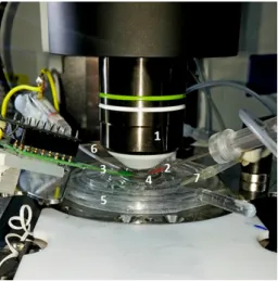
Figure 3:Close-up view of the simultaneous IC-EC recordings under the objective of the 2P microscope.
Labels are the following: Olympus 20x objective of the 2P microscope (1), patch-clamp pipette filled with IC solution (2), EC multi-channel probe (NeuroNexus comb probe shown here) with its connector (3), brain slice and holder ring (4), dual-perfusion chamber (5), reference electrode pellet (6), outlet of aCSF perfusion (7).
photon laser scanning microscope set-up developed by the Femtonics com- pany (Femtonics Ltd., Budapest, Hungary) from a transformed Olympus BX61 upright microscope, which has a NIR bright-field camera mode and a 2P fluorescent mode as well. Both the stage and two other lateral micro- manipulators were controlled by motorised wheels of the Luigs-Neumann (LN) controller system (Luigs&Neumann Feinmechanik und Elektrotechnik GmbH, Ratingen, Germany). We can have a close-up view of the set-up on Figure 3.
The patch-clamp system (Axon Instruments, USA) consists of several subunits. During the recording, the signal was first transferred to Multi- Clamp 700B amplifier, that maximizes signal-to-noise ratio and enables whole-cell voltage-clamp and current clamp recordings, then feed to the computer through Digidata 1550B A/D converter. MultiClamp Comman- der software was used to operate the glass pipette and set various param- eters and compensations. pClamp10 software consists of Clampex visual- ization software, which is suitable for acquiring digitized data, Ramp-test was also performed on this platform, and Clampfit analysis software, which provides a wide variety of tools for statistics, transforms and different lay- out designs for intracellular data. Extracellular signals from multi-channel silicon probes were transmitted to the Intan RHD2000 Evaluation system (Intan Technologies, USA), its task is to acquire, pre-amplify and digitize the raw data. It consists of a 32-channel pre-amplifier chip (RHD2132), a RHD2000 FPGA interface board, which is an open-source hardware with up to 128 channels for bio-potential recordings and thin, flexible interface ca- bles for connections. Intan RHD2000 software transfers data to the screen, operates the amplifiers and sampling frequency can be set here. The wide- band signals (from 0.1 Hz up to 7 kHz) were recorded with 20 kHz sampling rate.
3 Summary of novel scientific results with related publication
I. Thesis group: Development and testing of a novel, multi- channel spiky probe for extracellular recordings
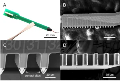
I.a. I have designed a novel multi-channel, laminar in vitro silicon probe with protruding, ’spiky’ contact sites for improved brain slice recordings. I have optically verified the fabricated arrowhead shape, protruding contact sites under scanning electron microscopy. Moreover, I have characterised the electrochemical impedance magnitudes and phase angles as well as the noise level of the spiky probe in physiological saline solution.
The spiky probe comprises a single silicon shank carrying 32 protruding Pt/Ir/IrOx contact sites with three possible spacing layouts of 25µm, 50 µm and 100µm. With the help of the optimally angled shank, I have shown that the spiky probes are compatible with large in vitro, water-immersion objectives used typically in two-photon microscopy imaging.
Figure 4:Optical photograph (A) and scanning electron microscopy (SEM) images of the spiky probe (B-D). Please note the arrowhead-shape, protruding contact sites.
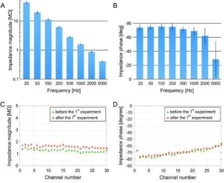
The high-density spiky probe (with inter-contact distance of 25µm) used in the comparative test showed an average impedance magnitude of 1.27± 0.1 MΩat 1 kHz across all contact sites with an average phase angle of -70± 5◦. In addition, I have tested the robustness and re-usability of spiky contact sites. Impedance changes were investigated after an extensive usage (7 ex- periments with 17 separate insertions, in total) by repeating the impedance test after the last experiment. The average impedance magnitude of all con- tact sites was slightly increased after the last experiment to 1.5±0.1 MΩ. The impedance phase angles did not change significantly (-69±5◦after ex- tensive testing).
Figure 5:Results for impedance spectroscopy (A-B). Please note the monotonically decreasing magni- tudes and phase angle values and the small standard deviations across channels. Stability and re-usability of spiky contact sites (C-D).
I have also measured the root mean square noise levels for the 32 contact sites. In the spike band (500 – 5000 Hz) noise level was 4.63±0.51µVr ms. Placing the probe onto the brain slice elevated measured noise levels by 10 - 20%.
I.b. With the aid of protruding contact sites I have proven quanti- tatively that the spiky probe provides higher neuronal yield and higher signal amplitude compared to a commercial surface probe. Moreover, I have also shown that the high-density spiky probe is suitable for separat- ing single unit clusters into putative cell types based on a spatio-temporal analysis of their extracellular waveforms oversampled by multiple adja- cent contact sites.
Figure 6:Quantitative comparison of recording performances. Box plots showing the distribution of the number of well-separated SUA clusters (A) and the distribution of the peak-to-peak amplitude of spike wave- forms (B) for the high-density spiky probe and the hockey-stick surface probe. **p=0.0078; ***p=0.0000049;
Welch’s t-test
The high-density spiky probe (with inter-contact distance of 25 µm) was compared to a commercially available, laminar surface probe called
’hockey-stick’ (in vitro U-type probe, Plexon Inc., Texas, USA). The aver- age single unit yield was 6.6 for spiky probe per position and 3.6 for the hockey-stick surface probe. The average signal amplitude was 139.2±96.4 µV in the case of spiky probe and 89.08 ± 30.2µV for the hockey-stick probe. The maximal measured signal amplitude was only 162.32 µV for the hockey-stick probe, while the spiky probe had a maximal amplitude of 576.79 µV. Consistent with prior studies, larger average spike amplitudes may correspond to closer cells, since extracellular spikes are decreased and flattened over distance [7, 3, 9, 1].
Figure 7: Clustering results of a putative interneuron (A,D) and pyramidal cell (C,E) based on their recorded extracellular waveforms. Bimodal distribution of trough-to-peak times of all recorded SUA wave- forms (B).
Using the close-packed contact sites of the high-density spiky probe (with 25 µm inter-contact distances) I have illustrated how this probe can spatio-temporally oversample single unit spikes. I have investigated further parameters in the case of two representative single units, namely trough-to-peak time, presence of the initial capacitive peak, features in the autocorrelogram and spatial spread. I clustered the two representative units into putative cell types using these extended, multi-channel level criteria.
Publication related to the I. Thesis group:[II].
II. Thesis group: Co-localised, simultaneous intra- and lami- nar extracellular recordings with corresponding morphology:
generation of a ground-truth dataset
II.a. I have created a novel measurement method providing ground- truth data for multi-channel electrophysiology, in vitro. Using this co-localised and simultaneous experimental protocol, both intra- and extracellular data can be acquired reliably from targeted neurons. I have also provided structural information for the ground-truth electrophysio- logical data by the reconstruction of the full 3D neuronal morphology and corresponding cell-electrode distances in the tissue. From these recordings I have constructed and released an online, open-source library of the recorded cells together with all the physiological and morphological in- formation listed throughout the thesis. The library and the corresponding tabular guide serves as an ever-expanding, constantly updated ground- truth data collection.
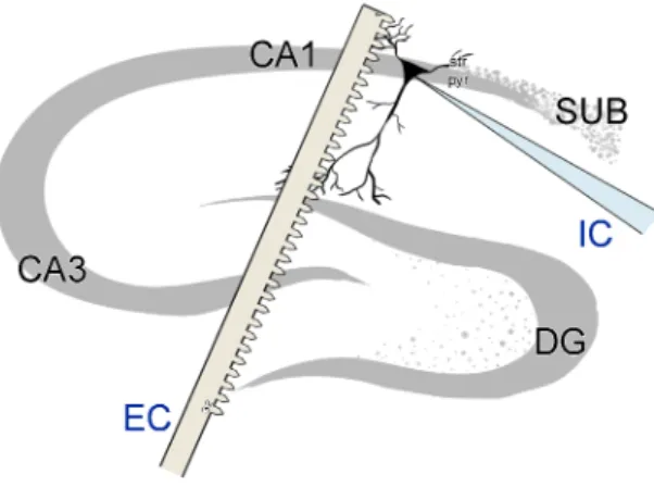
Figure 8:Schematics of the intra- and extracellular recording arrangements in the hippocampal CA1 pyra- midal region.
I have described the steps developed for the recording of co-localised and simultaneous intra- and extracellular signals with sub-millisecond accuracy. Extracellular recordings were complemented with a whole-cell patch-clamp recording in the closest vicinity of the silicon probe. Since these in vitroexperiments were designed under a two-photon microscope system, proper visual monitoring both for the electrode and for cell positions could
be achieved. Inclusion criteria for a successful simultaneous measurement were the following three conditions:
• I expected detection of at least 100 spike times for each 3-minute recording session to assure reasonable spike count in every data file (1).
• Holding current injected for maintaining stable spiking had to remain constant throughout each recording session and within a physiologi- cal range, ideally below 300 pA (2).
• After pre-processing, spikes of the patch-clamped cell should be de- tectable on at least one extracellular channel in the spike-triggered av- erage form (3).
Additionally, I have also performed a Ramp-test in the beginning of each whole-cell patch-clamp recording to characterise putative cell types and to determine various useful intracellular parameters of the patched cell.
Figure 9:2-D snapshots of two complete 3-D reconstructions of the complete neuronal morphologies and corresponding EC probe traces.
In addition to electrophysiology, concurrent two-photon imaging was included in the experimental protocol to take full advantage of the available multi-modal set-up. The field-of-view of the two-photon z-stack projection was set to cover both the track of the silicon probe and main cellular compartments (soma, apical trunk region, proximal apical and basal dendrites). Depth levels (z- dimension) typically ranged from -200 µm to 0 µm, (which is the surface of the slices). The resolution of the z-stack (or the step between stacks) was 2-3 µm, which was enough to capture small dendritic processes along the 3D morphology. In the end of the experimental protocol, patch pipette was carefully withdrawn, slice was removed from the dual perfusion chamber and fixated in paraformaldehyde solution forpost-hochistological reconstruction. Patch-clamped cells were passively filled with fluorescent markers (Alexa594 and Fluo-4) and with histological tracer molecule (Neurobiotin) via diffusion of the intracellular solution. In cases where all histological steps (fixation, reslicing, and the precipitation of Neurobiotin) were successful, neurons were chosen to be digitally reconstructed in 3D by the Neurolucida system. With the help of 3D reconstructions, Euclidean cell-electrode distances between contact sites and sub-cellular compartments could be calculated properly.
At the time of submission, a total of #16 neurons were successfully recorded from #12 animals by using the simultaneous and co-localised protocol introduced here in Thesis II.a. This library building is currently in progress and to be updated whenever a new successful, candidate data becomes available.
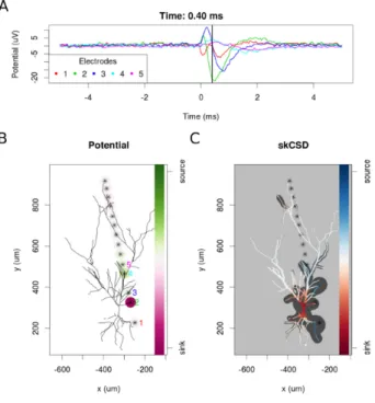
II.b. With the aid of my acquired simultaneous electrophysiology dataset, a novel single-cell source localisation method called skCSD (de- veloped in a collaborative study) was firstly tested and validated on real experimental data with reconstructed neuronal morphology.
Having all these structural data together with simultaneous electro- physiology made possible to support model-based single-cell calculations with real experimental validation. This was the first time in literature, when researchers were able to compute single-cell level current source densities (or the transmembrane sources of the extracellularly measured potentials) along a detailed, real neuronal morphology. The observed spatio-temporal dynamics was in accordance with previously estimated patterns, namely that somatic currents mostly dominate the process of spike generation and they are balanced by corresponding counter currents along the proximal
Figure 10:skCSD reconstruction of spike-triggered average for a CA1 pyramidal cell. Time course of the EC potentials for 5 selected channels (A). 2-D projection of the cell and silicon probe positions with overlaid EC potential dynamics (B). Reconstruction of current source densities based on the measured EC potentials and modelled cellular morphology (C).
dendritic tree. This example demonstrated the feasibility of the skCSD method on recorded experimental data and may help in planning further experiments (as an iterative ’experiment-data analysis cycle’), aiming to reveal the spatial distribution and temporal dynamics of the synaptic input currents which evoke the firing of a neuron.
Publication related to the II. Thesis group:[I].
III. Thesis group: Combination of multi-channel extracellu- lar recordings and two-photon laser scanning microscopy imaging for the experimental characterisation of photoelec- tric artefacts
III.a. I have designed and carried out experiments for acquiring both laser artefact-free and laser contaminated data in one experimental session. My generated dataset is suitable for the characterisation of the generated photoelectric artefact and for the comprehensive evaluation of any artefact suppression algorithm.
Figure 11:Imaging the close vicinity of the implanted silicon probe in a combined EC-2P recording.
Imaging reveals both activities of somas and dendtrites (examples are shown on the left) in the same FoV of EC contact sites (highlighted by yellow circles).
Firstly, I have proved experimentally, that the generated photoelectric artefact is not identical with a simple scanner noise and it has a varying nature across multiple contact sites. Moreover, I have carried simultaneous two-photon imaging and extracellular silicon probe recordings in the same field-of-view. Two-photon excitation was set to be sufficient for detecting re- liable transgenic Ca2+signals without causing damage on the metal-based extracellular probe. With the aid of this experiment, multi-cellular Ca2+sig- nals and co-localised extracellularly detected units become comparable.
Figure 12:Filtered signal (red) is overlaid on the original (unfiltered, green) data. It is visible that the laser generated photoelectric artefact was significantly reduced and SUAs emerged from the background noise.
III.b. I have shown that single unit clusters can be detectable even under contaminated signal conditions, as well as clustered spikes before-, under- and after laser scanning remained tractable based on their signal amplitudes, waveforms and autocorrelograms.
Spike sorted single unit activities remained tractable before and after the laser scanning period after applying an iterative artefact filtering process (which algorithm was developed in collaboration with other colleagues).
However, I also pointed out that in a few cases photoelectric artefact (due to laser scanning) may have some effects on the dynamics of scanned cells (e.g. indirect excitation mediated by photoelectric currents).
Publication related to the III. Thesis group:[III].
4 Application of results and future perspectives
All the three experimental problems I have addressed throughout the dissertation are very application oriented, even if their current stages of re- alisation are different.
Firstly, I have presented and tested penetrating a silicon-based multi- channel spiky probe. While the current shank design proved a significant im- provement forin vitrorecordings, it is certainly not designed for long-term recordings. Future developments may enable such devices to be further de- signed in both chronic and acutein vivoapplications via smaller modifica- tions. Another possible future application would be the investigation of dif- ferent tissue samples using the same probes, e.g. post-operative humanin vitrotissue samples. Dimensions of the spiky probes are variable in a wide range, and the longest spiky version (equipped with the largest inter-contact distances of 100µm) would cover a sufficiently large area to be applicable for human neocortical slices and for multi-layered structures.
Regarding the second Thesis group, multiple potential collaborators indi- cated their interest in using my dataset containing co-localised and simul- taneous intra- and extracellular recordings with corresponding morphol- ogy. However, as I have mentioned before, some of the intracellularly patch- clamped cells were not visible immediately on the raw extracellular signal, thus they prevented the application of blind spike sorting on the extracel- lular single unit activities. In these cases, the calculation of spike triggered averages was the only way for comparing intra- and extracellular signals. As a possible continuation of this study, my goal will be to get even closer to the cells with the silicon probes by further optimizing the measurement set-up and process and as a consequence, to record more cells with sortable spike amplitudes. I will also focus on recording more interneurons to expand the usability of the ground-truth dataset. The simultaneous experimental pro- tocol can also be applied on different regions, such as neocortex or human post-operative slices.
The first collaborative application of the dataset has already occurred within the framework of the theses. We have shown how the series of extracellu- lar recordings in combination with neuronal morphology can be used to estimate current source densities located on the cell contributing to the recorded extracellular potential. Unlike simple somatic patch-clamp record- ings, here we are able to obtain intracellular information along the whole cellular morphology, not just at a single point. This new approach may move the field a bit forward by opening a new experimental window into informa- tion processing by single cells allowing for their global monitoring, which
was not possible previously. It will be equally important from the theoretical side, as theoretical paradigms are informed by what we are able to extract from available measurements. Since it is now feasible experimentally to ob- tain the relevant data with the aid of my developed protocol, I believe that the data proposed here may find its uses in future works to constrain the biophysical models of the neuron membrane, as well as guide new discov- eries by giving a coherent view of the global synaptic bombardment within a targeted neuron.
Lastly, I have proposed an experimental solution for the investigation and elimination of photoelectric artefacts in co-localised two-photon imaging and extracellular silicon probe recordings. This study showed that it is possi- ble to get insight into the extracellular activity even under two-photon laser scanning. However, I have found putative modulations in spiking behaviour caused by the photoelectric artefact which were undesirable consequences of simultaneous scanning. Further investigations are to be performed to re- solve the indirect stimulation effect of laser generated artefacts. Regarding the application of the filtering algorithm, it would be beneficial to develop an automated and more robust software which is independent of the type of silicon probe used and can be applied to other layouts or different channel counts. The greatest challenge would be to search for correlations and causal roles between co-localised fluorescently active (or ’blinking’) cells and extra- cellularly detected single unit spikes. Similarly to the second Thesis group, the application of simultaneous, multi-modal measurements may give rise to novel findings in designing improved neural interfaces.
5 Author’s publications related to the theses
Peer-reviewed journal publications
I. D. Cserpán, D. Meszéna, L. Wittner, K. Tóth, I. Ulbert, Z. Somogyvári and D. Wójcik. Revealing the distribution of transmembrane currents along the dendritic tree of a neuron with known morphology from ex- tracellular recordings. eLIFE, 6:e29384, 2017. (IF: 7.73, Q1/D1) DOI:
10.7554/eLife.29384
II. D. Meszéna, B. P Kerekes, I. Pál, G. Orbán, R. Fiáth, T. Holzhammer, P.
Ruther, I. Ulbert and G. Márton. A silicon-based spiky probe providing improved cell accessibility for in vitro brain slice recordings.SENSORS
&ACTUATORS B – CHEM, 297C, 126649, 2019. (IF: 6.39, Q1/D1) DOI:
10.1016/j.snb.2019.126649
III. G. Orbán, D. Meszéna, K. R. Tasnády, I. Ulbert and G. Márton.
Method for spike detection from microelectrode array recordings contaminated by artifacts of simultaneous two-photon imaging.
PLOS ONE, 14(8): e0221510, 2019. (IF: 2.78, Q1) DOI: 10.1371/jour- nal.pone.0221510
Other publications of the author
Preprints and works in progress
• R. Fiáth, D. Meszéna, M. Boda, P. Barthó, P. Ruther and I. Ulbert.
Recording site placement on planar silicon-based probes affects neural signal quality: edge sites enhance acute recording perfor- mance.SCIENTIFIC REPORTS, 2020. (IF: 4.01, Q1/D1) (Under review) (Preprint onBioRxiv) DOI: 10.1101/2020.06.01.127308
• G. Dimitriadis, J. P. Neto, A. Aarts, [. . . ] G. Marton, D. Meszéna, S. Mitra, [. . . ] B. Raducanu, P. Ruther, T. Schroeder, W. Singer, P. Tiesinga, I. Ul- bert, S. Wang, M. Welkenhuysen, A. R Kampff. Why not record from every channel with a CMOS scanning probe?PNAS, 2020. (IF: 9.58, Q1/D1) (Under review) (Preprint onBioRxiv) DOI: 10.1101/275818
• T. Marek, G. Orbán, D. Meszéna, G. Márton, I. Ulbert, G. Mészáros and Zs. Keresztes. Optimization Aspects of Electrodeposition of Pho- toluminescent Conductive Polymer Layer onto Neural Microelectrode Arrays.MATERIALS CHEMISTRY AND PHYSICS, 2020. (IF: 2.78, Q2) (Under review)
Peer-reviewed journal publications
• G. Márton, E. Z. Tóth, L. Wittner, R. Fiáth, D.Pinke, G. Orbán, D. Meszéna, I. Pál, E. L. Gy˝ori, Z. Bereczki, Á. Kandrács, K. T. Hofer, A. Pongrácz, I. Ulbert and K. Tóth. The neural tissue around SU-8 im- plants: a quantitativein vivobiocompatibility study.MATERIALS SCI- ENCE&ENGINEERING C, 112C, 110870, 2020. (IF: 4.96, Q1/D1) DOI:
10.1016/j.snb.2019.126649
• A. Zátonyi, G. Orbán, R. Modi, G. Márton, D. Meszéna, I. Ulbert, A.
Pongrácz, M. Ecker, W. E. Voit, A. Joshi-Imre, and Z. Fekete. A soft- ening laminar electrode for recording single unit activity from the rat hippocampus.SCIENTIFIC REPORTS, vol. 9, no. 1, p. 2321, 2019. (IF:
4.12, Q1/D1) DOI: 10.1038/s41598-019-39835-6 Patent
• I. Ulbert, G. Márton, D. Meszéna, B.P. Kerekes, G. Orbán, K.R. Tasnády, D. Pinke. A design of an ionic conductance-based multielectrode sys- tem for mitigating photoelectric artefacts. Hungarian Patent Applica- tion (pending),Registration number: 45B01FEF1C, File number: P 17 00527, Date: 15th December 2017.
Selected talks and posters
• D. Meszéna, G. Orbán, K. R. Tasnády, I. Ulbert and G. Márton. Towards co- localised microelectrode array recordings and two-photon microscopy.Hun- Doc 2020, Szeged, Hungary, 2020. (Invited talk)
• Z. Somogyvári, D. Meszéna, D. Cserpán, L. Wittner and I. Ulbert. Spatio- temporal membrane potential and resistive current reconstruction from par- allel multielectrode array and intracellular measurements in single neurons.
10t hIBRO World Congress of Neuroscience, Daegu, Korea, 2019. (Poster)
• R. Fiáth, D. Meszéna, Mihály Boda and I. Ulbert. Impact of the recording site location on the recording performance of silicon probes in acute experiments.
FENS Regional Meeting, Belgrade, Serbia, 2019. (Poster)
• E. Z. Tóth, D. Meszéna, A Dublecz, D.Pálfi, K. Tóth, L. Er˝oss, A. Bagó, D. Fabó, I.
Ulbert and L. Wittner. Back-propagating action potentials in human neocor- tical pyramidal cells and interneurons: A preliminary study.Gordon Research Conference: Dendrites, Ventura, CA, US, 2019. (Poster)
• D. Meszéna, I. Pál, B. P. Kerekes, G. Marton, K. Tóth, L. Wittner, Z. Somogyvári and I Ulbert. Simultaneous intra- and linear extracellular recordings with cor- responding morphology: towards a ground-truth data for multichannel elec- trodes.SfN Neuroscience 2018, San Diego, CA, US 2018. (Poster)
• K. Tóth, E. Z. Tóth, L. Wittner, R. Fiáth, D. Meszéna, I. Pál, E. L. Gy˝ori, D. Pinke, Z. Bereczki, G. Orbán, A. Pongrácz, I. Ulbert and G. Márton. Biocompatibility of the SU-8 in the central nervous system.SfN Neuroscience 2018, San Diego, CA, US 2018. (Poster)
• G. Orbán, T. Marek, D. Meszéna, B. P. Kerekes, K. R. Tasnády, I. Ulbert, G.
Mészáros, Zs. Keresztes, G. Márton. Fluorescent conductive polymer coat- ing on implanted microelectrodes for visualization under two-photon micro- scopes. 11t hFENS Forum of Neuroscience, Berlin, Germany, 2018. (Poster)
• D. Meszéna, B. P. Kerekes, I. Pál, T. Holzhammer, P. Ruther, I. Ulbert and G.
Márton. A novel, silicon-based spiky probe providing improved cell accessi- bility for in vitro brain slice recordings.Gordon Research Conference: Neuro- electronic Interfaces, Galveston, Texas, US, 2018. (Poster)
• D. Cserpán, D. Meszéna, L. Wittner, K. Tóth, I. Ulbert, Z. Somogyvári and D. Wójcik. Revealing the Distribution of Transmembrane Currents along the Dendritic Tree of a Neuron with Known Morphology from Extracellular Recordings. 2ndNencki Symposium, Warsaw, Poland, 2017. (Poster)
• I Pál, KT. Hofer, B. P. Kerekes, K. Tóth, B. Rózsa, D. Meszéna and I. Ulbert. Mod- ulation of interictal-like and spontaneous population activity by microsurgi- cal intervention in rat brain slices, 10t hForum of Neuroscience, Copenhagen, Denmark, 2016. (Poster)
• D. Meszéna and I. Ulbert. Simultaneously recorded multimodal signals in the hippocampal CA1 region, in vitro.EMBO Practical Course in Advanced Optical Microscopy, Marine Biological Association, Plymouth, United Kingdom, 2016.
(Poster)
• D. Meszéna, E. Lakatos, G. Szederkényi. Sensitivity analysis and parameter es- timation of a human blood glucose regulatory system model. In.: Proceedings of the 11t hInternational Workshop on Computational Systems Biology, TISCP 64, pp. 28, Lisbon, Portugal, 2014. (Talk)
• E. Lakatos, D. Meszéna, G. Szederkényi. Identifiability analysis and improved parameter estimation of a human blood glucose control system model.LEC- TURE NOTES IN COMPUTER SCIENCE, A. Gupta and T.A. Henzinger (Eds.):
CMSB 2013, LNBI 8130 Springer, pp. 248-249, 2013. (Talk) (IF: 1.12, Q2), DOI:
10.1007/978-3-642-40708-6
• L. Négyessy, J. Minich, D. Meszéna, A. Buzás, B. Jákli, M. Bányai, E. Procyk, P.
Barone, F. Bazsó. From Neuronal Communication to the Flow of Information in the Cerebral Cortex. 11t hDigital Speech and Image Processing, Kovacica, Serbia, 2012. (Talk)
In Annual Proceedings of the PPCU FITB Doctoral School
• D. Meszéna. Towards a better understanding of intra- and extracellular neural signals and their relationships.in PhD Proceedings Annual Issues of the Doctoral School, Faculty of Information Technology and Bionics, Pázmány Péter Catholic University– 2017. G.
Prószéky, P. Szolgay Eds. Budapest: Pázmány University ePress, 2017, pp 27–27.
• D. Meszéna. Targeted simultaneous recordings on rat hippocampal CA1 cells, in vitro.in PhD Proceedings Annual Issues of the Doctoral School, Faculty of Information Technol- ogy and Bionics, Pázmány Péter Catholic University– 2016. G. Prószéky, P. Szolgay Eds.
Budapest: Pázmány University ePress, 2016, pp 69–71.
• D. Meszéna. Using two-photon imaging combined with simultaneous recordings to val- idate CSD analysis.in PhD Proceedings Annual Issues of the Doctoral School, Faculty of Information Technology and Bionics, Pázmány Péter Catholic University– 2015. G.
Prószéky, P. Szolgay Eds. Budapest: Pázmány University ePress, 2015, pp 81–84.
References
[1] Gyorgy Buzsaki, Costas A Anastassiou, and Christof Koch. The origin of extracellular fields and currents-EEG, ECoG, LFP and spikes.Nature re- views neuroscience, 13(6):407, 2012.
[2] Kenneth D Harris, Rodrigo Quian Quiroga, Jeremy Freeman, and Spencer L Smith. Improving data quality in neuronal population record- ings.Nature neuroscience, 19(9):1165, 2016.
[3] Marie Engelene J Obien, Kosmas Deligkaris, Torsten Bullmann, Douglas J Bakkum, and Urs Frey. Revealing neuronal function through microelec- trode array recordings.Frontiers in neuroscience, 8:423, 2015.
[4] Darrell A Henze, Zsolt Borhegyi, Jozsef Csicsvari, Akira Mamiya, Kenneth D Harris, and Gyorgy Buzsaki. Intracellular features predicted by extra- cellular recordings in the hippocampus in vivo.Journal of neurophysiol- ogy, 84(1):390-400, 2000.
[5] Woodrow L Shew, Timothy Bellay, and Dietmar Plenz. Simultaneous multi-electrode array recording and two-photon calcium imaging of neural activity.Journal of neuroscience methods, 192(1):75-82, 2010.
[6] Brian D Allen, Caroline Moore-Kochlacs, Jacob G Bernstein, Justin P Kin- ney, Jorg Scholvin, Luis F Seoane, Chris Chronopoulos, Charlie Laman- tia, Suhasa B Kodandaramaiah, Max Tegmark, et al. Automated in vivo patch-clamp evaluation of extracellular multielectrode array spike recording capability.Journal of neurophysiology, 120(5):2182-2200, 2018.
[7] Zoltan Somogyvari, Laszlo Zalanyi, Istvan Ulbert, and Peter Erdi. Model- based source localization of extracellular action potentials.Journal of neuroscience methods, 147(2):126-137, 2005.
[8] Zoltan Somogyvari, Dorottya Cserpan, Istvan Ulbert, and Peter Erdi. Lo- calization of single-cell current sources based on extracellular poten- tial patterns: the spike CSD method.European Journal of neuroscience, 36(10):3299-3313, 2012.
[9] Gaute T Einevoll, Christoph Kayser, Nikos K Logothetis, and Stefano Panzeri. Modelling and analysis of local field potentials for studying the function of cortical circuits.Nature reviews neuroscience, 14(11):770, 2013.