Chapter Title Fluorogenic Tetrazine-Siliconrhodamine Probe for the Labeling of Noncanonical Amino Acid Tagged Proteins
Copyright Year 2018
Copyright Holder Springer Science+Business Media, LLC
Author Family Name Kozma
Particle
Given Name Eszter
Suffix
Division Chemical Biology Research Group,
Research Centre for Natural Sciences, Institute of Organic Chemistry
Organization/University Hungarian Academy of Sciences
Address Budapest, Hungary
Author Family Name Paci
Particle
Given Name Giulia
Suffix
Division Structural and Computational
Biology Unit, Cell Biology and Biophysics Unit
Organization/University European Molecular Biology Laboratory
Address Heidelberg, Germany
Author Family Name Girona
Particle
Given Name Gemma Estrada
Suffix
Division Structural and Computational
Biology Unit, Cell Biology and Biophysics Unit
Organization/University European Molecular Biology Laboratory
Address Heidelberg, Germany
Author Family Name Lemke
Particle
Given Name Edward A.
Suffix
Structural and Computational Biology Unit
& Cell Biology and Biophysics Unit, Meyerhofstrasse 1, Heidelberg, 69117 Germany
Structural and Computational Biology Unit
& Cell Biology and Biophysics Unit, Meyerhofstrasse 1, Heidelberg, 69117 Germany
Chemical Biology Research Group,
Research Centre for Natural Sciences,
Institute of Organic Chemistry, Hungarian
Academy of Sciences, Magyar tudósok
krt 2, Budapest, 1117 Hungary
Division Structural and Computational Biology Unit, Cell Biology and Biophysics Unit
Organization/University European Molecular Biology Laboratory
Address Heidelberg, Germany
Corresponding Author Family Name Kele
Particle
Given Name Péter
Suffix
Division Chemical Biology Research Group,
Research Centre for Natural Sciences, Institute of Organic Chemistry
Organization/University Hungarian Academy of Sciences
Address Budapest, Hungary
Email kele.peter@ttk.mta.hu
Abstract Tetrazine-bearing fluorescent labels enable site-specific tagging of proteins that are genetically manipulated with dienophile modified noncanonical amino acids. The inverse electron demand Diels-Alder reaction between the tetrazine and the dienophile fulfills the criteria of bioorthogonality allowing fluorescent labeling schemes of live cells. Here, we describe the detailed synthetic and labeling protocols of a near infrared emitting siliconrhodamine-tetrazine probe suitable for super-resolution imaging of residue-specifically engineered proteins in mammalian cells.
Keywords (separated by “ - ”)
Bioorthogonality - Tetrazine - Fluorogenicity - NIR emission - Super-resolution microscopy
Unit, Meyerhofstrasse 1, Heidelberg, 69117 Germany
3 Departments of Biology and Chemistry, Pharmacy and Geosciences, Johannes Gutenberg-University Mainz, Johannes-von-Mullerweg 6
4 Institute of Molecular Biology (IMB), Mainz, 55128 Germany
Edward Lemke (ed.), Noncanonical Amino Acids: Methods and Protocols, Methods in Molecular Biology, vol. 1728, DOI https://doi.org/10.1007/978-1-4939-7574-7_22, © Springer Science+Business Media, LLC 2018
Chapter 22
Fluorogenic Tetrazine-Siliconrhodamine Probe
for the Labeling of Noncanonical Amino Acid Tagged Proteins
Eszter Kozma, Giulia Paci, Gemma Estrada Girona, Edward A. Lemke, and Péter Kele
Abstract
Tetrazine-bearing fluorescent labels enable site-specific tagging of proteins that are genetically manipu- lated with dienophile modified noncanonical amino acids. The inverse electron demand Diels-Alder reac- tion between the tetrazine and the dienophile fulfills the criteria of bioorthogonality allowing fluorescent labeling schemes of live cells. Here, we describe the detailed synthetic and labeling protocols of a near infrared emitting siliconrhodamine-tetrazine probe suitable for super-resolution imaging of residue-specif- ically engineered proteins in mammalian cells.
Key words Bioorthogonality, Tetrazine, Fluorogenicity, NIR emission, Super-resolution microscopy
1 Introduction
Emerging super-resolution microscopy (SRM) techniques have brought substantial progress in the exploration of biomolecular processes in the sub-diffraction range [1–3]. Live organisms can now be studied in fine details, yet, improvements can result in further increase of resolution and enable new biological insights.
To address current limitations that impede such improvements, small synthetic dyes with suitable spectral characteristics that allow site-specific tagging of intracellular structures even under in vivo conditions are needed [4, 5]. Preferred synthetically tai- lored, small-sized organic fluorophores are membrane perme- ant, photostable, brightly fluorescent, and allow minimal background labeling and autofluorescence in order to result in a high signal-to-noise ratio. The means by which such ideal probes are installed onto the biomolecule of interest is also cru- cial. The applied chemistry should be biocompatible and highly selective. Such chemical transformations are termed
1
2 3 4
5 6
7 8 9 10 11 12 13 14
15
16 17 18 19 20 21 22 23 24 25 26 27 28 29 30 31
bioorthogonal [6–8]. Most of the time, fast kinetics are also required. Inverse electron demand Diels-Alder (IEDDA) cyclo- addition of tetrazines and strained unsaturated ring systems enable fast and highly selective reactions [9, 10]. Most IEDDA labeling schemes rely on the use of tetrazine bearing fluorescent probes in combination with cyclooctyne or trans-cyclooctene modified biomolecules (e.g., by means of genetically encoded noncanonical amino acids, ncAAs). Furthermore, tetrazine scaf- folds can efficiently quench fluorescence of dyes giving rise to fluorogenic scaffolds [11–15].
In live cell imaging applications, phototoxicity and autofluo- rescence can be minimized if the spectral characteristics of the applied probe allow far-red/near-infrared (NIR) excitation/
emission. Since labeling schemes often apply large excess of the labeling species, background fluorescence of unreacted probes bound nonspecifically to hydrophobic surfaces is often encoun- tered, and several washing cycles are needed before imaging, which excludes labeling of, e.g., proteins with rapid turnovers.
So-called fluorogenic probes efficiently reduce background fluo- rescence as they are minimally fluorescent when bound nonspe- cifically but become intensely emitting upon the particular specific chemical reaction [16, 17].
Siliconrhodamines (SiRs) are widely used membrane-perme- able NIR dyes suitable for SRM applications [18–23]. Besides their high photostability and brightness, the unique environment-depen- dent fluorescence of carboxy-SiRs due to a polarity dependent lac- tone-formation offers an appealing opportunity to distinguish between specific and nonspecific labeling (polarity-based fluoroge- nicity) [18]. Carboxyl-SiRs exist in a fluorescent zwitterionic form when they are in polar environment such as near protein surfaces.
In non-polar microenvironments, however, they isomerize to their respective, non-fluorescent spirolactone form. These characteristics render carboxy-SiRs minimally emitting when bound nonspecifi- cally onto nonpolar surfaces. Despite their wide applications [21], there are only a few examples of SiR-based probes for site-specific labeling of proteins [18, 22, 23].
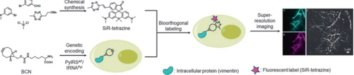
Herein, we describe the detailed synthesis of a bioorthogonally applicable, NIR-emitting, membrane permeable, double fluoro- genic carboxy-SiR suitable for SRM imaging, as well as the labeling scheme for genetically modified proteins in living cells (Fig. 1) [24].
We show the applicability of the method by labeling site-specifically an intracellular skeletal protein, vimentin, engineered with an IEDDA reactive ringstrained ncAA by means of Amber suppression using an orthogonal tRNA/tRNA synthetase system derived from Methanosarcia mazei as described previously [18, 25–30].We dem- onstrate the power of the developed SiR-dye in subsequent site- specific SRM imaging applications.
32 33 34 35 36 37 38 39 40 41 42 43 44 45 46 47 48 49 50 51 52 53 54 55 56 57 58 59 60 61 62 63 64 65 66 67 68 69 70 71 72 73 74 75 76 77 78
2 Materials
The laboratory should be equipped to perform basic chemical syn- thesis. All synthetic steps shall be performed in a chemical hood equipped with magnetic stirrer, nitrogen-line, and vacuum line.
The laboratory should be further equipped with a rotary evapora- tor (with a high vacuum pump), and a standard S1 cell culture. In this protocol specific examples of cell line, protein of interest, and microscopy setup are given, but the user should keep in mind that the method can easily be applied to other biological systems and that imaging can be performed on any fluorescence microscope.
1. Magnetic stirrer with a stand clamp.
2. 250 mL round bottom flask with a conical joint NS 29/32.
3. 500 mL round bottom flask with a conical joint NS 29/32.
4. 250 mL separatory funnel.
5. 500 mL Erlenmeyer flask.
6. Magnetic stir bar.
7. Bubbler filled with mineral oil.
8. Nitrogen gas.
9. Crystallizing dish.
10. Rotary evaporator.
11. High vacuum pump (like Vacuubrand Vario Pro PC3001 chemistry pumping unit).
12. Automated flash chromatographer (such as Teledyne Isco CombiFlash Rf+).
13. Empty solid load cartridge 65 g with three pieces of frit and a loading rod (like RedisepRf).
14. Silica gel (25–40 μm).
15. Analytical thin-layer chromatography (TLC) plates (such as silica gel 60 F254 precoated aluminum TLC plates from Merck).
2.1 Synthesis of 3,3′-(Dimethylsil- anediyl)bis(N,N- dimethylaniline) (2)
Fig. 1 Illustration of chemical synthesis, biorthogonal labeling, and subsequent imaging steps described in the protocol
79
80 81 82 83 84 85 86 87 88 89 90 91 92 93 94 95 96 97 98 99 100 101 102 103 104 105 106 107 108
16. 3-bromo-N,N-dimetylaniline (1, commercially available).
17. n-Butyllithium (n-BuLi, in 1.6 M solution in hexane, com- mercially available).
18. Dichlorodimethylsilane (SiCl2Me2, commercially available).
19. Absolute tetrahydrofurane (THF).
20. Dry ice.
21. Acetone.
22. Water (distilled or purified).
23. Ethyl acetate (EtOAc).
24. Saturated NaCl solution.
25. Anhydrous magnesium sulfate (MgSO4).
26. Dichloromethane (DCM).
27. Celite.
28. Hexane.
1. Magnetic stirrer with stand clamp and oil bath.
2. 250 mL round bottom flask with a conical joint NS 29/32.
3. Reflux condenser for with a conical joint NS 29/32.
4. Magnetic stir bar.
5. Rotary evaporator.
6. High vacuum pump (like Vacuubrand Vario Pro PC3001 chemistry pumping unit).
7. Fritted funnel.
8. Desiccator.
9. 5-bromophtalide (commercially available).
10. N-bromosuccinimide (NBS) (commercially available).
11. Azobisisobutyronitrile (AIBN) (commercially available).
12. 1,2-dichloroethane (DCE).
13. Water (distilled or purified).
1. Magnetic stirrer with stand clamp and oil bath.
2. 2–6 mL borosilicate microwave vial (like Anton Paar G10) with snap cap and PTFE-coated silicone septa.
3. Magnetic stir bar.
4. Sonicator (such as Selecta ultrasons 6.5 L).
5. Rotary evaporator.
6. High vacuum pump (like Vacuubrand Vario Pro PC3001 chemistry pumping unit).
7. Chromatography column with fused-in-frit and PTFE stopcock.
2.2 Synthesis of 4-Bromo-2- formylbenzoic acid (3)
2.3 Synthesis of 6′-Bromo-3,7- bis(dimethylamino)- 5,5-dimethyl-3′H,5H- spiro[dibenzo[b,e]
siline-10,1′-
isobenzofuran]-3′-one (SiR-Br, 4)
109 110 111 112 113 114 115 116 117 118 119 120 121 122 123 124 125 126 127 128 129 130 131 132 133 134 135 136 137 138 139 140 141 142 143 144 145 146 147
8. Silica gel (60–200 μM).
9. Preparative TLC plate (glass-backed such as Kiesegel 60 F254
20 × 20 cm, 2 mm).
10. Glass chamber for preparative TLC purification.
11. Analytical TLC plates (such as silica gel 60 F254 precoated alu- minum TLC plates from Merck).
12. Compound 2 (for preparation see Subheading 3.1).
13. Copper(II) bromide (CuBr2) (commercially available).
14. Compound 3 (for preparation see Subheading 3.2).
15. DCM.
16. Celite.
17. Hexane.
18. EtOAc.
19. Triethylamine (Et3N).
20. Methanol (MeOH).
1. Magnetic stirrer with stand clamp.
2. 250 mL round bottom flask with a conical joint NS 29/32.
3. 500 mL two-neck round bottom flask.
4. Magnetic stir bar.
5. Dropping funnel.
6. Analytical TLC plates (such as silica gel 60 F254 precoated alu- minum TLC plates from Merck).
7. Rotary evaporator.
8. High vacuum pump (like Vacuubrand Vario Pro PC3001 chemistry pumping unit).
9. 3-Hydroxypropionitrile (commercially available).
10. Acetyl chloride (commercially available).
11. Absolute ethanol (EtOH).
12. NaCl.
13. cc. H2SO4.
14. Diethyl ether (Et2O).
1. Magnetic stirrer with stand clamp.
2. 250 mL round bottom flask with a conical joint NS 29/32.
3. 100 mL round bottom flask with a conical joint NS 29/32.
4. 500 mL Erlenmeyer flask.
5. 250 mL separatory funnel.
2.4 Synthesis of Ethyl 3-hydroxypro- pionimidate
hydrochloride (9)
2.5 Synthesis of 2-(6-Methyl-1,2,4,5- tetrazin-3-yl)ethyl methanesulfonate (OMs-tet, 5)
148 149 150 151 152 153 154 155 156 157 158 159 160 161 162 163 164 165 166 167 168 169 170 171 172 173 174 175 176 177 178 179 180 181 182 183 184 185
6. Vacuum line (with vacuum pump like Vacuubrand rotary vane pump RZ 2.5).
7. Rotary evaporator.
8. High vacuum pump (like Vacuubrand Vario Pro PC3001 chemistry pumping unit).
9. Magnetic stir bar.
10. Bubbler filled with mineral oil.
11. Nitrogen gas.
12. Crystallizing dish for ice/water bath.
13. Automated flash chromatographer (such as Teledyne Isco CombiFlash Rf+).
14. Empty solid load cartridge 65 g with three pieces of frit and a loading rod (like RedisepRf).
15. Silica gel (25–40 μm).
16. Analytical TLC plates (such as silica gel 60 F254 precoated alu- minum TLC plates from Merck).
17. Compound 9 (for preparation see Subheading 3.4).
18. Acetonitrile (MeCN).
19. Hydrazine hydrate (N2H4 50–60%) (commercially available).
20. Sodium nitrite (NaNO2) (commercially available).
21. Distilled water.
22. Ice.
23. cc. HCl.
24. EtOAc.
25. Saturated NaCl solution.
26. Anhydrous MgSO4 (commercially available).
27. DCM.
28. Celite.
29. Hexane.
30. Methanesulfonyl chloride (MsCl, commercially available).
31. Et3N.
1. 2–6 mL borosilicate microwave vial (like Anton Paar G10) with snap cap and PTFE-coated silicone septa.
2. 50 mL round bottom flask with conical joint NS 29/32.
3. Magnetic stir bar.
4. Microwave reactor (such as Anton Paar Monowave 300).
5. Analytical TLC plates (such as silica gel 60 F254 precoated alu- minum TLC plates from Merck).
2.6 Synthesis of (E)-3,7-
Bis(dimethylamino)- 5,5-dimethyl-6′-(2-(6- methyl-1,2,4,5- tetrazin-3-yl) vinyl)-3′H,5H- spiro[dibenzo[b,e]
siline-10,1′-
isobenzofuran]-3′-one (SiR-tetrazine, 6)
186 187 188 189 190 191 192 193 194 195 196 197 198 199 200 201 202 203 204 205 206 207 208 209 210 211 212 213 214 215 216 217 218 219 220 221 222 223 224
6. Rotary evaporator.
7. High vacuum pump (like Vacuubrand Vario Pro PC3001 chemistry pumping unit).
8. Preparative TLC plate (such as glass-backed Kiesegel 60 F254
20 × 20 cm, 2 mm).
9. Glass chamber for preparative TLC.
10. Preparative HPLC system.
11. Preparative C18 column (such as Gemini 5 μm C18 110 Å LC column 150 × 21.2 mm).
12. Round bottom flask freeze-drier for lyophilization.
13. SiR-Br (compound 4, for preparation see Subheading 3.3).
14. OMs-tet (compound 5, for preparation see Subheading 3.5).
15. Tris(dibenzylideneacetone)dipalladium(0) (Pd2(dba)3, com- mercially available).
16. 1,2,3,4,5-Pentaphenyl-1′-(di-tert-butylphosphino)ferrocene (QPhos, commercially available).
17. Anhydrous dimethylformamide (DMF).
18. N,N-dicyclohexylmethylamine ((Cy)2NMe, commercially available).
19. DCM.
20. Hexane.
21. EtOAc.
22. MeCN.
23. Distilled water.
Note that many steps are similar, if not even identical to proce- dures described in Chapter 18, by Nikic et al. [31].
1. COS-7 (Sigma 87021302) cell line or any other cell line of interest.
2. 1× PBS (phosphate-buffered saline).
3. Dulbeccos’s modified Eagle medium (DMEM, Gibco 41965039) supplemented with 10% (v/v) fetal bovine serum (FBS, Sigma F7524), 1% (v/v) penicillin-streptomycin 10,000 U/mL (Gibco 15140122), 1% (v/v) 100 mM sodium pyruvate (Gibco 11360070), and 1% (v/v) 200 mM l-gluta- mine (Sigma G7513). Store at 4 °C (see Note 1). Other manu- facturers providing similar formulations could also be used.
4. Trypsin-EDTA (Gibco 25300054 or similar). Long-term storage at −20 °C. Once thawed, keep at 4 °C.
5. Hemocytometer.
2.7 Site-Specific Protein Engineering with the Ringstrained ncAA Bicyclo[6.1.0]
nonyne-Lysine (BCNendo): Cell Culture and Transfections
225 226 227 228 229 230 231 232 233 234 235 236 237 238 239 240 241 242 243 244 245 246 247 248 249 250 251 252 253 254 255 256 257 258 259 260 261 262 263 264
6. Cell culture incubator at 37 °C with a humidified 5% CO2
atmosphere.
7. Cell culture hood.
8. Water bath at 37 °C.
9. Sterile plastic ware (Falcon and microcentrifuge tubes, 100 mm cell culture dishes, serological pipettes).
10. 4-well Lab-Tek™ II Chambered Coverglass (Nunc™).
11. JetPrime transfection reagent (Polyplus, or other transfection reagents appropriate for the cell line of choice).
12. Eukaryotic expression vector for the Amber suppression machinery. In this protocol a vector with M. mazei NESPylRSAF/ tRNAPyl was used (available from Dr. Edward Lemke, EMBL, Heidelberg) (see Note 2).
13. Eukaryotic expression vector for the Amber mutant of the protein of interest. In this protocol pVimentinN116TAG-PSmOr- ange was used (see Notes 3 and 4).
14. Plasmid Maxiprep kit (low or endotoxin-free recommended, e.g., Qiagen 12362 or Invitrogen K210007).
15. Noncanonical amino acid stock: 100 mM endo BCN-l-Lysine (BCNendo, SiChem SC8014) in 15% (v/v) DMSO, 0.2 M NaOH. Store at −20 °C.
16. Sterile 1 M HEPES. Store at 4 °C.
17. Vortex.
18. Benchtop mini-centrifuge.
19. SiR-tetrazine dye of choice. Stock in DMSO at 0.5 mM.
20. 2% PFA: 2% paraformaldehyde in 1× PBS (see Note 5).
1. 20× TN buffer: 1 M Trizma base, 0.2 M NaCl, adjust pH to 8.0 with HCl and filter. Store at 4 °C.
2. 20% Glucose. Store at 4 °C for maximum 2 weeks.
3. 20× Glucose Oxidase/Catalase (GO/C) stock: 51% glycerol, 50 mM Tris–HCl pH 8.0, 800 μg/mL Catalase (Sigma C3155), 10 mg/mL Glucose Oxidase (Sigma G0543). Store at −20 °C.
4. MEA (Sigma 411000). Store at −20 °C, aliquotes can be reused if kept cold all the time.
5. GLOX-MEA buffer: 1× TN buffer, 10% glucose, 10 mM MEA, and 1× GO/C mix) in water (see Notes 6 and 7).
6. TIRF-microscope (e.g., Leica GSDIM) with appropriate laser and filter cube for dyes of choice (see Note 8) and software for data analysis (see Note 9).
2.8 Localization- Based Super- Resolution Imaging and Analysis
265 266 267 268 269 270 271 272 273 274 275 276 277 278 279 280 281 282 283 284 285 286 287 288 289 290 291 292 293 294 295 296 297 298 299 300 301 302 303 304
3 Methods
Perform all synthesis steps in a chemical hood (for safety instruc- tions, see Note 10) (see Scheme 1).
1. Weigh 5.0 g 3-bromo-N,N-dimethylaniline (1) with a glass pipette into a well-dried 250 mL round bottom flask with conical joint NS 29/32 equipped with a magnetic stir bar (see Note 11).
2. Close the flask with a fold over rubber septum, mount it above a magnetic stirrer using a stand clamp, and flush the flask carefully with nitrogen using a bubbler. Make sure the reaction is kept under nitrogen atmosphere until otherwise stated (see Note 12).
3. Transfer 100 mL absolute THF to the flask while stirring the solution.
4. Fill a large crystallizing dish with dry ice and add acetone very carefully until the temperature reaches −78 °C. Place the dish under the flask and make sure the temperature is kept constant during the reaction. Refill dry ice if needed.
5. When the temperature of the reaction mixture reaches −78 °C, add 17 mL n-butyllithium (in 1.6 M solution in hexane) drop- wise to the reaction mixture over the course of 15 min.
6. Stir the reaction at −78 °C under nitrogen atmosphere for 2 h.
7. Add 1.8 mL dichlorodimethylsilane dropwise.
8. Remove the cooling dish, and let the reaction mixture warm to room temperature. Keep stirring the reaction mixture for an additional 16 h (see Note 13).
3.1 Synthesis of 3,3′-(Dimethyl- silanediyl)bis(N,N- dimethylaniline) (2)
Scheme 1 Synthesis of SiR-tetrazine (6). Reaction conditions: (a) (1) n-BuLi, THF, −78 °C, 2 h; (2) SiCl2Me2, RT, 16 h, 71%; (b) CuBr2, 140 °C, 16 h, 21%; (c) Pd2(dba)3, QPhos, (Cy)2NMe, DMF, 50 °C, 40 min, MW, 48%
305
306 307 308 309 310 311 312 313 314 315 316 317 318 319 320 321 322 323 324 325 326 327 328 329
9. Remove the septum and very carefully quench the reaction with 30 mL of distilled water. Add only small portions of the water at once and wait until the bubbling stops before adding another portion.
10. Transfer the mixture to a 250 mL separatory funnel. Add 30 mL EtOAc and extract the aqueous phase (see Note 14). Repeat it two more times with 2 × 30 mL EtOAc. Combine the organic phases and wash it with saturated NaCl solution to remove any remaining water soluble components and impurities. In a 500 mL Erlenmeyer flask add anhydrous MgSO4 to the organic phase to remove water. Filter the solution to remove the drying agent.
11. Transfer the solution to a 500 mL round bottom flask with a conical joint NS 29/32 and remove the solvents under reduced pressure with a rotary evaporator set to 40 °C and 400 mbar.
Decrease the pressure until no additional solvent is evaporating.
12. Redissolve the oily residue in 20 mL DCM and add 5 g celite to the solution. Remove the solvent under reduced pressure set to 40 °C and 850 mbar on a rotary evaporator slowly decreasing the pressure until the celite is completely dry (see Note 15).
13. Fill a 65 g cartridge with silica gel up to 2/3, add a frit using a loading rod, transfer the celite from the previous step onto the top, place a second a frit on top of it, and place the column into an automated flash chromatographer. Start the chromatographic separation using a 1–15% EtOAc gradient in hexane over 25 min.
Use 254 and 280 nm detection wavelengths for fraction collec- tion. Check the collected fractions by TLC (see Notes 13 and 16).
14. Combine the fractions containing purely the product in a round bottom flask, remove the solvent under reduced pressure with a rotary evaporator set to 40 °C and 300 mbar. Further decrease the pressure until no additional solvent is evaporating.
15. Collect the light yellow oil formed and store it at 4 °C until further use (see Note 17).
1. Weigh 4.582 g 5-bromophtalide and add it to a 250 mL round bottom flask with a conical joint NS 29/32 equipped with a magnetic stir bar.
2. Weigh 4.209 g NBS and add it to the 250 mL round bottom flask.
3. Weigh 177 mg AIBN and mix it with the solid compounds in the 250 mL round bottom flask (see Notes 18 and 19).
4. Dissolve the solid in 100 mL dichloroethane (DCE) and mount the flask using a stand clamp and immerse it in an oil bath on a magnetic stirrer. Adjust a reflux condenser with a conical joint NS 29/32.
3.2 Synthesis of 4-Bromo-2-
formylbenzoic acid (3) (Scheme 2)
330 331 332 333 334 335 336 337 338 339 340 341 342 343 344 345 346 347 348 349 350 351 352 353 354 355 356 357 358 359 360 361 362 363 364 365 366 367 368 369 370 371 372 373
5. While continuously stirring, heat the reaction mixture to 88 °C until the solvent starts to reflux and keep the tempera- ture for an additional 2 h (see Note 20).
6. Let the reaction mixture cool to room temperature. Close the flask with a fold over rubber septum and keep it at −20 °C for 2 h.
7. Filter the precipitate and collect the filtrate.
8. Using a 250 mL round bottom flask, remove the solvent under reduced pressure with a rotary evaporator set to 40 °C and 200 mbar. Further decrease the pressure until no addi- tional solvent is evaporating. A white crystalline residue forms.
9. Suspend the crystals in 50 mL water in a 250 mL round bot- tom flask with a conical joint NS 29/32. Adjust a condenser with a conical joint NS 29/32 and mount the flask using a stand clamp and immerse it in an oil bath on a magnetic stirrer.
Start stirring the suspension.
10. Adjust the temperature to 100 °C and keep the suspension refluxed for 2 h under continuous stirring (see Note 21).
11. Let the reaction mixture cool to room temperature. Close the flask with a fold over rubber septum and keep it at 4 °C for 16 h.
12. Collect the precipitate using a fritted funnel by applying vac- uum. Wash the precipitate with 2 × 10 mL ice-cold water.
Remove as much water as possible from the precipitate and then place the funnel into a desiccator, supplied with a drying agent, under vacuum for 24 h to remove any residual water.
13. Remove the funnel from the desiccator and collect the white crys- talline powder. Store it at 4 °C until further use (see Note 17).
1. Weigh 641 mg compound 3 and transfer it to a microwave borosilicate glass vial (2–6 mL) equipped with a magnetic stir bar.
2. Weigh 13 mg CuBr2 and add it to the microwave vial.
3. Weigh 167 mg of compound 2 and add it to the vial using a pipette. Slightly mix the mixture.
4. Close the tube with a snap cap containing a PTFE-coated silicone septum and mount it using a stand clamp and immerse it in an oil bath (covered 2/3 in oil) on a magnetic stirrer. Start stirring.
3.3 Synthesis of 6′-Bromo-3,7- bis(dimethylamino)- 5,5-dimethyl-3′H,5H- spiro[dibenzo[b,e]
siline-10,1′-
isobenzofuran]-3′-one (SiR-Br, 4)
Scheme 2 Synthesis of 4-bromo-2-formylbenzoic acid (3). Reaction conditions:
(1) NBS, AIBN, DCE, reflux, 2 h; (2) water, reflux, 2 h; 90%
374 375 376 377 378 379 380 381 382 383 384 385 386 387 388 389 390 391 392 393 394 395 396 397 398 399 400 401 402 403 404 405 406 407 408 409
5. Heat the reaction to 140 °C and keep stirring for 16 h at this temperature (see Note 22).
6. Let the reaction mixture cool to room temperature.
7. Add 3 mL DCM and dissolve the reaction mixture by applying sonication (see Note 23).
8. Transfer the solution to a round bottom flask and add 4 g celite. Remove the solvent under reduced pressure with a rotary evaporator set to 40 °C and 850 mbar. Further decrease the pressure until no solvent is evaporating (see Note 15).
9. Purify the product by column chromatography. For this, sus- pend silica gel in a 4:1 mixture of hexane and EtOAc with 1%
(v/v) Et3N and transfer to a chromatography column with a fused-in frit and PTFE stopcock. Cover the silica gel with sand and then place the celite on the top. Perform chromatographic purification using a 4:1 mixture of hexane and EtOAc with 1%
(v/v) Et3N and collect the eluting fractions in test tubes. Check the presence of the product by TLC (Rf = 0.4) (see Note 24).
10. Combine fractions containing the product and remove the solvent under reduced pressure with a rotary evaporator set to 40 °C and 200 mbar. Further decrease the pressure once no additional solvent is evaporating. A yellow solid forms.
11. Perform preparative thin-layer chromatography purification (see Note 25). For this, preincubate a large glass chamber with 50:1 mixture of DCM and MeOH. Dissolve the yellow resi- due in 2 mL of DCM and slowly apply the solution onto glass- backed preparative TLC plates using a pipette near one edge of the plate. Let the DCM evaporate before placing the plate into the chamber to develop (see Note 26).
12. Remove the plate and scrape the silica gel containing the blue product. Suspend the silica gel in DCM to dissolve the prod- uct from the silica. Filter the silica gel and collect the solution.
In order to dissolve as much product from the silica as possi- ble, repeat this step twice.
13. Collect the filtrates in a round bottom flask and remove the solvent under reduced pressure with a rotary evaporator set to 40 °C and 850 mbar. A white crystalline residue forms. Store at 4 °C until further use (see Note 17).
1. To synthesize OMs-tet (5), start preparing compound 9 from commercially available 3-hydroxypropionitrile (8).
2. For this, weigh 1.4 mL 3-hydroxypropionitrile (8) and transfer it to a 250 mL round bottom flask with a conical joint NS 29/32 charged with a magnetic stirring bar. Dissolve the com- pound in 14 mL abs. EtOH and mount the round bottom flask above a magnetic stirrer using a stand clamp.
3.4 Synthesis of Ethyl 3-hydroxypro- pionimidate
hydrochloride (9) (Scheme 3)
410 411 412 413 414 415 416 417 418 419 420 421 422 423 424 425 426 427 428 429 430 431 432 433 434 435 436 437 438 439 440 441 442 443 444 445 446 447 448 449 450 451 452 453 454
3. While continuously stirring, add 3 mL acetyl chloride dropwise.
4. Prepare HCl gas in situ. To do this, fill a 500 mL two-necked round bottom flask with NaCl and a magnetic stir bar. Adjust a dropping funnel onto one neck and fill it with cc. H2SO4. Adjust a teflon tube to the other neck using a connecting adapter.
Insert a glass pipette at the other end of the teflon tube and insert it into the carefully stirred EtOH solution. Start drop- ping the cc. H2SO4 while continuously stirring the solution.
(For safety instructions during HCl gas evolution see Note 27).
5. Continue purging the reaction with HCl gas for 2 h at room temperature. Check for completion (disappearance of starting material) by TLC (starting material Rf = 0.3 in DCM:MeOH 30:1, the product is at Rf = 0).
6. Purge the solution with nitrogen gas to remove residual HCl gas and remove the solvent under reduced pressure with a rotary evaporator set to 40 °C and 150 mbar. Further decrease the pres- sure until no additional solvent is evaporating. Close the flask with a fold over rubber septum and keep it at −20 °C for 16 h.
7. Collect the formed off-white crystals on a vacuum-filter using a fritted funnel and wash the crystals with Et2O (2 × 10 mL).
Store at 4 °C until further use (see Note 17).
1. First, prepare the OH-tetrazine (10). For this, weigh 6.14 g compound 9 and transfer it to a 250 mL round bottom flask with a conical joint NS 29/32.
2. Suspend compound 9 in 24 mL acetonitrile (MeCN) and 40 mL hydrazine hydrate and start stirring.
3. Close the flask with a fold over rubber septum, mount it above a magnetic stirrer using a stand clamp and flush the flask care- fully with nitrogen using a bubbler. Keep the nitrogen flow throughout the reaction (see Note 12).
4. Keep stirring the solution for 2 h at room temperature (see Note 28).
5. Remove the septum. Weigh 34.5 g NaNO2 and dissolve it in 50 mL water. Add the solution to the reaction mixture care- fully while continuously stirring.
6. Prepare nitrous gases (NOx) in situ to oxidize the dihydrotet- razine to tetrazine in the reaction mixture: Cool the reaction 3.5 Synthesis
of 2-(6-Methyl-1,2,4,5- tetrazin-3-yl)ethyl methanesulfonate (OMs-tet, 5)
Scheme 3 Synthesis of mesyl-tetrazine (OMs-tet) (5). Reaction conditions: (a) EtOH, HCl gas, acetyl chloride, RT, 2 h, 83%; (b) (1) MeCN, hydrazine hydrate, N2, RT, 2 h; (2) NaNO2, cc. HCl; 30%; (c) MsCl, Et3N, DCM, 91%
455 456 457 458 459 460 461 462 463 464 465 466 467 468 469 470 471 472 473 474 475 476 477 478 479 480 481 482 483 484 485 486 487 488 489 490 491 492
flask with an ice/water bath (see Note 29). While vigorously stirring, very carefully add cc. HCl dropwise. Wait until the gas evolution stops and add more cc. HCl dropwise. Repeat it until pH reaches 3 (check it with universal pH paper) and no more gas is evolved (see Notes 27 and 30).
7. Add 50 mL EtOAc to the mixture and transfer to a separatory funnel. Separate the organic phase. Add 50 mL EtOAc to the aqueous phase (see Note 14) and extract it. Repeat the extrac- tion of the aqueous phase three more times each time with 50 mL EtOAc.
8. Combine the organic phases and extract it with 50 mL satu- rated NaCl solution to further remove water soluble compo- nents and impurities. Place the organic phase in an Erlenmeyer flask and add anhydrous MgSO4 to the organic phase to remove residual water. Filter the solution to remove the drying agent.
9. Transfer the solution to a round bottom flask and remove sol- vents under reduced pressure with a rotary evaporator set to 40 °C and 240 mbar. Further decrease the pressure until no additional solvent is evaporating (see Note 31).
10. To remove the volatile pink 3,6-dimethyl-1,2,4,5-tetrazine side-product, place the round bottom flask to a high vacuum line and apply vacuum for at least 10 h, or until no further pink residue is removed.
11. Dissolve the remaining pink product in 20 mL DCM and add 5 g celite. Remove the solvent under reduced pressure with a rotary evaporator set to 40 °C and 850 mbar. Further decrease the pressure until no solvent is evaporating (see Note 15).
12. Fill a 65 g cartridge with silica gel up to 2/3, add a frit using a loading rod, transfer the celite from the previous step to the top, place a second frit on top of it, and place the column into an automated flash chromatographer. Start the chromatogra- phy separation using a 0–70% EtOAc gradient in hexane over 25 min. Use 280 and 524 nm for fraction collection. Check the collected fractions using TLC (see Notes 16 and 32).
13. Combine the fractions containing the product and remove the solvent under reduced pressure with a rotary evaporator set to 40 °C and 300 mbar. Further decrease the pressure until no additional solvent is evaporating. Collect the resulting OH-tetrazine as pink oil. Keep it at 4 °C until further use.
14. Dissolve the pink oil in 20 mL DCM and transfer to a 100 mL round bottom flask with a conical joint NS 29/32 equipped with a magnetic stir bar. Mount the reaction flask above a magnetic stirrer using a stand clamp and cool it with an ice/
water bath. Start stirring the reaction mixture.
493 494 495 496 497 498 499 500 501 502 503 504 505 506 507 508 509 510 511 512 513 514 515 516 517 518 519 520 521 522 523 524 525 526 527 528 529 530 531 532 533 534 535 536
15. Measure 1.81 mL MsCl and add it to the reaction mixture dropwise.
16. Measure 3.26 mL Et3N and add it to the reaction mixture dropwise.
17. Keep stirring the solution for 10 min at room temperature.
Check for completion by TLC (see Note 33).
18. Transfer the reaction mixture into a separatory funnel and wash it with 50 mL water. Collect the organic phase. Extract the aqueous phase with an additional 3 × 30 mL DCM (see Note 34). Combine the organic phases in an Erlenmeyer flask and add anhydrous MgSO4 to the organic phase to remove residual water. Filter the solution to remove the drying agent.
19. Transfer the solution to a round bottom flask, add 3 g celite, and remove solvents under reduced pressure with a rotary evaporator set to 40 °C and 850 mbar. Further decrease the pressure until no additional solvent is evaporating.
20. Perform flash chromatography purification. Fill a 65 g car- tridge with silica gel up to 2/3, add a frit using a loading rod, transfer the celite from the previous step to the top, add a second frit on top of it and place the column into an auto- mated flash chromatographer. Start chromatography using a 5–55% EtOAc gradient in hexane over 20 min. Use 280 and 524 nm for fraction collection. Check the collected fractions using TLC (see Notes 16 and 33).
21. Combine the fractions containing the product in a round bot- tom flask and remove the solvent under reduced pressure with a rotary evaporator set to 40 °C and 300 mbar. Further decrease the pressure until no more solvent is evaporating.
Collect the resulting OMs-tetrazine as pink crystals. Keep it at
−20 °C until further use (see Notes 17 and 35).
1. Weigh 61 mg SiR-Br (4) and transfer it to a dry microwave boro- silicate glass vial (2–6 mL) equipped with a magnetic stir bar.
2. Weigh 24 mg OMs-tet (5) and add it to the vial.
3. Weigh 10 mg Pd2(dba)3 and transfer it to the vial.
4. Weigh 32 mg QPhos and add it to the solids in the vial.
5. Mix all the solids and dissolve them in 3 mL anhydrous DMF.
6. Add 94 μL (Cy)2NMe to the solution, purge the vial with nitrogen, and close it with a snap cap containing a PTFE- coated silicon septum.
7. Transfer the vial to a microwave reactor and start the reaction at 50 °C for 40 min. Check for completion by TLC (see Note 36).
8. Remove the reaction mixture from the vial and transfer it to a 50 mL round bottom flask. Remove the solvent under reduced 3.6 Synthesis
of (E)-3,7-
Bis(dimethylamino)- 5,5-dimethyl-6′-(2-(6- methyl-1,2,4,5- tetrazin-3-yl) vinyl)-3′H,5H- spiro[dibenzo[b,e]
siline-10,1′-
isobenzofuran]-3′-one (SiR-tetrazine, 6)
537 538 539 540 541 542 543 544 545 546 547 548 549 550 551 552 553 554 555 556 557 558 559 560 561 562 563 564 565 566 567 568 569 570 571 572 573 574 575 576 577 578 579 580
pressure with a rotary evaporator set to 45 °C and 30 mbar.
Further decrease the pressure until no additional solvent is evaporating.
9. Dissolve the residue in 2 mL DCM and perform preparative TLC purification. For this, preincubate a large glass chamber with 2:1 mixture of hexane and EtOAc. Slowly apply the solu- tion onto glass-backed preparative TLC plates using a pipette near one edge of the plate. Let the DCM evaporate before plac- ing the plate into the chamber to develop.
10. Remove the plate and scrape the silica gel containing the product (see Note 36). Suspend the silica gel in MeCN to dissolve the product (see Note 37). Filter the silica gel and collect the solution.
In order to dissolve as much product from the silica as possible, repeat this step two more times with fresh MeCN portions.
11. Collect the filtrates in a round bottom flask and remove the solvent under reduced pressure with a rotary evaporator set to 40 °C and 200 mbar.
12. Perform preparative HPLC purification (see Note 25). For this, dissolve the residue in MeCN:H2O 1:1. Purify the prod- uct using a C18 column with the following gradient: A = H2O B = MeCN, flow rate: 15 mL/min, 0 min 70% A, 70 min 0%
A. Use 220, 296, 520 and 620 nm detection wavelengths.
Analyze the fractions with MS (or LC-MS). Combine pure fractions and remove the solvent using a round bottom flask freeze-drier for lyophylization. Store the blue crystalline prod- uct at −20 °C until further use (see Note 17).
Note that many steps are similar, if not even identical to proce- dures described in Chapter 18, by Nikic et al. [31].
Perform cell seeding under aseptic conditions in a cell culture hood.
1. Warm up PBS and growth medium in a water bath at 37 °C.
2. Warm up trypsin-EDTA to room temperature (RT).
3. Take cells out of the cell culture incubator.
4. Aspirate off growth medium.
5. Rinse the 100 mm cell culture plate with 5–10 mL of PBS.
6. Aspirate off PBS.
7. Add 2 mL of trypsin-EDTA to one 100 mm plate.
8. Put the plate back to the incubator for 3–5 min.
9. Check if the cells are detached from the plate surface (see Note 38). When detached, inactivate trypsin-EDTA by adding 8 mL of growth medium.
3.7 Cell Seeding for Labeling Experiments
581 582 583 584 585 586 587 588 589 590 591 592 593 594 595 596 597 598 599 600 601 602 603 604 605 606 607 608 609 610 611 612 613 614 615 616 617 618 619 620 621 622
line 610: improper identation of the text
10. Pipette up and down a few times, rinsing the entire plate and homogeneously resuspend the trypsinized cells.
11. Transfer the cell suspension to a 15 mL falcon tube.
12. Count the number of cells with a hemocytometer.
13. Seed the appropriate number of cells required for the chosen culture surface. In this case, COS-7 cells are seeded in 4-well Lab-Teks at a density of 35,000 cells/well (see Note 39). Add the required volume of cell suspension to each well and add fresh medium to a total of 500 μL per well. Rock the Lab-Tek to distribute evenly. For multiple well seeding, prepare a mas- ter mix.
14. Incubate the cells in the cell culture incubator overnight.
Perform transfections under aseptic conditions in a cell culture hood. Note that many steps are similar, if not even identical to procedures described in Chapter 18, by Nikic et al.) [31].
1. Transfections are performed on the following day (15–20 h after the seeding).
2. Prepare the transfection mix according to the manufacturer’s recommendations. In this protocol, we used 0.5 μg of plasmid coding for NESPylRSAF/tRNAPyl and 0.5 μg of plasmid cod- ing for vimentin Amber mutant per well (see Note 40). For each well 50 μL of JetPrime buffer are mixed with the DNAs in a microcentrifuge tube. Please note that you can prepare a master mix by multiplying this amount with the number of wells that you want to transfect.
3. Vortex the tube for 10 s at maximum speed and then briefly spin it down using a mini-centrifuge.
4. Add JetPrime reagent to the tube using a 1:2 DNA to JetPrime ratio (w/v). Each well contains a total of 1 μg of the total DNA and therefore 2 μL of JetPrime reagent are added to the tube.
5. Vortex the tube for 10 s at maximum speed and then briefly spin it down using a mini-centrifuge.
6. Incubate the transfection mix for 10 min at RT.
7. After the incubation time is over, take the Lab-Tek with cells out of the cell culture incubator.
8. Add the transfection mix dropwise to the well.
9. Return the Lab-Tek to the incubator.
10. After 4–6 h the ncAA is added. First, prepare the ncAA working solution. For each well of the Lab-Tek, mix 1.25 μL of ncAA stock and 3.75 μL of 1 M HEPES in a tube (see Note 41). Prepare a master mix if working with several wells at the same time.
3.8 Transfections of the Amber Suppression System and the Protein of Interest
623 624 625 626 627 628 629 630 631 632 633 634 635 636 637 638 639 640 641 642 643 644 645 646 647 648 649 650 651 652 653 654 655 656 657 658 659 660 661 662 663 664
11. Aspirate off the medium containing the transfection mix from the Lab-Tek.
12. Add 500 μL of fresh, pre-warmed (37 °C) growth medium to the well.
13. Add 5 μL of the ncAA working solution per well.
14. Gently rock the Lab-Tek back and forth and from side to side.
15. Return the cells to the incubator and keep for 24 h.
16. After 24 h aspirate off the growth medium and add fresh pre- warmed medium to the Lab-Tek.
17. Incubate the cells in fresh medium without ncAA overnight in the cell culture incubator (see Note 42).
Perform the labeling under aseptic conditions in a cell culture hood. When handling the dye stock and solution, it is recom- mended to turn off the light of the cell culture hood and not expose the labeled sample to light. Please note that the steps dur- ing and after cell fixation do not require aseptic conditions. Note that many steps are similar, if not even identical to procedures described in Chapter 18, by Nikic et al. [31].
1. On the following day, proceed to label the transfected cells.
2. Pre-warm growth medium at 37 °C.
3. Take the cells out of the cell culture incubator.
4. Aspirate off the medium.
5. Rinse with growth medium once.
6. Prepare the dye solution by diluting the dye stock to 3 μM in growth medium.
7. Add 500 μL of dye solution to each well.
8. Return the Lab-Tek to the incubator and keep for 10 min (see Note 43).
9. Aspirate off the dye solution.
10. Rinse the well twice with fresh growth medium.
11. Return the Lab-Tek to the incubator and keep for 2 h for additional washing and better image contrast (see Note 43).
12. Before imaging, fix the cells.
13. Aspirate off the medium and rinse with PBS.
14. Add 500 μL of 2% PFA per well and incubate for 10 min at RT.
15. Aspirate off PFA.
16. Rinse with PBS twice.
17. Leave the cells in 500 μL/well PBS.
3.9 IEDDA-Click Chemistry-Based Live Cell Labeling
665 666 667 668 669 670 671 672 673 674 675 676 677 678 679 680 681 682 683 684 685 686 687 688 689 690 691 692 693 694 695 696 697 698 699 700 701 702 703
18. Proceed with imaging or keep the cells in the fridge (up to 2 days prior to imaging).
1. Once you are ready to image the cells, change the medium of the well you want to image to freshly prepared GLOX-MEA buffer (see Note 44).
2. Take the cells to the microscope.
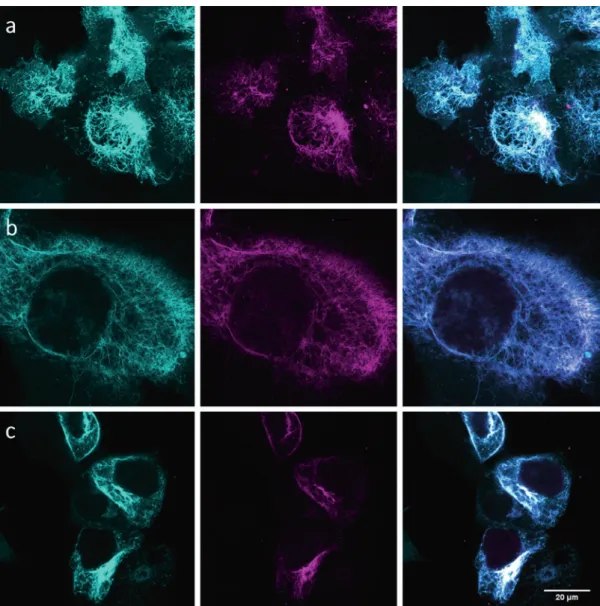
3. Use mOrange laser and excitation/emission filters to identify transfected cells. For optimal results, look for bright cells showing high expression levels and characteristic expression pattern of the target protein (see Note 45, Figs. 2 and 3).
4. Change to the laser of the dye used for the labeling. Select the appropriate filter set and check that the labeling has been success- ful (you should see signal colocalizing with the reference mOr- ange image).
5. Adjust the TIRF illumination angle (see Note 46).
6. Switch the laser to maximum power to bring the fluorophores to a dark state. You should see the signal becoming very bright at first and gradually bleach until individual blinking molecules appear (see Note 47).
7. Lower the laser power to an appropriate value (see Note 48), set the exposure time to 30 ms, and start the acquisition.
8. Acquire 10,000–30,000 frames. The optimal length depends on the sample quality, but recognizable features should already appear around 10,000 frames. With longer imaging a better signal-to-noise ratio could be achieved (see Notes 49 and 50).
9. Do further image processing in appropriate software (see Note 9).
10. To localize the spots, apply a threshold based on the maxi- mum likelihood ratio and perform fitting with a symmetrical 2D Gaussian function.
11. If desired, consolidate identical emitters (falling within one standard deviation of the spot fit) into a single intensity- weighed localization.
12. Reconstruct a final super-resolved image from binning all the detected events and convolving the resulting image with a Gaussian width according to the resolution determined by the Fourier ring correlation criterion [32] (Fig. 3).
4 Notes
1. Once the growth medium is prepared with all supplements, we do not recommend using it for longer than a month.
3.10 Super- Resolution Imaging and Data Processing
704 705 706 707 708 709 710 711 712 713 714 715 716 717 718 719 720 721 722 723 724 725 726 727 728 729 730 731 732 733 734 735 736 737 738 739 740
741
742 743
2. Several Amber suppression expression systems for eukaryotes exist. We use the NESPylRSAF/tRNAPyl system because of its enhanced efficiency and reduced background in imaging exper- iments [25].
3. When testing new reagents or labeling methods/conditions, we recommend using target proteins with very characteristic features, e.g., cytoskeletal proteins. However, any other pro- tein of interest can be used.
Fig. 2 Representative confocal images of live cell SiR labeling of vimentinBCNendo–mOrange with SiR-tetrazine (dye 6). Left to right: reference channel (mOrange, in cyan), labeling channel (SiR, in magenta), and merge. The labeling was performed in all cases at 37 °C with a dye concentration and reaction time of 1.5 μM for 10 min (a), 3 μM for 10 min (b), and 3 μM for 30 min (c—images scaled differently). Reprinted with permission from [24]
744 745 746 747 748 749 750 751
4. When testing new reagents or labeling methods/conditions, we recommend using a fusion of the target protein with a C-terminally installed fluorescent protein. This provides a direct readout obtained after a successful transfection and ncAA incorporation (only with successful Amber suppression full-length protein will be generated) that can be later on used as a reference for labeling.
5. PFA is a toxic reagent. Avoid inhalation or contact with skin and eyes. Wear protective gear while handling and follow the relevant institutional rules for using chemicals and discarding waste material.
6. The buffer composition is based on [33] and frequently used for blinking (localization-based) super-resolution microscopy.
7. We recommend always preparing the buffer freshly before starting the imaging experiment.
8. We used a commercial Leica GSDIM microscope (based on ground-state depletion and single molecule localization [34]) but any other TIRF microscope with appropriate lasers, cameras, Fig. 3 TIRF SRM imaging of vimentinBCNendo-mOrange labeled with SiR-tetrazine (dye 6). Panels a (mOrange, cyan) and b (SiR labeling, magenta) are used as a reference for protein expression and expected structure/
pattern. Corresponding SRM image from dye 6 labeling (3 μM for 30 min at 37 °C) is shown in panel c, with a resolution of 35 nm as determined by Fourier ring correlation (FRC) [32]. Reprinted with permission from [24]
752 753 754 755 756 757 758 759 760 761 762 763 764 765 766 767 768 769
and filter cubes can be used. Other localization-based micros- copy techniques (such as STORM) would also be suitable [2].
9. We used the Localizer package for IgorPro but any other soft- ware for localization-based microscopy, such as Leica’s GSDIM tools and various ImageJ plugins can be used. The following webpage (http://bigwww.epfl.ch/smlm) provides a bench- marking tool for developers to test different localization-based image analysis algorithms and provides an extensive list of tools available.
10. While performing chemical synthesis, follow general and insti- tutional safety rules. Perform all steps in a well-ventilating chemical hood. Always wear safety glasses, lab coat, protective gloves, and proper clothing. The laboratory has to be equipped with a fire extinguisher, safety shower, and eye wash device. If you do get a chemical in your eye rinse immediately with large quantities of water using the eye-wash station. If possible, col- lect halogenated and non-halogenated chemical waste sepa- rately. Specific instructions on highly hazardous steps are specified at each step.
11. Dry the 250 mL round bottom flask in an oven at 110 °C and let it cool to room temperature before reaction. Make sure that there is no water remaining in the flask before performing the reaction as it can destroy n-butyllithium.
12. Turn on the nitrogen flow so that a reasonably rapid stream of bubbles passes through the mineral oil in the bubbler. Flush the apparatus with a gentle flow of nitrogen delivered through a needle; another needle in the top serves as the gas outlet during purging. When adding reagents to the mixture under inert atmosphere, use a syringe and a needle and add it through the septum. Argon can be used instead of nitrogen if needed.
13. The reaction can be followed using thin-layer chromatogra- phy. In hexane:EtOAc 10:1 Rf(starting material) = 0.7, Rf(product) = 0.4.
14. The organic phase is the upper phase.
15. When transferring compound mixtures onto celite for chro- matography purification, make sure that the mixture is uni- formly distributed on the celite powder. If the celite is still oily or cannot be dried completely, resuspend it in an organic sol- vent (DCM or EtOAc for example), add more celite and remove the solvent under reduced pressure.
16. Here, flash chromatography is used to enhance separation by enabling gradient elution. Alternatively, you can use the classic column chromatography technique.
17. Compounds can be checked by nuclear magnetic resonance (NMR) or MS. For reference spectra see ref. 24.
770 771 772 773 774 775 776 777 778 779 780 781 782 783 784 785 786 787 788 789 790 791 792 793 794 795 796 797 798 799 800 801 802 803 804 805 806 807 808 809 810 811 812 813 814
18. AIBN is an explosive compound; handle with care, use an eye protector and protective gloves.
19. The reaction can be followed by thin-layer chromatography.
In hexane:EtOAc 3:1 Rf(starting material) = 0.55, Rf(first step product) = 0.76. In hexane:EtOAc 1:1 Rf(second step product) = 0.3.
20. The suspension transforms into a brown solution over the course of 2 h.
21. The white suspension becomes thick after 10–15 min when stirring may be challenging, and then a smooth white suspen- sion again. Check the reaction frequently and make sure that the stirring is continuous.
22. Once it reached 140 °C, the mixture starts to turn blue and it develops a dark blue color by the end of the reaction.
23. The mixture can be challenging to remove from the vial. Use prolonged (15–30 min) sonication on a high-performance sonicator to dissolve the blue residue. Methanol can be used as a co-solvent.
24. The product is blue when on silica gel (column and TLC), but colorless in solution (hexane:EtOAc 4:1 with 1% (v/v) Et3N) and forms white crystals as a solid.
25. The second purification step is optional. If the compound is pure after the first purification, omit this step.
26. The side-product to be separated is colorless on silica gel and runs just above the product.
27. Take all safety precautions for this step: wear gloves, safety glasses and the reaction must be performed in a ventilation hood. Concentrated H2SO4 is seriously corrosive. HCl is a pungent, irritating gas that can cause severe damage to the eyes, skin, lungs, and upper respiratory tract. NOx is harmful for the lung when inhaled.
28. The reaction can be exothermic. In that case, cool the reaction flask with ice/water bath.
29. Use fresh ice/water bath if the ice melted completely.
30. The orange suspension will turn to magenta.
31. The removed solvent may contain pink 3,6-dimethyl-1,2,4,5- tetrazine side product.
32. Rf(product) = 0.25 in hexane:EtOAc 1:1.
33. Rf(product) = 0.38 in hexane:EtOAc 1:1.
34. The organic phase is the bottom phase under the aqueous solution.
35. The pink crystals can be kept at −20 °C without any degrada- tion up to 9 months.
815 816 817 818 819 820 821 822 823 824 825 826 827 828 829 830 831 832 833 834 835 836 837 838 839 840 841 842 843 844 845 846 847 848 849 850 851 852 853 854 855 856 857