IV. 5. ON THE ORDER OF DISULFIDE BOND REDUCTION IN RIBONUCLEASE
F. H. White, Jr. and C. B. Anfmsen
Laboratory of Cellular Physiology and Metabolism, National Heart Institute, National Institutes of Health, Bethesda, Maryland
Page
I. Introduction 279 II. Methods 280
1. Partial Reduction of Ribonuclease 280 2. Carboxymethylation of Reduced Ribonuclease 281
3. Assay for Specific Disulfide Bonds in Reduced Ribonuclease . . . . 281
III. Results and Discussion . 283
IV. Summary 287
I. Introduction
Protein disulfide bonds may be reductively cleaved by a number of SH-containing reagents. Many early observations were made on the gross effects of reduction on proteins and have been adequately reviewed elsewhere (1-4)- More recently various studies on the relationship be- tween the extent of reduction of proteins and their biological activity have been reported. Kern (a) found that reduction of one-third of the disulfide bonds in pepsin had little effect on the enzymatic activity, which could only be abolished by cleavage of the remaining disulfide bonds. Liener (6), however, reported complete inactivation of trypsin with only partial re- duction. Complete reduction of lysozyme (7, 8) and ribonuclease (RNase)
(8, 9) yielded products devoid of enzymatic activity.
The reduction of protein disulfide bonds by SH-containing reagents is apparently specific (8), and therefore it should be ideal for the study of the contribution of disulfide bonds to the physical and catalytic proper- ties of proteins. With recent advances in the knowledge of protein struc- ture, the correlation of observed properties with specific disulfide bonds has become increasingly feasible. Lindley (10) has already obtained evi- dence that the intrachain disulfide bond of insulin is cleaved preferentially on partial reduction. Since hormonal activity is completely lost on par-
279
tial reduction, it would appear that this disulfide bond is at least in part responsible for maintaining activity.
The amino acid sequence of RNase is now essentially established {11-15), and the locations of the disulfide bonds have been reported (16, 17). Half-cystine residue No. 1 (counting from the N-terminal end) appears to be joined to half-cystine No. 6 , No. 3 to No. 7, No. 2 to No. 8 , and No. 4 to No. 5*. This information will ultimately make possible a comparison of the enzymatic properties of homogeneous samples of par- tially reduced RNase and those of the native protein in structural terms.
The problem of purifying partially reduced RNase remains to be solved.
Preliminary fractionation studies on partially reduced, carboxymethylated RNase in this laboratory have indicated a high degree of heterogeneity;
it has been suggested (18) that the mixed disulfide content of the partially reduced enzyme is largely responsible for this effect.
A prerequisite for the isolation of any product is the development of a suitable means for its assay. A method which may be used for determina- tion of the extent of homogeneity with respect to reduction of specific di- sulfide bonds in reduced RNase is described in the present paper. A sample of reduced, carboxymethylated RNase is subjected to further reduction under such conditions that complete cleavage of the remaining disulfide bonds should occur, and radioactive iodoacetic acid is employed for car- boxymethylation of this product. The resulting completely reduced and carboxymethylated RNase is degraded proteolytically, and, after separa- tion of the peptides, the degree of labeling of each radioactive peptide is taken as a measure of the extent to which the corresponding disulfide bond was intact in the original sample. This approach has been used in a pre- liminary investigation of the order of disulfide bond reduction by the assay of unfractionated, reduced, carboxymethylated RNase.
II. Methods 1. PARTIAL REDUCTION OF R N A S E
Reductions of bovine pancreatic RNase (Armour lot No. 3 8 1 - 0 5 9 ) to various levels (up to 4 SH groups per mole) were carried out as follows:
RNase was dissolved to a concentration of 1 0 - 1 2 mg. per ml. in a water solution of thioglycolate (previously adjusted to pH 8 . 5 with trimethyl- amine) of such concentration that the final molar ratio of thioglycolate to RNase was 4 0 0 : 1 . The reaction was allowed to proceed at room tem- perature with nitrogen bubbling through the mixture. After the desired
*Due to the possibility of some chemical rearrangement during isolation and characterization of cysteic acid peptides, some uncertainty remains in regard to the 2-8 and 3-7 bonds.
REDUCTION OF RIBONUCLEASE 281 time, the protein was precipitated by addition of ice-cold, acidified ace- tone and washed with this solvent and with ether as previously described
(9). To arrive at a level of reduction for which titration with p-chloro- mercuribenzoate (PCMB) indicated the presence of 2 SH groups per mole of protein, the reaction time was 20 minutes. For the "4 S H level" of reduction, the time was 40 minutes. A level of reduction approximating 6 SH groups per mole was achieved with a reaction time of 25 minutes in the presence of 8 M urea ; other conditions were as described above. Esti- mation of reduction levels was routinely made by spectrophotometric titration with P C M B by a modification of the technique of Boyer {19).
The exact details of the procedure used are given elsewhere (8).
2. CARBOXYMETHYLATION OF REDUCED R N A S E
Iodoacetate reacts with the SH groups that appear on reduction of RNase to yield S-carboxymethyleysteine (SCMC) residues. This reaction has been employed routinely to prevent the reformation of disulfide bonds by autoxidation. A molar ratio of 700:1 for iodoacetic acid to protein was used originally in this reaction (9). In subsequent investigations, however, a ratio of 70:1 was found to serve the same purpose and therefore was used throughout the work reported in this paper. Otherwise the procedure was that reported previously. The precipitation of protein at the end of the reaction and its washing were carried out as indicated above.
3. ASSAY FOR SPECIFIC DISULFIDE BONDS IN REDUCED R N A S E
Partially reduced, carboxymethylated RNase was subjected to com- plete reduction. The reaction mixture was 8 M with respect to urea and the reaction time was 4.5 hours. Other conditions were as described above for partial reduction. The product was carboxymethylated with iodoacetic acid 1 - C1 4 with a specific activity of 0.23 mc. per mM. After precipitation and washing the protein was redissolved in water and lyophilized. Five mg. of the dried protein were dissolved in 1 ml. of freshly prepared 0.1 M ammonium carbonate—HCl buffer of pH 8.0. The reaction mixture con- tained 0.005 ml. of a phenol red solution (0.1% in aqueous 20% ethanol) as a control on the pH. Ten μΐ. of a trypsin* solution (5 mg. per ml. in wa- ter) were added, and the mixture was incubated at 37° for 2 hours. Ten μ\. of a chymotrypsin* solution (5 mg. per ml. in water) were then added and incubation continued for another 2 hours. The digest was lyophilized for 18 hours to ensure removal of sufficient ammonium carbonate to elimi- nate the salt effect in the subsequent chromatography. The dried sample was redissolved in a small volume of water, and one-half of the final vol-
* Crystallized, Armour.
282
urne was dried as a small spot on Whatman No. 3 paper. The sample was chromatographed (descending) in the organic phase of butanol-acetic acid-water (4:1:5), prepared on the day of use. The paper was dried at room temperature and cut into two halves. Each half was subjected to electro- phoresis in a pyridine-acetic acid buffer of p H 6.5 under toluene by the technique of Michl (20) at 35 volts per cm. for 1.5 hours*. After the papers were dried in a ventilated oven at 80° for 15 minutes, they were dipped in 0.5% ninhydrin in absolute ethanol containing 1 ml. of collidine per 400 ml. of solution. The papers were oven-dried as before to develop the color. Radioautographs of the resulting fingerprints were prepared by exposure of X-ray film to the papers for 72 hours, followed by develop- ment of the film under standardized conditions.
For amino acid analysis, the radioautographs were prepared before development of the ninhydrin color. The labeled peptides, which were located by their positions on X-ray film, were excised from the fingerprints and eluted with 6 Ν HCl, in which they were hydrolyzed at 105° for 18 hours in sealed tubes. The hydrolyzates were dried in vacuo and chroma- tographed for 18 hours on Whatman N o . 1 paper in the butanol-acetic acid-water system. The papers were dried at room temperature and chromatographed in the second dimension with aqueous 80% pyridine. The papers were dried as before and developed by dipping in an absolute ethanol solution of 0.1% ninhydrin-glacial acetic acid-collidine (15:5:2) followed by oven-drying at 80°.
Approximations of the peptide concentration on the fingerprints and the extent of labeling were made by scanning of the ninhydrin spots and the corresponding spots on film, respectively, with a model 525 densitome- ter (Photovolt Corporation). Optical density curves were obtained in this way for each spot, and the area under each curve was taken as a measure of the peptide concentration or the extent of its labeling. The ratio of this area for the radioautographic spot to that of the corresponding nin- hydrin spot constituted a measure of the specific activity of the peptide under investigation. With knowledge of the amino acid content of the peptide it was possible to assign it a position in the RNase chain and thereby to conclude which of the half-cystine residues was present in the peptide (as SCMC). The specific activity of each peptide was used as an indication of the extent to which the corresponding disulfide bond was present in the original sample of reduced, carboxymethylated RNase.
* A similar procedure has been used in the "fingerprinting" of proteins by Ingram (21 ) who, however, employed electrophoresis first and chromatographed in the second dimension. A more detailed account of the present method is in preparation by Drs.
A. N. Katz and W. J. Dreyer of this laboratory.
REDUCTION OF RIBONUCLEASE 283
III. Results and Discussion
Several factors influenced the choice of proteolytic enzymes for the degradation of reduced RNase. Since the action of trypsin is restricted to peptide bonds involving lysine and arginine, there would be no formation of "families" of closely related peptides with the use of this enzyme. While chymotrypsin is not as ideal in this respect, it was used in combination with trypsin to obtain peptides small enough for convenient chromato- graphic and electrophoretic separation. All protein samples subjected to the action of these enzymes had been completely reduced prior to the final carboxymethylation and presumably differed only to the extent of
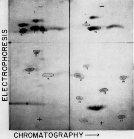
FIG. 1. Two dimensional separation of peptides from a trypsin and chymotrypsin digest of completely reduced, carboxymethylated ribonuclease. When iodoacetic acid 1-C14 was used for carboxymethylation, the encircled and numbered peptides were found to be labeled.
the labeling of their carboxymethyl groups. Therefore, any unusual cleavages by the proteases employed would be expected to occur in an identical way with all the samples.
The enzymatic digests of these samples, when analyzed two-dimen- sionally, always yielded the same ninhydrin-positive pattern as shown in Fig. 1. The results obtained thus far in amino acid analysis of the radio- active peptides from such fingerprints are given in Table I, with con-
T A B L E I
AMINO ACID ANALYSIS OF LABELED PEPTIDES
Peptide0
Amino acid content in
addition to SCMC Half-cystine
residue number6 Disulfide bond0
5 Lys, Asp, Glu, Ser, Ala
Val, Leu, His 3 3-7
7 Asp, Glu, Gly, Thr 5 4-5
9 Arg, Asp, Ser, Thr, Leud 6 1-6
12 Lys, Asp, Thr, Pro, Val 2 2-8
14 Tyr, Pro, Asp, Ala 7 3-7
° The peptides are numbered as shown in Fig. 1.
b The half-cystine residues are numbered from the N-terminal end of the ribonuclease chain.
c The disulfide bonds are given as reported previously (14, 15) (see footnote on page 280).
d Leucine and isoleucine are indistinguishable by the chromatographic technique used (described in text).
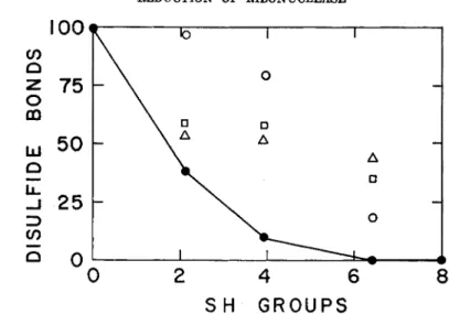
elusions as to which SCMC residue is contained by each peptide. From the observed specific activities of these peptides, the percentage of each disulfide bond was calculated for various levels of reduction (Tables II and III) and plotted in Fig. 2. The points for the 1-6 disulfide bond are connected to emphasize its complete disappearance at a level of reduction for which the remaining bonds are clearly in evidence. When the starting material was completely reduced, carboxymethylated RNase, no disulfide bonds were detected by this method. It is not considered possible, from these preliminary results, to arrive at any conclusion regarding the rela-
tive liabilities of the 2-8, 3-7, and 4-5 bonds. A correlation of RNase ac- tivity loss with reduction is presented in Fig. 3. A comparison of the ac- tivity loss with disappearance of disulfide bonds (Fig. 2) strongly sug- gests that the 1-6 bond is not essential for RNase activity.
Confirmation and extension of this work will be greatly facilitated by purification of partially reduced RNase. The homogeneity of a sample
R E D U C T I O N O F R I B O N U C L E A S E 285
S H G R O U P S
FIG. 2 . Disappearance of disulfide bonds in ribonuclease with reduction. The di- sulfide bond content is expressed as per cent of the specific activity of the correspond- ing peptide (see Table I ) , with the specific activity of this peptide for native ribo- nuclease taken as 100 (see Tables I I and I I I ) . The reduction levels were determined by titration of reduced ribonuclease with p-chloromercuribenzoate. The specific ac- tivities of peptides 5 and 1 4 were averaged to obtain points for the 3 - 7 disulfide bond. KEY: # , 1-6 disulfide bond; O , 4 - 5 disulfide bond; • , 3 - 7 disulfide bond;
Δ , 2 - 8 disulfide bond.
TABLE I I
CONCENTRATION AND RADIOACTIVITY MEASUREMENTS OP ISOLATED PEPTIDES
Radioactivity6 Concentration0
Peptide0 SH:<* 0 2.1 3 . 9 6 . 4 8 . 0 0 2.1 3 . 9 6 . 4 8 . 0
5 8 . 4 5 . 6 6 . 0 3 . 0 0 1 7 2 0 2 4 1 6 2 2
7 7 . 8 7.0 4 . 1 1.1 0 2 . 6 2 . 4 1.7 2 . 0 2.1
9 2 2 . 0 1 1 . 0 2 . 5 0 0 1.7 2.1 1.9 2 . 2 1.9
1 2 5 . 9 3 . 9 4 . 4 1.9 0 4 . 9 5 . 3 5 . 6 5 . 0 5 . 2
1 4 7 . 4 5 . 0 4 . 2 3 . 2 0 6 . 2 7.5 6.7 6 . 1 6 . 4
"The peptides are numbered as shown in Fig. 1.
6 The figures are proportional to areas under the curves (optical densitv versus mm.) resulting from photometric scanning of the corresponding radioautographic spots.
cT h e figures are proportional to areas under the curves (optical density versus mm.) resulting from photometric scanning of the corresponding ninhydrin-developed spots (see Fig. 1).
d Sulfhydryl groups per mole of reduced ribonuclease, determined by titration with p-chloromercuribenzoate.
Specific activity (reduced) Specific activity6 Specific activity (native) Peptide0 SH:<* 0 2 . 1 3 . 9 6 . 4 8 . 0 2 . 1 3 . 9 6 . 4 8 . 0
5 0 . 5 0 0 . 2 8 0 . 2 5 0 . 1 9 0 5 6 5 0 3 8 0
7 3 . 0 2 . 9 2 . 4 0 . 5 5 0 9 7 8 0 1 8 0
9 1 3 . 0 5 . 2 1.3 0 0 4 0 1 0 0 0
1 2 1.2 0 . 7 4 0 . 7 9 0 . 3 8 0 6 2 6 6 3 2 0
1 4 1.2 0 . 6 7 0 . 6 3 0 . 5 2 0 5 6 5 3 4 3 0
° The peptides are numbered as shown in Fig. 1.
b Specific activity was obtained by dividing the value for radioactivity by that for concentration (Table II) for each peptide.
c These values were plotted against reduction level (Fig. 2 ) .
d Sulfhydryl groups per mole of reduced ribonuclease, determined by titration with p-chloromercuribenzoate.
S H G R O U P S
FIG. 3 . Activity of ribonuclease at various stages of reduction (expressed as per cent of the specific activity of native ribonuclease) as a function of the number of moles of sulfhydryl per mole of enzyme. Ribonucleic acid was the substrate. Details of the experimental procedures are given elsewhere (9). KEY: A, reduction in absence of urea; φ , reduction in 8 M urea; • , reoxidation of fully reduced, inactive ribo- nuclease; O , reoxidation of samples containing more than six sulfhydryl groups per average molecule; V, reoxidation of samples containing about four sulfhydryl groups per average molecule. A curve has been drawn through the black points to indicate the approximate course of reduction. (It is noteworthy that the points representing the reoxidation experiments roughly follow the same curve, indicating a specific rather than random reformation of disulfide bonds.)
T A B L E I I I
SPECIFIC ACTIVITIES OF ISOLATED PEPTIDES
REDUCTION OF RIBONUCLEASE 287 of partially reduced protein with respect to the absence of certain disul- fide bonds would be indicated by the total absence of labeling in the pep- tides corresponding to the bonds in question. The technique reported here serves to illustrate the approach that m a y be used as an assay in future attempts to obtain homogeneous samples of partially reduced pro- teins.
IV. Summary
A method for following the reduction of specific disulfide bonds in ribonuclease has been outlined. This method has been used for the study of ribonuclease at various levels of reduction. The results indicate that the disulfide bond between half-cystine residues 1 and 6 is the most labile under the present conditions of reduction and that this bond is not essen- tial for ribonuclease activity.
REFERENCES
1. H. S. Olcott and H. Fraenkel-Conrat, Chem. Revs. 41, 151 (1947).
2. R. M. Herriott, Advances in Protein Chem. 3, 169 (1947).
3. F. W. Putnam, in "The Proteins" (H. Neurath and K. Bailey, eds.), Vol. 1, Part B, p. 893. Academic Press, New York, 1954.
4. E. S. G. Barron, Advances in Enzymology 11, 201 (1951).
5. H. L. Kern, Doctoral Dissertation, Johns Hopkins University, Baltimore, Mary- land, 1953.
6. I. E. Liener, J. Biol. Chem. 225, 1061 (1957).
7. H. Fraenkel-Conrat, A. Mohammad, E. D . Ducay, and D . K. Mecham, / . Am.
Chem. Soc. 73, 625 (1951).
8. M. Sela, F. H. White, Jr., and C. B. Anfinsen Biochim. et Biophys. Acta, in press.
9. M. Sela, F. H. White, Jr., and C. B. Anfinsen, Science 125, 691 (1957).
10. H. Lindley, J. Am. Chem. Soc. 77, 4927 (1955).
11. C. H. W. Hirs, S. Moore, and W. H. Stein, J. Biol. Chem. 219, 623 (1956).
12. C. H. W. Hirs, S. Moore, and W. H. Stein, J. Biol. Chem. 221, 151 (1956).
15. J. L. Bailey, S. Moore, and W. H. Stein, J. Biol. Chem. 221, 143 (1956).
14. C. B. Anfinsen and R. R. Redfield, / . Biol. Chem. 221, 385 (1956).
IB. C. H. W. Hirs, W. H. Stein, and S. Moore, personal communication.
16. D . H. Spackman, S. Moore, and W. H. Stein, Federation Proc. 16, 252 (1957).
17. C. B. Anfinsen and A. P. Ryle, Biochim. et Biophys. Acta 24, 633 (1957).
18. F. H. White, Jr., Federation Proc. 17, 334 (1958).
19. P. D . Boyer, J.Am. Chem. Soc. 76, 4331 (1954).
20. H. Michl, Monatsh. Chem. 82, 489 (1951).
21. V. M. Ingram, Nature 178, 792 (1956).
Discussion
BENESCH : Have you done any nonenzymatic hydrolyses?
WHITE: We have just used trypsin and chymotrypsin for hydrolysis of reduced,
carboxymethylated ribonuclease. Nonenzymatic hydrolysis would be nonspecific, and we would very likely get families of closely related peptides. This would make our job more difficult. Trypsin is especially good for hydrolysis because of its high de- gree of specificity.
BENESCH: Yes. The only reason why I asked is because in the case of insulin Sanger had to resort to acid hydrolysis because the proximity of the two disulfide bonds on the A chain made this particular region unsusceptible to trypsin.
WHITE : We have no such problem with ribonuclease. With the use of trypsin and chymotrypsin, the result is one half-cystine residue per peptide.
BROWN: Have you tried in your reduction studies to correlate the relationship between the SH values obtained by analysis and that obtained by any of the other titration methods?
WHITE: To what other methods are you referring?
BROWN: I am talking about the SH titration method such as the PCMB one. I wonder whether you tried to reduce one disulfide group or two disulfide groups, treated the protein with iodoacetamide, and checked this result versus a straight titration method for SH. Have you done anything like that?
WHITE: We have compared PCMB titrations after partial reduction of ribonu- clease with those after carboxymethylation and complete reduction of the same sam- ple. Thus far the results indicate that the sum of the two is close to the theoretical value of 8.
FRABNKEL-CONRAT: I think the question refers to the specificity of the iodoace- tamide reaction.
BROWN : It does.
FRAENKEL-CONRAT: We use both iodoacetic acid and other methods and find them to correspond very closely. In a limited time period at around pH 7 to 8 there is no reaction with groups other than sulfhydryl. So this is as good a method to tag sulf- hydryl groups as any.
KAPLAN: DO you have to use urea also to get oxidation of the disulfide?
WHITE: N O , that is not necessary. The way we have carried it out is with per- formic acid, and we do not need urea for that.
KAPLAN : How do you explain this?
WHITE: Thioglycolic acid is apparently specific for disulfide bonds. For complete reduction, urea must be used as a denaturing agent and appears to act by cleaving hydrogen bonds. Performic acid seems to have a sufficiently strong denaturing effect
of its own so that urea need not be used with it.