Metadata of the chapter that will be visualized online
Chapter Title The Microbiome as a Component of the Tumor Microenvironment Copyright Year 2020
Copyright Holder Springer Nature Switzerland AG
Author Family Name Kovács
Particle
Given Name Tünde
Suffix
Division Department of Medical Chemistry,
Faculty of Medicine Organization/University University of Debrecen
Address Debrecen, Hungary
Division
Organization/University MTA-DE Lendület Laboratory of Cellular Metabolism
Address Debrecen, Hungary
Author Family Name Mikó
Particle
Given Name Edit
Suffix
Division Department of Medical Chemistry,
Faculty of Medicine Organization/University University of Debrecen
Address Debrecen, Hungary
Division
Organization/University MTA-DE Lendület Laboratory of Cellular Metabolism
Address Debrecen, Hungary
Author Family Name Ujlaki
Particle
Given Name Gyula
Suffix
Division Department of Medical Chemistry,
Faculty of Medicine Organization/University University of Debrecen
Address Debrecen, Hungary
Organization/University MTA-DE Lendület Laboratory of Cellular Metabolism
Address Debrecen, Hungary
Author Family Name Sári
Particle
Given Name Zsanett
Suffix
Division Department of Medical Chemistry,
Faculty of Medicine Organization/University University of Debrecen
Address Debrecen, Hungary
Division
Organization/University MTA-DE Lendület Laboratory of Cellular Metabolism
Address Debrecen, Hungary
Corresponding Author Family Name Bai
Particle
Given Name Péter
Suffix
Division Department of Medical Chemistry,
Faculty of Medicine Organization/University University of Debrecen
Address Debrecen, Hungary
Division
Organization/University MTA-DE Lendület Laboratory of Cellular Metabolism
Address Debrecen, Hungary
Division Research Center for Molecular
Medicine, Faculty of Medicine Organization/University University of Debrecen
Address Debrecen, Hungary
Email baip@med.unideb.hu
processes. Changes in the composition and proportion of the microbiome are associated with metabolic diseases (Fulbright et al., PLoS Pathog 13:e1006480, 2017; Maruvada et al., Cell Host Microbe 22:589–599, 2017), psychiatric disorders (Macfabe, Glob Adv Health Med 2:52–66, 2013; Kundu et al., Cell 171:1481–1493, 2017), and neoplastic diseases (Plottel and Blaser, Cell Host Microbe 10:324–335, 2011; Schwabe and Jobin, Nat Rev Cancer 13:800–812, 2013; Zitvogel et al., Cell 165:276–287, 2016). However, the number of directly tumorigenic bacteria is extremely low. Microbial dysbiosis is connected to cancers of the urinary tract (Yu, Arch Med Sci 11:385–394, 2015), cervix (Chase, Gynecol Oncol 138:190–200, 2015), skin (Yu et al., J Drugs Dermatol 14:461–465, 2015), airways (Gui et al., Genet Mol Res 14:5642–5651, 2015), colon (Garrett, Science 348:80–86, 2015), lymphomas (Yamamoto and Schiestl, Int J Environ Res Public Health 11:9038–9049, 2014; Yamamoto and Schiestl, Cancer J 20:190–194, 2014), prostate (Yu, Arch Med Sci 11:385–394, 2015), and breast (Flores et al., J Transl Med 10:253, 2012; Fuhrman et al., J Clin Endocrinol Metab 99:4632–4640, 2014; Xuan et al., PLoS One 9:e83744, 2014; Goedert et al., J Natl Cancer Inst 107:djv147, 2015; Chan et al., Sci Rep 6:28061, 2016; Hieken et al., Sci Rep 6:30751, 2016; Urbaniak et al., Appl Environ Microbiol 82:5039–5048, 2016; Goedert et al., Br J Cancer 118:471–479, 2018). Microbial dysbiosis can influence organs in direct contact with the microbiome and organs that are located at distant sites of the body. The altered microbiota can lead to a disruption of the mucosal barrier (Plottel and Blaser, Cell Host Microbe 10:324–335, 2011) or promote or inhibit tumorigenesis through the modification of immune responses (Kawai and Akira, Int Immunol 21:317–337, 2009; Dapito et al., Cancer Cell 21:504–516, 2012) and microbiome- derived metabolites, such as estrogens (Flores et al., J Transl Med 10:253, 2012; Fuhrman et al., J Clin Endocrinol Metab 99:4632–4640, 2014), secondary bile acids (Rowland, Role of the gut flora in toxicity and cancer, Academic Press, London, p x, 517 p., 1988; Yoshimoto et al., Nature 499:97–101, 2013; Xie et al., Int J Cancer 139:1764–1775, 2016; Shellman et al., Clin Otolaryngol 42:969–973, 2017; Luu et al., Cell Oncol (Dordr) 41:13–24, 2018; Miko et al., Biochim Biophys Acta Bioenerg 1859:958–974, 2018), short-chain fatty acids (Bindels et al., Br J Cancer 107:1337–1344, 2012), lipopolysaccharide (Dapito et al., Cancer Cell 21:504–516, 2012), and genotoxins (Fulbright et al., PLoS Pathog 13:e1006480, 2017). Thus, altered gut microbiota may change the efficacy of chemotherapy and radiation therapy (McCarron et al., Br J Biomed Sci 69:14–17, 2012; Viaud et al., Science 342:971–976, 2013; Montassier et al., Aliment Pharmacol Ther 42:515–528, 2015; Buchta Rosean et al., Adv Cancer Res 143:255–294, 2019). Taken together, microbial dysbiosis has intricate connections with neoplastic diseases; hereby, we aim to highlight the major contact routes.
Keywords (separated by “ - ”)
Microbiome - Breast cancer - Tumor microenvironment - Bacterial metabolite
- Bacterial metabolism - Antitumor immunity - Tumor metabolism - Epithelial-
mesenchymal transition - Tumorigenesis - Metastasis - Chemotherapy
© Springer Nature Switzerland AG 2020
A. Birbrair (ed.), Tumor Microenvironment, Advances in Experimental Medicine and Biology 1225, https://doi.org/10.1007/978-3-030-35727-6_10
The Microbiome as a Component of the Tumor Microenvironment
Tünde Kovács, Edit Mikó, Gyula Ujlaki, Zsanett Sári, and Péter Bai
Abstract
Microbes, which live in the human body, affect a large set of pathophysiological pro- cesses. Changes in the composition and pro- portion of the microbiome are associated with metabolic diseases (Fulbright et al., PLoS Pathog 13:e1006480, 2017; Maruvada et al., Cell Host Microbe 22:589–599, 2017), psy- chiatric disorders (Macfabe, Glob Adv Health Med 2:52–66, 2013; Kundu et al., Cell 171:1481–1493, 2017), and neoplastic dis- eases (Plottel and Blaser, Cell Host Microbe 10:324–335, 2011; Schwabe and Jobin, Nat Rev Cancer 13:800–812, 2013; Zitvogel et al., Cell 165:276–287, 2016). However, the num-
ber of directly tumorigenic bacteria is extremely low. Microbial dysbiosis is con- nected to cancers of the urinary tract (Yu, Arch Med Sci 11:385–394, 2015), cervix (Chase, Gynecol Oncol 138:190–200, 2015), skin (Yu et al., J Drugs Dermatol 14:461–465, 2015), airways (Gui et al., Genet Mol Res 14:5642–
5651, 2015), colon (Garrett, Science 348:80–
86, 2015), lymphomas (Yamamoto and Schiestl, Int J Environ Res Public Health 11:9038–9049, 2014; Yamamoto and Schiestl, Cancer J 20:190–194, 2014), prostate (Yu, Arch Med Sci 11:385–394, 2015), and breast (Flores et al., J Transl Med 10:253, 2012;
Fuhrman et al., J Clin Endocrinol Metab 99:4632–4640, 2014; Xuan et al., PLoS One 9:e83744, 2014; Goedert et al., J Natl Cancer Inst 107:djv147, 2015; Chan et al., Sci Rep 6:28061, 2016; Hieken et al., Sci Rep 6:30751, 2016; Urbaniak et al., Appl Environ Microbiol 82:5039–5048, 2016; Goedert et al., Br J Cancer 118:471–479, 2018). Microbial dys- biosis can influence organs in direct contact with the microbiome and organs that are located at distant sites of the body. The altered microbiota can lead to a disruption of the mucosal barrier (Plottel and Blaser, Cell Host Microbe 10:324–335, 2011) or promote or inhibit tumorigenesis through the modifica- tion of immune responses (Kawai and Akira, Int Immunol 21:317–337, 2009; Dapito et al., Cancer Cell 21:504–516, 2012) and
AU2
T. Kovács · E. Mikó · G. Ujlaki · Z. Sári Department of Medical Chemistry, Faculty of Medicine, University of Debrecen,
Debrecen, Hungary
MTA-DE Lendület Laboratory of Cellular Metabolism, Debrecen, Hungary P. Bai (*)
Department of Medical Chemistry, Faculty of Medicine, University of Debrecen,
Debrecen, Hungary
MTA-DE Lendület Laboratory of Cellular Metabolism, Debrecen, Hungary
Research Center for Molecular Medicine, Faculty of Medicine, University of Debrecen,
Debrecen, Hungary e-mail: baip@med.unideb.hu
AU1
10
1 2
3 4
5 6 7 8 9 10 11 12 13 14 15 16 17 18 19
20 21 22 23 24 25 26 27 28 29 30 31 32 33 34 35 36 37 38 39 40 41 42 43 44 45 46 47 48 49 50 51
microbiome- derived metabolites, such as estrogens (Flores et al., J Transl Med 10:253, 2012; Fuhrman et al., J Clin Endocrinol Metab 99:4632–4640, 2014), secondary bile acids (Rowland, Role of the gut flora in toxicity and cancer, Academic Press, London, p x, 517 p., 1988; Yoshimoto et al., Nature 499:97–101, 2013; Xie et al., Int J Cancer 139:1764–1775, 2016; Shellman et al., Clin Otolaryngol 42:969–973, 2017; Luu et al., Cell Oncol (Dordr) 41:13–24, 2018; Miko et al., Biochim Biophys Acta Bioenerg 1859:958–974, 2018), short-chain fatty acids (Bindels et al., Br J Cancer 107:1337–1344, 2012), lipopolysac- charide (Dapito et al., Cancer Cell 21:504–
516, 2012), and genotoxins (Fulbright et al., PLoS Pathog 13:e1006480, 2017). Thus, altered gut microbiota may change the effi- cacy of chemotherapy and radiation therapy (McCarron et al., Br J Biomed Sci 69:14–17, 2012; Viaud et al., Science 342:971–976, 2013; Montassier et al., Aliment Pharmacol Ther 42:515–528, 2015; Buchta Rosean et al., Adv Cancer Res 143:255–294, 2019). Taken together, microbial dysbiosis has intricate connections with neoplastic diseases; hereby, we aim to highlight the major contact routes.
Keywords
Microbiome · Breast cancer · Tumor microen- vironment · Bacterial metabolite · Bacterial metabolism · Antitumor immunity · Tumor metabolism · Epithelial-mesenchymal transition · Tumorigenesis · Metastasis · Chemotherapy
10.1 The Human Microbiome
The human body harbors different kinds of sym- biotic, commensal, and pathogenic bacteria that live on the surface and the cavities of the body.
Microbiota is a collective term that refers to the group of microbes colonizing the human body, and the collection of genes they encode is known as our microbiome [36]. The number of coloniz- ing microbial cells (>1014) is 10 times more than
the total sum of human somatic and germ cells.
Therefore, their collective genome—called the metagenome—contains a large number of genes that exceed the human genome by 150 times.
This metagenome performs key functions rele- vant to human health [37].
Each anatomical niche possesses a unique mixture of microbial populations (gut, skin, vagina, mouth, nose, and conjunctiva) that have important and functionally relevant individual variability (at the levels of genus, species, and strain) [5]. The great majority of microorganisms live in the gastrointestinal (GI) lumen. These microbes compete and collaborate with other organisms in this niche, resulting in a function- ally and genetically plastic metagenome [5]. The GI microbiota plays a crucial role in digestion, maturation, immune response, protection against pathogen overgrowth, maintenance of intestinal barrier function, regulation of intestinal endo- crine functions, neurologic signaling, bone den- sity, biosynthesis of vitamins, neurotransmission, metabolism of bile salts, reaction or modification of drugs, elimination of exogenous toxins, and maintenance of the energy homeostasis of the host [38].
10.2 Bidirectional Microbiome- Host Connection
There is increasing evidence for complex and dynamic microbial interactions with hosts. The microbe-human symbiotic connection is a result of millions of years of coevolution, coadaptation, and codependence. Bacterial colonization begins at birth and progresses through childhood to adulthood. The adaptation process is nonrandom [39] and depends on the body habitat, lifestyle, physiological conditions, genotype of the host, and presence of other microbes in the niche [40].
The function and composition of the microbiome are determined by the diet of the host, probiotic or antibiotic consumption, stress, and short- or long-term travel. Besides these external factors, the host can affect the dynamics of the microbi- ome through its genetics, immune system, and personal hygiene [38]. Given the diverse func-
AU3
AU4 52 53 54 55 56 57 58 59 60 61 62 63 64 65 66 67 68 69 70 71 72 73 74 75 76 77 78
79 80 81 82 83 84 85
86
87 88 89 90 91 92 93 94
95 96 97 98 99 100 101 102 103 104 105 106 107 108 109 110 111 112 113 114 115 116 117 118 119 120
121 122
123 124 125 126 127 128 129 130 131 132 133 134 135 136 137 138 139
tional repertoire of the microbiome, it is not sur- prising that dysbiosis is associated with a broad range of diseases from neurological disorders to metabolic diseases and cancer [12]. Numerous studies highlight the relationship between changes in the function, composition, and pro- portion of microbes—also called microbial dys- biosis—and the progression of certain diseases.
Koch’s concept that one microbe is responsible for the formation of one disease (“one microbe- one disease hypothesis”) was shown to be an oversimplification. Recent advances have shown that the loss of balance in microbial communities and the global change in our microbiome are directly or indirectly connected to carcinogene- sis, rather than the presence of a single causative microbe [41]. Nevertheless, there are directly tumorigenic bacteria, although their number is extremely low, including about 10 species (e.g., Helicobacter pylori promote the development of gastric cancer). Dysbiosis is associated with can- cers of the urinary tract, cervix, skin, airways, colon, lymphomas, prostate, and breast [42].
However, it is still unclear whether cancer is the product of alterations of the microbiota or modi- fications in the “normal” microbiome are the consequences of cancer progression.
10.3 The Tumor
Microenvironment
Cancers are not just masses of homogenous malignant cells. Tumors have been recognized as complex organs, whose complexity may exceed that of normal healthy tissues. Interactions between malignant and recruited non- transformed cells create the tumor microenvironment (TME).
Nonmalignant cells include immune cells, cells of the vasculature and lymphatic system, cancer- associated fibroblasts, pericytes, and adipocytes [43]. The role of nonmalignant cells in the TME is to support cancer growth. Nonmalignant cells have a dynamic tumor-promoting function at all
stages of carcinogenesis. The communication between cell types is driven by an extremely complex network of cytokines, chemokines, growth factors, other inflammatory mediators, and matrix remodeling enzymes [44]. Cancer cell metabolism is strictly regulated by the tumor microenvironment. The microbiome is a new component of the tumor microenvironment that impairs tumor cell metabolism by maintaining a healthy barrier, inducing inflammation, and pro- ducing genotoxins and bacterial metabolites with different features. Below, we review the modali- ties of how dysbiosis interferes with carcinogen- esis (Fig. 10.1).
10.4 Bacteria-Driven Carcinogenesis
Through Physical Interaction The most relevant pathomechanism for microbiome- derived carcinogenesis is barrier failure. In healthy humans, numerous commensal bacteria are found in the intestinal lumen, where some bacteria are in direct association with the epithelium. The microbiota is vital in preserving the functional luminal barrier, by maintaining epithelial cell turnover, facilitating mucin pro- duction, and competing for resources and, thereby, suppressing the growth of pathogens [45]. The physical and chemical barrier of gut epithelial cells prevents microbial translocation to the underlying connective tissue. Defects in protein-coding genes (e.g., laminin) that are essential for the maintenance of a normal barrier, infections, inflammation, carcinogenesis, or microbial dysbiosis may induce barrier failure.
Inflammation and carcinogenesis may trigger barrier failure, but barrier failure also promotes inflammation and carcinogenesis, suggesting a forward-amplifying loop [6]. Breakdown of the intestinal barrier leads to translocation of bacteria and the development of a systemic inflammatory response [46].
140 141 142 143 144 145 146 147 148 149 150 151 152 153 154 155 156 157 158 159 160 161 162 163 164 165 166
167 168
169 170 171 172 173 174 175 176 177 178 179 180
181 182 183 184 185 186 187 188 189 190 191 192 193 194
195 196 197
198 199 200 201 202 203 204 205 206 207 208 209 210 211 212 213 214 215 216 217 218 219 220 221
10.5 Microbiome-Immune System Interactions
in Tumorigenesis
Microbiome-immune system interactions play multifaceted roles in tumorigenesis. The microbi- ome may promote tumorigenesis by inducing chronic inflammation, disrupting the balance between cell proliferation and cell death, and triggering immune responses. The physical loss of the natural gut epithelial barrier—barrier fail- ure—or the loss of the antibacterial defense sys- tem enables the movement of cellular components and microbes across the barrier, where they cause an innate inflammatory response. The mamma- lian immune system detects the presence of microbial infection through pattern recognition receptors (PRRs). Toll-like receptors (TLRs) and NOD-like receptors (NLR) belong to the PRR family and recognize different but overlapping microbial components. They are expressed in dif- ferent cellular compartments (cell surface, cyto- plasm, lysosome, and endosome) and activate specific signaling pathways that promote inflam- mation, tumor proliferation, or resistance to cell death [23].
TLRs are one of the most powerful pro- inflammatory stimuli. These structures recognize microbe-associated molecular patterns, such as
lipopolysaccharides (LPS), peptidoglycan, fla- gella, or microbial DNA/RNA. TLR2 recognizes peptidoglycan and lipoteichoic acid (bacterial cell wall components) and promotes gastric can- cer, while TLR4 detects LPS (Gram-negative cell wall component) and contributes to skin, pan- creas, liver, and colon cancer development [6].
Carcinogenesis is promoted through TLRs of epithelial cells, macrophages, and fibroblasts.
TLR induction leads to the production of pro- inflammatory cytokines, such as interleukins and TNFα. Downstream effectors of TLR signaling induce cell survival and suppress apoptosis through NF-κB (nuclear factor-κB) and STAT3 signaling, which is in line with the role of MYD88 mutations that induce NF-κB and STAT3 in many human lymphomas [24]. Tumor formation is reduced by pharmacologic inhibition of interleu- kins (IL-17 and IL-23), antibiotic treatment, or MYD88 inactivation [6].
Although a direct link between endogenous bacteria and tumor-associated angiogenesis has not been shown, the microbiome is required for normal development of the vasculature. LPS, produced by the microbiome, may promote angiogenesis through TLRs. IL-17 is produced by T-helper-17 (Th17), suggesting that bacteria also impact the tumor microenvironment by stim- ulating Th17 lymphocytes. A connection between
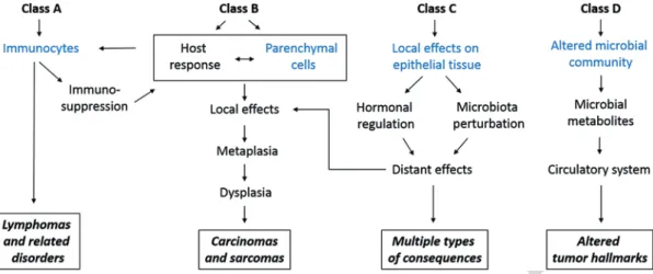
Fig. 10.1 Schematic picture of the classification of microbiota-associated human malignancies. Class A is defined by the involvement of the immune response, Class B requires direct microbial interactions with parenchymal cells, Class C covers distant effects from local interactions, and Class D shows the consequences of altered microbiome composi- tion. (Modified figure from [5])
222 223 224
225 226 227 228 229 230 231 232 233 234 235 236 237 238 239 240 241 242 243 244 245 246 247 248 249
250 251 252 253 254 255 256 257 258 259 260 261 262 263 264 265 266 267 268 269 270 271 272 273 274 275 276 277 278
breast cancer and immunoglobulins has been established. Secretory immunoglobulin A (IgA) helps to maintain the integrity of the mucosal barrier, attenuates the host immune response, and regulates the composition of the gut microbial community.
Several bacterial species induce immunity in tumor development. Lactococcus species help maintain the cytotoxic activity of natural killer (NK) cells, while Sphingomonas yanoikuyae have an important role in maintaining breast tis- sue health. Cytotoxic immune cells (cytotoxic T lymphocytes) are essential for identifying and destroying precancerous and cancerous cells;
Fusobacterium nucleatum destroy this protective mechanism and enable tumor progression, while others stimulate anticancer immunity.
Bifidobacterium, Bacteroides thetaiotaomicron, and Bacteroides fragilis enhance dendritic cell function and antitumor cytotoxic T cell immunity [1]. TLRs may also promote cancer cell prolifer- ation through different growth factor receptor ligands (amphiregulin, epiregulin, and hepato- cyte growth factors), which exert both local and long-distance effects.
In carcinogenesis, the microbiota induce acti- vation of NOD-like receptors (NLRs) as well.
Many studies focus on NOD2, because loss of NOD2 activity is connected with Crohn’s dis- ease. NOD2 has a key role in the activation of NF-κB signaling and the formation of a bacterial community. Thus, NOD2 loss-of -function muta- tions may lead to intestinal dysbiosis and an enhanced risk of developing colorectal carci- noma (CRC). Genetically induced CRC is also evoked by NOD1 deficiency, which plays an important role in intestinal defense against bacte- ria. NLRP6, another NLR, is important in microbiota- tumorigenesis interactions. NRRP6 is a component and key activator of inflamma- somes (multiprotein oligomers responsible for the activation of inflammatory responses), which are downregulated in dysbiosis-driven carcino- genesis, together with decreased IL-18 produc- tion [6].
Immunotherapy is used to eliminate residual cancer cells after chemotherapy or radiation ther- apy. In therapy, monoclonal antibodies target
molecules, such as anti-T-lymphocyte-associated antigen 4 (CTLA-4) and anti-programmed death 1 (PD-1) or its ligand anti-PD-L1. The advantage of immunotherapy is that it stimulates and sup- ports the immune system of the host to fight can- cer cells. The gut microbiome can stimulate the T cell response and improve inflammatory signal- ing through PRRs that potentiate the immune system to directly eliminate cancer cells.
Antibodies against immune checkpoints improve T cell function and proliferation and, thereby, improve the anticancer immune response, pro- viding an effective therapeutic approach in patients with various types of cancers, such as in advanced melanoma [47], renal cell carcinoma [48], or non-small cell lung cancer [49].
Alterations in commensal gut bacteria influence therapeutic responses to inhibition of CTLA-4 and PD-1. Following CTLA-4 therapy, the micro- bial composition shifts; Bacteroidales and Burkholderiales abundance decreases and Bacteroides and Clostridiales are enriched [50].
Bacteroides fragilis is capable of promoting T-helper 1 (Th1) responses and activating antigen- presenting cells (dendritic cells) through the induction of IL-12. Thus, an improvement in anti-CTLA-4 effectiveness may be partially due to the enrichment of Bacteroides fragilis.
Improved effectiveness of anti-CTLA-4 therapy was observed in melanoma patients with increased abundance of Bacteroides, Bacteroides thetaiotaomicron, and Bacteroides fragilis [50].
The main bacterial component driving these pro- cesses was found to be the LPS of Bacteroides species. Thus, inhibition of CTLA-4 can alter the composition of the gut microbiome that in turn influences responsiveness to immunotherapy.
Studies on anti-PD-1 or anti-PD-L1 therapy showed similar bacteria-driven differences in tumor outgrowth. In a mouse model of mela- noma, increased effectiveness of anti-PD-L1 therapy was associated with enhanced Bifidobacterium (Bifidobacterium longum and B.
breve) abundance in the gut and a consequent activation of dendritic cells [51]. In metastatic melanoma patients receiving anti-PD-1 and anti- PD- L1 treatment, patients with greater alpha diversity with an enrichment of Clostridiales,
279 280 281 282 283 284 285 286 287 288 289 290 291 292 293 294 295 296 297 298 299 300 301 302 303 304 305 306 307 308 309 310 311 312 313 314 315 316 317 318 319 320 321 322 323 324 325 326
327 328 329 330 331 332 333 334 335 336 337 338 339 340 341 342 343 344 345 346 347 348 349 350 351 352 353 354 355 356 357 358 359 360 361 362 363 364 365 366 367 368 369 370 371 372 373 374
Faecalibacterium, and Ruminococcaceae species and decrement in Bacteroidales had longer sur- vival. These beneficial effects were partly due to an enhanced T cell response (connected mainly to CD8+ T lymphocytes) and the upregulation of antigen-presenting pathways [52]. Increased CD8+ T cell activation was shown in another study in advanced melanoma patients. Patients that responded to anti-PD-L1 therapy had elevated levels of Bifidobacterium longum, Collinsella aerofaciens, and Enterococcus fae- cium. Moreover, all patients that responded to treatment carried Akkermansia muciniphila [53].
Better survival was shown in urothelial carci- noma, renal cell carcinoma, or non-small cell lung carcinoma patients undergoing anti-PD-1 treatment who did not receive antibiotics during or after treatment and carried elevated levels of Akkermansia and Alistipes species. These find- ings were mainly connected to CD4+ T cell acti- vation [54] and demonstrated that antibiotic-induced dysbiosis could negatively influence responses to immunotherapy.
However, the mechanisms that contribute to dysbiosis and changes in the microbial commu- nity are not well understood. Host-driven immune and inflammatory responses are important driv- ing factors that shape the bacterial community composition. The composition of the microbi- ome, innate immunity, and inflammation deter- mine the outgrowth of different types of specific bacteria by changing the production of metabo- lites, such as nitrate. Nitrate may provide a unique energy source for facultative anaerobic bacteria (e.g., Enterobacteriaceae). Inflammation may promote bacterial fitness and adaptation by inducing the expression of stress-response genes in bacteria (e.g., Escherichia coli) [6].
10.6 Genotoxins and Microbiota- Driven Genomic Instability Inflammation enhances tumorigenesis by induc- ing DNA damage and altering the mechanism of DNA repair. Macrophage release of reactive oxy- gen species (ROS) in response to inflammatory cytokines directly induces DNA breakage and
mutations, and their downstream pathways stim- ulate transcription factors (NRF2, NF-κB) that impair cellular growth to produce cancer [36].
Enterococcus faecalis can generate large amounts of superoxide, while Fusobacteria species and Deltaproteobacteria produce hydrogen sulfide;
both Fusobacteria species and
Deltaproteobacteria are associated with CRC.
Hydrogen sulfide is a product of sulfate reduc- tion from dietary taurine and sulfur-containing amino acids and has a wide effect on the host.
Hydrogen sulfide is highly inflammatory and toxic to colonocytes. Furthermore, hydrogen sul- fide can enhance colonocyte proliferation through the ERK1/2 pathway [55], inhibit mucus synthe- sis and butyrate oxidation while impairing the activity of cytochrome oxidase, and generate free radicals that lead to genotoxicity.
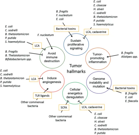
Although the ability of microorganisms to produce ROS [56] contributes to tumorigenesis, bacteria can also release specific toxins that induce DNA damage responses, which also con- tribute to tumorigenesis (Fig. 10.2). Damaged barrier function may also allow the bacteria to transfer or deliver toxins, including cytolethal distending toxin (CDT), colibactin, cytotoxic necrotizing factor 1 (CNF1), and Bacteroides fra- gilis toxin. CDT and colibactin are true genotox- ins, which directly damage the DNA and activate the ataxia signaling pathway and histone phos- phorylation, which lead to G2/M cell cycle arrest [6]. CDT is created by Gram-negative bacteria (E. coli, Helicobacter species, and Salmonella typhi) and is relevant to colorectal, gastric, and gallbladder cancer. Colibactin is produced by E.
coli, Enterobacteriaceae, Proteus mirabilis, and Klebsiella pneumoniae and is important in the development of CRC. Colibactin produced by E.
coli induces DNA double-strand brakes, cell cycle arrest, and improper cell division [1].
Bacteroides fragilis toxin activates the Wnt/β- - catenin signaling pathway, which promotes epi- thelial proliferation, by promoting the cleavage of the adhesion molecule, E-cadherin. The cleav- age of E-cadherin leads to β-catenin translocation to the nucleus and enables the transcription of proto-oncogene c-myc, leading to colonic epithe- lial hyperplasia [1].
375 376 377 378 379 380 381 382 383 384 385 386 387 388 389 390 391 392 393 394 395 396 397 398 399 400 401 402 403 404 405 406 407 408 409 410 411 412
413 414
415 416 417 418 419
420 421 422 423 424 425 426 427 428 429 430 431 432 433 434 435 436 437 438 439 440 441 442 443 444 445 446 447 448 449 450 451 452 453 454 455 456 457 458 459 460 461 462 463 464 465 466 467
10.7 Bacterial Metabolites in Carcinogenesis
A major pathway in microbiome-host signaling is the production of bacterial metabolites. These metabolites, which are synthesized by the microbiome, enter the circulation at the site of production and travel to distant organs, where they exert their biological effects [57]. Bacterial metabolites behave like human hormones in the sense that they are synthesized by an “organ” (the microbiome) and are then transferred to the site of action by the circulation [57].
Microbiota have the potential to metabolize hormones, such as estrogen. The gut microbiome is a key determinant of estrogen levels in the
body. β-Glucuronidases are the enzymes respon- sible for estrogen deconjugation. Deconjugation of excreted estrogen is important in estrogen reuptake and, thus, modulation of systemic estro- gen availability and the regulation of estrogen- associated pathways. Numerous bacterial species can express β-glucuronidases, including Firmicutes and Bacteroidetes: Alistipes, Bacteroides, Bifidobacterium, Citrobacter, Clostridium, Collinsella, Dermabacter, Edwardsiella, Escherichia, Faecalibacterium, Lactobacillus, Marvinbryantia, Propionibacterium, Roseburia, and Tannerella.
Thus, these bacterial species affect circulating and excreted estrogen levels. Reactivated estro- gen increases the serum estrogen levels and act
Fig. 10.2 The intestinal microbiota can modulate several hallmarks of cancer through different mechanisms
468 469
470 471 472 473 474 475 476 477 478 479 480 481 482
483 484 485 486 487 488 489 490 491 492 493 494 495 496 497 498
through estrogen receptors (ERα and ERβ) to modulate the expression of several genes, includ- ing mitochondrial genes. Elevated oxidative phosphorylation was shown to support metastasis [58], contribute to therapy failure [59], and, thereby, render the tumors more aggressive.
Taken together, bacterial estrogen deconjugation promotes breast cancer progression and changes the risk for development and progression of estrogen-dependent cancers [6, 57].
The fermentation of nondigestible carbohy- drates is beneficial for the host due to the genera- tion of short-chain fatty acids (SCFAs), such as acetate, butyrate, formate, lactate, and propio- nate. SCFAs are novel potential targets for the management of obesity, metabolic disorders, and lipomas, due to their ability to influence adipo- cyte differentiation [60]. SCFAs have known anti-inflammatory, antiproliferative, and antineo- plastic effects. In addition, SCFAs can regulate autophagy. Thus, SCFAs have a protective effect on the colonic mucosa and play a significant role in the protection against colon and liver cancer [6]. In the gut, acetate, butyrate, and propionate production are associated with a large group of bacteria. Acetate production is widespread, while the production of butyrate is connected to Faecalibacterium prausnitzii, Eubacterium hal- lii, Eubacterium rectale, Roseburia faecalis, Odoribacter, and Anaerotruncus species. The majority of propionate production is associated with Bacteroidetes, Lachnospiraceae, and Negativicutes species, as well as to Roseburia inulinivorans and Ruminococcus obeum. In line with this, the abundance of Akkermansia muciniphila, a propionate-producing bacterium, is associated with the richness of the gut microbi- ome [61]. SCFAs have both positive and negative effects on breast cancer. Stroma and cancer cells have free fatty acid receptors, through which SCFAs modulate several hallmarks of cancer:
cell proliferation, invasion, apoptosis, metabo- lism, and the expression level of certain genes.
Lactate can be used as a direct energy substrate;
thus, the inhibition of lactate metabolism reduces cancer cell viability. Butyrate enhances mito- chondrial ROS level, induces apoptosis, and
inhibits histone deacetylases, which lead to ele- vated anticancer activity [57].
The intestinal microbiota regulate bile acid metabolism and are involved in producing the secondary bile acids, deoxycholic acid (DCA) and lithocholic acid (LCA), through the deconju- gation, oxidation, and dehydroxylation of pri- mary bile acids. The enzyme responsible for the conversion of primary bile acids to secondary bile acids is 7α/β hydroxysteroid dehydrogenase (HSDH). Conversion to secondary bile acids increases the hydrophobicity of bile salts allow- ing recovery through the colonic epithelium.
Secondary bile acids have both pro- and antican- cer activity. The consumption of a high-fat diet changes the gut microbiome and enhances the level of DCA via 7/α-dehydroxylase, which is produced by bacteria, mainly clostridia. DCA is a promoter of carcinogenesis in certain cancers.
DCA-elicited cell signaling is connected to pro- tein kinase C and ERK1/2 signaling through epi- dermal growth receptors, resulting in enhanced cell proliferation. DCA is known to increase CRC development and promote colon and esoph- ageal cancers [6]. Moreover, bile acids disrupt cell membranes through their amphipathic prop- erties and the generation of ROS and reactive nitrogen species. Bile acids also exert antimicro- bial activity that changes the composition of the intestinal community. LCA is synthesized through 7α-dehydroxylation of chenodeoxycho- lic acid (CDCA) or 7β-dehydroxylation of urso- deoxycholic acid (UDCA). The enzyme responsible for LCA synthesis is encoded by the bile acid-inducible (baiH) operon and expressed by aerobic and anaerobic bacteria, including Bacteroides fragilis, Bacteroides intestinalis, Clostridium scindens, Clostridium sordellii, Clostridium hylemonae, and E. coli. These bacte- ria belong to the phyla Bacteroides, Firmicutes, and Proteobacteria. LCA inhibits the epithelial- to- mesenchymal transition, vascular endothelial growth factor (VEGF) production, and metastasis formation of breast cancer cells, changes the met- abolic features of the cells, and enhances antitu- mor immunity of the host [30]. In line with these observations, human serum levels of LCA and the ability of the microbiome to produce LCA are
499 500 501 502 503 504 505 506 507 508 509 510 511 512 513 514 515 516 517 518 519 520 521 522 523 524 525 526 527 528 529 530 531 532 533 534 535 536 537 538 539 540 541 542 543 544 545
546 547 548 549 550 551 552 553 554 555 556 557 558 559 560 561 562 563 564 565 566 567 568 569 570 571 572 573 574 575 576 577 578 579 580 581 582 583 584 585 586 587 588 589 590 591 592 593
largely reduced in breast cancer; this is most pro- nounced in in situ and early stage carcinoma (stages 0 and 1) [30]. LCA can potentially exert its effects through the farnesoid X receptor (FXR), liver X receptor (LXR), pregnane X receptor (PXR), constitutive androstane receptor (CAR), vitamin D receptor (VDR), and G-protein-coupled bile acid receptor 1 (TGR5).
In breast cancer, the main receptor is TGR5.
Activation of TGR5 signaling was shown to induce OXPHOS, mitochondrial biogenesis through NRF1, AMPK, and PGC-1β signaling.
The expression of mitochondrial proteins (cyto- chrome c, atp5g1, and ndufb5) consequently increases mitochondrial activity and exerts anti- Warburg effects in breast cancer models [30]. In supraphysiological concentrations (>1 μM), LCA was shown to inhibit fatty acid production and induce cell death and the expression of multidrug- resistant proteins [62].
When undigested dietary compounds reach the large intestine, they are fermented through anaerobic respiration. High protein consumption is associated with elevated colonic fermentation.
Bioactive products, similar to bile salts, can pro- duce or inhibit carcinogenesis. Cadaverine, a bio- genic amine, is synthesized from L-lysine by bacterial lysine decarboxylase enzymes (LdcC and CadA). Cadaverine also has a human origin, but it seems that bacterial production is more important as it highly exceeds human biosynthe- sis. The main cadaverine-producing bacteria include Aeromonas veronii, Clostridium perfrin- gens, E. coli, Enterobacteriaceae bacteria, Edwardsiella tarda, Hafnia alvei, Raoultella ornithinolytica, Staphylococcus, and Streptomyces species. These species belong to the Acinetobacteria, Bacteroides, Firmicutes, Fusobacteria, and Proteobacteria phyla. Trace amine-associated receptors (TAARs) were shown to be responsible for mediating cadaverine- elicited effects. Through TAARs, cadaverine inhibits epithelial-to-mesenchymal transition, proliferation, movement, and invasion of breast cancer cells. Moreover, cadaverine treatment inhibits primary tumor infiltration to the sur- rounding tissue and reduces the proportion of cancer stem cells [42].
Many bacteria in the GI tract have alcohol dehydrogenase activity, which enables the bacte- ria to metabolize ethanol and produce reactive and toxic acetaldehyde. The most important gas- tric pathogen, H. pylori, and some skin bacteria have high alcohol dehydrogenase activity. The colonic mucosa has a low aldehyde dehydroge- nase activity, resulting in acetaldehyde accumu- lation in the colon. High acetaldehyde levels contribute to the pathogenesis of alcohol-induced diarrhea and the increased risk of colon polyps and colon cancer [63] (Fig. 10.3).
10.8 The Interference of the Microbiome with Chemotherapy
Bacteria of the intestinal microbiome can inter- fere with therapeutic agents during cancer treat- ment and management. The microbiome can modulate the efficacy of both chemotherapy and radiotherapy. Bacteria can inactivate or activate chemotherapeutic drugs, alter immune responses, or interfere with the side effects of the therapy.
The relationship is reciprocal, as tumor therapy can influence the composition and function of the microbiome [57].
Chemotherapeutic compounds, such as cispla- tin or oxaliplatin, exert their cytotoxic effects through DNA damage, the upregulation of apop- totic pathways, or the promotion of antitumor immune responses (through a TLR4-dependent mechanism). The antitumor effects of platinum compounds significantly decrease upon broad- spectrum antibiotic treatment or in microbiota- deficient mice. In addition, tumor-infiltrating cells show reduced production of ROS after anti- biotic treatment [35]. In this scenario, commen- sal microbes prime tumor-infiltrating cells for ROS production through the connection to PRRs, with the involvement of MYD88 signaling (described previously) [6, 56]. Lactobacillus aci- dophilus supplementation can restore the antitu- mor effects of cisplatin in mice [11].
Cyclophosphamides have been used for antican- cer therapy for almost 60 years. In high doses, cyclophosphamides are immunosuppressive,
AU5 594
595 596 597 598 599 600 601 602 603 604 605 606 607 608 609 610 611 612 613 614 615 616 617 618 619 620 621 622 623 624 625 626 627 628 629 630 631 632 633 634 635 636 637 638 639 640 641
642 643 644 645 646 647 648 649 650 651 652 653
654 655 656
657 658 659 660 661 662 663 664 665 666 667 668 669 670 671 672 673 674 675 676 677 678 679 680 681 682 683 684 685 686
while in low doses, cyclophosphamides promote the antitumor immune response through activa- tion of cytotoxic T cells and induction of immu- nogenic cell death [33]. Cyclophosphamides are used in the therapy of breast cancer; however, cyclophosphamides cause damage to the gut mucosa, making the gut leaky and allowing gut bacteria to enter the circulation. A rich microbi- ome and elevated levels of Lactobacillus planta- rum are protective against cyclophosphamide-induced mucosal injury [57].
Cyclophosphamide treatment causes the overrep- resentation of Gram-negative species, such as Barnesiella intestinihominis that enhance effec- tor T cells (cytotoxic CD8+ T cell), and Enterococcus hirae, Gram-positive bacteria that enhance MYD88-dependent CD8+ T cell activa- tion in a tumor-specific manner. Both bacteria are regulated by intestinal NOD2 receptors that pro- mote a pro-inflammatory tumor environment and drive antitumor immune responses [35]. T cell- mediated immune responses against B. intestini- hominis and E. hirae have clinical relevance in chemotherapy-treated patients with lung and ovarian cancers.
In addition to cyclophosphamides, anthracy- clines, selective estrogen receptor modulators (SERMs), taxanes, and antimetabolites have key roles in breast cancer therapy. Anthracyclines are produced by Streptomyces species. Anthracyclines act mainly by intercalating into DNA and inter- fering with DNA metabolism and RNA produc- tion, or by generating excessive ROS. Anthracyclines can be bacteriostatic; they decrease the abundance of Acinetobacter species [32]. No bacterial drug metabolism was associ- ated with SERMs (tamoxifen, raloxifene).
Tamoxifen can modulate the composition of the microbiome, while tamoxifen resistance can also be modulated by the microbiome. SERMs are toxic to different species in the GI tract, including Acinetobacter baumannii, Bacillus stearother- mophilus, Enterococcus faecium, Klebsiella pneumoniae, Porphyromonas gingivalis, Pseudomonas aeruginosa, and Streptococcus mutans [57]. Taxanes (paclitaxel, docetaxel) are widely used as chemotherapy agents. Taxanes disrupt microtubule formation and, hence, block cell division and proliferation. Taxanes may change the composition of the microbial commu- nity or interfere with bacterial LPS, while activat-
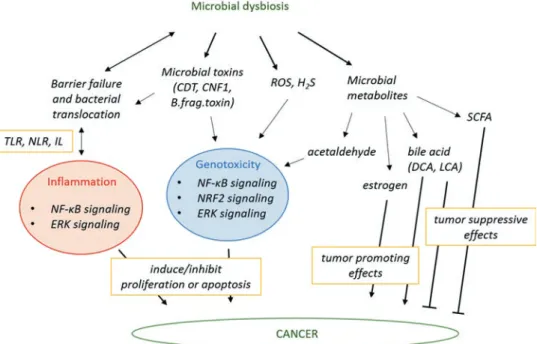
Fig. 10.3 Mechanisms by which microbial dysbiosis modulates carcinogenesis
687 688 689 690 691 692 693 694 695 696 697 698 699 700 701 702 703 704 705 706 707 708 709 710 711
712 713 714 715 716 717 718 719 720 721 722 723 724 725 726 727 728 729 730 731 732 733 734 735 736 737
ing the immune system. PARP inhibitors are drugs used in the treatment of ovarian cancer with a potential to be used for other neoplasias (e.g., breast cancer, prostate cancer). PARP inhibitors were shown to induce the diversity of the gut microbiome [64].
Drugs are often used in combinations to enhance treatment efficacy. Irinotecan is used to treat colon cancer and small cell lung carcinoma.
For treating colon cancer, irinotecan is generally used in combination with 5-fluorouracil (5FU), whereas for the treatment of small cell lung can- cer, irinotecan is combined with cisplatin.
Bacterial reactivation of irinotecan by bacterial β-glucuronidase leads to severe side effects, such as diarrhea, vomiting, bone marrow suppression, hair loss, shortness of breath, and fever. Antibiotic treatment or β-glucuronidase inhibition prevents most of these side effects [6]. When 5FU is used in combination with irinotecan, dysbiosis- induced mucositis leads to bacterial translocation from the GI tract. Both 5FU and gemcitabine undergo bacterial activation and bacterial deacti- vation. In human pancreatic ductal adenocarci- noma, Gammaproteobacteria was found to be the most important player in deactivating gem- citabine. In tumors, levels of Gammaproteobacteria were elevated in tumor patients as compared to healthy individuals, underlining its role in the regulation of gem- citabine availability. Both 5FU and gemcitabine have bactericidal properties; therefore, they can alter the composition of the GI microbial com- munity [57].
Chemotherapy is often not specific for one or two bacterial species, but change the proportion and diversity of the microbiome. After chemo- therapy, both the alpha diversity, which repre- sents species richness (the number of different species in a sample), and beta diversity, which refers to the diversity in the microbial community between different environments, are altered as compared to samples without chemotherapy.
These changes are independent of covariates (age, sex, previous antibiotic consumption, and previous chemotherapeutic treatment) and show increases in Citrobacter, Enterococcus, Klebsiella, Megasphaera, and Parabacteroides
species, while showing decrements in the abun- dance of Adlercreutzia, Anaerostipes, Bifidobacterium, Blautia, Clostridium, Collinsella, Coprococcus, Dorea, Lachnospira, Roseburia, and Ruminococcus species. Some bacteria showed resistance to chemotherapy; thus their abundance did not change upon treatment, including Actinomyces, Erysipelotrichaceae, Mobiluncus, Mitsuokella, Oxalobacter, Prevotella, Scardovia, and Slackia [34].
Besides inducing taxonomic dysbiosis, che- motherapy can disrupt microbial function.
Several metabolic pathways can be suppressed by chemotherapy, including amino acid, carbo- hydrate, and nucleotide metabolism, as well as the metabolism of vitamins and cofactors. Other pathways are enhanced by chemotherapy, includ- ing signal transduction, xenobiotic degradation, and glycan metabolism. Glycan metabolism, together with disrupted carbohydrate and amino acid metabolism, contributes to enhanced intesti- nal inflammation [65] and upregulation of nitro- gen, sulfate, and riboflavin pathways, which is associated with inflammatory diseases, increased ROS production, and bacterial translocation [66].
Moreover, chemotherapy increases bacterial motility proteins and flagella assembly (essential for bacterial pathogenesis, motility, adhesion, and invasion).
Dysregulated microbiota plays a significant role in the development of GI mucositis.
Mucositis is a painful inflammation of the mucous membranes of the digestive system, usu- ally as an unpleasant side effect of chemotherapy and radiotherapy for cancer. In the first step of this process, the microbiome enhances the activa- tion of NF-κB and TNFα signaling, leading to long-lasting inflammation. Several bacteria are reduced after chemotherapy, including Bifidobacterium, Coprococcus, Clostridium, Dorea, Faecalibacterium, Lachnospira, Roseburia, and Ruminococcus, which inhibit inflammation through blocking NF-κB and pro- duce mucosa-protecting metabolites (SCFAs), whereas Citrobacter and other species, which participate in LPS biosynthesis and enhance intestinal inflammation, are increased during chemotherapy [34]. Subsequently, GI mucositis
738 739 740 741 742 743 744 745 746 747 748 749 750 751 752 753 754 755 756 757 758 759 760 761 762 763 764 765 766 767 768 769 770 771 772 773 774 775 776 777 778 779 780 781 782 783 784 785
786 787 788 789 790 791 792 793 794 795 796 797 798 799 800 801 802 803 804 805 806 807 808 809 810 811 812 813 814 815 816 817 818 819 820 821 822 823 824 825 826 827 828 829 830 831 832 833
barrier dysfunction develops, leading to increased intestinal permeability, which coincides with a decrease in the amount of the previously men- tioned protective bacteria. The microbiome may modulate the composition of the mucus layer, as the terminal step of mucositis induction.
Citrobacter, which increases after chemotherapy, may participate in the degradation of the mucosal barrier through the expression of mucus- degrading enzymes (mucinase, glycosidase), and Enterobacteriaceae can disrupt the mucus layer.
Butyrate-producing bacteria protect the mucin layer, as butyrate increase mucin synthesis. A decrement in cysteine, proline, and methionine metabolism, which occurs during chemotherapy, can also be responsible for altered mucin compo- sition and the development of GI mucositis after chemotherapy [34].
Radiation therapy is used as a primary treat- ment in cancers that are localized to one area of the body to prevent tumor recurrence after sur- gery or applied together with chemotherapeutic agents. Radiation itself is genotoxic, resulting in cancer cell death. However, radiation can also abolish nontarget cells due to the activation of the immune system by radiation-induced inflamma- tion. The microbiota is known to be involved in these off-target effects due to intestinal mucosa damage and toxicity. Radiotherapy decreases both the diversity and the total amount of gut bac- teria, particularly Bacteroidetes, Enterobacteriaceae, Firmicutes, and Lactobacillus species, while enriching Fusobacterium and Proteobacteria, which are connected with increased production of pro- inflammatory cytokines [35].
10.9 Modulation
of the Microbiome to Enhance the Efficacy of Chemotherapy
Probiotics and prebiotics are widely used to shift the composition of the microbiome, and these interventions are potentially useful in restoring the microbiome after chemotherapy. Probiotics contain live bacteria that can be administered
orally, while prebiotics (dietary prebiotics) are compounds in food, which provide substrates that stimulate the growth or activity of advanta- geous bacteria colonizing the gut. Prebiotics and probiotics prevent infection and moderate the side effects of cancer treatment. Administration of various strains of Lactobacillus, such as Lactobacillus acidophilus, is associated with enhanced cisplatin sensitivity and longer survival in lung cancer [35]. Bifidobacterium bifidum, Lactobacillus acidophilus, Lactobacillus casei, and Lactobacillus rhamnosus decrease the toxic- ity associated with 5FU chemotherapy and, con- sequently, reduce abdominal discomfort and diarrhea. In addition, Bifidobacterium and Lactobacillus species in combination were able to moderate the side effects after radiation treat- ment. Current clinical trials are focused on the efficacy of probiotic treatment for colorectal, kid- ney, breast, gynecologic, and lung cancer [35].
Fecal microbiota transplantation (FMT), also known as stool transplantation, is the process of transplanting fecal bacteria from a healthy indi- vidual into a diseased subject. FMT is an effec- tive therapy to shift the composition of the microbiome. FMT is effective in the treatment of Clostridium difficile, where FMT is curative through enhancement of the diversity of the microbiome [67]. FMT could be potentially effective after chemotherapy or radiotherapy in cancer patients by avoiding gut toxicity or pre- venting infections. However, FMT has numerous side effects (fever, diarrhea, vomiting), including serious side effects, such as GI bleeding or perfo- ration, that limit its applicability in cancer patients [35].
As a developing future therapy, bacterial engi- neering offers the opportunity to treat cancer without reconfiguring the gut microbiome.
Biologically engineered bacteria could be applied effectively to target cancer cells or to deliver ther- apeutic agents, thereby avoiding serious side effect-eliciting anticancer therapies. Bacterial cells can be easily and rapidly transfected with vectors encoding interfering RNAs, cytokines, toxins, antiangiogenic factors, or antibodies.
Listeria and Shigella species could invade hypoxic tumor tissues, and, given their quick rep-
834 835 836 837 838 839 840 841 842 843 844 845 846 847 848 849 850 851 852 853 854 855 856 857 858 859 860 861 862 863 864 865 866 867 868 869
870 871 872 873
874 875 876 877 878
879 880 881 882 883 884 885 886 887 888 889 890 891 892 893 894 895 896 897 898 899 900 901 902 903 904 905 906 907 908 909 910 911 912 913 914 915 916 917 918 919 920 921 922 923 924 925 926
lication rate, these bacteria could amplify their transgene(s) within the tumor microenvironment.
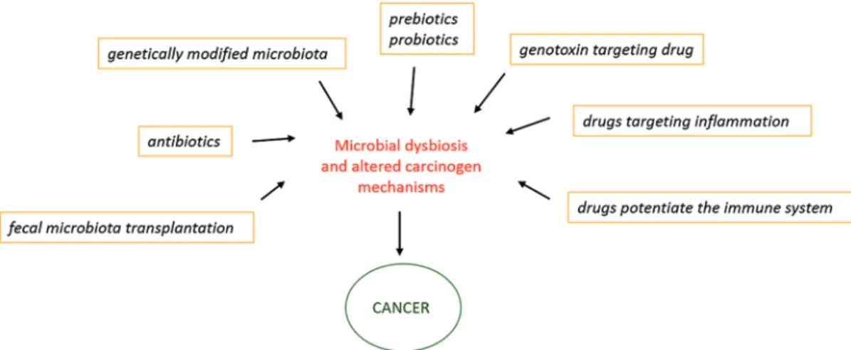
Upon the application of bacteria, finding a good balance is necessary; one must seed a sufficient number of bacteria to elicit therapeutic effect but should avoid suppressing the immune system at the same time [35] (Fig. 10.4).
10.10 Type of Cancers Related to Microbial Dysbiosis
Besides the GI tract, other organs are colonized by a unique microbial community, such as the skin, oral cavity, and germinal tracts. Growing evidence confirms a significant relevance of bac- terial microbiota in the carcinogenesis of the colon, liver, breast, lung, oral cavity, and pancreas.
The liver receives 70% of its blood supply from the intestinal vein. This close functional relationship between the liver and GI tract results in constant exposure to nutrients, toxins, micro- bial metabolites, and microbes. Various types of immune cells (NK cells, macrophages, lympho- cytes) defend this organ against harmful agents derived from the intestine. An altered microbi- ome may contribute to the development of hepa- tocellular carcinoma (HCC), which is preceded by chronic liver disease, fibrosis, and cirrhosis [68]. The disrupted microbiome may drive this process through the loss of intestinal barrier func-
tion, the activation of the NF-κB pathway, the production of pro-inflammatory cytokines, and increased anti-apoptotic signals.
Pancreatic cancer is an aggressive cancer type with low therapeutic success and survival rate.
Periodontal disease, low oral hygiene, obesity, smoking, and alcohol consumption are well- known risk factors for pancreatic cancer, because they facilitate the translocation of bacteria through disrupted barrier layers. Bacteria can reach the pancreas through the circulation.
Furthermore, although the pancreas does not have a microbiome, carcinogenesis of this organ is enhanced by distant dysbiotic microbiota [6], through the involvement of inflammatory responses, LPS expression, and TLR4 activation [69].
About 90% of all lung cancer cases are attrib- uted to smoking, while only 15% of smokers develop lung cancer, suggesting other mecha- nisms and influences. The interface of the lung is continuously connected to the outside environ- ment, and the microbiota of the lung reflect the microaspiration of oral microbiota. The lung has a unique microbiome with different species of Proteobacteria. The connection between lung cancer and chronic pulmonary disease is assigned to toxic pro-inflammatory and neoplasia-causing compounds. Different bacteria species, such as Moraxella catarrhalis, Haemophilus influenza, and Streptococcus pneumoniae, are associated with 50% of chronic pulmonary disease, and
Fig. 10.4 Targeting the microbiome for modulation of carcinogenesis
927 928 929 930 931 932 933
934 935
936 937 938 939 940 941 942 943 944 945 946 947 948 949 950 951 952 953 954 955
956 957 958 959 960 961 962 963 964 965 966 967 968 969 970 971 972 973 974 975 976 977 978 979 980 981 982 983 984 985 986 987