NAD(P)HX dehydratase (NAXD) deficiency:
a novel neurodegenerative disorder exacerbated by febrile illnesses
Nicole J. Van Bergen,
1,2,* Yiran Guo,
3,* Julia Rankin,
4,5,* Nicole Paczia,
6,*
Julia Becker-Kettern,
6,* Laura S. Kremer,
7,8Angela Pyle,
9Jean-Franc¸ois Conrotte,
6Carolyn Ellaway,
10,11,12Peter Procopis,
12,13Kristina Prelog,
14Tessa Homfray,
15Ju´lia Baptista,
4,5Emma Baple,
4,5Matthew Wakeling,
4Sean Massey,
1Daniel P. Kay,
6Anju Shukla,
16Katta M. Girisha,
16Leslie E. S. Lewis,
17Saikat Santra,
18Rachel Power,
19Piers Daubeney,
19,20Julio Montoya,
21Eduardo Ruiz-Pesini,
21Reka Kovacs-Nagy,
7,22Martin Pritsch,
23Uwe Ahting,
7David R. Thorburn,
1,2,24Holger Prokisch,
7,8Robert W. Taylor,
9John Christodoulou,
1,2,10,11,24,#Carole L. Linster,
6,#Sian Ellard
4,5,#and Hakon Hakonarson
3,#*,#These authors contributed equally to this work.
Physical stress, including high temperatures, may damage the central metabolic nicotinamide nucleotide cofactors [NAD(P)H], generating toxic derivatives [NAD(P)HX]. The highly conserved enzyme NAD(P)HX dehydratase (NAXD) is essential for intra- cellular repair of NAD(P)HX. Here we present a series of infants and children who suffered episodes of febrile illness-induced neurodegeneration or cardiac failure and early death. Whole-exome or whole-genome sequencing identified recessive NAXD variants in each case. Variants were predicted to be potentially deleterious throughin silico analysis. Reverse-transcription PCR confirmed altered splicing in one case. Subject fibroblasts showed highly elevated concentrations of the damaged cofactors S- NADHX, R-NADHX and cyclic NADHX. NADHX accumulation was abrogated by lentiviral transduction of subject cells with wild-type NAXD. Subject fibroblasts and muscle biopsies showed impaired mitochondrial function, higher sensitivity to metabolic stress in media containing galactose and azide, but not glucose, and decreased mitochondrial reactive oxygen species production.
Recombinant NAXD protein harbouring two missense variants leading to the amino acid changes p.(Gly63Ser) and p.(Arg608Cys) were thermolabile and showed a decrease in Vmaxand increase in KMfor the ATP-dependent NADHX dehydratase activity. This is the first study to identify pathogenic variants in NAXD and to link deficient NADHX repair with mitochondrial dysfunction. The results show that NAXD deficiency can be classified as a metabolite repair disorder in which accumulation of damaged metabolites likely triggers devastating effects in tissues such as the brain and the heart, eventually leading to early childhood death.
1 Murdoch Children’s Research Institute, Royal Children’s Hospital, Parkville, Melbourne, 3052, Australia 2 Department of Paediatrics, University of Melbourne, Parkville, Melbourne, 3052, Australia
3 Center for Applied Genomics, Children’s Hospital of Philadelphia, Philadelphia, PA 19104, USA 4 University of Exeter Medical School, Exeter, EX4 4QD, UK
5 Royal Devon Exeter NHS Foundation Trust, Exeter, EX4 4QD, UK
6 Luxembourg Centre for Systems Biomedicine, University of Luxembourg, Belvaux, L-4367, Luxembourg 7 Institute of Human Genetics, Technische Universita¨t Mu¨nchen, Munich, 81675, Germany
8 Institute of Human Genetics, Helmholtz Zentrum Mu¨nchen, Munich, 81675, Germany
Received June 1, 2018. Revised August 8, 2018. Accepted October 16, 2018. Advance Access publication December 20, 2018 ßThe Author(s) (2018). Published by Oxford University Press on behalf of the Guarantors of Brain. All rights reserved.
For permissions, please email: journals.permissions@oup.com
Downloaded from https://academic.oup.com/brain/article-abstract/142/1/50/5255623 by Hungary EISZ Consortium user on 07 August 2019
9 Wellcome Centre for Mitochondrial Research, Institute of Neuroscience, The Medical School, Newcastle University, Newcastle upon Tyne, NE2 4HH, UK
10 Western Sydney Genetics Program, Children’s Hospital at Westmead, Sydney, 2145, Australia 11 Discipline of Genetic Medicine, University of Sydney, 2145, Sydney, Australia
12 Neurology Department, Children’s Hospital at Westmead, Sydney, 2145, Australia 13 Discipline of Child and Adolescent Health, University of Sydney, 2145, Australia 14 Medical Imaging Department, Children’s Hospital at Westmead, Sydney, 2145, Australia 15 Royal Brompton and St George’s University Hospital, London, SW17 0RE, UK
16 Department of Medical Genetics, Kasturba Medical College and Hospital, Manipal Academy of Higher Education, Manipal, 576104, India
17 Department of Paediatrics, Kasturba Medical College and Hospital, Manipal Academy of Higher Education, Manipal, 576104, India
18 Birmingham Children’s Hospital, Birmingham, B4 6NH, UK 19 Royal Brompton Hospital, London, SW3 6NP, UK
20 National Heart and Lung Institute, Imperial College, London, SW3 6LY, UK
21 Departamento de Bioquimica y Biologia Molecular y Celular- CIBER de Enfermedades Raras (CIBERER)-Instituto de Investigacio´n Sanitaria de Arago´n (IISAragon), Universidad Zaragoza, Zaragoza, 50013, Spain
22 Department of Medical Chemistry, Molecular Biology and Pathobiochemistry, Semmelweis University, Budapest, 1085, Hungary 23 Department of Pediatric Neurology, DRK-Childrens-Hospital, Siegen, 57072, Germany
24 Victorian Clinical Genetics Services, Royal Children’s Hospital, Melbourne, 3052, Australia Correspondence to: Professor John Christodoulou
Neurodevelopmental Genomics Research Group, Murdoch Childrens Research Institute, 50 Flemington Rd Parkville, 3052, Victoria, Australia
E-mail: john.christodoulou@mcri.edu.au
Correspondence may also be addressed to: Dr Carole L. Linster
Luxembourg Centre for Systems Biomedicine (LCSB), Universite´ du Luxembourg, Campus Belval, 6, avenue du Swing, L-4367 Belvaux, Luxembourg
E-mail: carole.linster@uni.lu
Keywords:metabolite repair; mitochondria; febrile illness; dehydratase; epimerase
Abbreviations:NAD = nicotinamide adenine dinucleotide; NADP = nicotinamide adenine dinucleotide phosphate; NAXD = NAD(P)HX dehydratase; NAXE = NAD(P)HX epimerase; OXPHOS = oxidative phosphorylation
Introduction
Metabolism is traditionally viewed as an efficient and pre- cise system, supported by enzymes that are highly specific for their substrate and the type of reaction through which they convert the substrate. This view has been challenged over recent years by the identification of a growing list of enzymes that function to repair or remove metabolic side products, the latter of which are generated by side reactions of main metabolic enzymes. Metabolic side products can also be produced intracellularly by unwanted spontaneous chemical reactions. These non-canonical or ‘damaged’ me- tabolites can inhibit key metabolic reactions if they are left to accumulate. It is to precisely prevent the accumulation of potentially toxic small molecules that all organisms of all domains of life have most likely evolved a panoply of me- tabolite repair systems (Linster et al., 2013; Van Schaftingen et al., 2013).
The nicotinamide adenine dinucleotides (NAD) (reduced form NADH, oxidized form NAD + ) and nicotinamide ad- enine dinucleotide phosphate (NADP) (reduced form NADPH, oxidized form NADP + ) have essential roles in many cellular functions. NAD is involved in a series of
catabolic reactions and in mitochondrial energy production, whereas NADP is a key component of numerous biosyn- thetic processes as well as cellular antioxidant protection systems (Ying, 2008; Houtkooper et al., 2010). The nico- tinamide ring within these cofactors is prone to hydration, forming NADHX or NADPHX, which can be present asR or Sepimers and which can further degrade irreversibly to cyclic NAD(P)HX (Yoshida and Dave, 1975). NADHX can be slowly produced from NADH by GAPDH (Rafteret al., 1954); NAD(P)HX can also form spontaneously from the normal reduced cofactors under ‘stress’ conditions such as increased temperature or acidic pH (Rafter et al., 1954;
Yoshida and Dave, 1975). The damaged cofactors cannot act as electron carriers and have been shown in vitro to inhibit several key dehydrogenase enzymes (Yoshida and Dave, 1975; Prabhakar et al., 1998). In Saccharomyces cerevisiae, in vitro and in vivo evidence for an inhibitory effect of NADHX on 3-phosphoglycerate oxidoreductase, catalysing the initial step of the serine biosynthesis path- way, has been obtained recently (Becker-Kettern et al., 2018). Therefore, NAD(P)HX can be expected to be toxic to cells, and detoxification by a metabolite repair system is critical.
Downloaded from https://academic.oup.com/brain/article-abstract/142/1/50/5255623 by Hungary EISZ Consortium user on 07 August 2019
The nicotinamide nucleotide repair system consists of two partner enzymes: NAD(P)HX epimerase (NAXE, formerly APOA1BP; OMIM: 608862), which converts R-NAD(P)HX to S-NAD(P)HX, and NAD(P)HX dehydratase (NAXD, for- merly CARKD; OMIM: 615910), which converts S- NAD(P)HX back to NAD(P)H in an ATP-dependent manner (Marbaix et al., 2011). Both these enzymes are tar- geted to several subcellular compartments, including the mito- chondrion (Marbaix et al., 2014). They also have a ubiquitous tissue distribution (Marbaixet al., 2014) and are conserved across all taxa (Marbaixet al., 2011). The presence of the NAD(P)HX repair enzymes across all tissues and spe- cies, combined with the central metabolic roles of the cofac- tors that they function to preserve, suggest that the repair system is critical to sustain life. More specifically, it can be predicted that the brain, which has a very high and constant demand for energy supply generated by mitochondria, would be particularly vulnerable to impaired NAD(P)HX repair and as such, the NAXE and NAXD enzymes would be critical to support normal brain function. In apparent contradiction with those predictions, however, knocking out NAD(P)HX repair enzymes in other organisms such as yeast, bacteria and plants, has so far only revealed subtle growth pheno- types, if any, at least under standard growth conditions (Breslow et al., 2008; Hillenmeyer et al., 2008; Nichols et al., 2011; Colinas et al., 2014; Niehaus et al., 2014;
Becker-Kettern et al., 2018).
Recently, whole exome sequencing identified pathogenic variants in the epimeraseNAXE, which were associated with cases of a lethal neurometabolic disorder of early childhood (Kremeret al., 2016; Spiegelet al., 2016). In these subjects, it appeared that episodes of febrile illness aggravated the consequences of an already compromised metabolite repair system, resulting in rapid neurological deterioration and de- composition of other tissues with clinical observation of ataxia, muscular hypotonia, respiratory insufficiency and/or respiratory failure, nystagmus and skin manifestations followed by premature death. This provided the first cases of subjects with pathogenic variants in a key enzyme of the nicotinamide nucleotide repair pathway. Here we report on the first known cases with pathogenic variants in the dehydratase NAXD.
Materials and methods
Full materials and methods are available in online Supplementary material.
Subjects and variant analysis
All procedures were approved by human research ethics com- mittees. Primary cultures of fibroblasts were established from skin biopsies.
Mitochondrial analysis
Fibroblast extracts were analysed by immunoblotting using antibodies against representative oxidative phosphorylation
(OXPHOS) subunits. The complex I and IV activity was deter- mined using commercially available dipstick assays (MitoSciences). Mitochondrial stress was induced by culturing fibroblasts galactose media and determining cell growth rates.
Mitochondria reactive oxygen species production was mea- sured by dihydroethidium fluorescence.
LC-MS analysis of NAD(H),
NADP(H) and damaged derivatives
Wild-type NAXD including the mitochondrial targeting signal (mNAXD) or not (cNAXD starting at Met3) or a GFP control construct was introduced into patient fibroblasts by lentiviral delivery. Intracellular metabolites were extracted from cultured fibroblasts and concentrations were measured with an HRAM- RP-LC-MS (high resolution accurate mass reversed phase liquid chromatography mass spectrometry) method compared against chemically pure standards.
NADHX dehydratase activity assays and thermostability testing
Missense variants were introduced into wild-type cNAXD and proteins expressed and purified from a bacterial overexpres- sion system as detailed in the Supplementary material. The S-NADHX substrate was synthesized and purified as pre- viously described (Becker-Kettern et al., 2018) and NAXD enzyme kinetic properties determined using a spectrophoto- metric assay (Marbaix et al., 2011). Protein thermostability was tested by pre-incubation of purified protein at different temperatures (30–47C).
Statistical analysis
Statistical analyses were carried out using either a two-tailed Student’s t-test or one-way ANOVA corrected for multiple comparisons as appropriate (GraphPad PrismÕ Software).
Error bars represent the standard deviation of the mean [standard deviation (SD)]. AP-value50.05 was considered to be statistically significant.
Data availability
Derived data supporting the findings of this study are available from the corresponding author upon request.
Results
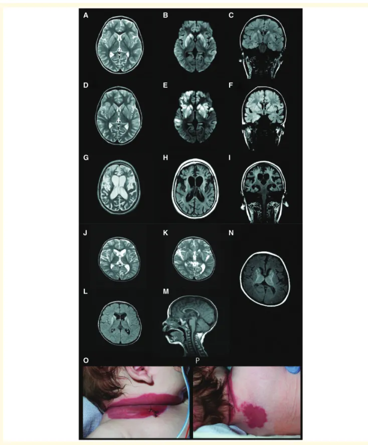
In the present study we report six unrelated individuals with homozygous or compound heterozygous variants in NAXD. The predominant clinical features included repeated episodes of regression often triggered by episodes of mild fever or infection, an infantile onset neurodegenera- tive condition (Fig. 1A–M) and skin lesions (Fig. 1O and P), ultimately leading to early death in all cases. The clinical features of our NAXD subjects therefore resembled those of the previously published cases of NAXE deficiency (Table 1), and detailed clinical reports for all cases are available in Supplementary material. The six subjects with
Downloaded from https://academic.oup.com/brain/article-abstract/142/1/50/5255623 by Hungary EISZ Consortium user on 07 August 2019
Figure 1 Neuroimaging findings and skin manifestations in children with NAXD mutations.Brain MRI scans for Case 1 at the age of 3 years and 7 months, 3 months after normal MRI scans: (A) axial T2, (B) diffusion (DWI) and (C) coronal T2FLAIR showed bilateral and symmetrical T2high signal and cytotoxic oedema of the basal ganglia and focal areas of cortical involvement in the temporal lobes. Progress MRI after 3 weeks: (D) axial T2, (E) DWI and (F) coronal T2FLAIR showed bilateral and symmetrical T2high signal and cytotoxic oedema of the basal ganglia persist with increasing areas of asymmetrical cortical involvement in the temporal lobes and frontal lobes. Follow-up MRI 3 years later:
(G) axial T2, (H) axial and (I) coronal T2FLAIR showed generalized cerebral atrophy, most marked in the frontal lobes and basal ganglia with exvacuo-dilatation of the lateral ventricles. The high signal is consistent with gliosis. Case 2 MRI (J–M) showed bilateral hyperintensity of striatal nuclei, which remained unchanged in subsequent scans. Case 4 brain MRI (N) showed bilateral basal ganglia changes suggestive of a mitochondrial disorder. Extensive skin lesions in Case 4 (OandP).
Downloaded from https://academic.oup.com/brain/article-abstract/142/1/50/5255623 by Hungary EISZ Consortium user on 07 August 2019
NAXD variants were identified individually by whole exome or whole genome sequencing as part of either inde- pendent international gene discovery cohorts, National Health Services diagnostic testing or through the Genematcher/Matchmaker database (Philippakis et al., 2015; Sobreira et al., 2015).
Sanger sequencing confirmed all variants identified through next generation sequencing (Supplementary Fig.
1). The human NAXD gene is predicted to generate four coding transcripts, leading to the expression of cytosolic, mitochondrial and endoplasmic reticulum (ER) protein iso- forms, as explained in detail in the Supplementary material (see also Supplementary Figs 2 and 3). For initial interpre- tation we focused on the most abundant transcript RefSeq isoform c (NM_001242882.1; Supplementary Table 1) whose existence is most strongly supported by EST (expressed sequence tag) analysis and from which mito- chondrial and cytosolic NAXD protein forms can be trans- lated (Supplemental material). In silico analyses predicted the variants to be pathogenic to each of the four NAXD transcript isoforms, and affect the structure and function of the NAXD protein (Supplementary Table 2, Supplementary Figs 2 and 3). By HSF3 analysis (Desmet et al., 2009) the splicing variant in Case 1 was predicted to lead to disrup- tion of the wild-type donor site, which was confirmed by RT-PCR studies, revealing aberrant splicing and skipping of Exon 9 (Supplementary Fig. 4).
We then used patient fibroblasts to determine the intra- cellular concentrations of NAD(P), NAD(P)H, S- NAD(P)HX, R-NAD(P)HX and cyclic-NAD(P)HX using HRAM RP-LC-MS with comparison against chemically pure standards (Supplementary Fig. 5). We detected no significant differences in NAD, NADH and NADP levels between control and subject-derived fibroblasts (NADPH levels were below the limit of quantification). Importantly, the damaged cofactor derivatives S-NADHX, R-NADHX and cyclic-NADHX were only detected in fibroblasts from Cases 1, 2 and 4, but not in any of our control cell lines (Fig. 2A and B). Quantification of these metabolites revealed a similar level of accumulation of S-NADHX
and R-NADHX in Cases 1, 2 and 4 (Fig. 2C).
Interestingly the levels detected in Case 3 were much lower than for Cases 1, 2 and 4 and only just above the detection threshold for LC-MS analysis (Fig. 2B).
Lentiviral transduction with either the cytosolic (cNAXD) or mitochondrial (mNAXD) wild-type NAXD cDNA completely prevented the accumulation of any of the NADHX derivatives in fibroblasts from Cases 1 and 2 (Fig. 2D) whereas a control GFP construct had no effect (Fig. 2D).
To address whether mitochondrial function is affected by NAXD deficiency in our subjects, we examined the expres- sion of representative protein subunits of the mitochondrial respiratory chain and enzyme activity of complexes I and IV in fibroblasts from Cases 1 and 2. There was a marked reduction in the expression of both NDUFB8 (complex I membrane subunit) and COXII (complex IV subunit) in Cases 1 and 2 compared to four paediatric control fibro- blast extracts (Fig. 3A and Supplementary Fig. 6). We found a significant reduction in cytochromecoxidase activ- ity in Cases 1 and 2 compared to controls (Fig. 3B), whilst complex I NADH oxidation activity was not affected (data not shown). Mitochondrial superoxide production revealed a significant decrease in rotenone-inhibited complex I superoxide production in both Cases 1 and 2 compared to controls (Fig. 3C). There was a significant decrease in the growth rate of fibroblasts in glucose-free, galactose- azide medium to induce mitochondrial stress from Cases 1 and 2 compared to control fibroblasts (Fig. 3D).
The NAXD missense variants [p.(Arg308Cys) and p.(Gly63Ser)] were introduced by site-directed mutagenesis into human NAXD cDNA, then expressed and purified as recombinant proteins. For the p.(Gly63Ser) variant, we found a 3.4-fold decrease in Vmax and a 3.3-fold increase in KM compared to the wild-type protein. For the p.(Arg308Cys) variant, we determined a 2.5-fold decrease in Vmax and a 2.2-fold increase in KM compared to wild- type (Supplementary Table 3). Thermostability analyses of recombinant NAXD revealed that both the p.(Gly63Ser) and the p.(Arg308Cys) proteins lost enzymatic activity Table 1 Clinical findings in individuals with variants inNAXDorNAXE
Clinical presentation NAXDcases (present study) NAXE(previously published)
Case 1 Case 2 Case 3 Case 4 Case 5 Case 6 Spiegelet al.
(2016)
Kremeret al.
(2016)
Gender M F F M F F 3F, 2M 2F, 4M
Episodes of fever/illness prior to deterioration Y Ya Y Y Y Y 5/5 4/6 (2 unclear)
Neurodegeneration Y Y N Y ?b Y 5/5 4/6 (2 unclear)
Skin lesions Y Y N Y N Y Not reported 4/6
Cardiac presentation N N N N Y N Not reported 2/6c
Early death Y Y Yd Y Y Y 4/5 6/6
aFever was associated with some, but not all episodes of deterioration.
bAbnormal MRI scan.
cTwo NAXE patients died from cardiovascular failure.
dSibling had mild developmental delay, mild anaemia, recurrent episodes of fever, and died after an episode of vomiting and lethargy at 1 year 7 months. N = no; Y = yes.
Downloaded from https://academic.oup.com/brain/article-abstract/142/1/50/5255623 by Hungary EISZ Consortium user on 07 August 2019
upon pre-incubation at temperatures higher than 30C while the wild-type protein activity resisted exposure to temperatures up to 47C (Fig. 3E). This thermolability was most pronounced for the p.(Arg308Cys) variant, with a more than 90% decrease in activity above 45C, while for the p.(Gly63Ser) variant this same treatment resulted in a40% activity loss (Fig. 3E).
Discussion
Here we report on the first known pathogenicNAXD var- iants, which affected six families, leading to a fever-induced severe multisystem disease and death within the first decade of life. To explore the consequences of NAXD deficiency, quantification of NAD(P)HX metabolites revealed a similar Figure 2 NADHX accumulation in subject fibroblasts and phenotypic rescue by lentiviral gene delivery of wild-type NAXD.
Representative examples of LC-MS extracted ion chromatograms of (A)S- andR-NADHX metabolites (XIC = 682.12) and (B) NADH and cyclic NADHX (XIC = 664.11). The chromatograms clearly show peaks ofS-,R- and cyclic NADHX in Cases 1 and 2, and not in a control. (C) Quantitative results of the NADHX measurements after cultivation of fibroblasts from Cases 1–4 under standard conditions at 37C for 96 h.
Note that these metabolites were not detected in four control fibroblast lines. Data are meanSD,n= 3. (D) Quantitative results of the NADHX measurements in lentiviral-rescued cells. There was a clear accumulation ofS-,R- and cyclic NADHX in Cases 1 and 2, independently of transduction with GFP-only vectors.S-,R- and cyclic NADXH were not detected in control fibroblasts. Lentiviral gene rescue with either the mNAXD or cNAXD construct completely prevented accumulation ofS-,R- and cyclic NADHX in subject cells. Data are meanSD,n= 3.
Downloaded from https://academic.oup.com/brain/article-abstract/142/1/50/5255623 by Hungary EISZ Consortium user on 07 August 2019
level of accumulation of S-NADHX and R-NADHX in Cases 1, 2 and 4 in the expected 60:40 S to R epimer ratio (Marbaix et al., 2011). This was completely reversed by lentiviral transduction with either cytosolic (cNAXD) or
mitochondrial (mNAXD) lentiviral constructs. Supposing an intracellular fibroblast volume of 2 pl, the approximate intracellular concentrations of S-NADHX (100–300mM) and R-NADHX (70–210mM) were quantitatively similar Figure 3 Mitochondrial impairment in NAXD subject fibroblasts and thermostability of recombinant NAXD protein variants.
(A) Mitochondrial OXPHOS proteins were separated by SDS-PAGE, probed for relative expression levels of key OXPHOS subunits by immu- noblotting, and expression levels normalized to GAPDH as a loading control. There was a significant decrease in the expression of complex I and complex IV proteins expression in Cases 1 and 2 compared to controls. (B) Mitochondrial OXPHOS enzyme activity was measured in cell extracts by immunocapture dipstick assays. There was a significant decrease in complex IV activity [monoclonal antibodies (mAbs)/mg protein] in Cases 1 and 2 relative to controls. (C) Relative rates (relative fluorescent units/time) of mitochondrial superoxide production in fibroblasts was measured with the superoxide sensitive probe dihydroethidium in the presence or absence of the complex I inhibitor rotenone. (D) Growth rate in medium devoid of glucose but containing 5 mM galactose and 50mM sodium azide was normalized to growth rate in basal medium to account for variation in the baseline growth rate of each cell line. The normalized growth rate in Cases 1 and 2 was significantly lower than controls. (E) Purified recombinant NAXD protein, without or with missense mutations p.(Gly63Ser) and p.(Arg308Cys), was pre-incubated at the indicated temperature for 30 min prior to addition to a reaction mixture for the spectrophotometric assay of NADHX dehydratase activity. Data are meanSD,n43 per measurement from at least two independent experiments. Statistical significance was determined using one-way ANOVA with Bonferroni correction for multiple comparisons; *P50.05, **P50.01, ***P50.001.
Downloaded from https://academic.oup.com/brain/article-abstract/142/1/50/5255623 by Hungary EISZ Consortium user on 07 August 2019
to values induced by a complete loss of function for NAXD in an experimental cell line (Becker-Kettern et al., 2018).
The levels in Case 3 were only just above the detection threshold, which may be due to the frameshift mutation in this patient preventing expression of the mitochondrial and endoplasmic reticulum targeted NAXD isoforms (early truncation), but allowing for expression of the cytosolic NAXD protein from the ATG residing in exon 2 (Met3 in Supplementary Fig. 3). The same reasoning would also apply to Case 5, for which fibroblasts were, however, not available for measurements. These results provide strong support that the NADHX accumulation is specifically caused by NAXD mutations.
NAD(P)H are essential cofactors for many cellular reac- tions, particularly in the mitochondria, therefore accumula- tion of the non-canonical NAD(P)HX derivatives may impede multiple cellular functions including key mitochon- drial dehydrogenases (Yoshida and Dave, 1975; Prabhakar et al., 1998). Subjects with pathogenic variants in NAXE had decreased complex I activity (Kremer et al., 2016) indicating impaired mitochondrial function as a conse- quence of a deficiency in one of the NAD(P)HX repair enzymes. Respiratory chain activity was impaired in NAXD patients; fibroblasts had a significant decrease in the expression of complex I and IV, and activity of com- plex IV and muscle enzymology showed reduced respira- tory chain activity (complex II + III, Case 2; complex I, Case 4; complex I and IV, Case 6). We also demonstrated reduced superoxide production in NAXD subjects, which may be explained by decreased expression of specific com- plex I subunits subsequently affecting holocomplex stabi- lity, but partial enzyme activity may still remain because of preservation of NADH oxidation in the matrix arm of complex I. Mitochondrial dysfunction can also be revealed by culturing cells under galactose growth conditions (Robinson et al., 1992) in the presence of the complex IV inhibitor sodium azide (Swalwellet al., 2011), which limits ATP production by glycolysis, forcing cells to rely on mito- chondrial OXPHOS. There was a significant decrease in the growth rate of fibroblasts from Cases 1 and 2 compared to control fibroblasts under galactose conditions, further sup- porting that mitochondrial function in fibroblasts from NAXD subjects is compromised.
To determine the effect of the NAXD missense variants [p.(Arg308Cys) and p.(Gly63Ser)] on NADHX dehydratase activity, the missense variants were introduced by site-direc- ted mutagenesis into the cytosolicNAXDcDNA since simi- lar kinetic properties were previously obtained for both mitochondrial and cytosolic NAXD/Carkd (mouse homo- logue of human NAXD) (Marbaix et al., 2011) and the cytosolic protein gave greater yield. Analysis of the kinetic properties for the two missense variants revealed signifi- cantly reduced Vmax and increased KM compared to the wild-type protein. Thermostability analysis revealed loss of enzymatic activity upon pre-incubation temperatures above 30C, and the thermolability was more pronounced for the p.(Arg308Cys) variant. The Gly63 residue is highly
conserved from bacteria to humans (Marbaixet al., 2011);
the Arg308 residue is also conserved in the mouse and yeast homologs of NAXD, but not in the Escherichia coli homologue (Marbaix et al., 2011). This arginyl residue is relatively close to the C-terminus of the protein, as are the splicing and frameshift mutations found in Cases 1 and 2, respectively, suggesting that this may be a critical domain for NAXD protein stability. In addition, the thermolability of the NAXD missense variants found in Cases 1 and 2 may at least in part explain the coincidence of deterioration in the subjects sometimes occurring after episodes of fever.
Those individuals who had infections may have had unre- ported fevers secondary to the viral infection, which likely precipitated rapid decompensation. In summary, our in vitro analyses of recombinant NAXD demonstrated that the missense variants, while retaining residual enzyme activity, show a markedly decreased stability espe- cially at higher temperatures.
This report highlights the importance of the NAD(P)HX repair system to preserve cellular and overall health in humans. We could show high intracellular NADHX accu- mulation for three of the four individuals where fibroblast lines were available for analysis, and impaired mitochon- drial function. The missense variants found in Cases 1 and 2 led to partial loss of enzyme activity and a significant decrease in thermostability. More particularly, NAXD defi- ciency appears to have devastating effects in key tissues such as the brain, which are critically dependent on efficient energy metabolism and are exquisitely sensitive to abnor- mal metabolite accumulation. After 2-hydroxyglutaric acid- uria (Van Schaftingen et al., 2009) and NAXE deficiency (Kremeret al., 2016), NAXD deficiency represents now the third known disorder of metabolite repair. We also suggest that NAXD deficiency should be included in the growing list of genetic disorders associated with fever-induced neu- rological deterioration (Powers and Scheld, 1996; Longo, 2003).
Acknowledgements
We thank the families for their support.
Funding
We thank the Crane and Perkins families and the Lions International Club Esch-sur-Alzette for donations to this research. The research conducted at the Murdoch Children’s Research Institute was supported by the Victorian Government’s Operational Infrastructure Support Program. J.B.K. and N.P. were supported by an AFR-PhD grant (4044610) and a CORE junior grant (C16/
BM/11339953), respectively, of the Fonds National de la Recherche Luxembourg. Sequencing, data analysis and Sanger validation of one sample (Case 1) were done in the Center for Applied Genomics at the Children’s
Downloaded from https://academic.oup.com/brain/article-abstract/142/1/50/5255623 by Hungary EISZ Consortium user on 07 August 2019
Hospital of Philadelphia through research funding from Aevi Genomic Medicine Inc. This study was partly sup- ported by the German Bundesministerium fu¨r Bildung und Forschung (BMBF) through the German Network for mitochondrial disorders (mitoNET, 01GM1113 for H.P.) and through the E-Rare project GENOMIT (01GM1603 for H.P.). H.P. is supported by EU Horizon2020 Collaborative Research Project SOUND (633974). This work was supported by grants from Instituto de Salud Carlos III (PI17/00021); Departamento de Ciencia, Tecnologı´a y Universidad del Gobierno de Arago´n (Grupos de Referencia B33_17R). R.W.T. is supported by the Wellcome Centre for Mitochondrial Research (203105/
Z/16/Z), the Medical Research Council (MRC) Centre for Translational Research in Neuromuscular Disease and Mitochondrial Disease Patient Cohort (UK) (G0800674), the Lily Foundation and the UK NHS Highly Specialised Service for Rare Mitochondrial Disorders of Adults and Children. S.E. is a Wellcome Trust Senior Investigator.
Competing interests
The authors report no competing interests.
Supplementary material
Supplementary material is available at Brain online.
References
Becker-Kettern J, Paczia N, Conrotte JF, Zhu C, Fiehn O, Jung PP, et al. NAD(P)HX repair deficiency causes central metabolic pertur- bations in yeast and human cells. FEBS J 2018; 285: 3376–401.
Breslow DK, Cameron DM, Collins SR, Schuldiner M, Stewart- Ornstein J, Newman HW, et al. A comprehensive strategy enabling high-resolution functional analysis of the yeast genome. Nat Methods 2008; 5: 711–8.
Colinas M, Shaw HV, Loubery S, Kaufmann M, Moulin M, Fitzpatrick TB. A pathway for repair of NAD(P)H in plants. J Biol Chem 2014; 289: 14692–706.
Desmet FO, Hamroun D, Lalande M, Collod-Beroud G, Claustres M, Beroud C. Human splicing finder: an online bioinformatics tool to predict splicing signals. Nucleic Acids Res 2009; 37: e67.
Hillenmeyer ME, Fung E, Wildenhain J, Pierce SE, Hoon S, Lee W, et al. The chemical genomic portrait of yeast: uncovering a pheno- type for all genes. Science 2008; 320: 362–5.
Houtkooper RH, Canto C, Wanders RJ, Auwerx J. The secret life of NAD + : an old metabolite controlling new metabolic signaling path- ways. Endocr Rev 2010; 31: 194–223.
Kremer LS, Danhauser K, Herebian D, Petkovic Ramadza D, Piekutowska-Abramczuk D, Seibt A, et al. NAXE mutations disrupt the cellular NAD(P)HX repair system and cause a lethal neurometa- bolic disorder of early childhood. Am J Hum Genet 2016; 99: 894–
902.
Linster CL, Van Schaftingen E, Hanson AD. Metabolite damage and its repair or pre-emption. Nat Chem Biol 2013; 9: 72–80.
Longo N. Mitochondrial encephalopathy. Neurol Clin 2003; 21: 817–
31.
Marbaix AY, Noel G, Detroux AM, Vertommen D, Van Schaftingen E, Linster CL. Extremely conserved ATP- or ADP-dependent enzym- atic system for nicotinamide nucleotide repair. J Biol Chem 2011;
286: 41246–52.
Marbaix AY, Tyteca D, Niehaus TD, Hanson AD, Linster CL, Van Schaftingen E. Occurrence and subcellular distribution of the NADPHX repair system in mammals. Biochem J 2014; 460: 49–58.
Nichols RJ, Sen S, Choo YJ, Beltrao P, Zietek M, Chaba R, et al.
Phenotypic landscape of a bacterial cell. Cell 2011; 144: 143–56.
Niehaus TD, Richardson LG, Gidda SK, ElBadawi-Sidhu M, Meissen JK, Mullen RT, et al. Plants utilize a highly conserved system for repair of NADH and NADPH hydrates. Plant Physiol 2014; 165:
52–61.
Philippakis AA, Azzariti DR, Beltran S, Brookes AJ, Brownstein CA, Brudno M, et al. The matchmaker exchange: a platform for rare disease gene discovery. Hum Mutat 2015; 36: 915–21.
Powers JH, Scheld WM. Fever in neurologic diseases. Infect Dis Clin North Am 1996; 10(1): 45–66.
Prabhakar P, Laboy JI, Wang J, Budker T, Din ZZ, Chobanian M, et al. Effect of NADH-X on cytosolic glycerol-3-phosphate dehydro- genase. Arch Biochem Biophys 1998; 360: 195–205.
Rafter GW, Chaykin S, Krebs EG. The action of glyceraldehyde-3- phosphate dehydrogenase on reduced diphosphopyridine nucleotide.
J Biol Chem 1954; 208: 799–811.
Robinson BH, Petrova-Benedict R, Buncic JR, Wallace DC.
Nonviability of cells with oxidative defects in galactose medium: a screening test for affected patient fibroblasts. Biochem Med Metab Biol 1992; 48: 122–6.
Sobreira N, Schiettecatte F, Valle D, Hamosh A. GeneMatcher: a matching tool for connecting investigators with an interest in the same gene. Hum Mutat 2015; 36: 928–30.
Spiegel R, Shaag A, Shalev S, Elpeleg O. Homozygous mutation in the APOA1BP is associated with a lethal infantile leukoencephalopathy.
Neurogenetics 2016; 17: 187–90.
Swalwell H, Kirby DM, Blakely EL, Mitchell A, Salemi R, Sugiana C, et al. Respiratory chain complex I deficiency caused by mitochon- drial DNA mutations. Eur J Hum Genet 2011; 19: 769–75.
Van Schaftingen E, Rzem R, Marbaix A, Collard F, Veiga-da-Cunha M, Linster CL. Metabolite proofreading, a neglected aspect of inter- mediary metabolism. J Inherit Metab Dis 2013; 36: 427–34.
Van Schaftingen E, Rzem R, Veiga-da-Cunha M. L: -2-Hydroxyglutaric aciduria, a disorder of metabolite repair. J Inherit Metab Dis 2009;
32: 135–42.
Ying W. NAD + /NADH and NADP + /NADPH in cellular functions and cell death: regulation and biological consequences. Antioxid Redox Signal 2008; 10: 179–206.
Yoshida A, Dave V. Inhibition of NADP-dependent dehydrogenases by modified products of NADPH. Arch Biochem Biophys 1975; 169:
298–303.
Downloaded from https://academic.oup.com/brain/article-abstract/142/1/50/5255623 by Hungary EISZ Consortium user on 07 August 2019