Downloadedfromhttps://journals.lww.com/md-journalbyBhDMf5ePHKav1zEoum1tQfN4a+kJLhEZgbsIHo4XMi0hCywCX1AWnYQp/IlQrHD3tIQ5gQCIeyyUV9lmA4CJqvXkl5H2Ij7w5uIKfk/25g8=on09/02/2018
Downloadedfrom https://journals.lww.com/md-journalby BhDMf5ePHKav1zEoum1tQfN4a+kJLhEZgbsIHo4XMi0hCywCX1AWnYQp/IlQrHD3tIQ5gQCIeyyUV9lmA4CJqvXkl5H2Ij7w5uIKfk/25g8=on
09/02/2018
Dependent Catastrophic Antiphospholipid Syndrome: A Case Report
Andreas Kronbichler, MD, Renate Frank, MD, Michael Kirschfink, MD, A´gnes Szila´gyi, PhD, Dorottya Csuka, PhD, Zolta´n Proha´szka, PhD, Peter Schratzberger, MD, Karl Lhotta, MD,
and Gert Mayer, MD
Abstract:Catastrophic antiphospholipid syndrome (CAPS) is a rare but devastating complication in patients with antiphospholipid syn- drome (APS) with a high morbidity and mortality.
We describe a case of a 30-year old female patient with immuno- globulin A (IgA) deficiency who underwent splenectomy because of idiopathic thrombocytopenic thrombocytopenia. Subsequently, an APS and finally systemic lupus erythematosus was diagnosed. After an uncomplicated pregnancy that was terminated by cesarean section, the patient developed severe CAPS with cerebral, myocardial, renal, and pulmonary involvement.
Because of IgA deficiency, standard therapy consisting of plasma- pheresis and intravenous immunoglobulins in addition to steroids was not tolerated. After 8 sessions of immunoadsorption (IAS), massive pulmon- ary hemorrhage was controlled but relapsed twice whenever IAS was terminated. As other immunosuppressive agents were considered danger- ous because of the risk of infections in the face of severe hypogamma- globulinemia, we administered eculizumab, an inhibitor of the terminal complement pathway, which led to a persistent control of her disease.
Interestingly, eculizumab therapy was associated with a further decline of complement C3 and C4 serum levels. The patient developed a subsequent flare of her systemic lupus erythematosus, potentially indicating that complement inhibition by eculizumab is not effective in preventing lupus flares.
Taken together, we describe a unique case of life-threatening and difficult-to-treat CAPS with a good clinical response after terminal complement complex inhibition with eculizumab. Further controlled trials are necessary to investigate the value of eculizumab in patients with CAPS.
(Medicine93(26):e143)
Abbreviations: CAPS = catastrophic antiphospholipid syndrome, IAS = immunoadsorption, IgA = immunoglobulin A, IgG = immunoglobulin G, IgM = immunoglobulin M, LDH = lactate dehydrogenase, SLE = systemic lupus erythematosus.
INTRODUCTION
C
atastrophic antiphospholipid syndrome (CAPS) is a potentially life-threatening and rare variant of the anti- phospholipid syndrome (APS), characterized by vascular thrombosis in, among others, the brain, lung, heart, and kidney, ultimately leading to multiorgan failure. Most patients develop antiphospholipid antibodies and thrombocytopenia at the time of onset, whereas initially hemolytic anemia, dis- seminated intravascular coagulation, and the presence of schistocytes can be missing. Although diagnostic and thera- peutic approaches improved over the last years, the morbidity and mortality of patients with CAPS is still high.1Pregnancy and puerperium, per se predisposing to thrombotic events because of the induction of a procoagulatory state, are well- established triggers of the catastrophic variant,2 especially when complicated by preeclampsia. Mutations of complement regulatory proteins including membrane cofactor protein, complement factor I, and complement factor H have also been observed in patients with systemic lupus erythematosus (SLE) and antiphospholipid antibody positivity.3CASE REPORT
We report a 30-year-old woman, in whom splenectomy was necessary because of idiopathic thrombocytopenic throm- bocytopenia in 1997. Primary APS was diagnosed in 2004 after onset of deep venous thrombosis with antibodies against anticardiolipin (>90 U/mL, immunoglobulin M [IgM] and immunoglobulin G [IgG] positive) along with anti-beta 2-glycoprotein (>90 U/mL), and she finally fulfilled the diag- nostic criteria of SLE4in 2010 with predominance of muscu- loskeletal and hematologic involvement. During her first pregnancy, she was on antimalarial therapy with chloroquine and low-molecular weight heparin because of APS. After cesarean section and delivery in April 2013, confusion, acute renal failure, myocardial ischemia with heart failure, severe thrombocytopenia, and hemolytic anemia attributed to CAPS developed. Dialysis was initiated and high-dose corticosteroid therapy including initial bolus methylprednisolone (250 mg daily for 3 days) followed by oral methylprednisolone (1.5 mg/kg body weight), rituximab (1 g with a repeated admin- istration after 4 weeks), and plasmapheresis was started. Plasma exchange had to be stopped because of severe intolerance reactions, which were attributed to a selective immunoglobulin Editor: Worawit Louthrenoo.
Received: July 22, 2014; revised: September 1, 2014; accepted: September 4, 2014.
From the Department of Internal Medicine IV (Nephrology and Hyperten- sion) (AK, PS, GM); Department of Radiology (RF), Medical University Innsbruck, Innsbruck, Austria; Institute of Immunology (MK), University of Heidelberg, Heidelberg, Germany; 3rd Department of Medicine (AS, DC, ZP), Research Laboratory, Faculty of Medicine, Semmelweis University, Budapest, Hungary; and Department of Nephrology and Dialysis (KL), Academic Teaching Hospital Feldkirch, Feldkirch, Austria Correspondence: Andreas Kronbichler, MD, Anichstraße 35, 6020
Innsbruck, Austria (e-mail: andreas.kronbichler@i-med.ac.at).
All interventions given were part of normal health care and thus ethical approval was neither obliged, nor sought. However, approval from the patient was obtained to publish the case report.
The authors have no funding and conflicts of interest to disclose.
Copyright#2014 Wolters Kluwer Health | Lippincott Williams & Wilkins.
This is an open access article distributed under the Creative Commons Attribution License 4.0, which permits unrestricted use, distribution, and reproduction in any medium, provided the original work is properly cited.
ISSN: 0025-7974
DOI: 10.1097/MD.0000000000000143
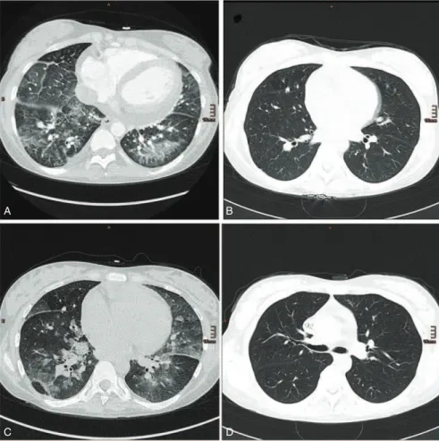
A (IgA) deficiency, which also precluded high-dose intravenous immunoglobulin therapy. The patient’s condition deteriorated and she developed respiratory distress. A computed tomography scan showed diffuse alveolar hemorrhage (Figure 1A). Immu- noadsorption (IAS) therapy using the Life 18 (Miltenyi Biotec, Bergisch Gladbach, Germany) was started with a total of 8 sessions. Treatment ameliorated thrombocytopenia and led to a resolution of the lung injury (Figure 1B). However, the patient was still dependent on dialysis. A renal biopsy revealed typical microangiopathic injury. After recurrence of pulmonary hemorrhage despite continuous high-dose methylprednisolone therapy, 10 additional daily IAS sessions were performed with clinical success. However, lung failure recurred again within 4 days after IAS withdrawal (Figure 1C) together with a rise in lactate dehydrogenase, thrombocytopenia, anemia, and a schis- tocyte count of 19 per mille. Thus, 4 additional sessions of IAS were necessary to control the disease again (Figure 1D). Due to low leukocyte counts and persistently low immunoglobulin levels (IgG 37 mg/dL and IgM 14 mg/dL, respectively), cyto- toxic therapy was considered dangerous because of the risk for serious infections. It was, therefore, decided to administer eculizumab, a monoclonal antibody against the complement component C5, which prevents the activation of the terminal complement pathway. Within 4 days, respiratory failure
completely resolved and signs of hemolytic anemia disappeared despite cessation of IAS. Finally, therapeutic anticoagulation with low molecular heparin could be commenced. The patient was discharged dialysis dependent, but with increasing amounts of urine 71 days after admission in July 2013 with a methyl- prednisolone dosage of 60 mg/day and on eculizumab treatment (weekly administration of 900 mg 4 times, followed by 1200 mg fortnightly). Laboratory values at the onset of the disease, after 3 weeks, at the time of eculizumab initiation, and after achieve- ment of stable remission are depicted in Figure 2.
Two weeks after the discharge, the patient presented again with signs of hemolysis after the fourth and fifth eculizumab infusion. In addition, the serum levels of complement C3 and C4 were consistently reduced. Serum levels of both complement components further declined in the days following eculizumab infusion (data not shown). The patient received oral anticoagula- tion with acenocoumarol. Eculizumab concentrations measured prior and after application revealed efficacious serum concen- trations, assured complete blockage of the terminal complement pathway, and neutralizing antibodies could not be detected.
Addition of mycophenolate mofetil sufficiently abrogated hemo- lysis, which was finally attributed to activity of the underlying SLE, indicating a lack of efficacy of eculizumab in preventing a lupus flare in this patient. The application of eculizumab could be
A B
C D
FIGURE 1. (A) Diffuse pulmonary hemorrhage in both lower lobes, which resolved after another initiation of (B) IAS. (C) After discontinuation of IAS, recurrence of pulmonary hemorrhage could be detected. These findings prompted us to initiate yet another series of IAS together with administration of eculizumab. (D) Complete resolution was detected in a control computed tomography 4 days later. IAS¼immunoadsorption.
stopped after 3 months in September 2013 after a total of 9 infusions without recurrence of thrombotic microangiopathy despite a persisting positive test for antiphospholipid antibodies.
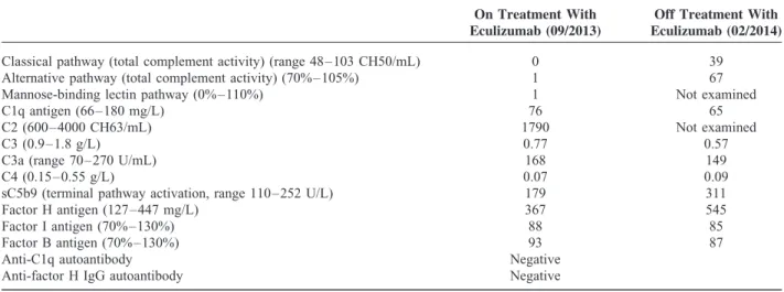
Several measurements of C3d and the terminal complement complex (sC5bC9) while under treatment revealed normal serum levels (data not shown). Examination of the complement regu- latory protein factors H and I as well as the alternative pathway component factor B revealed values within a normal range, indicative of a normal alternative complement pathway. How- ever, persistently reduced C3 and C4 serum levels along with a low C1q (normal in one measurement and slightly reduced in another) are suspicious for a consumption of the classical comp- lement pathway (Table 1). In addition, to exclude inherited predisposing factors in complement C3 or C4, genetic analysis was performed. A rare heterozygous synonymous variation in exon 13 of theC3gene (c.1677C>T; p.C559C) was detected, which was predicted as disease causing by MutationTaster (http://
www.mutationtaster.org/) with a high probability (0.99) (Table 2). Analysis of C4revealed the presence of 2 copies of the C4Aand 1 copy of the C4Bgenes (data not shown).
One year after the initial insult, the patient is still dialysis dependent. Steroid reduction was well tolerated without signs of active thrombotic microangiopathy. The patient is listed for renal transplantation. Her current medication consists of mycophenolate mofetil 250 mg twice a day, methylprednisolone 10 mg every second day, blood pressure-lowering medication, diuretics, thyroid hormone substitution, and acenocoumarol with a target international normalized ratio of 2 to 3.
DISCUSSION
Eculizumab has already shown encouraging results in a patient with recurrence of CAPS with reversal of thrombocy- topenia and prevention of further clinical episodes of
0 200 400 600 800 1000 1200 1400 1600 1800
Thrombocytes Schistocytes
Lactate dehydrogenase
Onset of symptoms Week 2 (IAS discontin
uation)
Relapse (3 w
eeks after onset)
Eculizumab star t (5 w
eeks after onset)9-w eek f
ollo w-up
FIGURE 2. Laboratory values at the onset of the disease and at the time point of stable remission following eculizumab administration.
Reference values of the respective parameters: thrombocytes (150–380 G/L), schistocytes (<5 per mille), and lactate dehydrogenase (100–250 U/L). Lactate dehydrogenase and schistocytes returned to normal values in the ninth week after onset time point and 4 weeks after initiation of eculizumab.
TABLE 1. Complement Analysis After Application of Eculizumab and 6 mo After Cessation of Therapy
On Treatment With Eculizumab (09/2013)
Off Treatment With Eculizumab (02/2014)
Classical pathway (total complement activity) (range 48–103 CH50/mL) 0 39
Alternative pathway (total complement activity) (70%–105%) 1 67
Mannose-binding lectin pathway (0%–110%) 1 Not examined
C1q antigen (66–180 mg/L) 76 65
C2 (600–4000 CH63/mL) 1790 Not examined
C3 (0.9–1.8 g/L) 0.77 0.57
C3a (range 70–270 U/mL) 168 149
C4 (0.15–0.55 g/L) 0.07 0.09
sC5b9 (terminal pathway activation, range 110–252 U/L) 179 311
Factor H antigen (127–447 mg/L) 367 545
Factor I antigen (70%–130%) 88 85
Factor B antigen (70%–130%) 93 87
Anti-C1q autoantibody Negative
Anti-factor H IgG autoantibody Negative
IgG¼immunoglobulin G.
thrombosis.5Its successful use has also been reported in recur- rence of CAPS after renal transplantation.6,7Transplantation in CAPS is also possible with prophylactic eculizumab adminis- tration.8In addition, murine models reveal a pivotal role of the complement system in antiphospholipid antibodies-induced thrombosis along with endothelial cell and platelet activation.
Prevention of terminal complement formation by using a monoclonal antibody against C5 inhibited thrombophilia induced by antiphospholipid antibodies.9 Complement acti- vation and consumption was also confirmed in our patient by the finding that serum levels of C3 and C4 were significantly lowered while disease was highly active. Immunohistochemical analysis of the kidney biopsy specimen revealed a strong staining for C1q and IgM, while deposition of C3, IgG, and IgA was sparsely present. Interestingly, administration of ecu- lizumab was associated with a further decrease in C3 and C4 serum levels. Moreover, sufficient eculizumab levels and comp- lement inhibition was not capable of preventing a flare of her underlying SLE. Evidence that eculizumab may not be effective in SLE is still lacking, since there is limited data coming from a phase I single-center trial. No changes in laboratory values and systemic lupus erythematosus disease activity index have been observed in this preliminary clinical trial. However, the cohort of patients had low disease activity, thus precluding consider- ations on the therapeutic efficacy of eculizumab.10
Our case confirms previous reports that it is possible to discontinue eculizumab in CAPS after complete remission despite a continuing positive test for antiphospholipid anti- bodies. Persistently, reduced C4 and C3 levels in our patient can be explained by continuous activation of the classical complement pathway induced by immune complexes and antiphospholipid antibodies that cannot be influenced by ecu- lizumab. Furthermore, a lowered C1q serum level after ecu- lizumab cessation and strong deposition in the kidney biopsy also supports a consumption of the classical complement pathway. The finding that eculizumab application was fol- lowed by a further decrease in C4 and C3 serum levels is intriguing. One possible explanation could be that eculizumab interfered with clearance of immune complexes and thus caused a further stimulation of the classical pathway. Genetic analysis revealed a rare heterozygous variation in exon 13 of the C3 gene. This mutation does not cause an amino acid change in the C3 protein, so it is likely not causative; however,
a potential influence cannot be excluded, as its predicted functional relevance showed a high probability as a disease- causing factor.
CAPS remains a severe variant of the APS with a high mortality rate of approximately 50%.1 However, patients achieving disease control are prone to a persisting morbidity as reported herein. This case is unique since the patient pre- sented with a continuum of autoimmune disorders and had underlying IgA deficiency. Due to the latter, the standard therapy for CAPS was contraindicated or not tolerated by the patient. The patient had a good clinical response toward IAS combined with high doses of methylprednisolone, whereas she was refractory to rituximab. Inhibition of the terminal comp- lement pathway by eculizumab led to a persistent clinical remission without evident recurrence of disease.
CONCLUSION
Targeting complement activation may provide a new therapeutic option in the treatment of CAPS, especially in patients refractory or unable to tolerate standard therapy. In our exceptional case, stabilization of the disease course was possible after eculizumab was commenced. Further clinical investigations are necessary to clarify whether it can be gener- alized that blockade of the terminal complement activation is efficacious in the treatment of CAPS.
REFERENCES
1. Cervera R, Font J, Gomez-Puerta JA, et al. Validation of the preliminary criteria for the classification of catastrophic antipho- spholipid syndrome.Ann Rheum Dis.2005;64:1205–1209.
2. Gomez-Puerta JA, Sanin-Blair J, Galarza-Maldonado C. Pregnancy and catastrophic antiphospholipid syndrome.Clin Rev Allergy Immunol.2009;36:85–90.
3. Salmon JE, Heuser C, Triebwasser M, et al. Mutations in comple- ment regulatory proteins predispose to preeclampsia: a genetic analysis of the PROMISSE cohort.PLoS Med.2011;8:e1001013.
4. Kronbichler A, Mayer G. Renal involvement in autoimmune connective tissue diseases.BMC Med.2013;11:95.
5. Shapira I, Andrade D, Allen SL, et al. Brief report: induction of sustained remission in recurrent catastrophic antiphospholipid TABLE 2. Genetic Analysis of Complement C3
C3
Exon Protein cDNA position SNP ID according to dbSNP Type Genotype Minor allele
13 p.C559C c.1677C>T — Coding-synonymous Heterozygous T
Tool Qualitative
prediction
Quantitative prediction/
Probability of prediction
Explanation
MutationTaster Disease causing Probability: 0.99 The MutationTaster
probability value is the probability of the prediction,
that is, a value close to 1 indicates a high ‘‘security’’
of the prediction.
cDNA and protein positions are indicated according to transcript (NM_000064.2) and protein (NP_000055.2), respectively. The prediction of the functional relevance of p.C559C was performed by MutationTaster (http://www.mutationmaster.org/).
syndrome via inhibition of terminal complement with eculizumab.
Arthritis Rheum.2012;64:2719–2723.
6. Hadaya K, Ferrari-Lacraz S, Fumeaux D, et al. Eculizumab in acute recurrence of thrombotic microangiopathy after renal transplantation.
Am J Transplant.2011;11:2523–2527.
7. Canaud G, Kamar N, Anglicheau D, et al. Eculizumab improves posttransplant thrombotic microangiopathy due to antiphospholipid syndrome recurrence but fails to prevent chronic vascular changes.
Am J Transplant.2013;13:2179–2185.
8. Lonze BE, Singer AL, Montgomery RA. Eculizumab and renal transplantation in a patient with CAPS.N Engl J Med.
2010;362:1744–1745.
9. Pierangeli SS, Girardi G, Vega-Ostertag M, et al. Requirement of activation of complement C3 and C5 for antiphospholipid antibody- mediated thrombophilia.Arthritis Rheum.2005;52:2120–2124.
10. Barilla-Labarca ML, Toder K, Furie R. Targeting the complement system in systemic lupus erythematosus and other diseases.Clin Immunol.2013;148:313–321.