Complement Activation Related Pseudoallergy and Its Animal Models
PhD thesis
Dr. Péter Bedőcs
Basic Medicine Doctoral School Semmelweis University
Supervisor: Dr. Miklós Tóth, D.Sc.
Dr. János Szebeni, D.Sc.
Official reviewers:
Dr. László Cervenak, Ph.D Dr. Kristóf Nékám, Ph.D
Head of the Final Examination Committee:
Dr. Zoltán Prohászka, D.Sc
Members of the Final Examination Committee:
Dr. Lilian Varga, Ph.D Dr. László Barkai, D.Sc
Budapest, 2013
Table of Contents
Abbreviations 3
Introduction 5
Hypersensitivity, anaphylaxis, pseudo-anaphylaxis 5
Complement system 9
CARPA 13
Drugs activating the complement system 15
Liposomes 17
Polymers 18
In vitro testing of the complement system 20
In vivo models of CARPA 23
Objectives 24
Methods 25
Preparation and characterization of test substances 25
In vivo tests of complement activation related hemodynamic reactions 29
In vitro complement assay 32
Results 33
In vivo complement activation by Doxil 33
Tolerance after first Doxil injection 40
Utilizing tachyphylaxis to prevent severe CARPA reaction 46
Other examples of the utility of the in vivo model 53
Predicting complement activation by polymers 53
Alternative species for CARPA testing 60
Discussion 64
Conclusions 70
Summary 71
Összefoglalás 72
References 74
Author’s Publications 82
Acknowledgements 84
Abbreviations
Ab - antibody
ADCC – antibody-dependent cell-mediated cytotoxicity Ag - antigen
C - complement
CARPA – complement activation related pseudo-anaphylaxis CH – complement hemolysis
CO – cardiac output CR – complement receptor CrEL – chremophor-EL CVP – central venous pressure CVR – coronary vascular resistance DAF – decay accelerating factor DLS – dynamic light scattering
ELISA – enzyme linked immunosorbent assay ETCO2 – end-tidal carbon dioxide
FDA – United States Food and Drug Administration GBM – glomerular basal membrane
HR – heart rate
IgE – immune globulin E IgG – immune globulin G IgM – immune globulin M i.v. – Intravenous
LPS – Lipopolysaccharide
LVEDP – left ventricular end diastolic pressure M – mast cells
MAC – membrane attack complex
MASP – mannose-binding lectin associated serine protease MBL – mannose-binding lectin
MLV – multilamellar vesicle
NHS – normal human serum NS – normal saline
PAP – pulmonary arterial pressure PEG – polyethylene glycol
PEI – polyethylene imine
PIM – pulmonary intravascular macrophage PL – phospholipid
PMN cells – polymorphonuclear cells PVR – pulmonary vascular resistance RBCs – red blood cells
SAP – systemic arterial pressure SRBC – sheep red blood cells SVR – systemic vascular resistance TCC – terminal complement complex Tc cells – cytotoxic T cells
Th1 cells –T helper 1 cells TXA2 – thromboxane A2
USP – United States Pharmacopeia WBCs – white blood cells
Introduction
A large variety of chemical substances as well as medicinal products have been suspected or proven to provoke adverse immunological reactions. One of the most common side effects of drugs is hypersensitivity reaction that often prevents or limits their use. These reactions also have been a major cause of drug withdrawal from the market in the past few decades. Their prevention and treatment is of utmost importance.
Hypersensitivity, anaphylaxis, pseudo-anaphylaxis
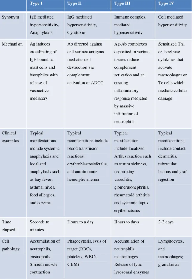
Hypersensitivity means adverse reactions of the normal immune system. These range from a mild localized rash to detrimental effects on vital systems. The classification by Coombs and Gell distinguishes 4 types1,2 (Table 1):
Type 1 – Allergy (immediate), mediated by IgE (and IgG4)
Type 2 – Cytotoxic, antibody-dependent, mediated by IgM or IgG (complement) Type 3 – Immune complex disease, mediated by IgG
Type 4 – Delayed type hypersensitivity, T-cell-mediated immune memory response, antibody-independent.
+1 – Sometimes used as a distinction from type 2. Instead of binding to cell surface components, the antibodies recognize cell surface receptors. Often these reactions are classified as type 2, or a subcategory of type 2.
Table 1. Characteristics of the four classical types of hypersensitivity reactions.
Type I Type II Type III Type IV
Synonym IgE mediated hypersensitivity, Anaphylaxis
IgG mediated hypersensitivity, Cytotoxic
Immune complex mediated hypersensitivity
Cell mediated hypersensitivity
Mechanism Ag induces crosslinking of IgE bound to mast cells and basophiles with release of vasoactive mediators
Ab directed against cell surface antigens mediates cell destruction via complement activation or ADCC
Ag-Ab complexes deposited in various tissues induce complement activation and an ensuing
inflammatory response mediated by massive infiltration of neutrophils
Sensitized Th1 cells release cytokines that activate macrophages or Tc cells which mediate cellular damage
Clinical examples
Typical manifestations include systemic anaphylaxis and localized anaphylaxis such as hay fever, asthma, hives, food allergies, and eczema
Typical
manifestations include blood transfusion reactions,
erythroblastosisfetalis, and autoimmune hemolytic anemia
Typical manifestation include localized Arthus reaction such as serum sickness, necrotizing vasculitis,
glomerulonephritis, rheumatoid arthritis, and systemic lupus erythematosus
Typical manifestations include contact dermatitis, tubercular lesions and graft rejection
Time elapsed
Seconds to minutes
Hours to a day Hours to days 2-3 days
Cell pathology
Accumulation of neutrophils, eosinophils.
Smooth muscle contraction
Phagocytosis, lysis of target (RBCs, platelets, WBCs, GBM)
Accumulation of neutrophils, macrophages.
Release of lytic lysosomal enzymes
Lymphocytes, and
macrophages;
granulomas
Anaphylaxisis a severe,potentially life-threatening hypersensitivity reaction to an allergen. The first documented individual who developed an anaphylactic reaction was Pharaoh Menes who died from a wasp sting in 2640 BC3. Many years later, in 1902, Portier and Richet used the term anaphylaxis to describe acute reactions developing in dogs after repeated injections of the sea anemone toxin4.
Upon first exposure to a substance the immune system becomes sensitized, causing an allergic reaction to occur on repeated exposure. During this quick reaction histamine and other mediators are released, symptoms develop rapidly, within seconds or minutes. These include abdominal pain and cramping, abnormal breathing, wheezing, anxiety, confusion, cough, diarrhea, difficulty swallowing, fainting, light-headedness, dizziness, hives, itchiness, nasal congestion, nausea, vomiting, palpitations, skin redness, rash, mottling, slurred speech. Further clinical signs are arrhythmia, pulmonary edema, hypotension, and angioedema. Anaphylaxis is an emergency condition and requires immediate treatment, often tracheostomy, endotracheal intubation, epinephrine, antihistamines and corticosteroids. In the most severe cases airway occlusion, respiratory and/or cardiac arrest and shock can develop. Common causes include drug allergies, food allergies, insect bites or stings.
Pseudo-anaphylaxishas very similar clinical symptoms asanaphylaxis, without detectable immunological sensitization (antibodies or sensitized cells). It is also called pseudo-allergic or anaphylactoid reactions. ―Non-immune anaphylaxis‖ is the term currently used by the World Allergy Organization for classification, although this nomenclature is somewhat misleading, as for example the complement system, which is often involved in these reactions, is part of the immune system.
The mechanisms of these reactions are not well understood and include direct liberation of vasoactive mediators (e.g. histamine), general mast cell or basophil activation with release of other mediators, activation of the complement or other plasma protein systems (coagulation, kinin-kallikrein) as well as neuro-psychogenic reflex mechanisms.5
Classical hypersensitivity requires a pre-sensitized state of the immune system.
However, in some cases adverse immune reaction can occur on the first exposure to the
drug.6 Unlike IgE-mediated allergy, these reactions arise without prior sensitization and symptoms often lessen or disappear on later treatments (Table 2).
Possible mechanisms of pseudo-allergic reactions:
Direct release of mediators (e.g. histamine) Direct activation of complement system Activation of the coagulation system Interaction with kinin-kallikrein system
Shift in eicosanoid metabolism toward leukotriene formation Platelet activation
Psycho-neurogenic reactions
Table 2. Differences between classical anaphylaxis and pseudo-anaphylactic reactions Anaphylactic reactions Pseudo-anaphylactic
reactions
Is sensitization required? Yes No
Can reaction occur on first
exposure? No Yes
Is reaction predicted by
allergy skin test? Yes No
Complement system
The complement system is part of the humoral arm of the innate, nonspecific immune system (Figure 1). It consists of about 30-35 proteins, some bound to cell membranes and others found in the blood plasma, most of which are synthesized by the liver. A numberof these proteins circulate as inactive precursors and form a cascade that can be activated typically by antibody bound to an antigen.
Figure 1. Components of the immune system
The purpose of the complement system is to assist, or ―complement‖ the action of antibodies in defense against bacteria and rid the body of antibody-coated antigens (antigen-antibody complexes). When the complement cascade is triggered, activation of one of the three pathways leads to the formation of the terminal complement complex (TCC), along with the generation of anaphylatoxins (C5a and C3a) and the release of vasoactive mediators.
The terminal complement complex – also known as membrane attack complex (MAC) – has the conformation of a cylinder that is inserted into the cell wall/membrane
Immune System
Specific (adaptive) Nonspecific (native)
Humoral induced antibodies
Cellular Committed T cells
Cellular PMN Macrophages (RES)
complement system act as opsonins or chemo-attractants, making the recognition and clearance of the pathogens by other components of the immune system more efficient.
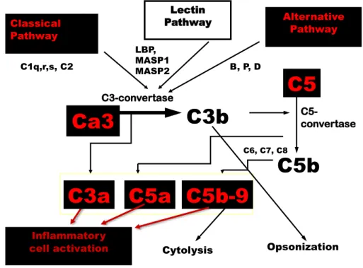
The three pathways that activate the complement system are the classical complement pathway, the alternative complement pathway and the mannose-binding lectin pathway (Figure 2).They function as an enzymatic cascade: inactive proteases are cleaved to form activated proteins with the capacity to cleave downstream proteins.7
Figure 2. The most important complement factors and the three complement activation pathways
Classical Pathway
C1q,r,s, C2
Lectin Pathway LBP,
MASP1 MASP2
Alternative Pathway B, P, D
C6, C7, C8
C5a C5b-9
C5b C5
C5-
convertase
Inflammatory
cell activation Cytolysis Opsonization
C3a Ca3 C3b
C3-convertase
Activation of the classical pathway is initiated by specific binding of the component C1q to the Fc region of the antibodies that are present in immune complexes or on the surface of pathogens. C1q cannot be directly activated by free antibodies, only by antibodies bound to various sites, such as on the surface of pathogens. IgM, then IgG1 and IgG3 are the most effective antibodies at activating the complement. C1q is activated after binding to the Fc region of antibodies and in turn activates the normally inactive serine proteases C1r and C1s. C1s interacts with C4 to form C4a and C4b, then with C2 which is cleaved into C2a and C2b. C2b and C4b form the complex C4bC2b, which is the classical C3 convertase.
The extent of complement activation by the classical pathway is under control of the C1 inhibitor, which dissociates C1r and C1s from C1q, and inactivates the spontaneous low activation of C1q. Other regulatory proteins include the C4-binding protein that controls activation of the classical C3 convertase.
The alternative pathway is initiated by the spontaneous cleavage of C3 - the most abundant complement protein in the plasma - to C3(H20) which binds to factor B.
Factor B is then cleaved to Bb by factor D. C3(H20)Bb is a soluble convertase which cleaves C3 into C3a and C3b. A fraction of formed C3b binds to factor B, which is then cleaved to Ba and Bb by factor D. C3bBb is the alternate C3 convertase.
Regulatory proteins of the alternative pathway are found either in the plasma or on cell membranes. Complement receptor 1 (CR1) and the decay-accelerating factor (DAF or CD55) compete with factor B to prevent its binding to C3b. Factor I together with CR1 and the membrane co-factor of proteolysis (MCP or CD46) cleave C3b to the inactive protein iC3b. Finally, factor H binds to C3b and prevents the formation of C3bBb by competing with Bb.
Activation of the third, mannose-binding lectin pathway is initiated by a protein very similar to C1q, the mannose-binding protein, which binds specifically to mannose and other sugars on the surface of pathogens. In turn, the bound protein forms a complex with two proteases very similar to C1r and C1s, MASP1 and MASP2, which are activated to cleave C4 and C2 with the resulting formation of the C4bC2b convertase.
One result of complement activation is opsonization. C3b is covalently bound to the surface of pathogens, and is recognized by phagocytes via CR1. A large amount of C3b can be deposited on the surface of pathogens, facilitating uptake and elimination.
Another result is the generation of anaphylatoxins, i.e. C3a, C4a and C5a. These activate mast cells and basophils causing the release of inflammatory mediators, such as histamine, thromboxane, tryptase, etc.
Another major consequence is the formation of the membrane attack complex (MAC). The binding of C3b to C4bC2b or Bb produces the C5 convertase, which cleaves C5 to C5a and C5b. C5b initiates the assembly of the MAC and results in the direct lysis of the pathogen cells.
Activation of the complement system also enhances the removal of circulating immune complexes that cannot be engulfed by phagocytes. The number of IgG molecules in immune complexes is sufficient to bind and activate C1q, then C4 and C3.
Immune complexes tagged with C4b and C3b are bound to CR1 on the surface of erythrocytes so that macrophages in the spleen and the liver can degrade immune complexes.
CARPA
Szebeni at al. coined the term complement activation related pseudo-anaphylaxis for this new class of hypersensitivity reaction in 19998, when they examined the role of complement in pseudo-allergic cardiopulmonary reactions to intravenously administered liposomes. By the time of those studies 4 liposomal drugs encapsulating doxorubicin, daunorubicin, and amphotericin B were already in clinical use in several countries, and many other liposomal preparations were in advanced stages of clinical trials.9 Adverse events reported during the use of these formulations draw attention because of their unusual characteristics. Some of these infusion reactions appeared on the first exposure to the drugs, immediately after the start of the infusion, and symptoms included dyspnea, tachycardia, chest pain, hypotension or hypertension, and back pain.
The reactions were categorized as pseudo-allergy, to distinguish these hypersensitivity reactions from the classical IgE mediated allergy.10-15
The relatively high frequency of such reactions was also worrying. Among 705 patients treated with Doxil 6.8% exhibited symptoms of some degree of pseudoallergy.16The underlying mechanism being unknown, it was impossible to specifically predict, prevent, or treat these reactions, some of which were severe, occasionally life threatening, and excluded these patients from further therapy with these drugs.
Based on the fact that certain liposomes can activate the complement systems17and that complement activation can lead to similar cardiovascular and pulmonary symptoms as described above18,19, the authors investigated the possible involvement of the complement system in these hypersensitivity reactions provoked by liposomal drug delivery systems.
Szebeni et al. proved that the complement system has a causal role in the cardiopulmonary distress exhibited in this porcine model of pseudo-allergy and a hypothetical reaction sequence (Figure 3) was described explaining hemodynamic changes following intravenous injection of liposome boluses in the pigs.
Figure 3. Liposomes recognized by IgM and IgG activate the complement system (C) and anaphylatoxins are formed (C5a and C3a), along with the assembly of the membrane attack complex (C5b-9). Anaphylatoxins activate mast cells (M), platelets and polymorphonuclear cells (PMN). As a result thromboxane (TXA2) is released from mast cells, and the platelets and PMN form occlusions in the microcirculation of the lung. The coronary vascular resistance (CVR) and the pulmonary vascular resistance (PVR) and pulmonary arterial pressure (PAP) increases, causing ischemia and acute pulmonary hypertension. The central venous pressure (CVP) increases, the left ventricular end diastolic pressure (LVEDP) drops. These all lead to compromised cardiac output (CO) with diminished systemic circulation and systemic arterial pressure (SAP). In some cases the consequential compensatory mechanisms, i.e. increase in heart rate (HR) and in systemic vascular resistance (SVR) may result in elevated SAP.
Drugs activating the complement system
In the past decade several drugs and chemicals were shown to have a potential to trigger complement activation related pseudo-anaphylaxis (CARPA). These include but are not limited to particulate radio contrast media20, drug delivery systems, carbon nanotubes21, liposomes22 and micellar solvents23, such as Cremophor EL (CrEL) in Taxol24. The monitoring of CARPA became an important aspect in the development of these pharmaceuticals. Underlying the importance of this new type of hypersensitivity, in vitro and in vivo testing of complement activation became a recommended toxicology test by the US Food and Drug Administration25.
Nanomaterials are expected to revolutionize materials science, technology and a wide range of industries, including medicine. By controlling the structure of materials on a super-fine scale, nanotechnologies will improve functions and characteristics of materials as well as creation of new functions26. By definition, a nanoparticle is a particle having one or more dimensions of the order of 100nm or less. Some representatives of this class are liposomes and polymers. There is an extensive amount of evidence attesting that the infusion of nanoparticulate systems, including regulatory- approved stealth nanomedicines, in some individuals is associated with cutaneous, respiratory and circulatory disturbances.27,28
The harmful effects of nanoparticles arise from the combination of various factors, two of which are particularly important: (i) the high surface area, and (ii) the intrinsic toxicity of the surface29. In contrast with conventional particles of larger mean diameter, nanoparticles under 100 nm can potentially be more toxic to the lung (portal of entry), can redistribute from their site of deposition, may escape from the normal phagocytic defenses and can modify the structure of proteins. Therefore, nanoparticles can activate inflammatory and immunological responses and may affect the normal tissue function.
Recently, the critical parameters determining the toxicity of nanoparticles have been proposed30:
Particle size, size distribution, shape, surface area, redox potential and properties, purity, identity of contaminants, catalytic activity and generation of reactive oxygen species;
Interaction with biologically critical macromolecules such as DNA, membranes and cytoskeleton elements;
Potential for unintended carriage of toxic molecules (toxic chemicals that may be present in the environment and are loaded along with therapeutic drugs on the surface of the nanoparticles);
Nanoparticle escape from the normal phagocytic defences and redistribution from site of deposition (translocation);
Agglomeration state (pro-agglomeration factors, size, structure and toxic effects of nanoparticle agglomerates before and after biomodification);
Chemical composition (surface charge, shape, area, reactivity and solubility).
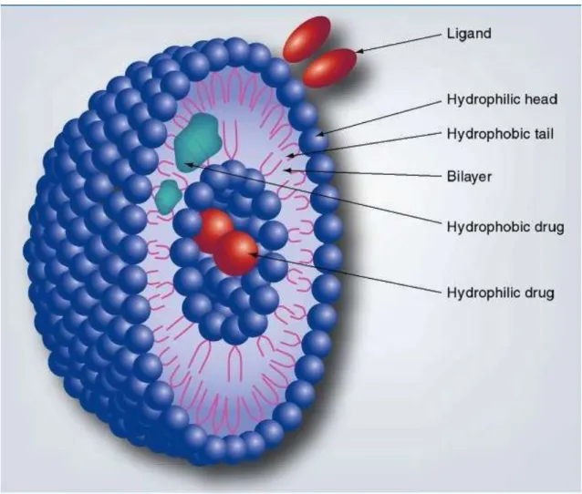
Liposomes
Figure 4. Cross section of a liposome
Since their discovery in the 1960s liposomes were used as a model to study biological membranes and as drug delivery systems for both hydrophilic and maybe even more importantly, for hydrophobic molecules. They are vesicles made of phospholipid bilayers, and can contain small amounts of other molecules, e.g.
cholesterol (Figure 4). The three types of liposomes are small unilamellar liposomes and large unilamellar liposomes consisting of one bilayer, while multilamellar vesicles are formed by multiple bilayers of phospholipid molecules. Their clinical utility comes from the amphiphilic characteristic of phospholipids. Because of this, liposomes can carry both hydrophilic and hydrophobic molecules, and can even be targeted by surface
However, shortly after their discovery it became apparent, that their similarity to biological membranes, a characteristic that provides opportunities to use them as a model system, can as well lead to the activation of the immune system.31 On one hand, this potential can be harvested with the use of liposomes as immune adjuvants.32 On the other, unintended hypersensitivity reactions can be detrimental for their use as drug delivery systems.22
Polymers
Figure 5. Polymeric nanoparticles
'Polymer therapeutics' (Figure 5) is a collective term used to describe polymeric drugs, polymer–drug conjugates, polymer–protein conjugates, polymeric micelles to which a drug is covalently bound, and multicomponent polyplexes that are being developed as non-viral vectors.33 The idea of pairing up a bioactive agent, the drug proper with a carrier and transport vehicle dates back to the 1970s. Utilization of the nearly infinite compositional versatility of synthetic macromolecules and the potentials of synthetic chemistry allows tailoring of these drug delivery systems based on need.
Polymeric drug delivery systems may feature (i) subunits facilitating cell entry;
(ii) other subunits equipped with intra- or extrachain water-solubilizing groups; (iii) still other units acting as a homing device capable of directing the polymer-drug conjugate to the target tissue; and (iv), most importantly, units equipped with functional groups suitable for the critical conjugation step involving bioreversible drug binding to the polymer.34
In vitro testing of the complement system
A detailed understanding of the interaction between nanomaterials/nanoparticles and the complement system is essential, as it could lead to innovation in the design and engineering of safer nanomedicines as well as development of safe clinical practices for their application.
Indications for the assays of complement system:
Clarification of complement defects Detection of activation of the system Verification of adequate regulation
In the context of nanomaterial and nanomedicines safety, the most important is the monitoring of complement activation. There is a wide range of tests currently used for biocompatibility testing.
In the static in vitro model test materials are incubated with serum (e.g. NHS = normal human serum, defined and complement-tested standard serum) under certain circumstances (e.g. 37 °C water bath) for a defined period of time (e.g. 1 hour). The reaction is stopped and complement activation products (C3a, C5a, C4d, Bb, SC5b-9) are measured (e.g. with ELISA). These are highly sensitive assays and identification of various activation products helps differentiation between the activation pathways involved. C4d and Bb measure activation via the classical and the alternative pathway, respectively, and SC5b-9 is a factor representing the common terminal part of all the activation chains.
A similar test can be carried out with whole blood. The presence of blood cells provides an environment that models the conditions found in the human circulation closer than serum or plasma tests. However, exceptional care should be exercised when using anticoagulants, as these can have significant effect on the complement cascade.
EDTA and citrate may inhibit complement activation; heparin has inhibitory or
stimulating effect depending on its concentration. Lepirudin (hirudin) is the only anticoagulant that has no known impact on the complement activation.35
These kits may also be used for ex vivo detection of complement factors and cleavage products in samples taken from individuals exposed to the drugs or test substances prior to collection.
Conversely, analysis of animal serum and plasma samples is more challenging.
There are only a very limited number of assays available for the detection of animal complement factors and activation products. Rat and mouse C5a ELISA kits are commercially available, and methods for the quantification of porcine C5a have also been described in the literature36. However, the use of C5a as a marker for in vivo C activation is problematic because of the rapid clearance of C5a from blood by C5aR- carrying cells (WBC, platelets, macrophages, etc.), while specific ELISAs for the measurement of stable byproducts are not available.
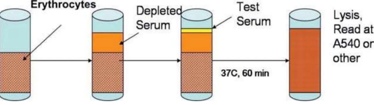
The classical C hemolytic (CH50) assay fills this gap as it measures total hemolytic activity in a species-independent manner. The traditional CH50 is a measure of total complement activation through the ability of the complement system to lyse sensitized sheep red blood cells. The CH50 reflects the ability of the complement in the sample to activate. However, this is only an indirect measure of activation. When testing serum samples drawn from subjects, the CH50 measures the remaining complement activation capability of the sample, giving indirect information about the consumption of the complement factors in the subject. Hence the more activation in the assay, the less of the complement factors was consumed prior to the collection of the sample.
With the use of specific complement depleted sera, the test can be made more specific, focusing on a single protein. These assays are called CxH50, detecting the functional activity of a specific complement component in the sample, for example C3H50 using C3 depleted serum for the detection of C3 function in the sample.
Figure 6. Principle of CxH50 test
The CH50 is a screening assay for the activation of the classical complement pathway and it is sensitive to the reduction, absence and/or inactivity of any component of the pathway. The CH50 tests the functional capability of serum complement components of the classical pathway to lyse sheep red blood cells (SRBC) pre-coated with rabbit anti-sheep red blood cell antibody (hemolysin). When antibody-coated SRBC are incubated with test serum, the classical pathway of complement is activated and hemolysis results. If a complement component is absent, the CH50 level will be zero; if one or more components of the classical pathway are decreased, the CH50 will be decreased. A fixed volume of optimally sensitized SRBC is added to each serum dilution. After incubation, the mixture is centrifuged and the degree of hemolysis is quantified by measuring the absorbance of the hemoglobin released into the supernatant at 540nm. The amount of complement activity is determined by examining the capacity of various dilutions of test serum to lyse antibody coated SRBC.
The assay can be modified to measure the activity of a specific complement factor in the test serum. In the CxH50 test the test serum is added to a serum depleted of the complement component of interest (Cx). (Figure 6)
In vivo models of CARPA
Animal models are the closest experimental alternatives to the human conditions. The interaction of the complex homeostatic systems is kept intact, allowing the analysis of physiological and pathophysiologic processes as they occur in a living organism. Animals, however, are not exactly like humans, and the selection of the appropriate model, and understanding of its limitations is of great importance. Care should be exercised with the interpretation of the results in order to correctly relate the findings to the human situation.
The advantage of rodents like mice and rats for complement research is the availability of biochemical assays for a wider range of complement factors and activation products. Also, genetically engineered animals, like knock out mice have potential utility for investigation of specific proteins, like complement factors and regulators.37 However, phylogenetically these species are farther from humans.
Additionally, due to their size, special equipment is required, and their low blood volume limits repeated blood sampling.
Non-human primates are the species developmentally closest to humans, however, their use raises some unique challenges due to legal limitations and safety, ethical, and financial considerations38,39. In some cases their utility as research animals is also limited compared to other species.40
Other larger animals used in research include cats, dogs and swine. Pigs are useful and relevant laboratory animals for several reasons. There is high sequence and chromosome structure homology between pigs and humans, indicating that the majority of the orthologous genes are conserved between the two species.41 Also, due to the size of the pig, instrumentation, monitoring, repeated blood sampling and pharmacokinetics closely resemble standard human treatment. Furthermore, the anatomy and physiology of pigs, that is particularly important for the study of the cardiovascular system and hemodynamics, is also very similar to humans.42 Considering these advantages and limitations, we decided to use the pig as a model for our in vivo studies.
Objectives
The endeavors to monitor and understand complement-activating properties of various drugs, implants, etc. may eventually provide laboratory tests for screening individuals and predicting risk of CARPA in patients who need advanced medical interventions utilizingnanomedicines, functionalized particulate systems and biomaterials. Furthermore, if effective preventive methods would be developed to avoid adverse events associated with immune-reactogenicity of certain drug delivery systems, even sensitive individuals might be able to benefit from these state of the art pharmacotherapies.
In our attempt to study complement activation by various nanoparticles and improve the safety and utility of these compounds, we had the following specific aims:
1. Establish an in vivo animal model for the testing of complement activation related pseudo-anaphylaxis by intravenously administered substances.
2. Identify the benefits of the in vivo model compared to in vitro complement testing.
3. Examination of the hemodynamic effects of serial intravenous injections of Doxil, a clinically used liposomal drug.
4. Based on the observations during in vivo testing of Doxil, develop a method for the prevention of complement activation related pseudo-anaphylaxis to the drug.
5. Evaluate the differences between in vitro and in vivo methods for the prediction of complement activation related pseudo-anaphylaxis provoked by polyethylene imine polymer nanoparticles.
6. Test utility of alternative animal species for studying complement activation by nanomedicines.
Methods
Preparation and characterization of test substances
Doxil
Commercial Doxil (Ben Venue Laboratories, Inc.) was obtained from the pharmacy of Semmelweis University. It contains doxorubicin HCl, 2 mg/ml, 4.22 mM;
fully hydrogenated soy phosphatidylcholine (HSPC), 9.58 mg/ml; cholesterol (Chol), 3.19 mg/ml; N-carbamyl-poly(ethylene glycol methyl ether)-1,2-distearoyl-sn-glycerol- 3-phospho–ethanol–amine triethyl ammonium salt with a polyethylene glycol (PEG) moiety of 2000 Da (2K-PEG-DSPE), 3.19 mg/ml; ammonium sulfate (≈0.2 mg/ml);histidine, 10 mM, pH 6.5, and sucrose, 10%. Total phospholipid content of Doxil is 12.8 mg/ml (13.3 mM).
Doxebo
Dimyristoylphosphatidylcholine (DMPC), dimyristoylphosphatidylglycerol (DMPG), cholesterol (Chol), and egg yolk phosphatidylcholine (EPC) were purchased from Avanti Polar Lipids Inc. (Alabaster, Alabama). Fully hydrogenated soy phosphatidylcholine (HSPC) and soy phosphatidylglycerol (HSPG) were from Lipoid Inc. (Ludwigshafen, Germany). All lipids had a purity of ≥97%. The negatively charged N-carbamyl-poly(ethylene glycol methyl ether)-1,2-distearoyl-sn-glycerol-3-phospho- ethanolamine triethyl ammonium salt (PEG-DSPE), having PEG moieties of 350 Da, 2 kDa, and 12 kDa (0.35K- PEG-DSPE; 2K-PEG-DSPE; 12K-PEG-DSPE, respectively), and the uncharged 3-methoxy polyethylene glycol-oxycarbonyl 3-amino-1,2-propandiol distearoyl ester having a PEG moiety of 2 kDa (2K-PEG-DS), were from Alza Corp.
(Mountain View, California). The uncharged 3-methoxy-polyethelene glycol 1,2- distearoyl glycerol (2K-PEG-DSG) was from NOF Corp. (Tokyo, Japan).
The freeze-dried lipid components (originally dissolved in tertiary butanol) were hydrated in 10 ml sterile pyrogen-free normal saline (NS) by vortexing for 2-3 minutes at 70°C to form multilamellar vesicles (MLVs). The MLVs were downsized through 0.4- and 0.1-μm polycarbonate filters in two steps, 10 times through each, using a 10-ml extruder barrel from Northern Lipids (Vancouver, British Columbia, Canada) at 62°C.
Liposomes were suspended in 0.5 M NaCl/5 mMhistidine buffer (pH 6.5). Micelles were prepared by extensive vortex mixing of 2K-PEG-DS-PE or 2K-PEG-DS in saline at 2 mg/ml, followed by filtration through 0.22-μm filters.
The phospholipid concentration of preparations was determined using a modification of Bartlett’s procedure.43The procedure is based on determination of the level of phosphorus, the common denominator for all phospholipids. To be specific for phospholipids it is necessary to extract the phospholipids from all other compounds that contain inorganic or organic phosphorus. The majority of phospholipid classes used for the preparation of liposomes (with the exception of cardiolipin, which contains 2) contain exactly 1 mole of phosphorus per mole of phospholipid; therefore, the phospholipid concentration can be derived directly from a quantification of the lipid phosphorus content of the sample. Standards/samples in aqueous phase were adjusted to the final sample volume with highly pure H2O based on the sensitivity range selected (Table 3).
Table 3. Total phospholipid determination at three sensitivity ranges Sensitivity range (nmol)
Addition 20-500 8-150 4-70
HCIO4a 1.0 ml 0.4 ml 0.2 ml
H2O 3.3 ml 1.2 ml 0.6 ml
Ammonium molybdateb 0.6 ml 0.2 ml 0.1 ml
Reducerc 150 μl 50 μl 30 μl
Final volume 4.75 ml 1.73 ml 0.87 ml
a Reagent 1
b Reagent 2
c Reagent 3
A few acid-washed silicon carbide boiling stones (Thomas Scientific, Swedesboro, NJ) were added to each sample to ensure safe boiling and to prevent loss.HCIO4 (reagent 1) was added according tothe selected sensitivity range. Samples were heated to the boiling point (180–200°C) for 30 min, and then cooled to room temperature.Water, then reagent 2, and then reagent 3 were added to fit the desired sensitivity range (Table 3) and mixed well after each addition. Immediate mixing is important to obtain low and reproducible blanks because ammonium molybdate may contain a small amount of phosphorus. The test tubes were heated at 100°C for 7 min, and cooled to room temperature. The samples were read in a spectrophotometer at 830 nm for high sensitivity or at 660 nm for low sensitivity, against the reagent blank. Each determination was performed in triplicate and included at least a partial calibration curve and reagent blanks.
Particle size distribution was determined by dynamic light scattering (DLS), using an ALV-NIBS/HPPS High Performance Particle Sizer with ALV-5000/ EPP multiple digital correlator (ALV-Laser Vertriebsgesellschaft GmbH, Langen, Germany). Liposome surface potential was determined by measuring 4-heptadecyl-7- hydroxycoumarin ionization over a broad range of pH values as described earlier.44 Table 4 shows the essential characteristics of all preparations used. (Table 4)
Table 4. Physicochemical characteristics of preparations
*Mean size was determined by DLS in 5% (w/v) dextrose, triplicate measurements with SD<10%. ND, not done.
Name Lipid mole
ratio
Mean size*
(nm)
Surface potential (mV)
Zeta potential (mV)
Doxil 57:38:5 108 ND* -13.3
Doxebo 57:38:5 124 -52 -10.1
In the results section, injected or infused doses of Doxil and Doxebo are given as mg phospholipid content per kg bodyweight. The doxorubicin : phospholipid w/w ratio for Doxil is 1 : 6.385.
Determination of bacterial endotoxin (LPS) in liposome dispersions
The LPS content of liposomes prepared for this study was determined by a Limulus amebocyte lysate assay (PYROGENT Plus, Cat. No. N294-06, Cambrex Bio Science Walkersville, Inc., Walkersville, Maryland), after dissolving (96% ethanol) and separating (ultrafiltration using 20 kDa cutoff membrane) the lipids from LPS.45 Acceptance criterion as pyrogen-free was ≤ 0.5 endotoxin units (EU)/ml (0.01-0.25 ng LPS/ml).
Morphological analysis of liposomes by differential interference contrast (DIC) and cryotransmission electron microscopy (cryo-TEM)
A light microscope with DIC (Nomarski) optics was used to examine the presence of aggregates in various liposome preparations. A small drop of the liposome stock solutions was placed in a concave well of a glass slide and covered with a coverglass. The cryo-TEM analysis of liposomes was performed by methods described earlier.46,47
Polymers
A collaborator in Germany prepared the polymers used in our studies. Branched poly(ethylene imine) with a molecular weight of 25 kDa (PEI 25 kDa, PolyminTM) and a molecular weight of 5 kDa (PEI 5 kDa) were gifts from BASF (Ludwigshafen, Germany). The block copolymers poly(ethylene glycol)-graft- poly(ethylene imine) PEI(25k)-PEG(20k)1 and PEI(25k)-PEG(2k)10 were synthesized as described earlier.48Polymer solutions were p -
rfelden-Walldorf, Germany). Schematic structures of the tested polymers and block copolymers are shown in Figure24A.
USP bacterial endotoxins test of the polymers
The Limulus Amoebocyte Lysate (LAL) test kit PyrotellTM
rfelden-Walldorf, Germany. The assay was performed according to the manufacturer’s protocol. Briefly, polymer solutions were diluted to 10 mg/ml with WFI of which 100 µl was mixed with 100 µl reconstituted Pyrotell in the designated glass test tubes. A positive control of 2λ Control Standard Endotoxin (CSE) and a negative control of WFI were treated like the polymer samples.
The mixtures were incubated for 60 min at 37°C before the test tubes were flipped upside down and checked for gel retention at the bottom of the test tube.
Zymosan
Zymosan was from Sigma Chemical Co. (St. Louis, Missouri). A stock solution with a concentration of 10 mg/ml was freshly prepared before the experiments with normal saline.
In vivo tests of complement activation related hemodynamic reactions
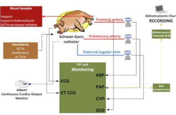
Instrumentation
Experiments using pigs and dogs were performed at the Semmelweis Medical University in Hungary and at the Uniformed Services University of the Health Sciences (USUHS) in the USA, and were approved by the local Animal Subject Review Committees and followed their guidelines, treating the animals humanely.
Swine (25-40 kg) and mongrel dogs (20-40 kg) of both sexes were purchased from approved local vendors. They were sedated with i.m. ketamine (500 mg) and intubation was carried out with a 6.5 Fr tracheal tube to maintain free airway, and to enable controlled ventilation if needed. The animals were anesthetized with 1-2%
isoflurane, or with i.v.xylasine/ketamine mixture and Nembutal (pentobarbital, 30 mg/kg for induction and 5-10 mg/kg/h for maintenance), via the ear vein. Fluid (Salsol A or Ringer) supply maintaining circulatory stability was provided via the left external jugular vein. Ventilation (upon isoflurane anesthesia) was maintained using the anesthesia machine or (during pentobarbital anesthesia) was assisted by a Harvard ventilator (Harvard Apparatus, Cambridge, MA).
Surgery was performed to cannulate the right external jugular vein for drug injections and a Schwan-Ganz catheter was floated through the right atrium and right ventricle to the pulmonary artery for pulmonary arterial pressure (PAP) and central venous pressure (CVP) measurement. The right femoral artery was also cannulated for blood sampling and to measure systemic arterial pressure (SAP). The ECG was traced at the standard Einthoven’s 3-lead detection points. Hemodynamic parameters (PAP, SAP, CVP), heart rate (HR) and ECG were continuously monitored throughout the experiments.Additionally, continual respiratory rate, end-tidal carbon dioxide (ETCO2), and rectal temperature were monitored with an M1026A Gas Analyzer and Model 68 clinical monitor (Hewlett-Packard, Andover, MD) when available.
Test materials were injected through the Swan-Ganz catheter into the pulmonary circulation and flushed with 5 ml saline solution. Between injections of test and/or reference material, a resting period of at least 20 minutes was maintained.
SAP, PAP, CVP, standard ECG leads I, II, and III and – instrumentation permitting - ETCO2were recorded continuously with the SPEL Advanced Haemosys data acquisition system (Experimetria Ltd., Budapest, Hungary) or an ADInstrumentPowerLab Recorder and LabChartTM software (ADInstruments, Bella Vista, NSW, Australia) at a sampling rate of at least 100 Hz. Other details of surgery, instrumentation and hemodynamic analysis were described previously8,50,51. (Figure 7)
Figure 7. A schematic figure of the instrumentation setup, the monitored parameters, and the experimental endpoints in the swine model
Cardiopulmonary data analysis
The pulmonary and systemic blood pressure was registered continuously at 100- 1000 Hz. We averaged the mean blood pressure every 30 seconds before and after the injections of the various substances and plotted against time. We also registered the maximal increase of mean pulmonary and systemic arterial pressure after each injection and compared the changes provoked by the various test substances. When comparing groups in the results section, the blood pressure values are expressed as medians.
Blood collection and analysis:
Blood samples of 4-6 ml were collected in tubes coated with EDTA (BD Vacutainer, BD Franklin Lakes, NJ USA). Samples were drawn at 0 (baseline) and at 3, 6, 9 and 12 minutes after each injection, leaving at least 20 minutes rest periods between
instrumentation and baseline sampling, and between injections.
Statistical analysis
The significance of PAP and SAP changes caused by Doxil was computed by the Wilcoxon matched-pairs signed rank test, (confidence interval 95%), or Mann- Whitney test (confidence interval 95%), as indicated. Differences between groups were considered significant at P<0.05. Data are reported as median and interquartile range.
In vitro complement assay
Human serum samples from healthy volunteer donors, obtained through an institutionally approved phlebotomy protocol, were stored at -70°C until use. The ELISA-based method for quantification of serum S-protein-bound C terminal complex (SC5b-9) and levels of the catalytic subunit of Complement factor B (Bb) was performed as described earlier.49,50 In brief, the test polymers, at the concentration determined to be the IC50 value, and control compounds were incubated with different human sera for 45 min at 37°C in a shaking water bath (shaking rate of 80 rpm). After terminating the reaction by adding chilled specimen diluent (provided in the ELISA kits) at a 20-fold volume, samples were tested for SC5b-9 and Bb levels using the respective ELISA kits (TCC and Bb kits, Quidel Co, San Diego, CA), following the manufacturer’s instructions. All reactions were tested in duplicates.
Results
In vivo complement activation by Doxil
To test complement-activating properties of Doxil, 12 pigs were administered 0.01 mg/kg Doxil, the equivalent of 0.06385 mg phospholipid per kg bodyweight (0.06385 mg PL/kg), as first injection. This represents minute amounts of Doxil, far below the doses clinically administered to patients, which underscores the importance of the anaphylactic reactions observed during these experiments.
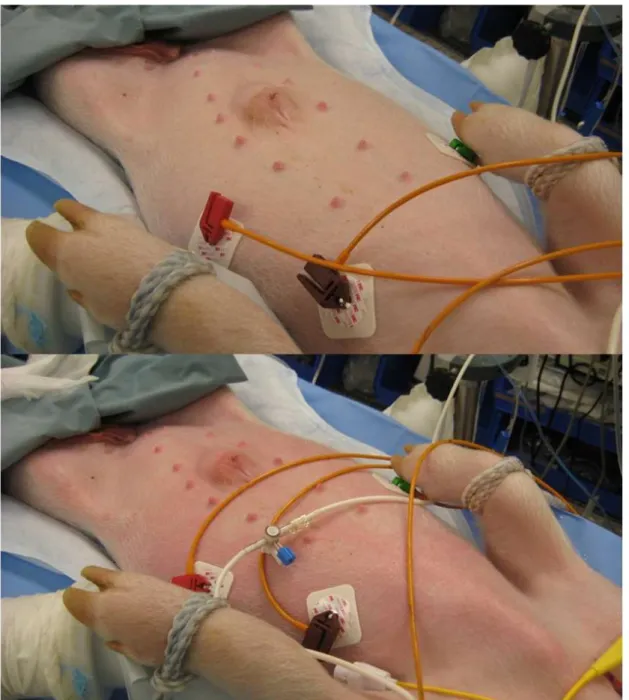
Hemodynamic parameters were continuously monitored and recorded. We were paying focused attention to typical physiological changes characteristic to complement activation related pseudo-anaphylactic reaction, which include increase of pulmonary arterial pressure, increase or decrease of systemic arterial blood pressure, increase or decrease of heart rate, compromised cardiac output, dyspnea, apnea, increased or decreased end tidal CO2, decreased arterial oxygen saturation measured by pulse oximetry, skin mottling, rash, etc. (Figure 8 and Figure 9)
Figure 8. Typical monitoring screens (A) during baseline and (B) during CARPA reaction.
This figure illustrates typical hemodynamic symptoms of CARPA: increase in pulmonary arterial blood pressure, increase or decrease in systemic arterial blood pressure, arrhythmias including tachycardia, and signs of ischaemia (ST segment depression or elevation, increased or inverted T waves)
Figure 9: Skin reaction during CARPA.Above: normal skin before injection Below:
rash, erythema, following injection of 0.06385 mg PL/kg Doxil. Cutaneous symptoms occur during severe reactions, with near lethal drop in systemic blood pressure, substantial increase in pulmonary arterial pressure, and often apnea.
These symptoms vary depending on the severity of the reaction. The most consistent sign of anaphylaxis in the pig model is acute pulmonary hypertension. In mild cases of CARPA the systemic arterial pressure increases or slightly decreases.
However, during severe reactions, due to compromised cardiac output and/or peripheral vasodilation, the mean arterial pressure can drop to very low levels, occasionally even below the minimum required for the perfusion of vital organs. This is also reflected in the end tidal CO2.On one hand high end tidal CO2 could mean hypoventilation.
However, if the cardiac output is insufficient to maintain proper perfusion of the lungs to enable gas exchange, the end tidal CO2 drops. Heart rate can also change in both directions. During minor challenge heart rate slightly increases. However, during serious CARPA events paradoxical bradycardia can be observed. Described in detail in a previous paper by our group, this has been shown to be mediated by adenosine release from the ischemic heart.52 Also, if the animal can recover from a state of severe cardiovascular compromise with low cardiac output and blood pressure, usually a high amount of catecholamine is released into the circulation resulting in a ―rebound‖
tachycardia with high blood pressure.
The physiological changes observed in the pig model also correspond to clinical symptoms of CARPA: dyspnea, light-headedness, chest pain, etc.
After the injection of Doxil, we observed acute pulmonary hypertension in all animals, within a few minutes following the injection. The mean pulmonary arterial pressure (PAP) increased significantly (p=0.0005, n=12) from a median of 16.5 (15- 20.25) mmHg to 42 (34.5-48) mmHg. (Figure 10)
Figure 10. PAP changes following 1st injection of Doxil (0.06385 mg PL/kg)
Each pair of connected points represents one animal, and pre-injection baseline and post-injection maximum pressure values are shown. p=0.0005, n=12, PAP baseline 16.5 (15-20.25) mmHg, PAP maximum change 42 (34.5-48) mmHg
In our model, the symptom of CAPRA can be an increase or decrease of systemic mean arterial pressure. Because the Wilcoxon matched-pairs signed rank test used for analysis of our data compares the medians of two paired groups, it is possible that individual variations among the animals cancel out each other during analysis. As a result, the baseline and post-injection median of the SAP values (99 and 106 mmHg, respectively) was not significantly different (p=0.2061),because in some cases the pressure increased, in other cases it decreased.However, there were some animals where the blood pressure dropped to life-threateningly low levels. Notably, we observed changes from 111 to 32 mmHg, from 83 to 30 mmHg, from 63 to 31 mmHg, from 98 to 52 mmHg (Figure 11)
PAP base
PAP max 0
20 40 60
Pulmonary arterial pressure (mmHg)
Figure 11. SAP changes following 1st injection of Doxil (0.06385 mg PL/kg)
p=0.2061, n=11, SAP baseline 99 (84-116) mmHg, SAP after injection106 (32-118) mmHg
The dynamics of a typical reaction in one of the animals is illustrated in Figure 12.The pulmonary arterial pressure (PAP) increased from a baseline of 16 to a maximum of 48 mmHg.This was followed by a decrease in systemic arterial pressure (SAP)from a baseline of 111 mmHg to a minimum of 32 mmHg. The drop in SAP is also accompanied by paradoxical bradycardia, i.e. at the time of the blood pressure reaching 31 mmHg, representing life-threatening hypotension, instead of compensatory tachycardia we see a temporary decrease in heart rate. This is likely mediated by adenosine release from the ischemic myocardium.
Systemic arterial pressure (mmHg)
SAP base
SAP after 0
50 100 150
Figure 12. Typical hemodynamic changes following injection of Doxil. The representative data is from an experiment following the administration of 0.06385 mg PL/kg Doxil at time 0 min on the graph.
These changes resembled typical CARPA reactions, and were comparable to the reactions observed following the administration of Zymosan, a potent known complement activator used as positive control.
Summary:
First injection of Doxil causes clinically significant acute pulmonary arterial hypertension.
We compared baseline and maximum of PAP after 1st Doxil injection in the control group using Wilcoxon matched-pairs signed rank test, with confidence interval of 95%.
PAP increased from a median of 16.5 mmHg to 42 mmHg (p=0.0005, n=12)
SAP change was statistically and clinically not significant (99 to 106 mmHg), except for some cases.
0 20 40 60 80 100 120 140
0 20 40 60 80 100 120 140
0 5 10 15 20 25
Heart rate (1/min)
Mean arterial pressure (mmHg)
Time following injection (min)
PAP SAP HR
Tolerance after first Doxil injection
To test whether the first reaction can be repeated with subsequent doses, in seven animals the first Doxil injection was followed by a second identical dose of Doxil injection (0.06385 mg PL/kg). Although the change in PAP from a median of 17 (14- 19) mmHg to 19 (16-20) mmHg was statistically significant (p=0.0177, n=7), such a slight increase is clinically irrelevant, and hence we don’t consider it to be a serious anaphylactic reaction. The systemic arterial pressure did not change (88.5 (74- 95.5)mmHg at baseline and 89 (80.25-98.75) mmHg following injection).
Also, as it can be seen in Figure 13 below,that following the second injection of Doxil there were no individual animals that suffered acute pulmonary hypertension or a drop in systemic blood pressure.
Figure 13. (A) PAP and (B) SAP changes following the 2nd injection of Doxil.
Each point represents one animal, and pre-and post-injection pressure values for the same animal are connected with a line. It’s apparent that in contrast to the first injection, there are no steep lines representing a major increase or decrease in the blood pressures of any of the subjects.(A) PAP: p=0.0177, n=7, baseline 17 (14-19) mmHg, maximum change 19 (16-20) mmHg; (B) SAP: p=0.2932, n=6, baseline 88.5 (74-95.5) mmHg, maximum change 89 (80.25-98.75) mmHg
PAP base 2
PAP max 2 0
20 40 60
Pulmonary arterial pressure (mmHg)
SAP base 2
SAP after 2 0
50 100 150
Systemic arterial pressure (mmHg)
A B
To test whether tolerance can be breached with a higher dose, six animals received an additional five-fold bolus dose of Doxil (0.31925 mg PL/kg) as a third injection. No statistical difference could be shown between pre- and post-injection pressures either in the pulmonary arterial pressure (17 (12.75-19.5) and 20 (17.75- 22.25) mmHg, respectively), or in the systemic arterial pressures (89.5 (77.75-97.75) and 86 (74.5-99.75) mmHg, respectively). (Figure 14)
Figure 14.(A) PAP ands (B) SAP changes following third injection of Doxil with 5x dose (0.31925 mg PL/kg)
There were no significant changes, even at the individual level. Tolerance was shown to 5x dose. (A) PAP: p=0.0579, n=6, baseline 17 (12.75-19.5) mmHg, maximum change 20 (17.75-22.25) mmHg; (B) SAP: p=0.625, n=6, baseline 89.5 (77.75-97.75) mmHg, maximum change 86 (74.5-99.75) mmHg.
PAP base 3
PAP max 3 0
20 40 60
Pulmonary arterial pressure (mmHg)
SAP base 3
SAP after 3 0
50 100 150
Systemic arterial pressure (mmHg)
A B
Another compelling evidence for tachyphylaxis is that comparing the maximal PAP values in the animals that have received all 3 consecutive Doxil injections, we found significantly higher pulmonary pressure after the 1st than after the 2ndinjection (44.5 (31.5-49.75) versus 17.5 (15.5-20.75) mmHg, respectively; p=0.0355), while the baselines were not different. This is illustrated in Figure 15. Comparison of the PAP maximum values between the 1st and 3rd injections showed similar difference (44.5 (31.5-49.75) vs. 20 (17.75-22.25) mmHg, respectively; p=0.0355).
Figure 15.Comparison of (A) baselines and (B) post-injection maximums of pulmonary arterial pressure.
Baselines were not different (p=0.2795 for 1 vs. 2, and p=0.2809 for 1 vs. 3, n=6), while maximum change after 1st injection was higher than after the 2nd (p=0.0355) and 3rd (p=0.0355) injections.
To test whether tolerance is specific to Doxil, after the Doxil injections we administered two sequential doses of 0.5 mg/kg Zymosan, a known complement activator as positive control. The first injection caused a significant increase in PAP from 17 (14-18) mmHg to 46 (30-50) mmHg (p=0.0156, n=7) suggesting that the tolerance to Doxil is specific, and reactivity to Zymosan is preserved. The PAP also changed after the second injection of an identical dose of 0.5 mg/kg Zymosan from 21 (17-24.25) mmHg to50.5 (42.25-57.25) mmHg, (p=0.0355, n=6), suggesting that Zymosan does not induce self-
PAP base 1
PAP base 2
PAP base 3 0
20 40 60
Pulmonary arterial pressure (mmHg)
0.06385 mg PL/kg
0.06385 mg PL/kg
0.31925 mg PL/kg 0
20 40 60
Pulmonary arterial pressure (mmHg)
A B
tolerance under these circumstances. (Figure 16)
Figure 16. PAP changes following (A) firstand (B) second injection of Zymosan (0.5 mg/kg) after Doxil injections.
PAP increased from 17 to 46 mmHg after the 1st injection (p=0.0156, n=7) and from 21 to 50.05 mmHg (p=0.0355, n=6) after the 2nd injection.
Summary:
There is tachyphylaxis for subsequent Doxil injections and they do not cause clinically significant acute pulmonary arterial hypertension or changes in systemic blood pressure.
a) We compared baseline and maximum of PAP after 2nd Doxil injection in control group using Wilcoxon matched-pairs signed rank test, with confidence interval of 95%.
PAP increased from a median of 17 mmHg to 19 mmHg (p=0.0177, n=7). This is statistically significant, but biologically not relevant.
b) Wecompared baseline and maximum of PAP after 3rd Doxil injection in control group using Wilcoxon matched-pairs signed rank test, with confidence interval of 95%.
PAP before and after injection was 17 and 20 mmHg, respectively; the difference was not significant (p=0.0579, n=6)
PAP base z1
PAP max z1 0
20 40 60
Pulmonary arterial pressure (mmHg)
PAP base z2
PAP max z2 0
20 40 60
Pulmonary arterial pressure (mmHg)
A B