T cell and chemokine level change in systemic lupus erythematosus with pulmonary
manifestation
PhD thesis
Krisztina Vincze MD
Doctoral School of Clinical Medicine Semmelweis University
Supervisor: Veronika Müller MD, DSc.
Official reviewers: Viola Tamási MD, Ph.D., Krisztina Bogos MD, Ph.D.
Head of the Final Examination Committee:
Edit Buzás MD, DSc.
Members of the Final Examination Committee:
Györgyi Műzes MD, Ph.D.
György Böszörményi Nagy MD, Ph.D.
Budapest 2017
1. INTRODUCTION
Systemic lupus erythematosus (SLE) is the most common systemic autoimmune disease. SLE predominantly affects females in child-bearing age.
Lupus is characterized by the presence of auto antibodies and immuncomplexes targeting multiple organs, therefore clinical symptoms are very heterogeneous. In Hungary SLE affects about 5-6000 persons. The prognosis is largely dependent on gender, age, organ manifestations, the degree of active inflammation and irreversible organ damage. SLE diagnosis was based on the SLICC/ACR criteria system, which was revised by an international clinical research team in 2012. SLE diagnosis requires at least 4 criteria (minimum 1 clinical and 1 laboratory). The most important aspect of the treatment is the presence of organ manifestations, evidence of active inflammations and extent of the irreversible organ damage. SLE therapy is primarily immunosuppressive treatment, the latest recommendations represent organ specific guidelines.
There are only few data available on the prevalence of pulmonary manifestations in SLE, indicating approximately 4.9-30% of SLE patients.
One study verified that ~48% of all SLE patients had respiratory symptoms.
Serositis, also included into the diagnostic criteria, represents the most common lung involvement. Pleuritis appears often as first symptom, associated with pleural effusion. The detection of antinuclear antibody (ANA) and anti-double stranded (ds) DNA from pleural fluid confirms SLE diagnosis.
One in five SLE patients have vascular involvement, mainly including pulmonary hypertension, pulmonary embolism, pulmonary vasculitis and the extreme rarely acute reversible hypoxemia. Parenchymal involvements are
acute lupus pneumonitis, which is often lethal, and chronic interstitial lung disease (ILD). The acute cricoarytenoiditis, bronchiectasis and bronchiolitis obliterans are the airway manifestations of SLE. Shrinking lung syndrome (SLS) is relatively rare, but a specific musculoskeletal involvement of SLE.
In SLE, as other autoimmune diseases, T helper(Th)1/Th2 ratio and Th17/T regulatory (Treg) ratio in the peripheral blood were demonstrated to change.
The local cytokine/chemokine milieu (polarization of Th0 cells) is most important for the development of organ involvement in autoimmune diseases.
Changes in the dominant Th cell subpopulation are organ specific the dominant Th cell mediated immune response and the produced cytokine/chemokine pattern in certain autoimmune diseases differ. In SLE the dominance of peripheral Th1 cells and increased production of associated cytokines (TNF-α, INFγ) and chemokines (IP-10, MCP-1) was confirmed. By contrast decreased level of peripheral Th2 cells in SLE can be observed. Th17 cells were increased in several autoimmune processes with increased production of proinflammatory cytokines (IL-17, IL-21, IL-22, IL-23). The Treg cells have a very important role in SLE pathogenesis, but it is not clear how peripheral Treg cells change in SLE. Our studies were aimed the assessment of pulmonary manifestations in SLE, furthermore at T cell and cytokine and chemokine changes in SLE patients with lung involvement.
2. AIMS
1. What are the pulmonary manifestations in SLE, which age group is mainly affected and what are the most important clinical symptoms in SLE patients with pulmonary involvement?
2. Are there differences in lung function parameters in SLE patients with pulmonary involvement as compared to SLE patients without pulmonary involvement?
3. Assessment of changes in peripheral blood Th1, Th2, Th17 and Treg cell ratios in SLE patients with and without pulmonary manifestation.
4. Evaluation of relationship between peripheral Th cell changes and functional parameters in pulmonary manifestation of SLE.
5. Assessment of cytokine/chemokine changes in plasma characteristic for pulmonary involvement of SLE.
3. METHODS
Patient recruitment
Study subjects included 28 SLE patients with mean age of 35.4±2.9 years.
Two groups were investigated: SLE patients with pulmonary manifestation (SLEpulm) and SLE patients without pulmonary manifestation (SLEc). Patients in SLEpulm group had higher mean age, longer duration of their SLE diagnosis, included more smokers and patients with more active disease than SLEc. Immunosuppressive therapy significantly differed in the daily steroid use, as patients in SLEpulm had higher dose as SLEc patients. The CD4+ T cell subpopulation prevalence was taken from 22 SLE patients at the time of screening. Patients in SLEpulm group (N=9, age: 44.0±4.7 years, female: male ratio: 7:2) had higher scores of SLE activity index (SLEDAI), than patients in SLEc group (N=13; age: 24.8±3.7 years, female: male ratio: 10:3).
Furthermore samples of 40 healthy blood donors (age: 35.5±5.6 years, range:
20-70 years, female:male ratio: 36:4) were used as population control for peripheral T cell analysis. For the second study investigating plasma cytokine and chemokine changes 9 SLEpulm patients (age: 38.23±5.46 years, female:male ratio: 7:2,), 9 SLEc patients (age: 50.48±2.69 years, female: male ratio: 9:0) and 9 healthy control volunteers were included.
Pulmonological examinations
All SLE patients were asked about their respiratory symptoms, underwent physical examination, chest X-ray, electrocardiography, blood gases analysis (Stat Profile® pHOx Plus®, Nova Biomedical Corporation, Waltham MA, USA), spirometry with body pletysmography and CO diffusion capacity
measurement (PDD-301/s, Piston, Budapest, Hungary) and routine laboratory tests.
T cell subpopulation analysis
For T cell analysis 6 ml sodium heparinized venous blood from SLE patients and healthy controls was analyzed. Peripheral blood mononuclear cells (PBMC) were isolated and frozen - 80°C. Identification of Th cells was done using surface markers. (Table 1.)
Table 1. Surface markers used for CD4+ T cell identification.
Marker FITC PE PECy5/PerCP PECy7 APC APCCy7
Th1 CCR4 CD62L CD4 CXCR3 CD8
Th2 CD45RA CD45RO HLA-DR CD4 CD69 CD8
Th17 CCR4 CCR6 CD4 CXCR3 CD8
Treg CD25 CD4 CD127 CD8
Flowcytometry measurements (BD FACS Aria, Beckton Dickinson & Co, Plymouth, UK) were performed in the laboratory of the Research Group of Pediatrics and Nephrology, Ist Department of Pediatrics of Semmelweis University and Hungarian Academy of Sciences. CD4+ cells were plotted with dot-plot and histogram. The histogram showed the distribution of CD4+ cells on signal intensity.
Cytokine/chemokine analysis
The plasma from peripheral blood was stored at -80 C until cytokine and chemokine measurements. The measurements were performed in the research laboratory of the 3d Department of Internal Medicine, Semmelweis University.
Custom made human Cytokine Antibody Array Kits (RayBiotech Inc., Norcross, GA, USA) were used to determine plasma cyto- and chemokine levels. The following factors were measured in parallel on a single array per patient: 4BB-1, interferon gamma-induced protein 10 (IP-10), interleukin-12 , p70 (IL12p70), interleukin-17 (IL-17), CC chemokine ligand 14a (CCL14), CC chemokine ligand 21 (CCL21), CC chemokine ligand 28 (CCL28), monocyte chemotactic protein 1 (MCP-1), monocyte chemotactic protein 4 (MCP-4), interferon-induced T cell alpha chemoattractant (I-TAC). Signal intensities were recorded and sorted using a chemoluminescence imaging system (Syngene Chemigenius 2 and GeneTools, Synoptics Ltd. Cambridge, UK).
Statistical analysis
Statistical analyses were performed applying Graph Pad Prism software 5.0 (Graph Pad Prism 5.0 by Graph Pad Software Inc., San Diego, USA).
Normally distributed data were analyzed by unpaired t-test and ANOVA.
After one-way ANOVA, if significant difference (p< 0.05) was found, the Neuman-Keuls multiple comparison post-hoc test was used for further analysis. Categorical data were analyzed by χ2 and Fisher’ s exact test.
Parametric data were compared using Student’s t-test. Unless otherwise specified, data were reported as mean±SEM for continuous variables and as number (percent) for categorical variables. For all statistical analyses p< 0.05 was considered statistically significant.
4. RESULTS
4.1. PLEUROPULMONARY MANIFESTATIONS IN SLE
SLEpulm patients had significantly more pulmonary complaints than their SLEc
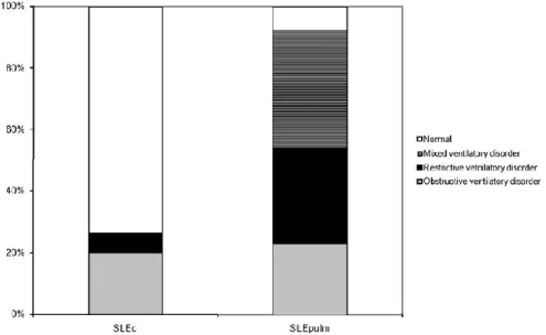
counterparts. Dyspnea was the leading symptom in SLEpulm and was associated with significantly decreased pO2 in these patients (effort dyspnea SLEpulm: 85% vs. SLEc: 7%; pO2 SLEpulm: 70,23±3,51Hgmm vs. SLEc: 92.73±2.28 Hgmm). Significantly increased number of serositis, SLS, pulmonary vascular disease were noted in SLEpulm patients, however it was the most pronounced in respect to lung function abnormalities (SLEpulm: 92% vs SLEc: 33%, p<0.0001) and fibrosis (SLEpulm: 54% vs. SLEc: 0%, p<0,0001).
(Figure 1.)
%
Figure 1. Prevalence of pulmonary manifestations at the time of pulmonary examinations in SLEpulm and SLEc patients. Pulmonary embolism represents patients on continuous anticoagulant treatment as the result of previously confirmed pulmonary embolism. * p<0.0001 vs. SLEpulm.
Figure 2. Distribution of lung function abnormalities in SLEc and SLEpulm patients. p=0.0018 vs. SLEpulm.
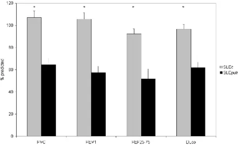
Significantly decreased values in the SLEpulm compared to SLEc were measured for forced vital capacity (FVC), forced exspiratory volume in one second (FEV1) and forced exspiratory flow (FEF25-75), carbon monoxide diffusing capacity (DLCO) (96.7±4.1 vs. 61.8±5.1; p<0.01) and lung volume adjusted KLCO (102.9±7.1 vs. 72.5±3.4; p<0.01). (Figure 3.)
Figure 3. Pulmonary function parameters in SLEc and SLEpulm patients. Mean values ± SEM of FVC% predicted, FEV1 % predicted, FEF25-75% predicted and DLCO.
* p <0.05 vs. SLEpulm.
4.2. PERIPHERAL CD4+ T CELL SUBPOPULATION PREVALENCE IN SLE PATIENTS
No change in absolute circulating number of Th1 cells was measured in SLE patients while significantly decreased number of Th2 cells resulted into elevated Th1/Th2 ratio as compared to controls. There was no difference between SLEpulm and SLEc subgroups in respect to Th1, Th2 cells or Th1/Th2 ratio. (Figure 4. /Part A.)
Figure 4. [A]: Th1/Th2 ratio; [B]: Th17/Treg ratio; [C]: Treg/CD4+CD25hi+ cell ratio SLEc, SLEpulm, all SLE and control patients. *p<0.05 vs. SLEpulm; # p < 0.05 vs. control.
Significant decrease in Th17 and Treg (CD4+CD25hi+CD127-) was present in SLE patients as compared to controls, without differences according lung involvement. Interestingly, Th17/Treg ratio was also lower in lupus patients as in healthy subjects (Figure 4./ Part B). Additional analysis using CD4+CD25hi+ cells revealed significantly increased cell number in SLEpulm
compared to SLEc or controls. Similarly decreased relative proportion of Treg in CD4+CD25hi+ cells in SLEpulm to SLEc and controls suggests pleuropulmonary specific increase of other, non „real” Treg cell in SLEpulm
probably representing an other subgroup of activated T cells. (Figure 4./Part C)
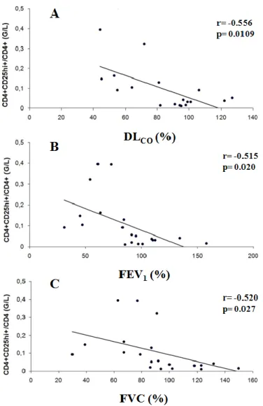
Significant association was demonstrated regarding dynamic lung function parameters (FVC, FEV1) or DLCO and CD4+CD25hi+ or Treg/CD4+CD25hi+
ratio. This is further emphasizing a possible lung specific CD4+CD25hi+ T cell subset, not incorporating „real” Treg cells. (Figure 5.)
Figure 5. Correlation of lung function parameters and CD4+CD25hi+ cell number and Treg/CD4+CD25hi+ ratio in all SLE patients.
[A]: DLCO (% predicted); r=-0.556, p=0.0109;
[B]: FEV1 (%predicted); r=-0.515, p=0.020;
[C]: FVC (%predicted); r=-0.520, p=0.027.
4.3. CYTOKINE/ CHEMOKINE PLASMA LEVEL CHANGE IN SLEpulm AND SLEc GROUPS
In most plasma samples the levels of 4BB-1, IL12p70, IL-17, CCL28, MCP-4 and I-TAC were below the detection limit of the array, therefore only data obtained for the remaining factors CCL21, IP-10, MCP-1 and CCL14 were used in the analysis.
As presented in Figure 6. the chemiluminescent intensity of CCL21 was significantly higher in SLEpulm than in patients without lung involvement;
however, no significant difference to the control group has been identified (4949±1481 vs. 1272±236.1 vs. 1581±382.1 U; p<0.05; Figure 6./Part A).
Significantly increased chemiluminescent intensity was detected for circulating IP-10 in the SLEpulm group as compared to SLE patients and healthy volunteers. (5923±1278 vs. 5759±926 vs. 10762±1584 U; p<0.05) (Figure 6. / Part B).
Figure 6. Comparison of CCL21 [A] and IP-10 [B] in healthy volunteers and in SLE patients with (SLEpulm) and without pulmonary involvement (SLEc).
[A]: SLEpulm vs. control and SLEc; p <0.05, [B]: SLEpulm vs. control and SLEc; p<0.05.
Significant association was demonstrated regarding lung function parameters FVC and FEV1 and IP-10; MCP-1; CCL14, as shown in Figure 7.
Figure 7. Correlation of lung funtion parameters: FVC (% predicted) and FEV1 (% predicted) and chemokine levels: IP-10 (Part A and B), MCP-1 (Part C and D) and CCL-14 (Part E and F) in SLE patients.
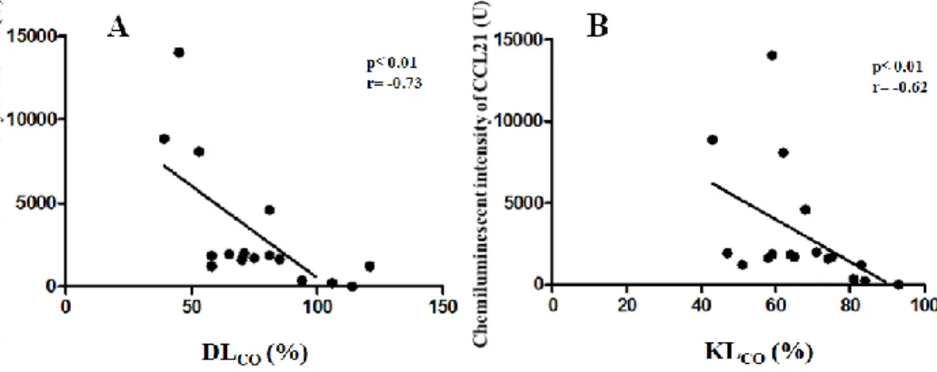
Correlation analyses revealed positive correlation between the chemiluminescent intensity of CCL21 and diffusion parameters, including DLCO (r=-0.73; p<0.01) and KLCO (r= -0.62; p<0.01). (Figure 8.)
Figure 8. Correlation between the chemiluminescent intensity of CCL21 and diffusion parameters: DLCO (Part A) and KLCO (Part B).
5. CONCLUSION
1. High prevalence of lung manifestations in SLE, especially in older patients was described. In our study mainly ILD, SLS and small airway involvement were confirmed. The main respiratory symptoms were dyspnea and reduced exercise capacity.
2. The majority of patients in SLEpulm group showed lung function abnormalities (92%), including decrease in FVC, FEV1, FEF25-75, DLCO and pO2 values.
3. Changes in Th1/Th2 or Th17/Treg in SLE patients were observed confirming previous results, however no differences according pleuropulmonary manifestations were recorded.
4. Our data first described a significant positive correlation between peripheral CD4+CD25hi+ activated T cells and lung function parameters (FVC, FEV1) and DLCO indicating that these cells might represent lung specific immunological activity in SLE.
5. Pleuropulmonary involvement in SLE patients was associated with significant increase in CCL21 and IP-10 and significantly correlated to lung functional parameters.
6. THE BIBLIOGRAPHY OF THE CANDIDATE’S PUBLICATIONS
International publications related to the dissertation:
1. Vincze K, Kovats Z, Cseh A, Pasti K, Kiss E, Polgar A, Vasarhelyi B, Szabo AJ, Bohacs A, Tamasi L, Losonczy G, Muller V. Peripheral CD4+ cell prevalence and pleuropulmonary manifestations in systemic lupus erythematosus patients. Respiratory Medicine 2014;108(5): 766-774. IF: 3,086
2. Odler B, Bikov A, Streizig J, Balogh C, Kiss E, Vincze K, Barta I, Horvath I, Muller V. CCL21 and IP-10 as blood biomarkers for pulmonary involvement in systemic lupus erythematosus patients.
Lupus. 2017;26(6):572-579. IF: 2,118
Hungarian publications related to the dissertation:
1. Vincze K, Odler B, Müller V. A szisztémás lupus erythematosus pulmonalis manifesztációi. Orv Hetilap 2016;157(29): 1154-1160.
International publications not related to dissertation:
1. Tamasi L, Muller V, Eszes N, Kardos T, Budai M, Vincze K, Losonczy G, Szilasi M. Patterns of erythropoiesis-stimulating agent use for chemotherapy-induced anemia in lung cancer: results of a retrospective Hungarian real-life clinical data analysis.Exp Op Drug Safety. 2011;10(4):503-507. IF: 3,015
2. Müller V, Vincze K, Eszes N, Zahorecz G, Bohács A, Losonczy G, Tamási L. Supportive care of palliative chemotherapy for advanced stage lung cancer patients: Summary for the daily practice. Journal of Nursing Education and Practice 2013;4(3):101-111.
Hungarian publications not related to the dissertation:
1. Ivancsó I, Vincze K, Juhász M, Tamási L. Sarcoidosis atípusos mycobacteriosissal
Medicina Thoracalis 2011; 64:(6):317-321.
2. Müller V, Horváth G, Tamási L, Eszes N, Bohács A, Vincze K, Kováts Z, Lázár Z, Odler B, Kornafeld A, Erdély T, Somogyi V, Losonczy G. Obstruktív tüdőbetegségekben alkalmazott inhalációs gyógyszerek célpontjai: receptorok a légúti felszín sejtjein Medicina Thoracalis 2015; 68:(1): 6-12.

![Figure 4. [A]: Th1/Th2 ratio; [B]: Th17/Treg ratio; [C]: Treg/CD4+CD25hi+ cell ratio SLE c , SLE pulm , all SLE and control patients](https://thumb-eu.123doks.com/thumbv2/9dokorg/1383935.114354/11.629.70.564.78.451/figure-ratio-treg-ratio-treg-ratio-control-patients.webp)
![Figure 6. Comparison of CCL21 [A] and IP-10 [B] in healthy volunteers and in SLE patients with (SLE pulm ) and without pulmonary involvement (SLE c )](https://thumb-eu.123doks.com/thumbv2/9dokorg/1383935.114354/14.629.46.580.104.794/figure-comparison-ccl-healthy-volunteers-patients-pulmonary-involvement.webp)