Non-identical twins: Different faces of CR3 and CR4 in myeloid and lymphoid cells of mice and men
Anna Erdei a b, Szilvia Lukácsi a b , Bernadett Mácsik-Valent a b, Zsuzsa Nagy-Baló b, István Kurucz a, Zsuzsa Bajtay ab
a MTA-ELTE Immunology Research Group, Department of Immunology, Eötvös Loránd University, Budapest, Hungary
b Department of Immunology, Eötvös Loránd University, Budapest, Hungary
* Corresponding author
Anna Erdei
Department of Immunology, Eötvös Loránd University, Pázmány Péter s. 1/C, Budapest H-1117, Hungary Phone: (+36)-1-3812-175
Fax: (+36)-1-3812-176
e-mail: anna.erdei@ttk.elte.hu, anna.erdei@freemail.hu
Abstract
Integrins are cell membrane receptors that are involved in essential physiological and serious pathological processes. Their main role is to ensure a closely regulated link between the extracellular matrix and the intracellular cytoskeletal network enabling cells to react to environmental stimuli. Complement receptor type 3 (CR3, αMβ2, CD11b/CD18) and type 4 (CR4, αXβ2, CD11c/CD18) are members of the β2-integrin family expressed on most white blood cells. Both receptors bind multiple ligands like iC3b, ICAM, fibrinogen or LPS. β2- integrins are accepted to play important roles in cellular adhesion, migration, phagocytosis, ECM rearrangement and inflammation. Several pathological conditions are linked to the impaired functions of these receptors.
CR3 and CR4 are generally thought to mediate overlapping functions in monocytes, macrophages and dendritic cells, therefore the potential distinctive role of these receptors has not been investigated so far in satisfactory details. Lately it has become clear that a functional segregation has evolved between the two receptors regarding phagocytosis, cellular adhesion and podosome formation. In addition to their tasks on myeloid cells, the expression and function of CR3 and CR4 on lymphocytes have also gained interest recently. The picture is further complicated by the fact that while these β2-integrins are expressed by immune cells both in mice and humans, there are significant differences in their expression level, functions and the pathological consequences of genetic defects. Here we aim to summarize our current knowledge on CR3 and CR4 and highlight the functional differences between these receptors, involving their expression in myeloid and lymphoid cells of both men and mice.
Keywords:CR3, CR4, β2integrins, human, mouse
1. Introduction
The name “integrin” was given to point out that these integral cell membrane proteins ensure and maintain the integrity of the cytoskeletal-extracellular matrix (ECM) linkage [1].
Integrins are cell adhesion molecules that mediate cell-cell, cell-extracellular matrix, and cell- pathogen interactions. They play critical roles in several immune phenomena where leukocyte trafficking and migration are involved, immunological synapses are formed, costimulation and phagocytosis take place. These hetero-dimeric glycoproteins consist of non-covalently coupled α and β chains. Integrins are ancestral receptors with early evolutional history, since both the α- and β- subunits can be found throughout the invertebrates from nematodes and fruit flies with remarkably well conserved sequences [2]. The integrin α and β subunits have
developed separately during evolution. In vertebrates, the molecules of the integrin family are composed of 18 α and 8 β subunits, that can assemble into 24 different heterodimeric receptors, each with different ligand-binding specificities and a diverse tissue distribution [3].
In mammals, nine of the 18 α subunits have the “inserted” or “αI” domain, forming the ligand recognition part of the receptor. Four αI domain containing integrins are collagen receptors, while the other five recognize E-cadherins, ICAMs or VCAMs and two of them (CR3, CR4) also bind plasma proteins such as fibrinogen, FactorX or component iC3b [4]. Both subunits, namely the α and the β chain have a large extracellular ligand binding region, a single transmembrane domain and a short cytoplasmic tail (Figure 1.).
Recently the interest has been revived in the physiological and pathological role of CR3 and CR4, members of the β2-integrin family. Therefore we aim to give an overview on the structure, expression and diverse functions of these complement receptors on myeloid as well as lymphoid cells, suggesting a „division of labour” regarding their distinct roles on phagocytes. We also aim to highlight similarities and differences between the human and murine systems.
2. Complement receptors CR3 (CD11b/CD18) and CR4 (CD11c/CD18)
Complement receptor type 3 (CR3) and type 4 (CR4) belong to the β2-integrin family (Figure 1.). As it will be discussed later in detail, they are expressed on all myeloid cells and certain lymphoid populations. The members of this family differ in their specificity and avidity to the different ligands. Associated with the common β2 chain (CD18) the four members of this family are the following: αLβ2 integrin (also known as LFA-1 or CD11a/CD18), αMβ2 integrin (Macrophage antigen-1 (Mac-1), complement receptor 3 (CR3 or CD11b/CD18), αXβ2 integrin (p150,95, CD11c/CD18 or complement receptor 4 (CR4)), and αDβ2 integrin (CD11d/CD18) [5, 6]. Among the complement receptors binding various C3-fragments CR3 and CR4 have the unique property that their ligand binding is metal-ion dependent.
CD11c is closely related to CD11b. It is 66% identical and 77% homologous to Mac-1 over the β-propeller and the ligand-binding I domains. They bind similar ligands, including iC3b, ICAM and fibrinogen [7-10] via the metal-ion-dependent adhesion site (MIDAS) in the presence of a bivalent cation [11].
Sequence analysis of human cDNA encoding the α chains of CR3 and CR4 (165 and 150 kD Mw) show a homology of 87% [12, 13], however these two integrins bind iC3b at distinct sites [14]. A greater structural difference is present in the cytoplasmic domains of
CR3 and CR4 α chains. The cytoplasmic tail of CR3 α (CD11b) has only 56% homology with the cytoplasmic domain of CR4 α (CD11c), and is less than two-third of its size. This suggests that functional differences between CR3 and CR4 may be derived from differences in their ability to associate with cytoplasmic components such as signalling or actin-binding proteins [13]. The common β-subunit extracellularly has an N-terminal I-like domain, a sandwich hybrid domain, a cysteine-rich PSI (plexin-semaphorin-integrin) domain, four EGF-like repeats (I-EGF1, -2, -3, -4) and a β-tail domain (βTD) (Figure 1.).
CR3 and CR4 are named complement receptors as they readily interact with the iC3b fragment of the major component C3, generated during activation of the complement cascade (Figure 2.). However, as indicated in the figure these receptors interact with several additional structures, including ICAMs, the cell surface adhesion molecules, fibronectin, the adhesion molecule in extracellular matrix, and fibrinogen, the coagulation factor.
The most studied functions of these receptors are phagocytosis, adherence and podosome formation. The earliest investigations identified CD11b/CD18 as a phagocytic β2-integrin specific to iC3b opsonized antigen, which was identical with the Mac-1 antigen expressed by mouse and human myeloid cells as well [13, 15]. While the main role of CR3 is to promote phagocytosis and cytotoxicity, it is also known to enhance the function of several effector molecules such as FcγR, uPAR, and CD14 [16]. It was demonstrated that association of macrophage cytoskeleton with CR3 and CR4 regulates receptor mobility and phagocytosis of iC3b-opsonized erythrocytes [13]. The α chain of CR4, CD11c is a cell surface marker which can be used to identify human myeloid cell subsets.
3. Expression of CR3 and CR4
Both CR3 and CR4 are widely expressed on most of the myeloid and lymphoid cell types both in men and mice, although the extent of their appearance varies. These receptors are usually upregulated on activated cells, irrespective of their naive expression status. In Table 1. we summarize available data and indicate the presence of these β2-family integrins using the plus/minus sign.
Table 1. Expression of CR3 and CR4 on human and mouse myeloid and lymphoid cells.
HUMAN MOUSE
CR3 CR4 CR3 CR4
MYELOID CELLS
Monocyte + [17, 18] + [17, 18] + [19, 20] +/- [20, 21]
Macrophage +++ [17, 22] +++ [17, 22] + [23] +/- [23-25]
Dendritic cell +++ [17, 26] +++ [17, 26] +/- [27, 28] +++ [24, 29]
Neutrophil + [18, 30] + [18] + [31] +/- [32]
LYMPHOID CELLS
NK cell ++ [33, 34] +/- [35] +/- [20, 36] +/- [37]
T cell +/-* [33, 38] +/-* [18, 39] +/-* [40, 41] +/-* [40, 42-45]
B cell +/-* [33, 46-48] +/-* [46, 47, 49, 50] +/- [50, 51] +/- [50, 52]
+/- appears on certain subpopulations
+/-* appears on activated and leukemic cells
3.1. Expression of CR3 and CR4 on human myeloid cells
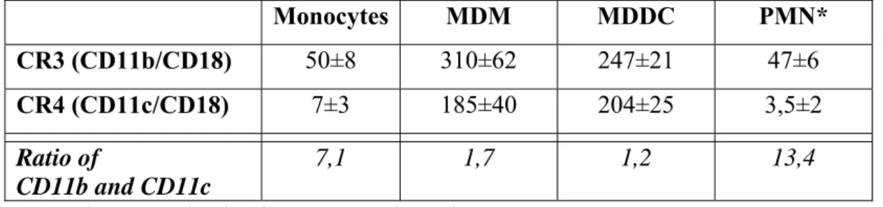
These integrins can be found on all myeloid cells, namely neutrophil granulocytes, monocytes, macrophages and dendritic cells [17, 18, 30]. An important progress has been made recently, when the absolute number of these receptors has been assessed on human monocytes, monocyte-derived macrophages (MDM), monocyte-derived dendritic cells (MDDC) [17] and neutrophils (Table 2.). Data shown reveals that the expression level of the two receptors varies between cell types. Comparing the relative number of CR3 and CR4 on the same cell types there is a strong shift in favour of CR3 in the case of monocytes and neutrophils, whereas on MDMs and MDDCs the CD11b:CD11c ratio is close to 1:1. The
notable differences in receptor number most probably contribute to the functional diversity observed in the case of these cell types. Furthermore, the availability of ligand epitopes may also influence the competition between these receptors, determining the outcome of the interactions.
Table 2. The average number of CR3 and CR4 expressed by normal human myeloid cells (receptor number x103 /cell)
Monocytes MDM MDDC PMN*
CR3 (CD11b/CD18) 50±8 310±62 247±21 47±6
CR4 (CD11c/CD18) 7±3 185±40 204±25 3,5±2
Ratio of
CD11b and CD11c
7,1 1,7 1,2 13,4
*Personal communication by Dr. Noémi Sándor.
3.2. Expression of CR3 and CR4 on human lymphoid cells
Among lymphoid cells of healthy donors all NK cells are known to express CD11b, while CD11c can be found only on a subset of these leukocytes [33-35]. Approximately 10% of human peripheral T lymphocytes - mainly CD8+ cells -, express CD11b, while CD4+ T cells become positive only after activation. The majority of CD11b+ T cells express CD56, too [33]. T cells – similarly to NK cells - contain preformed CR3, which is transferred rapidly to the surface following exposure to phorbol ester [33, 38]. Stimulation of T cells yielded up to 28% CD11b+ T cells [38]. CD11c is not expressed on peripheral T lymphocytes, but it appears on both CD4+ and CD8+ CTL clones [18, 39].
The picture is more complex in the case of B lymphocytes. Muto et al. found that approximately 20% of peripheral blood B cells express CR3 [33]. In contrast to this Postigo and colleagues found no significant expression of CD11b on tonsillar or peripheral B lymphocytes, even after activation [46], similarly to Uzonyi et al. [47] In contrast to this, Rothstein et al. detected CD11b+ on a small subset of B-1 cells [48]. In the case of chronic lymphocytic leukemia (CLL), B cells express diverse levels of CD11b, varying widely among patients [47]. Regarding the appearance of CD11c on human B lymphocytes there are only sporadic and inconsistent data available. In 1990 Wormsley et al. found two subsets of peripheral B cells expressing CD11c [49], but Postigo et al. found no significant expression of
this integrin on non-stimulated tonsillar or peripheral B lymphocytes. After activation with PMA for three days however, most B cells became CD11c+ [46]. Recently Rubtsov et al.
defined a CD11c+ population of B cells (called Age Associated B Cells, ABCs) which is represented in low numbers in the blood of healthy donors but is expanded in older women with autoimmune diseases [50]. Furthermore, CD11c was found clearly expressed by leukemic B cells, although to various extent [47].
3.3. Expression of CR3 and CR4 on mouse myeloid cells
CD11b is unequivocally expressed on mouse monocytes [19, 20] similarly to macrophages [23]. This is in contrast to CD11c, which is not present on monocytes newly released from the bone marrow, however appears upon maturation of the cells [21].
According to Helft et al. CD11c is expressed by mouse macrophaghes [24], and the expression increases in multinucleated giant cells [23], while Gorgani et al. showed that mouse residential peritoneal macrophages lack CD11c [25].
Mouse DC are highly positive for CD11c, therefore this molecule is often used to identify these cells. On dendritic cells (DC) CD11b is expressed in different levels, defining CD11bhigh and CD11blow or CD11b- subsets. CD11b is accepted as a marker of myeloid DC in mice [27, 28]. Interestingly however, mouse DC down-regulate cell surface CD11c upon activation in sharp contrast to human DC [29, 53].
Mouse neutrophil granulocytes also express CD11b, which increases after activation [31]. According to Matsushima et al. these cells can also acquire a CD11c positive phenotype in certain conditions [32].
3.4. Expression of CR3 and CR4 on mouse lymphoid cells
According to Chiossone et al. murine NK cells have a CD11blow phenotype in the beginning of their differentiation and become CD11bhigh when fully developed [36].In contrast to this Sunderkotter et al. showed that the CD11bhigh population of peripheral blood contain no NK cells [20]. Regarding the expression of CD11c, a distinct subset of NK cells was found to be positive, particularly in the liver [37].
In the case of murine B cells, Ghosn et al. described, that during ontogeny, CD11b- B- 1 cells precede CD11b+ B-1 cells [51]. The CD11c+, age associated B cells were found to express CD11b, too [50]. Recently Naradikian and colleagues demonstrated that some memory B cells also express CD11c due to a unique cytokine milieu and/or infections [52].
With regard to murine T cells CD11b expression correlates with their level of activity [41]
and is often co-expressed with CD11c on activated T cells [42, 43, 45]. A number of studies revealed, that only activated T cells express CD11c [42-45]. A specific cell type, called TDC
also expresses CD11b along with CD11c [40].
4. Integrin activation and signalling
Generally, integrins are present on the cell membrane in an inactive state, which allows free circulation of leukocytes in blood vessels. They undergo conformational changes induced by intracellular and extracellular stimuli following inside-out and outside-in signalling which leads to the separation of the α and β cytoplasmic tails. This activation is achieved by cytosolic proteins connecting integrins to the actin cytoskeleton [54].
A major breakthrough in integrin research was brought by the explanation of the switchblade-like model of integrin activation based on electron microscopic and crystallization studies [55, 56]. The three possible conformations of β2-integrins are the following: i/ bent, inactive, ii/ extended with intermediate affinity and iii/ extended, open conformation, with high affinity [55, 57, 58] (Fig. 1.). In the inactive form both subunits are bent, positioning the I domain near the cell membrane. The extension of the integrin legs enables a swing-out movement for the PSI and hybrid domains, inducing the conformational change in the I domain required for a high affinity, open state [59]. It has become clear by now that the structure of integrins is functionally relevant, therefore the access of the specific monoclonal antibodies –such as mAb KIM127 or mAb 24 for CD18 and CBRM1/5 for CD11b such as mAb KIM127, mAb 24 – makes possible to differentiate the conformation dependent functions [60, 61].
During the past few years several details of the molecular mechanisms of integrin activation have been elucidated. The small GTPase Rap1 (Ras-proximate-1), was shown to participate in the talin dependent inside-out activation of many different integrins, including β2-integrins [62, 63]. At least two effectors of Rap1 have been found: RapL (Rap-ligand) and RIAM (Rap1-interacting adaptor molecule). RAPL acts on the α subunit, while RIAM is proposed to stimulate the binding of talin to the cytoplasmic tail of the β-subunits [64, 65].
Macrophages and neutrophils from RIAM deficient mice show disrupted β2-integrin dependent adhesion and spreading [66]. Using the human THP-1 monocytic cell line, Lim et al. found, that siRNA silencing of RIAM did not impair phagocytosis and spreading [67].
Later it has been shown that the Regulator of G-Protein Signalling-14 (RGS14) plays a role in
the activation of CR3 during phagocytosis in the murine J774.A1 cell line [68]. However, Medraño-Fernández et al. revealed, that RIAM is important for complement mediated phagocytosis in the case of HL-60 and THP-1 cell lines and human monocyte derived macrophages [69]. Data obtained from the KO mice models suggest, that the effect of RIAM might be β2-specific, as the function of platelet αIIbβ3 integrin [70] and β1 integrins expressed by PMNs [66] is not disrupted in these mice. Additionally, talin deficient mice show a more severe phenotype compared to RIAM KO mice [66], underlining that the absence of talin affects all integrins, but the absence of RIAM does not.
The extracellular binding of ligands to activated integrins stimulates outside-in signalling pathways, including recruitment of multiple structural (vinculin, paxillin and actin) and signalling (p130Cas, FAK and Rho GTPases) proteins. These processes result in rearrangement of the actin cytoskeleton, and generation of adhesion structures promoting various biological processes including cellular adherence, migration and phagocytosis.
Little is known about the molecules interacting with the CD11b and CD11c chains, however it has been shown that the kinetics of activation of αLβ2 and αMβ2 integrins are different and the specific signals derived from the integrin α tail is assigned to distinct cytoplasmic molecules [71].
A remarkable property of CR3 is its ability to contribute to the function of other immune receptors like FcγRIIA, FcγRIIIB, CD14, β1-integrins, Dectin-1, and TLRs. These interactions are dynamic and the functional consequences depend on the activation state of the integrin [16, 72].
5. Function of CR3 and CR4 on myeloid cells
In this section we review the present knowledge on the function of CR3 and CR4 in different cell types of humans and mice. In Table 3. available information is summarized, which is followed by subsections with more detailed description of the several roles played by these β2-integrins on a wide variety of cell types.
Table 3. The role of CR3 and CR4 in different cell types of mice and men
Receptor Cell type
Role in human systems Role in mouse systems
CR3 Mo adherence to fibrinogen [17] and endothelium [73], phagocytosis [74- 78]
adhesion to endothelium [79]
phagocytosis [20]
MF podosome formation [78]
phagocytosis [78, 80, 81]
adherence [82], opsonic [83] and non- opsonic [76, 84, 85] phagocytosis DC podosome formation [78, 86]
phagocytosis [53, 78, 87]
phagocytosis of apoptotic cells [88]
PMN chemotaxis [89], adherence [90]
opsonic [78, 91] and non-opsonic [92, 93] phagocytosis
adherence, degranulation [94]
phagocytosis [95]
NK enhancement of cytotoxicity [96-98] enhancement of cytotoxicity [99]
T cell inhibition of proliferation and IL-2 release [38]
homing of CD8+ T cells [100, 101]
amelioration of ICG [102]
involvement in T cell development [103], boosting EAE [104]
B cell migration [105]
spreading of CLL B cells [47]
negative regulation of BCR signalling [106]
regulation of Ig responses [107]
amelioration of EAH [108]
CR4 Mo adherence to fibrinogen [17]
migration [39]
n. d.
MF podosome formation [78] adherence to fibrinogen [17]
phagocytosis [75, 77, 91, 109]
phagocytosis [95]
DC podosome formation [78, 86]
adherence to fibrinogen [17]
role in uptake [87]
phagocytosis of apoptotic cells [110]
PMN adhesion to endothelium [73]
enhancement of anti-bacterial activity [111]
n. d.
NK n. d. n. d.
T cell cell-mediated cytolysis by a subset of CD8+ cells [39]
boosting EAE [104, 112]
B cell proliferation [46]
attachment to fibrinogen [46]
spreading of CLL B cells [47]
n. d.
Mo: monocyte; MF: macrophage; DC: dendritic cell; PMN: polymorphonuclear cell; NK:
natural killer cell
5.1. Involvement of CR3 and CR4 in phagocytosis
Well accepted roles of CR3 and CR4 are to mediate cellular adhesion and phagocytosis of pathogen microbes, tumor- and apoptotic cells. In phagocytosis both receptors are clearly involved, as it had been shown by transfection of non-phagocytic CHO cells by CR3 and CR4 [113]. However, depending on the level of expression and the possibly different signalling partners, their participation in phagocytosis varies among cell types. A study carried out in whole blood showed that the CD14high CD16+ subset of monocytes, expressing the highest levels of CD11c and activated CD11b, phagocytosed Plasmodium falciparum infected erythrocytes more efficiently than other monocyte subsets. Phagocytosis could be inhibited on these monocytes more effectively with anti-CD11b than with CD11c blocking antibodies [74]. Further experiments employing monocytes also proved the dominant role of CR3 in the phagocytosis of Mycobacterium leprae [75] and Borrelia burgdorferi [76] and no significant contribution of CR4 to the CR3 mediated uptake of Mycobacterium tuberculosis [77] and Staphylococcus aureus [78] was found.
Downregulation of CR3 and CR4 by cytomegalovirus infection in human macrophages reduced their phagocytic capacity for Candida albicans, however the extent of contribution of the two receptors was not clarified [22]. While CR3 clearly mediates phagocytosis [80] and the phagocytic removal of desyalinated neurits by macrophages [81], CR4 is shown to be the major receptor mediating the uptake of Mycobacterium tuberculosis by alveolar macrophages [77]. Experimental data show that on MDM both CR3 and CR4 are involved in binding and/or phagocytosis of opsonised Cryprococcus neoformans [109], Mycobacterium leprae [114] and Francisella tularensis [91]. These studies however, do not clearly differentiate between uptake and digestion of the pathogens. Recently we examined these two sequential processes separately using S. aureus labelled with the pH sensitive pHrodo Green dye, which becomes highly fluorescent only in the acidic milieu of phagolysosomes, indicating the digestion of bacterial particles. Using this method we proved that in MDM only CR3 takes part in the ingestion of iC3b opsonized Staphylococcus aureus, while CR4 participates only in the surface binding of the pathogen [53, 78]. According to our studies human DC use exclusively CR3 as a phagocytic receptor and CR4 does not take part in this process [53, 78]. Pathogens can exploit CR3 to evade the host DC, like in the case of HIV, where complement-opsonized particles were shown to enhance infection [115]. In contrast to our findings suggesting a divergent function of CR3 and CR4 on human DC, Ben Nasr et al. demonstrated that blocking either CR3 or CR4 inhibited the phagocytosis of
opsonized, live Francisella tularensis [87]. This contradiction might be due to differences in the pathogens employed in the experiments and the capacity of live Francisella tularensis to utilize CR4 as well to get into the host cells.
Neutrophils use CR3 but not CR4 for serum-mediated phagocytosis of Francisella tularensis [91] and iC3b opsonized S. aureus [78]. In the case of human neutrophils CR3 is also involved in nonopsonic phagocytosis e.g. of Salmonella enterica [92] or Mycobacterium kansasii [93].
The role of CR3 in the process of phagocytosis by mouse macrophages has been demonstrated by several authors. Pan et al. showed that CR3 mediates the uptake of complement opsonized Trypanosoma congolense [83], while others demonstrated the role of this integrin in the uptake of non-opsonized Histoplasma capsulatum [84], Porphyromonas gingivalis [85] and Borellia burgdorferi [76]. The participation of CR4 in phagocytosis by murine macrophages is debated. Milde et al. showed, that in macrophage derived multinucleated giant cells CR4 is rather involved in adhesion, while phagocytosis of opsonized particles is mediated by CR3 [23]. By contrast, according to Jawhara et al. CR4 is the dominant receptor in phagocytosis and killing of Candida albicans or Escherichia coli by mouse macrophages [95]. However, this could be explained by the fact that involvement of complement receptors in phagocytosis may be different not only among various species and cell types, but can depend on the serotype of bacteria. For instance human MDM recognize Salmonella typhimurium but not S. typhi via CR3. By contrast, murine macrophages bind S.
typhi through CR3 but not S.typhimurium [116].
While mainly CR3 is involved in the phagocytosis of apoptotic cells by mouse BMDC [88] and marginal zone DC, CR4 was also shown to contribute to this process, however, to a lesser extent. The higher level of CD11b expression on splenic DC correlates with a higher phagocytic capacity for apoptotic cells in the presence of complement factors [110].
Nevertheless, the phagocytosis of Porphyromonas gingivalis was not dependent on CR3 by BMDC [117], whereas the uptake of the same pathogen is mediated by CR3 in macrophages [85].
Challenging PMN isolated from wild type and CR3 or CR4 deficient mice with Candida albicans showed that only CD11b-deficient PMN had reduced antifungal activity, lack of CD11c did not affect their phagocytic capacity [95]. These data suggest that mouse neutrophils use only CR3 but not CR4 in phagocytosis, similarly to the human cells.
The uptake of apoptotic cells is a physiological process that occurs without the induction of inflammation. CR3 was shown to induce an anti-inflammatory response,
contributing to the maintenance of immune tolerance [118, 119]. The ligation of CR3 by iC3b opsonized apoptotic cells down-regulates costimulatory molecule and MHC-II expression [120] and induce a tolerogenic phenotype in dendritic cells [121].
5.2. Involvement of CR3 and CR4 in cellular adherence and podosome formation
In addition to promote phagocytosis, CR3 and CR4 are known for long to mediate cell adhesion, spreading and migration through the establishment of cell-cell and cell-extracellular matrix connections. In most of the studies the functions of LFA-1 and CR3 – but not that of CR4 - are investigated in detail, moreover the commonly used mouse models are either CD11b- or CD18-deficient [31, 94]. Nevertheless, studies on human granulocytes and monocytes showed that LFA-1, CR3 and CR4 all contribute differently to endothelial adhesion. It has been demonstrated that depending on the cell type and the stimuli used, the participation of these receptors in adhesion may be different. The adherence of unstimulated monocytes to endothelial cells was mostly dependent on LFA-1, but PMA-induced granulocyte adhesion was only inhibited by antibodies against CR3 [73, 90]. Thacker &
Retzinger showed that human MDDC bind to fibrinogen and secrete cytokines in a CD18- dependent manner, however, they did not identify the β2-integrin involved [122].
Georgakopoulos et al. showed, that CR3 and CR4 contribute differently to adhesion on fibrinogen and their participation depends on the experimental conditions. Whereas freshly isolated monocytes stimulated with GM-CSF and fMLP utilized CD11b, cells cultured in medium supplemented with normal human serum prior to stimulation used both CD11b and CD11c for adhesion [123].
Recently we set out to dissect the role of CR3 and CR4 in adhesion, employing human MDM and MDDC along with antibodies specific to the ligand binding site of the receptors.
We proved that CR4 is the main receptor that mediates adhesion to fibrinogen, and blocking of CR4 decreased the force of adhesion on both cell types. Studying the spreading of macrophages we found that anti-CR4 also inhibited this function of these human cells. [17, 124, 125]. Immune cells of the monocytic lineage use specific adhesive structures for cell migration, called podosomes. These actin rich structures are known to mediate short-lived adhesion spots that are formed and quickly remodelled during migration. Cell movement can be achieved with podosome disassembly at the uropod and formation at the leading edge [126]. These structures have an F-actin core surrounded by an adhesion ring, and can be found
on the contact surface of adherent cells [127-129]. The importance of β2-integrins in podosome formation and podosome mediated adhesion has been proven, but the individual role of these receptors had not been studied so far. Burns et al. found, that β2- integrins are specifically recruited to podosomes in human MDDCs on a fibronectin surface (a ligand for both β1- and β2-integrins), whereas β1- integrins show a disperse distribution [86]. In a β2- integrin-null mouse model, Gawden-Bone et al. showed that in the absence of β2-integrins podosome assembly is disrupted [130]. Our group demonstrated recently that both CR3 and CR4 are located in the adhesion ring of podosomes in human MDMs and MDDCs attached to fibrinogen [78], suggesting that both receptors might be equally important for the formation of these adhesive structures.
5.3 Pathological implications of CR3 and CR4
There are pathological conditions linked to the impairment of cell attachment, migration and the formation of adhesive structures. The most severe of these, leukocyte adhesion deficiency (LAD) is a rare autosomal recessive human disorder characterized by recurrent bacterial and fungal infections and impaired wound healing [131]. This deficiency has several types, with various mutations affecting certain steps of the cellular adhesion cascade [132]. The key role of β2 integrins against microbial antigens is illustrated by patients with the type 1 LAD syndrome (LAD-I), which is caused by a variety of mutations in the ITGB2 gene, coding the β2-integrin subunit [133]. Patients having the mutated gene show reduced expression of all β2 -integrins, including LFA-1, CR3 and CR4 [131]. Neutrophils and mononuclear cells from LAD-I patients fail to adhere and migrate to the site of inflammation, and show defects in the phagocytosis of complement opsonized particles [134- 137].
In the context of impaired immune cell motility the Wiskott–Aldrich Syndrome (WAS) should be mentioned, which is an X-linked complex human disorder caused by the lack of Wiskott–Aldrich Syndrome protein (WASP). WASP is a regulator of actin polymerisation and its presence is restricted to the haematopoietic lineage [138]. As the assembly and disassembly of podosomes is a dynamic process mediated by fast actin reorganisation, it is not surprising, that WASP is required for their formation in myeloid cells [139, 140], and the integrins forming their adhesion rings also require a cytoskeletal connection [141]. Podosome assembly [139, 140, 142] and the clustering of β2-integrins [86,
143] are disrupted in PMN, macrophages and DC of WAS patients and WASP-deficient mice [142].
CR3 was also shown to have a role in the pathogenesis of systemic lupus erythematosus (SLE). A single nucleotide polymorphism in the CD11b chain (rs1143679) is identified as a risk factor in SLE [144]. The expression of cell surface CD11b is not altered, and the receptor can go under activation induced conformational changes [6, 145]. Reduced iC3b mediated phagocytosis and adhesion was observed in monocytes and monocyte-derived macrophages isolated from patients with this mutation [145, 146].
6. Function of CR3 and CR4 on lymphoid cells
As of now, the function of CD11b and CD11c on lymphoid cells has scarcely been studied, and available data are not always concordant. However, interest in this topic is increasing and summarizing data presently available might be useful to boost further investigations. Here we will discuss results obtained in human and mouse systems simultaneously.
6.1. B cells
Regarding human cells, in 2005 Kawai et al. characterised a CD11b-expressing peripheral blood memory B cell population and demonstrated the extensive contribution of CD11b to the high migratory potential of these cells [105]. Using a trans well assay they observed a significantly reduced number of migrating B lymphocytes when CD11b blocking antibody was added to the system. A few years later the same group also showed that epigallocatechin gallate (EGCG), the major component of tea catechin exerted its anti-allergic and anti-inflammatory effects by inhibiting the CD11b-induced adhesion and migration of peripheral blood B cells [147]. Besides this, a CD11b+ population of human B cells with high IL-10 producing capacity and T cell regulator property was identified by Griffin et al. [148].
The direct participation of CD11b in the observed regulatory functions however, was not investigated in these studies.
The role of CD11c in the activation and adhesion of PMA stimulated human B lymphocytes was demonstrated by Postigo et al., who revealed that the ligation of CD11c with receptor specific antibodies triggers proliferation and blocks attachment to fibrinogen [46].
The appearance of CD11b and CD11c has also been reported in various human B cell malignancies [149, 150]. It is assumed that this might lead to the elevated adhesive and migratory behaviour of leukemic B cells to retain in the bone marrow or reach different organs effectively. Recently, our group also showed that both CD11b and CD11c take part in the spreading of CpG-activated CLL B cells on fibrinogen [47]. Correlation between the expression of CD11b/CD11c and malignant disease progression has been suggested by Legac et al. [151], but clinical studies also produced contradictory data [152].
In a mouse model, Ding et al. showed that CD11b negatively regulates BCR signalling via the Lyn-CD22-SHP-1 pathway to maintain autoreactive B cell tolerance, since CD11b- KO autoreactive B cells exhibit enhanced proliferation, activation, survival and autoantibody production [106]. These authors also suggest that this might have a connection with the pathogenesis of SLE, given the known association of the rs1143679 CD11b gene variation and disease susceptibility [153, 154]. Besides, it was demonstrated by Rubtsov et al. that a population of CD11b and CD11c double positive B cells – called age-associated B cells (ABCs) – might have a direct role in autoantibody production and the development of autoimmunity. This special population of B cells was found not only in mice, but in the peripheral blood of some elderly women with autoimmune disease [50]. In a recently published study, the activation induced expression of CD11b on mouse B-2 B cells seemed to be required for an adequate antibody response. Namely, CD11b regulated the immunoglobulin heavy chain class switch recombination and the somatic hyper mutation of B-2 B cells via the NF-κβ dependent induction of the activation-induced cytidine deaminase (AID) [107]. The role of CD11b was also studied by Liu et al., who characterized a population of mouse B cells expressing CR3 which can suppress the severity of Experimental Autoimmune Hepatitis (EAH) through the inhibition of CD4+ T cell responses. They also proved that CD11b is necessary for this regulatory function [108].
6.2. T cells
In the case of human T cells several studies showed that their subpopulations with killer properties acquire CD11b [33, 155]. The direct link between CR3 expression on T cells and their cytotoxicity however, has not been evidenced so far. Furthermore, it was also demonstrated that CD11b mediates inhibition on anti-CD3-induced T cell proliferation and IL-2 release [38] and facilitates homing of CD8+ T cells to sites of inflammation [100, 101].
The exact role of CD11c in human T lymphocyte functions is still unclear, but correlation with high migratory properties and a greater IFN-γ secretory potential has been
described [45]. Besides this Keizer et al. found direct evidence of CD11c mediated cytotoxicity of CD8+ T cell clones [39].
In knock out mice CD11b seemed to be protective against complement-mediated immune complex glomerulonephritis (ICG) via diminishing the number of infiltrating, reactive CD4+ T cells to the inflamed kidney [102]. Moreover, in mice with the same genetic background decreased proliferation of T cells stimulated with the commonly used superantigen, staphylococcal enterotoxin was observed, probably due to phenotypic abnormalities. This result suggests that CD11b is required for the appropriate development of mouse T cells [103].
CD11c in mice appears on some subpopulations of CD8+ regulatory T cells, which participate in the amelioration of collagen-induced arthritis (CIA) [156], experimental autoimmune uveoretinitis (EAU) [157] and colitis [44], presumably through IFN-γ mediated suppression of CD4+ T cell induced proinflammatory responses. The effector function of CD11c+ CD8+ T cells in viral [42] and parasitic [158] infection models is also demonstrated.
However, the direct contribution of CD11c to these protective mechanisms has not yet been revealed.
Results from adoptive transfer experiments and T cell proliferation assays in β2- integrin deficient mice indicate that the expression of both CD11b and CD11c on mouse T lymphocyte subsets is critical in the development of experimental autoimmune encephalomyelitis (EAE), the animal model for multiple sclerosis [104].
6.3. NK cells
Regarding human NK cells, early results indicated that CD11b was essential in the C3- fragment mediated enhancement of cytotoxicity by promoting the interaction of complement- coated target cells with the lytic effector NK cells [96]. The exact function of CD11c on human NK cells has not yet been clarified, however a differentiated CD11c+ NK cell population with increased IFN-γ producing capacity, tumor cell cytotoxicity and γδ T cell proliferation inducing property was characterized recently [159]. In mice, subsets of CD11c+ NK cells were identified as the main IFN-γ producers at the sites of infection [37, 160] and in a mouse model of SLE [161] they were also described.
Conclusions
The recognition of complement opsonized, iC3b-labelled targets by the structurally very similar leukocyte integrins M2 (CR3) and X2 (CR4) is essential for various effector functions, including phagocytosis. Since the early description of Mac-1 (CR3), as a phagocytic β2-integrin binding to iC3b opsonized antigen, and the description of p150,95 (CR4) as a molecule with similar properties; several studies have indicated, that in spite of the undoubtedly overlapping structures and functions, a „division of labour” exists between the two receptors. CR3 and CR4 play critical roles in diverse immune phenomena where leukocyte phagocytosis takes place, trafficking and migration are involved, immunological synapses are formed and co-stimulation is implemented. Because of the significance of these β2-type integrin receptors, research in the field is intense and covers a wide range of topics.
Studies on their expression and function both in human and mouse leukocytes in healthy, diseased and genetically modified subjects, as well as examination of the molecular details of their working are expanding in recent years.
From an evolutionary point of view it looks wasteful to express two different receptors for the same task by one cell. Moreover, CR3 and CR4 bind to different regions of iC3b and the intracellular domain of CD11b and CD11c differ in length and amino acid sequence, which suggest differences in their functions. Regarding myeloid cells, recent data highlight clearly separable roles of CR3 and CR4, while in the case of lymphoid cells their distinct task has not yet been systematically investigated. It will be important to see whether we can find a similar “separation of duties” in the case of lymphocytes and NK cells, too.
Acknowledgement
This work was supported by the Hungarian Academy of Sciences (01008 grant to A.E) and by the Hungarian Science Fund (OTKA) grants K 112011 and 104838. The authors thank Dr. Noémi Sándor for generously providing unpublished data on CR3 and CR4 receptor numbers expressed by neutrophil granulocytes (Table 2.).
Legend to Figures
Fig. 1. Domain structure and conformational changes of β2-integrins.
The ligand binding affinity of integrins is regulated by activation dependent conformational changes. Integrins can convert between an inactive bent and an activated extended conformation. Both subunits are bent in the inactive form, positioning the head domains near the cell membrane. Inside-out signalling results in the extended conformation, orienting the I domain away from the membrane. Ligand binding to the activated integrin triggers the
outside-in signalling pathway, recruiting various cytosolic activator proteins. The combination of a lateral force exerted by the actin cytoskeleton and the resistance provided by the bound ligand stabilizes the high affinity, open state.
Fig.2. Generation of complement-derived ligand for CR3 and CR4
The complement cascade activated by either of the three pathways (classical, lectin-dependent and alternative) leads to the activation of the central component, C3. The larger cleavage product C3b, can be further processed to generate iC3b, which remains covalently attached to the activating surface (eg. microbes, apoptotic cells, immune complexes) and binds to complement receptors CR3 (CD11b/CD18) and CR4 (CD11c/CD18). Additional ligands of these β2-type integrin family receptors are listed in the ellipsoid with a grey background.
Fig.3. CR3 and CR4 mediated functions in human myeloid cells
CR3 is the dominant receptor to mediate uptake and/or digestion of iC3b-opsonized antigen by human Mo, MDM, MDDC and PMN, while the prominent role of CR4 on MDM and MDC has been demonstrated in the process of adherence to fibrinogen. For podosome formation CR3 and CR4 participate equally in MDM and MDDC. For further information please consult Table 3.
Fig.4. Involvement of CR3 and CR4 in various functions of T and B lymphocytes and NK cell in mice and men.
In the figure only those functions are shown where involvement of CR3 and/or CR4 had been directly proven. For further information please consult Table 3.
References
[1] R.O. Hynes, The emergence of integrins: a personal and historical perspective, Matrix biology : journal of the International Society for Matrix Biology 23(6) (2004) 333‐40.
[2] M.S. Johnson, N. Lu, K. Denessiouk, J. Heino, D. Gullberg, Integrins during evolution: evolutionary trees and model organisms, Biochimica et biophysica acta 1788(4) (2009) 779‐89.
[3] R.O. Hynes, Integrins: bidirectional, allosteric signaling machines, Cell 110(6) (2002) 673‐87.
[4] B.L. Myones, J.G. Dalzell, N. Hogg, G.D. Ross, Neutrophil and monocyte cell surface p150,95 has iC3b‐receptor (CR4) activity resembling CR3, The Journal of clinical investigation 82(2) (1988) 640‐51.
[5] C. Xie, J. Zhu, X. Chen, L. Mi, N. Nishida, T.A. Springer, Structure of an integrin with an alphaI domain, complement receptor type 4, The EMBO journal 29(3) (2010) 666‐79.
[6] M. MacPherson, H.S. Lek, A. Prescott, S.C. Fagerholm, A systemic lupus erythematosus‐associated R77H substitution in the CD11b chain of the Mac‐1 integrin compromises leukocyte adhesion and phagocytosis, The Journal of biological chemistry 286(19) (2011) 17303‐10.
[7] D.I. Beller, T.A. Springer, R.D. Schreiber, Anti‐Mac‐1 selectively inhibits the mouse and human type three complement receptor, The Journal of experimental medicine 156(4) (1982) 1000‐9.
[8] C.A. Bilsland, M.S. Diamond, T.A. Springer, The leukocyte integrin p150,95 (CD11c/CD18) as a receptor for iC3b. Activation by a heterologous beta subunit and localization of a ligand recognition site to the I domain, Journal of immunology 152(9) (1994) 4582‐9.
[9] J. Xie, R. Li, P. Kotovuori, C. Vermot‐Desroches, J. Wijdenes, M.A. Arnaout, P. Nortamo, C.G.
Gahmberg, Intercellular adhesion molecule‐2 (CD102) binds to the leukocyte integrin CD11b/CD18 through the A domain, Journal of immunology 155(7) (1995) 3619‐28.
[10] S.D. Wright, J.I. Weitz, A.J. Huang, S.M. Levin, S.C. Silverstein, J.D. Loike, Complement receptor type three (CD11b/CD18) of human polymorphonuclear leukocytes recognizes fibrinogen, Proceedings of the National Academy of Sciences of the United States of America 85(20) (1988) 7734‐8.
[11] M. Michishita, V. Videm, M.A. Arnaout, A novel divalent cation‐binding site in the A domain of the beta 2 integrin CR3 (CD11b/CD18) is essential for ligand binding, Cell 72(6) (1993) 857‐67.
[12] A.L. Corbi, T.K. Kishimoto, L.J. Miller, T.A. Springer, The human leukocyte adhesion glycoprotein Mac‐1 (complement receptor type 3, CD11b) alpha subunit. Cloning, primary structure, and relation to the integrins, von Willebrand factor and factor B, The Journal of biological chemistry 263(25) (1988) 12403‐11.
[13] G.D. Ross, W. Reed, J.G. Dalzell, S.E. Becker, N. Hogg, Macrophage cytoskeleton association with CR3 and CR4 regulates receptor mobility and phagocytosis of iC3b‐opsonized erythrocytes, Journal of leukocyte biology 51(2) (1992) 109‐17.
[14] S. Xu, J. Wang, J.H. Wang, T.A. Springer, Distinct recognition of complement iC3b by integrins alphaXbeta2 and alphaMbeta2, Proceedings of the National Academy of Sciences of the United States of America 114(13) (2017) 3403‐3408.
[15] I.L. Graham, H.D. Gresham, E.J. Brown, An immobile subset of plasma membrane CD11b/CD18 (Mac‐1) is involved in phagocytosis of targets recognized by multiple receptors, Journal of immunology 142(7) (1989) 2352‐8.
[16] W. Xue, A.L. Kindzelskii, R.F. Todd, 3rd, H.R. Petty, Physical association of complement receptor type 3 and urokinase‐type plasminogen activator receptor in neutrophil membranes, Journal of immunology 152(9) (1994) 4630‐40.
[17] N. Sandor, S. Lukacsi, R. Ungai‐Salanki, N. Orgovan, B. Szabo, R. Horvath, A. Erdei, Z. Bajtay, CD11c/CD18 Dominates Adhesion of Human Monocytes, Macrophages and Dendritic Cells over CD11b/CD18, PloS one 11(9) (2016) e0163120.
[18] L.J. Miller, R. Schwarting, T.A. Springer, Regulated expression of the Mac‐1, LFA‐1, p150,95 glycoprotein family during leukocyte differentiation, Journal of immunology 137(9) (1986) 2891‐900.
[19] E. Lagasse, I.L. Weissman, Flow cytometric identification of murine neutrophils and monocytes, Journal of immunological methods 197(1‐2) (1996) 139‐50.
[20] C. Sunderkotter, T. Nikolic, M.J. Dillon, N. Van Rooijen, M. Stehling, D.A. Drevets, P.J. Leenen, Subpopulations of mouse blood monocytes differ in maturation stage and inflammatory response, Journal of immunology 172(7) (2004) 4410‐7.
[21] L. Xu, X. Dai Perrard, J.L. Perrard, D. Yang, X. Xiao, B.B. Teng, S.I. Simon, C.M. Ballantyne, H. Wu, Foamy monocytes form early and contribute to nascent atherosclerosis in mice with hypercholesterolemia, Arteriosclerosis, thrombosis, and vascular biology 35(8) (2015) 1787‐97.
[22] V. Gafa, O. Manches, A. Pastor, E. Drouet, P. Ambroise‐Thomas, R. Grillot, D. Aldebert, Human cytomegalovirus downregulates complement receptors (CR3, CR4) and decreases phagocytosis by macrophages, Journal of medical virology 76(3) (2005) 361‐6.
[23] R. Milde, J. Ritter, G.A. Tennent, A. Loesch, F.O. Martinez, S. Gordon, M.B. Pepys, A. Verschoor, L.
Helming, Multinucleated Giant Cells Are Specialized for Complement‐Mediated Phagocytosis and Large Target Destruction, Cell reports 13(9) (2015) 1937‐48.
[24] J. Helft, J. Bottcher, P. Chakravarty, S. Zelenay, J. Huotari, B.U. Schraml, D. Goubau, C. Reis e Sousa, GM‐CSF Mouse Bone Marrow Cultures Comprise a Heterogeneous Population of CD11c(+)MHCII(+) Macrophages and Dendritic Cells, Immunity 42(6) (2015) 1197‐211.
[25] N.N. Gorgani, J.Q. He, K.J. Katschke, Jr., K.Y. Helmy, H. Xi, M. Steffek, P.E. Hass, M. van Lookeren Campagne, Complement receptor of the Ig superfamily enhances complement‐mediated phagocytosis in a subpopulation of tissue resident macrophages, Journal of immunology 181(11) (2008) 7902‐8.
[26] K. Shortman, Y.J. Liu, Mouse and human dendritic cell subtypes, Nature reviews. Immunology 2(3) (2002) 151‐61.
[27] B. Pulendran, J. Lingappa, M.K. Kennedy, J. Smith, M. Teepe, A. Rudensky, C.R. Maliszewski, E.
Maraskovsky, Developmental pathways of dendritic cells in vivo: distinct function, phenotype, and
localization of dendritic cell subsets in FLT3 ligand‐treated mice, Journal of immunology 159(5) (1997) 2222‐31.
[28] B.J. Masten, G.K. Olson, D.F. Kusewitt, M.F. Lipscomb, Flt3 ligand preferentially increases the number of functionally active myeloid dendritic cells in the lungs of mice, Journal of immunology 172(7) (2004) 4077‐83.
[29] H. Singh‐Jasuja, A. Thiolat, M. Ribon, M.C. Boissier, N. Bessis, H.G. Rammensee, P. Decker, The mouse dendritic cell marker CD11c is down‐regulated upon cell activation through Toll‐like receptor triggering, Immunobiology 218(1) (2013) 28‐39.
[30] M. Berger, J. O'Shea, A.S. Cross, T.M. Folks, T.M. Chused, E.J. Brown, M.M. Frank, Human neutrophils increase expression of C3bi as well as C3b receptors upon activation, The Journal of clinical investigation 74(5) (1984) 1566‐71.
[31] Z.M. Ding, J.E. Babensee, S.I. Simon, H. Lu, J.L. Perrard, D.C. Bullard, X.Y. Dai, S.K. Bromley, M.L.
Dustin, M.L. Entman, C.W. Smith, C.M. Ballantyne, Relative contribution of LFA‐1 and Mac‐1 to neutrophil adhesion and migration, Journal of immunology 163(9) (1999) 5029‐38.
[32] H. Matsushima, S. Geng, R. Lu, T. Okamoto, Y. Yao, N. Mayuzumi, P.F. Kotol, B.J. Chojnacki, T.
Miyazaki, R.L. Gallo, A. Takashima, Neutrophil differentiation into a unique hybrid population exhibiting dual phenotype and functionality of neutrophils and dendritic cells, Blood 121(10) (2013) 1677‐89.
[33] S. Muto, V. Vetvicka, G.D. Ross, CR3 (CD11b/CD18) expressed by cytotoxic T cells and natural killer cells is upregulated in a manner similar to neutrophil CR3 following stimulation with various activating agents, Journal of clinical immunology 13(3) (1993) 175‐84.
[34] J.R. Ortaldo, S.O. Sharrow, T. Timonen, R.B. Herberman, Determination of surface antigens on highly purified human NK cells by flow cytometry with monoclonal antibodies, Journal of immunology 127(6) (1981) 2401‐9.
[35] T. Aranami, S. Miyake, T. Yamamura, Differential expression of CD11c by peripheral blood NK cells reflects temporal activity of multiple sclerosis, Journal of immunology 177(8) (2006) 5659‐67.
[36] L. Chiossone, J. Chaix, N. Fuseri, C. Roth, E. Vivier, T. Walzer, Maturation of mouse NK cells is a 4‐
stage developmental program, Blood 113(22) (2009) 5488‐96.
[37] B.M. Burt, G. Plitas, J.A. Stableford, H.M. Nguyen, Z.M. Bamboat, V.G. Pillarisetty, R.P. DeMatteo, CD11c identifies a subset of murine liver natural killer cells that responds to adenoviral hepatitis, Journal of leukocyte biology 84(4) (2008) 1039‐46.
[38] C. Wagner, G.M. Hansch, S. Stegmaier, B. Denefleh, F. Hug, M. Schoels, The complement receptor 3, CR3 (CD11b/CD18), on T lymphocytes: activation‐dependent up‐regulation and regulatory function, European journal of immunology 31(4) (2001) 1173‐80.
[39] G.D. Keizer, J. Borst, W. Visser, R. Schwarting, J.E. de Vries, C.G. Figdor, Membrane glycoprotein p150,95 of human cytotoxic T cell clone is involved in conjugate formation with target cells, Journal of immunology 138(10) (1987) 3130‐6.
[40] M. Kuka, I. Munitic, J.D. Ashwell, Identification and characterization of polyclonal alphabeta‐T cells with dendritic cell properties, Nature communications 3 (2012) 1223.
[41] H.I. McFarland, S.R. Nahill, J.W. Maciaszek, R.M. Welsh, CD11b (Mac‐1): a marker for CD8+
cytotoxic T cell activation and memory in virus infection, Journal of immunology 149(4) (1992) 1326‐
33.
[42] M. Beyer, H. Wang, N. Peters, S. Doths, C. Koerner‐Rettberg, P.J. Openshaw, J. Schwarze, The beta2 integrin CD11c distinguishes a subset of cytotoxic pulmonary T cells with potent antiviral effects in vitro and in vivo, Respiratory research 6 (2005) 70.
[43] Y.H. Kim, S.K. Seo, B.K. Choi, W.J. Kang, C.H. Kim, S.K. Lee, B.S. Kwon, 4‐1BB costimulation enhances HSV‐1‐specific CD8+ T cell responses by the induction of CD11c+CD8+ T cells, Cellular immunology 238(2) (2005) 76‐86.
[44] D. Fujiwara, L. Chen, B. Wei, J. Braun, Small intestine CD11c+ CD8+ T cells suppress CD4+ T cell‐
induced immune colitis, American journal of physiology. Gastrointestinal and liver physiology 300(6) (2011) G939‐47.