Interaction of SZV 1287, a novel oxime analgesic drug candidate, and its metabolites with serum albumin
Eszter Fliszár-Nyúl
a,b, Zelma Faisal
a,b, Violetta Mohos
a,b, Diána Derdák
c,d,e, Beáta Lemli
c,d, Tamás Kálai
c,d, Cecília Sár
c,d, Balázs Z. Zsidó
f, Csaba Hetényi
f, Ádám I. Horváth
f,g, Zsuzsanna Helyes
f,g,h,i, Ruth Deme
j, Dóra Bogdán
j, Andrea Czompa
j, Péter Mátyus
k,l, Miklós Poór
a,b,⇑aDepartment of Pharmacology, Faculty of Pharmacy, University of Pécs, Szigeti út 12, Pécs H-7624, Hungary
bLab-on-a-Chip Research Group, János Szentágothai Research Centre, University of Pécs, Ifjúság útja 20, Pécs H-7624, Hungary
cInstitute of Organic and Medicinal Chemistry, Medical School, University of Pécs, Szigeti út 12, H-7624 Pécs, Hungary
dGreen Chemistry Research Group, János Szentágothai Research Centre, University of Pécs, Ifjúság útja 20, Pécs H-7624, Hungary
eDepartment of General and Physical Chemistry, Faculty of Sciences, University of Pécs, Ifjúság 6, H-7624 Pécs, Hungary
fDepartment of Pharmacology and Pharmacotherapy, Medical School, University of Pécs, Szigeti út 12, H-7624 Pécs, Hungary
gMolecular Pharmacology Research Group & Centre for Neuroscience, János Szentágothai Research Centre, University of Pécs, Ifjúság útja 20, H-7624 Pécs, Hungary
hPharmInVivo Ltd., Szondi György u. 10, H-7629 Pécs, Hungary
iAlgonist Gmbh, 1030 Karl-Farkas-Gasse 22, Wien, Austria
jDepartment of Organic Chemistry, Faculty of Pharmacy, Semmelweis University, H}ogyes u. 7, H-1092 Budapest, Hungary
kInstitute of Digital Health Sciences, Semmelweis University, Faculty of Health and Public Services, Ferenc tér 15, H-1094 Budapest, Hungary
lE-Group Ict Software Zrt, Kacsa u. 11, H-1027 Budapest, Hungary
a r t i c l e i n f o
Article history:
Received 30 September 2020 Revised 6 March 2021 Accepted 18 March 2021 Available online 22 March 2021
Keywords:
SZV 1287 Oxaprozin Serum albumin
Albumin-ligand interaction Pharmacokinetics
a b s t r a c t
SZV 1287 is our novel multi-target analgesic, which seems to be a promising drug candidate for the treat- ment of neuropathic pain. Therefore, the drug development process has been started in 2016. Since the pharmacokinetic characterization of a drug candidate is essential and albumin binding of drugs can strongly affect their pharmacokinetic properties, herein we provided the detailed investigation and char- acterization of the interaction of SZV 1287 and its known metabolites with serum albumin. In a prelim- inary animal study, we demonstrated the appearance of SZV 1287, oxaprozin, L 2799, L 2805, and L 2811 in the circulation after theper osadministration of the parent compound to rats. Then albumin-ligand interactions were examined employing fluorescence spectroscopy, affinity chromatography, ultrafiltra- tion, ultracentrifugation, and molecular modeling. Finally, we tested the potential species dependent dif- ferences in the albumin binding of SZV 1287, employing human, bovine, porcine, and rat serum albumins.
Our results demonstrated that SZV 1287 and its metabolites form highly stable complexes with albumin (Ka= 105to 106L/mol). Furthermore, SZV 1287 occupies Sudlow’s Site II on human serum albumin.
Therefore, it is reasonable to hypothesize that SZV 1287-albumin interaction is an important issue regarding the pharmacokinetics of this drug candidate.
Ó2021 The Author(s). Published by Elsevier B.V. This is an open access article under the CC BY license (http://creativecommons.org/licenses/by/4.0/).
1. Introduction
SZV 1287 (3-(4,5-diphenyl-1,3-oxazol-2-yl)propanal oxime;
Fig. 1), our novel multi-targeting analgesic candidate, is currently being developed for the treatment of neuropathic pain [1]. SZV 1287 is the oxime analog of oxaprozin, a non-steroidal anti- inflammatory drug which has been used in the therapy for a long time[2]. Originally, the compound was designed on the bases of
the metabolism-activated multi-targeting (MAMUT) concept, where the synergistic pharmacological effects of the parent drug and its active metabolite(s) are utilized [3,4]. The parent drug directly blocks amine oxidase copper containing 3 (AOC3), also known as vascular adhesion protein-1. AOC3 is a semicarbazide- sensitive amine oxidase, it produces noxious mediators (e.g., formaldehyde, methylglyoxal, and hydrogen peroxide) during the oxidative deamination of primary amines (e.g. methylamine and aminoacetone)[2]. An active metabolite of SZV 1287, the cyclooxy- genase (COX) inhibitor oxaprozin is formed at acidic pH in the gas- tric juice or as a result of biotransformation [3]. Based on our concept, the inhibition of both AOC3 and COX (by SZV 1287 and
https://doi.org/10.1016/j.molliq.2021.115945
0167-7322/Ó2021 The Author(s). Published by Elsevier B.V.
This is an open access article under the CC BY license (http://creativecommons.org/licenses/by/4.0/).
⇑Corresponding author at: Department of Pharmacology, Faculty of Pharmacy, University of Pécs, Szigeti út 12, H-7624 Pécs, Hungary.
E-mail address:poor.miklos@pte.hu(M. Poór).
Contents lists available atScienceDirect
Journal of Molecular Liquids
j o u r n a l h o m e p a g e : w w w . e l s e v i e r . c o m / l o c a t e / m o l l i q
oxaprozin, respectively) may help to develop a new type of broad- spectrum anti-inflammatory drugs. Furthermore, the direct antag- onistic effects of SZV 1287 on the pain-sensing transient receptor potential vanilloid 1 (TRPV1) and ankyrin 1 (TRPA1) receptors on capsaicin-sensitive peptidergic sensory neurons provide analgesic effect in addition to its anti-inflammatory action [5]. Moreover, we also discovered the analgesic effects of SZV 1287 in different pain models, particularly with neuropathic mechanisms [6,7];
therefore, the drug development process has been started in 2016. In vitro and in vivopharmacokinetic characterization of a drug candidate is essential for preclinical drug development. As a part of pharmacokinetic studies of SZV 1287, here we provided the detailed investigation and characterization regarding the inter- action of SZV 1287 and its known metabolites with serum albumin.
Human serum albumin (HSA) forms highly stable complexes with numerous endogenous compounds and xenobiotics in the cir- culation[8]. The tight binding of drugs to HSA can strongly affect their tissue distribution and the speed of their elimination [8,9];
therefore, albumin binding can be an important parameter among the pharmacological properties of medications. The two most rele- vant drug binding sites in HSA are Sudlow’s Site I (SSI) and Sud- low’s Site II (SSII), located in subdomain IIA and IIIA, respectively [8]. The interaction of SZV 1287 with albumin has not been inves- tigated yet. However, some data are available regarding the com- plex formation of its metabolite oxaprozin with HSA. Previous studies suggest the formation of highly stable complex of oxapro- zin with the SSII region of the protein[10–12]. Clinical data also support the high relevance of oxaprozin-HSA interaction: At ther- apeutic concentrations, approximately 99.5% of oxaprozin is albumin-bound, resulting in the low volume of distribution (0.07–0.33 L/kg, which approximates plasma volume) and long elimination half-life (t1⁄2b= 50–60 h) of the drug[13]. Nevertheless, based on our current knowledge, the association constant of oxaprozin-HSA complex has not been determined.
In this study, the interactions of SZV 1287 and its metabolites identified in rats (oxaprozin, L 2799, L 2805, and L 2811;Fig. 1) with HSA were investigated, employing fluorescence spectroscopy, affinity chromatography, ultrafiltration, ultracentrifugation, and molecular modeling. Since SZV 1287 and its metabolites exert flu- orescence, the association constants of albumin-ligand complexes were determined based on the albumin-induced increase in their emission signals. Thereafter, to confirm these results, HSA-ligand interactions were examined by high-performance affinity chro- matography (HPAC). Binding sites were evaluated based on the dis- placing ability of SZV 1287 and its metabolites vs. SSI and SSII
markers (warfarin and naproxen, respectively). Furthermore, molecular modeling studies were applied to identify the possible binding positions of SZV 1287 and oxaprozin on HSA as well as to specify amino acids that participate in the interaction. The free fractions of SZV 1287 and oxaprozin in spiked human plasma sam- ples were tested employing ultracentrifugation, after which the concentrations of the ligands in the protein-free fraction were examined employing HPLC-FLD. Finally, the species-dependent alternations regarding the albumin binding of SZV 1287 were examined using human, bovine (BSA), porcine (PSA), and rat (RSA) serum albumins. Our results demonstrate that SZV 1287 and its metabolites form stable complexes with albumin, and likely occupy the SSII region as their high-affinity binding site on HSA.
Plasma protein binding is a highly important parameter in drug development because it can strongly affect the pharmacokinetic and pharmacodynamic properties. Therefore, our study contributes to the early development phase of the novel analgesic drug SZV 1287, and include the detailed investigation of its albumin binding.
2. Materials and methods 2.1. Reagents
SZV 1287[14], oxaprozin[15], as well as L 2799 and L 2805[16]
were synthesized as described previously. The synthesis of L 2811 is described in the Supplementary (Suppl. 1). Human serum (pro- duct code: P2918), racemic naproxen (NAP), racemic warfarin (WAR), and serum albumins (human, bovine, porcine, and rat) were purchased from Sigma-Aldrich (St. Louis, MO, US). Other chemicals used were analytical or HPLC grade. Phosphate- buffered saline (PBS, pH 7.4) contained NaCl (137 mmol/L), KCl (2.7 mmol/L), NaH2PO4(8 mmol/L), and K2HPO4(1.5 mmol/L) dis- solved in ultrapure water (conductivity < 0.1
l
S/cm).2.2. Animal studies
The experiment was performed on 10–11 week-old male Wistar rats. Rats were purchased from Toxi-Coop Ltd. (Balatonfüred, Hun- gary) and kept in the Laboratory Animal House of the Department of Pharmacology and Pharmacotherapy (Medical School, Univer- sity of Pécs) at 24–25°C, under 12-h light–dark cycle and provided with standard rodent chow and waterad libitum. The experiment was carried out in accordance to the European legislation (Direc- tive 2010/63/EU) and Hungarian Government regulation Fig. 1.Chemical structures of SZV 1287 and its metabolites (oxaprozin, L 2799, L 2805, and L 2811).
(40/2013., II. 14.) regarding the protection of animals used for sci- entific purposes. The study was approved by the Ethics Committee on Animal Research of University of Pécs (license No: BA 02/2000–
67/2017).
To verify the appearance of the parent compound and its metabolites (oxaprozin, L 2799, L 2805, and L 2811) in the circula- tion, three rats were orally treated with 20 mg/kg of SZV 1287, which was administered in enteric capsules (Torpac, Fairfield, NJ, USA) to prevent the degradation of the compound under the acidic environment of the gastric juice. Rats were starved for 16 h before the experiment, but the food was returned 1 h after the single administration of the capsule. Blood samples were collected from the tail vein into ice-cold tubes containing 8
l
L of 50 mg/mL pH 7.5 EDTA (Reanal, Budapest, Hungary) before and after (1, 2, 4, 8, and 24 h) the treatment. Then blood samples were centrifuged for 5 min at 1,000 rpm and for 10 min at 10,000 rpm, and plasma samples were stored at80°C until HPLC analyses.In an Eppendorf tube, 150
l
L acetonitrile was added to 50l
L ratplasma and vortexed for 1 min. After centrifugation (10 min, 14,000 g, 4 °C), the supernatant was removed and diluted with water, then SZV 1287, oxaprozin, L 2799, L 2805, and L 2811 were quantified by HPLC-FLD (see details in 2.3).
2.3. HPLC analyses
Samples were analyzed employing an integrated HPLC system (Jasco, Tokyo, Japan) which was built up from an autosampler (AS-4050), a binary pump (PU-4180), and a fluorescence detector (FP-920). Chromatograms were evaluated with ChromNAV soft- ware. Samples (20
l
L) were driven through a Phenomenex Security GuardTM(C18, 4.03.0 mm) guard column coupled to a Gemini NX- C18(1504.6 mm, 3l
m; Phenomenex, Torrance, CA, US) analyt- ical column. The elution was performed with 1.0 mL/min flow rate at room temperature, using acetonitrile and 10 mM sodium phos- phate buffer pH 7.0 (30:70 v/v% in eluent A, and 55:45 v/v% in elu- ent B) as mobile phases. The following gradient program was applied: 0–4 min 100% eluent A, 4–4.5 min linear change to 100%eluent B, 4.5–14.9 min 100% eluent B. SZV 1287 and its metabolites were detected at 368 nm (kex= 320 nm).
Limit of detection (LOD) and limit of quantification (LOQ) values were determined as the lowest concentration where the signal-to- noise ratio were 3 and 10, respectively. The LOD and LOQ values of hydroxylated metabolites (L 2799, L 2805, and L 2811) were 2 nM and 5 nM, respectively. Furthermore, LOD and LOQ of SZV 1287 and oxaprozin were 20 nM and 50 nM, respectively. The linearity was examined in the 0–500 nM concentration range, R2 was 0.999 for oxaprozin, L 2799, L 2805, and L 2811; while it was 0.992 for SZV 1287. Intra-day precision values (coefficient of vari- ation from seven replicates) were in the 0.6–2.9% range.
2.4. Spectroscopic studies
UV–vis and fluorescence emission spectra were recorded employing Specord Plus 210 spectrophotometer (Analytik Jena, Jena, Germany) and Fluorolog
s
3 spectrofluorimeter (Jobin-Yvon/SPEX, Longjumeau, France), respectively. Measurements were car- ried out at 25°C in the presence of air. The inner filter effect was corrected using the following equation[17,18]:
Icor¼IobseðAexþAemÞ=2 ð1Þ
whereIcoris the corrected fluorescence emission intensity,Iobsis the observed emission signal, whileAexandAemdenote the absorbance of test compounds at the excitation and emission wavelengths used, respectively.
Fluorescence emission spectra of SZV 1287 and its metabolites (each 2
l
M) were recorded in the absence and presence of albu- mins (0–10l
M), using 305 nm excitation wavelength. Data were evaluated applying the Hyperquad2006 program package [19], the association constants (Ka) were determined as described previ- ously[18,20].2.5. Ultracentrifugation studies
To investigate the free fraction of SZV 1287 and oxaprozin, human serum and 40 g/L HSA solution (in PBS, pH 7.4) were spiked with SZV 1287 or oxaprozin (both 2
l
M). These samples (3 mL) were transferred to Ultra-ClearTMtubes (14 mL, 1495 mm, open end, thin wall; Beckman Coulter, Brea, CA, US) and centrifuged for 16 h at 170,000 g (20°C) using a Beckman Optima LE-80 K Ultra- centrifuge with swinging bucket rotor (SW40Ti, Beckman Coulter, Brea, CA, US), similarly to the previously reported study [21].Thereafter, the protein free fraction was carefully removed and directly analyzed by HPLC-FLD (see details in 2.3).
2.6. High-performance affinity chromatography measurements
Interaction of SZV 1287 and its metabolites with HSA was also examined employing HPAC. The HPLC system used was described in 2.3. Samples (5
l
L) were driven through the affinity column coated with immobilized HSA (Chiralpak HSA, 50 3.0 mm, 5l
m; West Chester, PA, US) with 0.5 mL/min flow rate at room temperature. The mobile phase contained sodium phosphate buf- fer (0.01 M, pH 7.0) and isopropanol (80:20 v/v%). The isocratically eluted analytes were detected at 368 nm (kex= 320 nm).2.7. Ultrafiltration studies
Displacing ability of SZV 1287 and its metabolites vs. SSI and SSII markers (warfarin and naproxen, respectively) were examined with ultrafiltration, using the methods described previously [22,23]. Before ultrafiltration, the test compounds (10 and/or 20
l
M) were added to warfarin-HSA (1 and 5l
M, respectively) or naproxen-HSA (1 and 1.5l
M, respectively) mixtures in PBS (pH 7.4). Then samples were filtered (10 min, 7500 g, 25°C, fixed angle rotor) using Pall Microsep Advance centrifugal devices (30 kDa molecular weight cut-off value; VWR, Budapest, Hungary).The concentrations of site markers in the filtrates were quantified by HPLC as it has been reported[22,23], without modifications.
2.8. Modeling studies
Ligand preparation. Ligand structures of SZV 1287 and oxapro- zin were built in Maestro (Schrödinger Release 2019–3: Maestro, Schrödinger, LLC, New York, NY, 2019) and energy-minimized with a quantum chemistry program package, MOPAC[24]with a PM7 parametrization[25]. Force calculations were also performed using MOPAC with the gradient norm set to 0.001, the force constant matrices were positive definite. Gasteiger-Marsilli partial charges were assigned in AutoDock Tools[26]. Flexibility was allowed on the ligand at all active torsions. These prepared structures were used for docking.
Target preparation. Atomic coordinates of HSA were obtained from the Protein Data Bank (PDB) with PDB code 1ao6, according to previous studies [18,20,22,27]. The target molecule was equipped with polar hydrogen atoms and Gasteiger-Marsilli partial charges in AutoDock Tools.
Docking. Ligands were docked to the SSII (subdomain IIIA)[28]
using AutoDock 4.2.6[26]. The number of grid points was set to 808080 at a 0.375 Å grid spacing. The center coordinates of the grid box were the following: 16.000, 28.000, and 22.000.
Lamarckian genetic algorithm was used for global search, with the flexibility of all active torsions allowed on the ligand. Ten docking runs were performed, and the resulted ligand conformations were ranked by their free binding energy values. Representative docked ligand conformations were used for subsequent evaluations and collection of the interacting target amino acid residues with a 3.500 Å cut-off distance calculated for heavy atoms. Blind docking was also performed, the center coordinates of the grid box were 29.535, 31.826, and 23.500. The number of grid points was set to 126126126 at a 0.575 Å grid spacing. 100 docking runs were performed, similarly to the focused docking, with an exception to the cut-off distance, which was set to 5.000 Å.
2.9. Statistics
Each experiment was performed at least in triplicates. Data rep- resent mean and standard error of the mean (±SEM) values. Statis- tical significance was evaluated applying one-way ANOVA and Tukey’s post-hoc test (p < 0.05 and p < 0.01), employing SPSS Statistics software (IBM, Armonk, NY, US).
3. Results and discussion
3.1. Testing the presence of SZV 1287 and its metabolites in the circulation after the per os administration of SZV 1287 to rats
In this preliminary experiment, we aimed to investigate the appearance of SZV 1287 and its suspected metabolites (oxaprozin, L 2799, L 2805, and L 2811) in the circulation. Therefore, three rats were treated with 20 mg/kg SZV 1287, then blood samples were collected before and after (1, 2, 4, 8, and 24 h) the administration.
Before the treatment, we did not find interfering peaks with the detection of SZV 1287 or its metabolites in the blood of the ani- mals. However, after administration, SZV 1287, oxaprozin, L 2799, L 2805, and L 2811 appeared in the circulation, showing high variations regarding their plasma concentrations in the three rats.
The highest plasma concentrations of SZV 1287 as well as its metabolites were observed 8 h after the administration: 0.063 (±0.050)
l
M SZV 1287, 1.371 (±0.828)l
M oxaprozin, 0.314 (±0.188)l
M L 2799, 1.939 (±1.136)l
M L 2805, and 0.126 (±0.094)l
M L 2811. Only lower concentrations of SZV 1287 and L 2811 appeared in the blood, while oxaprozin and L 2805 proved to be the major circulating metabolites with micromolar plasma concentrations. Thus, the preliminary animal study demonstrated that, after the oral administration of SZV 1287, the parent com- pound as well as oxaprozin, L 2799, L 2805, and L 2811 appear at detectable concentrations in the circulation.3.2. Association constants of ligand-HSA complexes based on fluorescence spectroscopic studies
First, we examined the interactions of SZV 1287 and its metabo- lites with HSA by fluorescence quenching studies[18,22]. Emission spectrum of HSA (2
l
M) was recorded in the presence of the ligand molecules (0–10l
M) in PBS (pH 7.4), using 295 nm excitation wavelength. However, under the applied conditions, each com- pound tested exerted significant fluorescence (Suppl. 2), which strongly interfered with the evaluation of the data at the emission wavelength maximum of HSA (340 nm).Since the precise determination ofKavalues with quenching studies seemed to be highly questionable, we examined the albumin-induced increase in the fluorescence of SZV 1287 and its metabolites. Using 305 nm excitation wavelength, HSA exerted only minor fluorescence at the emission wavelength maxima of the ligand molecules (370–395 nm; data not shown). Therefore,
after the correction of its background fluorescence, the albumin- induced enhancement in the fluorescence of SZV 1287 and its metabolites was evaluated. Emission spectra are demonstrated in Fig. 2. HSA caused a considerable, concentration dependent eleva- tion in the emission signal of each compound tested, showing the largest relative increase in the fluorescence of oxaprozin (5.5-fold) followed by SZV 1287 (3.7-fold) then L 2811, L 2799, and L2805 (1.8- to 2.2-fold) in the presence of 5-fold excess of HSA (10
l
M)vs. the ligands (Fig. 2F). This phenomenon can be likely explained by the known quenching effects of water molecules on the fluores- cence of aromatic fluorophores [29]. Since the interaction with albumin disrupts the hydration shell of the fluorophore, the quenching effects of water molecules decrease, and consequently the emission signal of the albumin-bound form is higher[30,31].
Based on the HSA-induced changes in the emission signal of the compounds tested,Kavalues of ligand-HSA complexes were deter- mined by non-linear fitting, employing the Hyperquad software [18]. With a 3106L/mol association constant, oxaprozin formed the most stable complex with HSA, other compounds showed 2.1- (L 2805), 3.3- (SZV 1287), 3.7- (L 2799), and 6.9-fold (L 2811) lower affinity toward HSA than oxaprozin (Table 1). Our observation that Kavalues are in the 105to 106L/mol range (Table 1) suggests that these compounds form highly stable complexes with HSA similarly to phenylbutazone, ibuprofen, or naproxen[9]. Therefore, it is rea- sonable to hypothesize their very low free fraction in the circula- tion. Furthermore, it is important to note that the extent of the fluorescence enhancement (e.g. in the presence of the highest albu- min concentration used) does not directly correlate with the bind- ing affinity of the ligand toward the protein, as it has also been demonstrated in previous studies[20,31]. Since the steepness of the concentration-intensity curve determines the affinity, even a stronger enhancer can bind to the protein with lower binding constant.
3.3. Interactions of SZV 1287 and oxaprozin with HSA based on ultracentrifugation
To determine the free fractions of SZV 1287 and oxaprozin, undiluted human serum samples and 40 g/L HSA solutions were spiked with these ligands (2
l
M). After ultracentrifugation (see in 2.5), the concentrations of SZV 1287 or oxaprozin were analyzed in the protein-free fraction. The concentration of oxaprozin was below the detection limit (20 nM) suggesting that its free fraction is lower than 1%, which is in agreement with the previously reported data [13]. However, we detected SZV 1287 in the protein-free fraction of both spiked serum samples and HSA solu- tions. Since its concentration only slightly exceeded the detection limit (20 nM), we cannot give precise data. Nevertheless, we can estimate the very low fraction (1.0 to 1.5%) of free SZV 1287 in the circulation. These observations are in accordance with the high Ka values (105-106 L/mol) of SZV 1287-HSA and oxaprozin-HSA complexes determined using fluorescence spectroscopy (Table 1).Furthermore, the free fractions of SZV 1287 in human serum and HSA solution (with 40 g/L albumin content, approximating the HSA concentration of the blood) were same, demonstrating that albumin is responsible for the plasma protein binding of SZV 1287.
3.4. Interaction of SZV 1287 and its metabolites with HSA based on high-performance affinity chromatography studies
To confirm the results of fluorescence spectroscopic studies, the interactions of SZV 1287 and its metabolites with HSA were also examined by affinity chromatography. Since the formation of ligand-HSA complexes delays the elution of ligand molecules from the HSA-coated column, the higher affinity of the ligand molecule toward HSA leads to its longer retention time. We observed the fol-
Table 1
Association constants (Ka± SEM) of HSA-ligand complexes.
SZV 1287 Oxaprozin L 2799 L 2805 L 2811
Ka[106L/mol] 0.91 ± 0.07 3.03 ± 0.13 0.82 ± 0.10 1.47 ± 0.03 0.44 ± 0.02
Fig. 2.Effects of HSA on the fluorescence signal of SZV 1287 and its metabolites. Emission spectra of SZV 1287 (A), oxaprozin (B), L 2799 (C), L 2805 (D), and L 2811 (E; each 2lM) in the presence of increasing concentrations of HSA (0.0, 0.5, 0.75, 1.0, 1.5, 2.0, 3.0, 4.0, 5.0, 7.0 and 10.0lM) in PBS (pH 7.4;kex= 305 nm). HSA-induced increase in the fluorescence of SZV 1287 and its metabolites (F;kem= 370 nm for SZV 1287 and oxaprozin, 390 nm for L 2799, 385 nm for L 2805, and 395 nm for L 2811).
lowing order of compounds based on their retention times:
oxaprozin > L 2805 > L 2799SZV 1287 > L 2811 (Fig. 3). In agree- ment with these observations, spectroscopic studies (Table 1) also demonstrated that oxaprozin forms the most stable complex with HSA followed by L 2805, then SZV 1287 and L 2799 bind with sim- ilar affinity, and L 2811-HSA complex is the least stable.
3.5. Testing the binding site of SZV 1287 and its metabolites with ultrafiltration
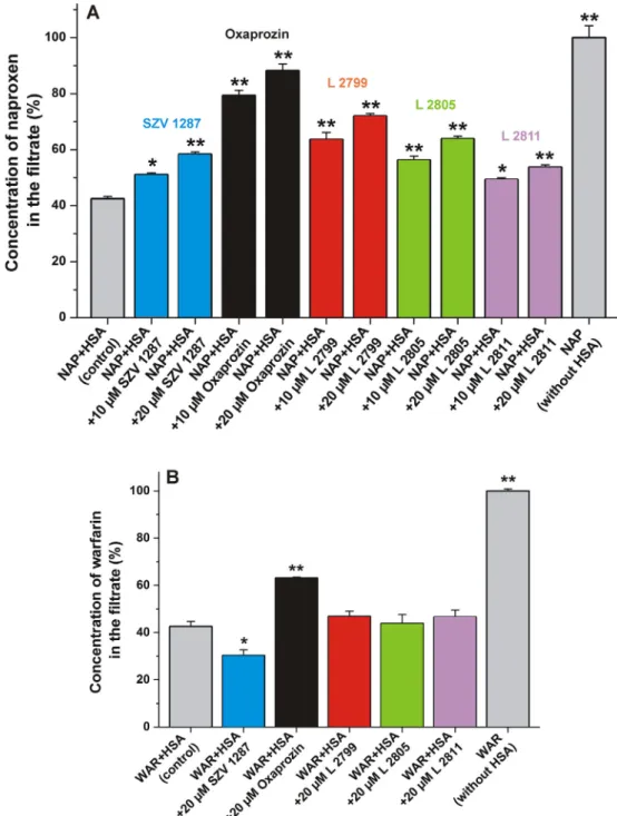
Molecular displacement of a ligand molecule from albumin can influence the pharmacokinetic and pharmacodynamic properties of strongly plasma protein-bound drugs. Ultrafiltration is a power- ful tool to examine the displacement of site markers from serum albumin [22,23]. It is based on the separation of free and albumin-bound molecules [32]. Since HSA is a large (66.5 kDa) macromolecule, albumin and albumin-bound compounds are unable to pass through the filter unit with 30 kDa molecular weight cut-off value. Therefore, the displacement of site markers from HSA results in their increased concentration in the filtrate.
Using these principles, the displacement of warfarin (SSI) and naproxen (SSII) from HSA by SZV 1287 and its metabolites was examined. The filtered fractions of site markers were quantified in the absence and presence of 10 and/or 20
l
M concentrations of compounds tested. As it is demonstrated inFig. 4A, each ligand caused significant increase in the filtered fraction of naproxen, in a concentration dependent fashion. The displacement of naproxen from HSA suggests that the high-affinity binding site of SZV 1287 and its metabolites is located in SSII. In agreement with this hypothesis, earlier reports also identified SSII as the binding site of oxaprozin on albumin [10–12]. Some non-steroidal anti- inflammatory drugs (NSAIDs), including diclofenac, ibuprofen, and flurbiprofen also occupy SSII; while other NSAIDs (e.g. aspirin,phenylbutazone, and indomethacin) preferentially bind to SSI[12].
Compared to our previous studies performed using the same experimental model [22,23], oxaprozin caused strong displace- ment of naproxen, which is in agreement with its high affinity toward HSA (Table 1).
Only SZV 1287 and oxaprozin modified the filtered fraction of warfarin (Fig. 4B). Interestingly, oxaprozin decreased the bound fraction of the SSI marker while SZV 1287 induced the opposite effect. The latter result suggests that SZV 1287 increased the bound fraction of warfarin, similarly to mycotoxin zearalenone and some of its metabolites[18,33]. Furthermore, it is important to note that oxaprozin displaced much lower amount of warfarin compared to its displacing ability vs. naproxen (Fig. 4). Considering these obser- vations, it is reasonable to hypothesize that SZV 1287 and its metabolites occupy another binding site (likely SSII) and some of these compounds can influence the binding affinity of the SSI ligand warfarin through allosteric modulation.
3.6. Binding sites of SZV 1287 and oxaprozin on HSA based on modeling studies
Molecular modeling is a suitable tool to gain a deeper insight into the albumin-ligand interactions at the molecular level. Thus, it helps to characterize which amino acids are involved in the com- plex formation, and to understand the differences in binding posi- tions and binding affinities of the ligand molecules, including the parent compound and its metabolites [18,20,22]. SZV 1287 and oxaprozin were docked to SSII on HSA. The binding modes were de novo described in the present study. Even if oxaprozin glu- curonide is a known[28]ligand to SSII, there is no atomic resolu- tion image of its binding. In the top 1st rank, oxaprozin binds to HSA with an affinity of7.25 kcal/mol. Furthermore, in the top 3rd rank, SZV 1287 binds with great resemblance to the position Fig. 3. HPAC chromatograms of SZV 1287, oxaprozin, L 2799, L 2805, and L 2811 with affinity column coated with immobilized HSA (Chiralpak HSA, 503.0 mm, 5lm; see experimental details in 2.6).
of oxaprozin with weaker affinity of6.72 kcal/mol, which is in agreement with the results of spectroscopic studies, where higher binding constant was determined for the oxaprozin-HSA vs. SZV 1287-HSA complex (Table 1). SZV 1287 found its final binding posi- tion through two prerequisite steps around the SSII. We hypothe- sized the top 3rd rank binding position to be the final of SZV 1287, since its accurate match to that of the top 1st rank binding position of oxaprozin (Fig. 5), which known to be a ligand of SSII [10–12]. The interacting residues were L398, N405, A406, V409, L529, T540, K541, L544, and K545 for oxaprozin (Fig. 5C). Further- more, L398, K402, N405, A406, V409, K413, L529, T540, K541, L544, K545, and M548 were involved in SZV 1287-HSA interaction (Fig. 5B). Our results suggest that lysine residues (K413 and
K545;Fig. 5B) might stabilize the binding of SZV 1287 through cation-
p
interactions with the benzene rings of the ligand, and the carboxyl group of oxaprozin might participate in an ionic inter- action with the K545 lysine positive charge (Fig. 5C).According to Kosa et al.[34], the Site II in HSA is a hydrophobic cleft, approximately 16 Å deep and 8 Å wide, with its hydrophobic amino acid residues being well conserved among the species. It was also demonstrated that the negatively charged carboxyl group of ibuprofen is separated from its hydrophobic part, both struc- tures play important role in the complex formation with albumin.
Similarly, oxaprozin has also a carboxyl group separated from its heterocyclic core (by two C atoms). Whereas, SZV 1287 contains a C = N-OH head group, and its protonated form does not contain Fig. 4. (A)Concentration of naproxen in the filtrate: Before ultrafiltration, samples contained naproxen and HSA (1.0 and 1.7lM, respectively) in the absence and presence of SZV 1287 or its metabolites (10 or 20lM) in PBS (pH 7.4).(B)Concentration of warfarin in the filtrate: Before ultrafiltration, samples contained warfarin and HSA (1.0 and 5.0lM, respectively) in the absence and presence of SZV 1287 or its metabolites (20lM) in PBS (pH 7.4; *p < 0.05, **p < 0.01).
a negative charge. However, the complex formation between albu- min and diazepam (and other similar compounds) is mainly stabi- lized by hydrophobic interactions [34]. Based on X-ray crystallographic studies, the binding of ibuprofen and diazepam are accessible under the PDB codes: 2bxg and 2bxf, respectively [35]. Even in the wider binding cavity of the holo structure, the C atom of L387 and the O atom of S489 are 4.7 Å apart. In the apo structure, the amino acid sidechain of R410 protrudes into the entrance of the cavity, further narrowing the binding site. Ibupro- fen and diazepam contain 15 and 20 heavy atoms, respectively;
thus, they are smaller molecules than SZV 1287 or oxaprozin (both containing 22 heavy atoms). Furthermore, oxaprozin and SZV 1287 have elongated and branching shape, while diazepam is a compact molecule with fused rings. Our docking calculations suggest that the position of oxaprozin and SZV 1287 is located nearby to the entrance of the tight cavity which accommodates ibuprofen and diazepam [35]. Given the size and shape differences of the above-listed molecules, it is fairly rational that oxaprozin and SZV 1287 cannot enter and/or fill this cavity.
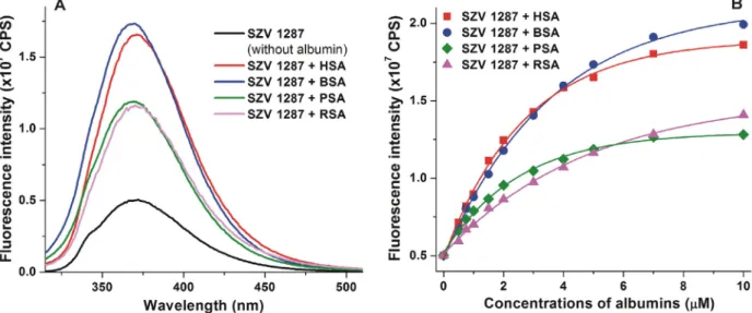
3.7. Interactions of SZV 1287 with albumins from different species
Finally, to test the potential species dependent alternations regarding the albumin binding of SZV 1287, its interactions with albumins from different species (bovine, porcine, and rat) were compared to the SZV 1287-HSA complexation. Inter-species differ- ences regarding the albumin binding of SSI and SSII ligand drugs can be resulted from the subtle alterations in the microenviron- ment (e.g. the differences in the interacting amino acid residues or the size of the hydrophobic cleft)[34]. Moreover, the presence and the location of the binding sites may also vary, which can explain certain differences in binding properties of the ligands [34]. Each albumin induced a considerable increase in the emission signal of SZV 1287: BSA and HSA caused almost four-fold, while PSA (2.5-fold) and RSA (2.8-fold) resulted in lower elevation (Fig. 6). Similarly to section 3.2,Ka values of SZV 1287-albumin complexes were determined based on the albumin-induced increase in the fluorescence of ligand molecules. As it is demon-
strated inTable 2, SZV 1287 formed the most stable complex with HSA, followed by PSA, BSA, and RSA. Nevertheless, the largest dif- ference betweenKavalues (SZV 1287-HSA vs. SZV 1287-RSA com- plexes) was only three-fold, which does not mean a large species difference. For example, mycotoxins ochratoxin A and zearalenone show one order of magnitude differences between the stability of some of their albumin complexes[18,20]. Therefore, we can con- sider only minor species dependent alternations regrading SZV 1287-albumin interactions.
4. Conclusions
In this study, the interactions of SZV 1287 and its metabolites (oxaprozin, L 2799, L 2805, and L 2811) with serum albumin were examined. Fluorescence spectroscopic and HPAC studies demon- strated the formation of stable ligand-HSA complexes (Ka = 105 to 106L/mol). These findings were also supported by ultracentrifu- gation experiments, showing the very low free fraction of SZV 1287 and oxaprozin in human plasma and in 40 g/L HSA solution.
Ultrafiltration studies with SSI and SSII markers and molecular modeling suggest that the high-affinity binding site of SZV 1287 and its metabolites is located in SSII. Furthermore, experiments with human, bovine, porcine, and rat albumins represented only minor species-dependent differences regarding the albumin bind- ing of SZV 1287. Considering the above-listed observations, albu- min binding seems to be a highly relevant parameter in the pharmacokinetics of SZV 1287 and its metabolites. In addition, we also provided a new insight into the pharmacokinetics of oxa- prozin by the detailed characterization of its interaction with HSA.
Funding
The research was supported by the 2017-1.2.1-NKP-2017- 00002 (NAP-2; Chronic Pain Research Group), GINOP 2.3.2-15- 2016-00050 ‘‘PEPSYS”, and GINOP-2.2.1-15-2016-00020 projects.
C. Hetényi’s work was supported by the Hungarian National Research, Development and Innovation Office (K123836) and by the European Union, co-financed by the European Social Fund Fig. 5. The binding mode of SZV 1287 (A; space-filling, pink) as docked to the SSII on HSA (teal cartoon). The close-up of binding mode of SZV 1287 (B; pink sticks) and oxaprozin (C; blue sticks) as docked to the SSII on HSA. Most important interacting amino acids are labeled and shown as gray sticks.
under the title Comprehensive Development for Implementing Smart Specialization Strategies at the University of Pécs EFOP- 3.6.1.-16-2016-00004.
CRediT authorship contribution statement
Eszter Fliszár-Nyúl:Methodology, Formal analysis.Zelma Fai- sal:Methodology, Formal analysis.Violetta Mohos:Methodology, Formal analysis. Diána Derdák: Methodology, Formal analysis.
Beáta Lemli: Methodology, Formal analysis. Tamás Kálai:
Resources. Cecília Sár:Resources. Balázs Z. Zsidó:Methodology, Formal analysis. Csaba Hetényi: Methodology, Formal analysis, Funding acquisition.Ádám I. Horváth:Methodology, Formal anal- ysis. Zsuzsanna Helyes: Conceptualization, Funding acquisition.
Ruth Deme:Resources.Dóra Bogdán:Resources.Andrea Czompa:
Resources. Péter Mátyus: Conceptualization, Resources. Miklós Poór:Conceptualization, Methodology, Formal analysis.
Declaration of Competing Interest
The authors declare that they have no known competing finan- cial interests or personal relationships that could have appeared to influence the work reported in this paper.
Acknowledgements
The authors thank to Katalin Fábián (Department of Pharmacol- ogy, Faculty of Pharmacy, University of Pécs) for her excellent assistance in HPLC analyses. Special thanks to Éva Tóth-Sarudy for elaborating the first synthesis of SZV 1287 at the Department of Organic Chemistry, Semmelweis University. We also thank to Tamás Gáti for his support in NMR spectroscopy, to Beáta Polgár (Department of Medical Microbiology and Immunology, Medical School, University of Pécs) and Tamás Béres (Department of Med- ical Biology, Medical School, University of Pécs) for their assistance
in ultracentrifugation studies, to Szilárd Pál and Györgyi Wittmer (Institute of Pharmaceutical Technology and Biopharmacy, Faculty of Pharmacy, University of Pécs) for the preparation of enteric cap- sules for animal studies, and to Dóra Ömböli and Kata Bölcskei (Department of Pharmacology and Pharmacotherapy, Medical School, University of Pécs) for their assistance in animal studies.
The work was supported by the ÚNKP-20-5 and ÚNKP-20-3-I New National Excellence Program of the Ministry for Innovation and Technology, and by the János Bolyai Research Scholarship of the Hungarian Academy of Sciences. We acknowledge the grant of computer time from the Governmental Information Technology Development Agency, Hungary.
Appendix A. Supplementary material
Supplementary data to this article can be found online at https://doi.org/10.1016/j.molliq.2021.115945.
References
[1]Z. Helyes, P. Mátyus, V. Tékus, B. Scheich, Semicarbazide-sensitive amine- oxidase inhibitors, as analgesics in traumatic neuropathy and neurogenic inflammation, WO/2015/159112 (2015).
[2] M. Salmi, S. Jalkanen, Vascular adhesion protein-1: A cell surface amine oxidase in translation, Antioxid. Redox Signal. 30 (2019) 314–332,https://doi.
org/10.1089/ars.2017.7418.
[3] P. Mátyus, C.L.L. Chai, Metabolism-activated multitargeting (MAMUT): An innovative multitargeting approach to drug design and development, ChemMedChem 11 (2016) 1197–1198, https://doi.org/10.1002/
cmdc.201500406.
[4] P. Mátyus, Multi-targeting drugs: past, present and future, Orv. Hetil. 161 (2020) 523–531,https://doi.org/10.1556/650.2020.31703.
[5] M. Payrits, É. Sághy, P. Mátyus, A. Czompa, R. Ludmerczki, R. Deme, Z. Sándor, Z. Helyes, É. Sz}oke, A novel 3-(4,5-diphenyl-1,3-oxazol-2-yl)propanal oxime compound is a potent Transient Receptor Potential Ankyrin 1 and Vanilloid 1 (TRPA1 and V1) receptor antagonist, Neuroscience 324 (2016) 151–162, https://doi.org/10.1016/j.neuroscience.2016.02.049.
[6] Á. Horváth, A. Menghis, B. Botz, É. Borbély, Á. Kemény, V. Tékus, J.Z. Csepregi, A.
Mocsai, T. Juhász, R. Zákány, D. Bogdán, P. Mátyus, J. Keeble, E. Pintér, Z. Helyes, Analgesic and anti-inflammatory effects of the novel semicarbazide-sensitive Table 2
Association constants (Ka± SEM) of SZV 1287-albumin complexes regarding human, bovine, porcine, and rat albumins.
HSA BSA PSA RSA
Ka[105L/mol] 9.09 ± 0.77 3.50 ± 0.19 6.83 ± 0.22 2.87 ± 0.07
Fig. 6. (A)Fluorescence emission spectrum of SZV 1287 (2lM) in the presence of human (HSA), bovine (BSA), porcine (PSA), and rat (RSA) serum albumins (each 5lM) in PBS (pH 7.4;kex= 305 nm).(B)Albumin-induced (0.0, 0.5, 0.75, 1.0, 1.5, 2.0, 3.0, 4.0, 5.0, 7.0 and 10.0lM) increase in the fluorescence emission signal of SZV 1287 (kex= 305 nm;
kem= 370).
amine-oxidase inhibitor SzV-1287 in chronic arthritis models of the mouse, Sci. Rep. 7 (2017) 39863,https://doi.org/10.1038/srep39863.
[7] Á. Horváth, V. Tékus, N. Bencze, N. Szentes, B. Scheich, K. Bölcskei, É. Sz}oke, A.
Mocsai, E. Tóth-Sarudy, P. Mátyus, E. Pinter, Z. Helyes, Analgesic effects of the novel semicarbazide-sensitive amine oxidase inhibitor SZV 1287 in mouse pain models with neuropathic mechanisms: Involvement of transient receptor potential vanilloid 1 and ankyrin 1 receptors, Pharmacol. Res. 131 (2018) 231–
243,https://doi.org/10.1016/j.phrs.2018.02.006.
[8] G. Fanali, A. di Masi, V. Trezza, M. Marino, M. Fasano, P. Ascenzi, Human serum albumin: From bench to bedside, Mol. Asp. Med. 33 (2012) 209–290,https://
doi.org/10.1016/j.mam.2011.12.002.
[9] K. Yamasaki, V.T. Chuang, T. Maruyama, M. Otagiri, Albumin-drug interaction and its clinical implication, Biochim. Biophys. Acta 2013 (1830) 5435–5443, https://doi.org/10.1016/j.bbagen.2013.05.005.
[10] A.F. Aubry, N. Markoglou, M.H. Adams, J. Longstreth, I.W. Wainer, The effect of co-administered drugs on oxaprozin binding to human serum albumin, J.
Pharm. Pharmacol. 47 (1995) 937–944, https://doi.org/10.1111/j.2042- 7158.1995.tb03274.x.
[11] F. Zsila, Z. Bikadi, D. Malik, P. Hari, I. Pechan, A. Berces, E. Hazai, Evaluation of drug-human serum albumin binding interactions with support vector machine aided online automated docking, Bioinformatics 27 (2011) 1806–1813, https://doi.org/10.1093/bioinformatics/btr284.
[12] F. Zsila, Subdomain IB is the third major drug binding region of human serum albumin: Toward the three-sites model, Mol. Pharm. 10 (2013) 1668–1682, https://doi.org/10.1021/mp400027q.
[13] N.M. Davies, Clinical pharmacokinetics of oxaprozin, Rev. Clin. Pharmacokinet.
35 (1998) 425–436,https://doi.org/10.2165/00003088-199835060-00002.
[14]P. Mátyus, K. Magyar, M. Pihlavista, K. Gyires, Y. Wang, P. Woda, P. Dunkel, É.
Tóth-Sarudy, G. Túrós, Compounds for inhibiting semicarbazide-sensitive amine oxidase (SSAO)/vascular adhesion protein-1 (VAP-1) and uses thereof for treatment and prevention of diseases, WO/2010/029379 (2010).
[15] G. Breviglieri, G. Bruno, S. Contrini, C. Assanelli, Process for the preparation of 4.5-diphenyloxazole-2-propanoic acid, US Patent US6096896A/2000.
[16] A.J. Lewis, R.P. Carlson, H. Fletcher, Hydroxy substituted 4,5-difenyl-2-oxazole propanoic acid, US Patent US4659728/1987.
[17]J.R. Lakowicz, Principles of Fluorescence Spectroscopy, 3rd ed., Springer, New York, NY, USA, 2008.
[18] Z. Faisal, B. Lemli, D. Szerencsés, S. Kunsági-Máté, M. Bálint, C. Hetényi, M.
Kuzma, M. Mayer, M. Poór, Interactions of zearalenone and its reduced metabolites a-zearalenol andb-zearalenol with serum albumins: species differences, binding sites, and thermodynamics, Mycotoxin Res. 34 (2018) 269–278,https://doi.org/10.1007/s12550-018-0321-6.
[19] P. Gans, A. Sabatini, A. Vacca, Investigation of equilibria in solution, Determination of equilibrium constants with the HYPERQUAD suite of programs, Talanta 43 (1996) 1739–1753,https://doi.org/10.1016/0039-9140 (96)01958-3.
[20] Z. Faisal, D. Derdák, B. Lemli, S. Kunsági-Máté, M. Bálint, C. Hetényi, R. Csepregi, T. K}oszegi, F. Sueck, B. Cramer, H.U. Humpf, M. Poór, Interaction of 2’R- ochratoxin a with serum albumins: binding site, effects of site markers, thermodynamics, species differences of albumin-binding, and influence of albumin on its toxicity in MDCK cells, Toxins 10 (2018) 353,https://doi.org/
10.3390/toxins10090353.
[21] D.W. Boulton, U.K. Walle, T. Walle, Extensive binding of the bioflavonoid quercetin to human plasma proteins, J. Pharm. Pharmacol. 50 (1998) 243–249, https://doi.org/10.1111/j.2042-7158.1998.tb06183.x.
[22] V. Mohos, E. Fliszár-Nyúl, G. Schilli, C. Hetényi, B. Lemli, S. Kunsági-Máté, B.
Bognár, M. Poór, Interaction of chrysin and its main conjugated metabolites chrysin-7-sulfate and chrysin-7-glucuronide with serum albumin, Int. J. Mol.
Sci. 19 (2018) 4073,https://doi.org/10.3390/ijms19124073.
[23] E. Fliszár-Nyúl, V. Mohos, T. Bencsik, B. Lemli, S. Kunsági-Máté, M. Poór, Interactions of 7,8-dihydroxyflavone with serum albumin as well as with CYP2C9, CYP2C19, CYP3A4, and xanthine oxidase biotransformation enzymes, Biomolecules 9 (2019) 655,https://doi.org/10.3390/biom9110655.
[24] J.J.P. Stewart, MOPAC: a semiempirical molecular orbital program, J. Comput.
Aided Mol. Des. 4 (1990) 1–103,https://doi.org/10.1007/BF00128336.
[25] J.J.P. Stewart, Optimization of parameters for semiempirical methods VI: More modifications to the NDDO approximations and re-optimization of parameters, J. Mol. Model. 19 (2013) 1–32,https://doi.org/10.1007/s00894- 012-1667-x.
[26] G.M. Morris, R. Huey, W. Lindstrom, M.F. Sanner, R.K. Belew, D.S. Goodsell, A.J.
Olson, AutoDock4 and AutoDockTools4: automated docking with selective receptor flexibility, J. Comput. Chem. 30 (2009) 2785–2791,https://doi.org/
10.1002/jcc.21256.
[27] M. Poór, B. Lemli, M. Bálint, C. Hetényi, N. Sali, T. K}oszegi, S. Kunsági-Máté, Interaction of citrinin with human serum albumin, Toxins 7 (2015) 5155–
5166,https://doi.org/10.3390/toxins7124871.
[28] D.S. Wells, F.W. Janssen, H.W. Ruelius, Interactions between oxaprozin glucuronide and human serum albumin, Xenobiotica 17 (1987) 1437–1449, https://doi.org/10.3109/00498258709044004.
[29] G.E. Dobretsov, T.I. Syrejschikova, N.V. Smolina, On mechanisms of fluorescence quenching by water, Biophysics 59 (2014) 183–188,https://doi.
org/10.1134/s0006350914020079.
[30] M. Poór, G. Boda, S. Kunsági-Máté, P.W. Needs, P.A. Kroon, Beáta Lemli, Fluorescence spectroscopic evaluation of the interactions of quercetin, isorhamnetin, and quercetin-30-sulfate with different albumins, J. Lumin.
194 (2018) 156–163,https://doi.org/10.1016/j.jlumin.2017.10.024.
[31] E. Fliszár-Nyúl, B. Lemli, S. Kunsági-Máté, L. Dellafiora, C. Dall’Asta, G. Cruciani, G. Peth}o, M. Poór, Interaction of mycotoxin alternariol with serum albumin, Int. J. Mol. Sci. 20 (2019) 2352,https://doi.org/10.3390/ijms20092352.
[32] G.A. Ascoli, E. Domenici, C. Bertucci, Drug binding to human serum albumin:
Abridged review of results obtained with high-performance liquid chromatography and circular dichroism, Chirality 18 (2006) 667–679, https://doi.org/10.1002/chir.20301.
[33] M. Poór, S. Kunsági-Máté, M. Bálint, C. Hetényi, Z. Gerner, B. Lemli, Interaction of mycotoxin zearalenone with human serum albumin, J. Photochem.
Photobiol. B 170 (2017) 16–24, https://doi.org/10.1016/j.
jphotobiol.2017.03.016.
[34] T. Kosa, T. Maruyama, M. Otagiri, Species differences of serum albumins: I.
Drug binding sites, Pharm. Res. 23 (1997) 1607–1612,https://doi.org/10.1023/
a:1012138604016.
[35] J. Ghuman, P.A. Zunszain, I. Petitpas, A.A. Bhattacharya, M. Otagiri, S. Curry, Structural basis of the drug-binding specificity of human serum albumin, J.
Mol. Biol. 353 (2005) 38–52,https://doi.org/10.1016/j.jmb.2005.07.075.