ORIGINAL ARTICLE
Aflatoxin production and in vitro toxicity of Aspergilli section Flavi isolated from air samples collected from different environments
Daniela Jakšić1&Sándor Kocsubé2&Ottó Bencsik2&Anita Kecskeméti2&András Szekeres2&Dubravko Jelić3&
Nevenka Kopjar4&Csaba Vágvölgyi2&János Varga2&MajaŠegvićKlarić1
Received: 14 August 2018 / Revised: 4 February 2019 / Accepted: 22 February 2019 / Published online: 12 March 2019
#Society for Mycotoxin (Research Gesellschaft für Mykotoxinforschung e.V.) and Springer-Verlag GmbH Germany, part of Springer Nature 2019
Abstract
Aspergilli sectionFlavi, originally isolated from air samples collected from inhabited apartments (AP), unoccupied basements (BS), and processing facilities of a grain mill (GM), were analyzed for their potential to produce aflatoxin B1(AFB1) on solid media. The isolates were further characterized with regard to their cytotoxic, genotoxic, and pro-inflammatory properties in vitro.
Aspergilli were identified based on partial calmodulin (CaM) gene sequencing; the producing capacities of isolates were analyzed by HPLC/FLD and confirmed by genes in biosynthesis (aflR,norA,omtA). In the grain mill, the Aspergilli sectionFlavi(up to 1.3 × 10⁶cfu/m3) dominated by AFB1-producingAspergillus flavus(71%, 4.5–5254 ng/ml) which showed a serious health risk for workers. Living environments were not relevant sources of exposure. After 24 h, AFB1(1–100μmol/l) reduced cell viability (MTT test) in both A549 cells and THP-1 macrophage-like cells without reaching IC50. In A549 cells, the extract of the AFB1- producingA. flavussignificantly decreased cell viability but not below 50%. THP-1 macrophage-like cells were more sensitive to both extracts, but IC50was obtained only for the AFB1-producing strain (0.37 mg/ml; AFB12.78μmol/l). AFB1(1 and 10μmol/
l) induced significant DNA damage (tail intensity, alkaline comet assay) in A549 cells in contrast to Aspergilli extracts. AFB1
elevated IL-6 and IL-8, while Aspergilli extracts increased IL-1β, TNF-α, and IL-17 release in THP-1 macrophages (ELISA).
Chronic exposure to AFB1and/or other metabolites in airborne A. flavusfrom occupational environments may stimulate epithelial damage of airways accompanied by lowered macrophage viability.
Keywords Airborne fungi . Aflatoxin B1. Cytotoxicity . DNA damage . Cytokines
Introduction
TheAspergillussectionFlavicomprises several species wide- ly distributed in the environment. These species can be sepa- rated into two groups based on their economic importance and impact on human and animal health. The first group includes the non-producing aflatoxin species Aspergillus oryzae,
Aspergillus sojae, and Aspergillus tamariiused in oriental food fermentation (Campbell-Platt1994). Genetically modi- fiedA. oryzaestrains are used for the production of enzymes, including lactase, pectin esterase, lipase, protease, and xylanase (Pariza and Johnson 2001). The second group in- cludes aflatoxin-producing species Aspergillus flavus, Aspergillus parasiticus, andAspergillus nomius, which con- taminate and damage various pre-harvested and stored agri- cultural products (Perrone et al.2014). In the past decade, the production of aflatoxins was also reported in newly described species Aspergillus arachidicola, Aspergillus bombycis, Aspergillus minisclerotigenes,Aspergillus parvisclerotigenus, Aspergillus pseudocaelatus, Aspergillus pseudonomius, Aspergillus pseudotamari,Aspergillus togoensis,Aspergillus mottae,Aspergillus sergii, and Aspergillus transmontanensis (Varga et al.2011; Soares et al.2012).
Aflatoxins are known hepatotoxic mycotoxins with carci- nogenic, genotoxic, and teratogenic properties in both humans and animals (Bennett et al.2003). Among these mycotoxins,
* MajaŠegvićKlarić msegvic@pharma.hr
1 Department of Microbiology, Faculty of Pharmacy and Biochemistry, University of Zagreb, Schrottova 39, 10000 Zagreb, Croatia
2 Department of Microbiology, Faculty of Science and Informatics, University of Szeged, H-6726, Szeged Közép fasor 52, Hungary
3 Fidelta Ltd., Prilaz baruna Filipovića 29, 10000 Zagreb, Croatia
4 Mutagenesis Unit, Institute for Medical Research and Occupational Health, Ksaverska cesta 2, 10000 Zagreb, Croatia
aflatoxin B1(AFB1) is the most toxic as well as the most frequently occurring aflatoxin in agricultural products (Varga et al.2011).
In general, aflatoxins are principally produced byA. flavus andA. parasiticus; A. flavus mainly produces AFB1 and AFB2, while mostA. parasiticusisolates produce AFG1and AFG2in addition to AFB1and AFB2. However,A. flavusis considered more invasive and out-competes A. parasiticus when both species are present in soil (Perrone et al.2014).
Aflatoxigenic strains ofA. flavusare most widespread in trop- ical and subtropical areas. However, in recent years, a more frequent occurrence of aflatoxin producers has been detected in temperate-climate regions, associated with current climate change. Air humidity changes (abundant rain, droughts), in- crease in temperature, and increased CO2concentration direct- ly affect the expression of regulatory (aflR) and structural genes (aflD) involved in the biosynthesis of AFB1and, thus, correlate with its biosynthesis (Paterson and Lima 2010;
Medina et al.2014).
Next toA. fumigatus,A. flavusis the second most frequent cause of invasive and non-invasive aspergillosis in humans; it may also cause sinusitis, cutaneous and wound aspergillosis, as well as otitis and keratitis (Hedayati et al. 2007;
Manikandan et al.2013).
Taking into account the aflatoxigenic ability of the listed Aspergilli sectionFlavi, particularlyA. flavus, as well as its role in the etiology of several diseases in humans, an accurate identification of the species would provide fundamental infor- mation concerning their aflatoxigenic and pathogenic proper- ties (Godet and Munaut2010). Several molecular genetic techniques have been developed to distinguish the Aspergillusspecies in the section Flavi; Varga et al. (2011) proposed a polyphasic approach, including morphological characters, extrolite data, and partial calmodulin (CaM),β- tubulin (BenA), and ITS sequences as well as the presence of genes involved in aflatoxin biosynthesis.
Since the discovery of aflatoxins, the hepatotoxic action of AFB1has been mainly studied in various experimental models through AFB1-contaminated food consumption (Bennett et al.
2003). However, a link between AFB1inhalation in an indus- trial setting and liver or lung cancer incidence and mortality has been suggested by several investigations (Olsen et al.
1988; Viegas et al.2012). Taking into account the lack of data on human exposure to fungal burden in urban homes and occupational settings in our country, recently, we conducted a year-round investigation of airborne fungi in living (apart- ments, basements) and occupational (grain mill) indoor envi- ronments in Croatia with special attention toAspergillussec- tionsFlavi,Nigri, andVersicoloresdue to their known patho- genic and/or mycotoxin-producing properties (JakšićDespot andŠegvićKlarić2014,b). Based on morphological methods, Aspergilli sections Flaviand Nigriwere recovered in the highest frequencies (50–100% and 15–55%, respectively)
from samples taken in the grain mill, while Aspergilli section Versicoloreswas more abundant in apartments and basements (10–65%) (JakšićDespot andŠegvićKlarić2014,b). Before appropriate molecular identification methods for species assigned to these sections, indoor air isolates were usually reported as A. flavus, A. niger, or A. versicolor (Samson et al.2014). As we previously noted, accurate identification of theAspergillusspecies is fundamental with regard to their toxigenic and/or pathogenic properties (Godet and Munaut 2010). Thus, herein, we described the species diversity of Aspergillus sections Nigri (Jakšić et al. 2018) and Versicolores (JakšićDespot et al. 2016) using calmodulin sequence-based methods, their ability to produce fumonisin B2and sterigmatocystin, respectively, and to explore toxic potential of Aspergilli extracts in comparison with these my- cotoxins alone. To further extend knowledge onAspergillus sectionFlavibiology and toxicology, the present study aimed to explore the species diversity of isolated strains using a polyphasic approach (morphological methods, CaM se- quence, and presence of genes taking part in aflatoxin biosyn- thesis) and confirm aflatoxin-producing abilities in Aspergilli extracts by HPLC/FLD.
Some studies showed that AFB1alone induces several tox- ic events in human airways, including damage of airway epi- thelial cells, decrease of the ciliary beat frequency (Lee et al.
2016), DNA damage due to formation of exo-AFB1-8,9-ep- oxide by human lung microsomes (Kelly et al.1997) as well as takes a part in inflammation (Lee et al.2016; Luongo et al.
2014). However, the effects of inhaled aflatoxin-producing fungi on the airway epithelium have not been well- characterized so far.
AsA. flavuswas among the most abundant species in the grain mill, it was justified to explore the cytotoxic, genotoxic, and immunomodulatory properties of AFB1-producing and AFB1-non-producingA. flavusextracts in contrast to AFB1
alone in order to clarify whether particular toxic properties of Aspergilli could be attributed to AFB1. This was carried out using human lung adenocarcinoma A549 cells and mac- rophages derived from human leukemic monocyte THP-1 cells as experimental models.
Materials and methods
Chemicals and mediaMethanol, acetonitrile, methylene chloride, ethyl acetate, and n-hexane for sample preparation and eluents were purchased from VWR (Debrecen, Hungary). Formic acid, trifluoroacetic acid, and aflatoxin B1standard were obtained from Sigma- Aldrich (Budapest, Hungary). Deionized water for both sam- ple preparation and high-performance liquid chromatography (HPLC) runs was produced by Merck Millipore Milli-Q
Gradient A10 water purification equipment (Budapest, Hungary). Dichloran 18% glycerol agar (DG-18), Czapek Yeast Agar (CYA), and Malt Extract Agar (MEA) were pur- chased from Oxoid (Hampshire, UK). Media for cell culture maintenance, including RPMI 1640, fetal bovine serum (FBS), trypsin-EDTA, phosphate-buffered saline (PBS; Ca2+
and Mg2+free), penicillin, and streptomycin were from Lonza (Basel, Switzerland). MTT reagent [3-(4,5-dimethylthiazol-2- yl)-2,5-diphenyltetrazolium bromide], phorbol 12-myristate 13-acetate (PMA) ethidium bromide, acridine orange, normal melting point (NMP) agarose, low melting point (LMP) aga- rose, Triton X-100, Tris buffer, and dimethyl sulfoxide (DMSO) were purchased from Sigma-Aldrich (Deisenhofen, Germany). Dimethyl sulfoxide (DMSO) ethanol HCl, NaCl, Na2EDTA, and NaOH were from Kemika (Zagreb, Croatia).
Sampling areas
The samples of airborne fungi were collected over a one-year period (2012) in two-month intervals at the processing facili- ties of a grain mill (GM) situated near Zagreb, Croatia, as well as in residential locations in the city which included two inhabited apartments (AP) and two unoccupied basements (BS) as well as outdoor air (ODA) as described in detail in JakšićDespot andŠegvićKlarić(2014,b). In each period of sampling in GM, twenty samples were collected during mill- ing operations at the site of grain/flour exchange, site of flour storage, site of sieving, and site of milling. In each AP (ap- proximately 70 m2), samples were taken at five locations, including the kitchen, dining room, living room, bedroom, and bathroom, in duplicate. BSs were located in the same buildings as the AP, and residents used these spaces to store various items (e.g., wooden, metal, plastic, or glass materials).
In each BS, samples were taken at five locations in duplicate.
Samples of outdoor air were taken in proximity to indoor locations, and, during each sampling period, ten ODA sam- ples were collected.
The sampling approach included collecting 20 indoor sam- ples and 10 outdoor samples per each location and at a given time point. Altogether, 420 individual samples were taken.
Sample collection
Airborne fungi were sampled using an Airsampler Mas-100 Eco (Merck, Berlin, Germany) with 400 holes (hole to agar impactor, impaction velocity 10.8 m/s, and airflow rate 100 l/
min) and DG-18 agar plates (Samson et al.2010). Because of the high contamination level, a volume of 10 l was chosen for sampling in the GM, while a volume of 50 l was applied at the other sampling locations. The plates were incubated at 25 °C
± 0.2 °C in the dark for five days, after which the developed fungal colonies were counted, and results were expressed as colony-forming units per cubic meter (cfu/m3). Aspergilli
were isolated on CYA and MEA agar plates (Samson et al.
2010). Isolates assigned to theAspergillussectionFlaviwere purified and cultivated on CYA and MEA plates at 25 °C in the dark for seven days. Morphological identifications were carried out according to literature (Samson et al.2010).
Determination of Aspergilli sectionFlavigenotypes
Isolation of genomic DNA from mycelia grown in liquid YPD medium (1% Bacto yeast extract, 1% Bacto peptone, 1% D- glucose) for five days was performed by Masterpure™yeast DNA purification kit (Epicentre Biotechnologies, Madison, WI, USA) according to the manufacturer’s instructions. The isolated DNA was diluted to < 1.000 ng and used as template DNA in PCR reactions. A fragment of the calmodulin gene (CaM), part of the transcriptional regulator of aflatoxin bio- synthesis (aflR), norsolorinic acid reductase (norA or aflE), and O-methyltransferase gene (omtA,aflP) (Yu et al.2004) were amplified using the primers specified in Table1. Each PCR reaction mixture (20μl) contained 0.2 mmol/l deoxyri- bonucleotide triphosphate (dNTP), 0.2μmol of primers, 1 U DreamTaq DNA Polymerase (Thermo Scientific, Madison, WI, USA) with the respective buffer (with 1.5 mmol/l MgCl2) (Thermo Scientific, Madison, WI, USA), nuclease- free water, and template DNA. Amplifications were per- formed using a Thermocycler T-100 (BioRad, Budapest, Hungary). The PCR cycling protocol consisted of 35 cycles, including an initial denaturation at 95 °C for 2 min in the first and 20 s in the following runs, annealing temperature as spec- ified in Table1for 20 s, elongation at 72 °C for 40 s, and 2 min for the final elongation. The amplification products were an- alyzed by electrophoresis on 2% agarose gels using fluores- cent dye GR Green (Excellgen, Budapest, Hungary), and the banding patterns were visualised under ultraviolet light (254 nm). In case of PCR products assigned toaflR,norA, andomtA, the bands were scored as either present or absent for eachA. flavusisolate. PartialCaMsequences were determined at the LGC Genomics GmbH (Berlin, Germany). Sequence analysis was performed by nucleotide–nucleotide BLAST similarity search at the website of the National Center for Biotechnology Information (http://www.ncbi.nlm. nih.gov/
BLAST), and sequences were, also, compared with our own sequence database. Species identification was determined from the lowest expected value of the BLAST output (Altschul et al.1990).
Preparation of Aspergilli sectionFlaviextracts and aflatoxin analysis
Extracts ofA. flavusisolates (n= 65) were prepared and ana- lyzed as described previously (Baranyi et al.2015). Briefly explained, following a microextraction procedure according to Frisvad et al. (2007), three agar plugs (6 mm in diameter
each) were extracted using a mixture of organic solvents meth- anol/dichloromethane/ethyl-acetate (1/2/3,v/v/v) supplement- ed with 1% of formic acid. The extracts were ultrasonicated, and the organic phases were separated, filtered through, and evaporated to dryness under a slight stream of nitrogen. Dry extracts were derivatized with trifluoroacetic acid (Fazekas et al.2005). HPLC (Shimadzu, Japan) equipped with a fluo- rescence detector set at an excitation wavelength of 365 nm and an emission wavelength of 430 nm was used for AFB1
analysis. Separations were achieved on a LiChroCART 250 × 4 mm, 5 μm (Merck, Hungary) column coupled with a LiChrospher 100 RP-18 (Merck, Hungary) guard column and an injection volume of 5μl. The isocratic mobile phase composition was water/methanol/acetonitrile 65/17.5/17.5 (v/
v/v), and the flow rate was maintained at 1 ml/min, while the column temperature was 40 °C. For the quantification of AFB1, the linear calibration curve was used in a concentration range of 6.25 to 100 ng/ml and the equation of resulted cali- bration curve was y = 4056.2x−7321.2 (R2= 0.9994).
Cytotoxicity, genotoxicity, and immunomodulation of AFB1vs Aspergilli sectionFlaviextracts
Cell cultures
Human lung adenocarcinoma cells A549 and human leukemia monocytes THP-1 (European Collection of Cell Cultures, Salisbury, UK) were grown in 75 cm2flasks in RPMI supple- mented with 2 mmol/l glutamine, 10% heat-inactivated FBS, penicillin (100 IU/ml; 1 IU 67.7μg/ml), and streptomycin (100μg/ml). Cultures were maintained in a moisturized atmo- sphere with 5% CO2at 37 °C and 95% relative humidity.
Preparation of tested compounds
A stock solution of AFB1(0.01 mol/l) was prepared in DMSO/70% ethanol mixture 80/20v/v. Weighed dried fungal extracts were dissolved in 100% DMSO. The final concentra- tions of AFB1and fungal extracts as well as DMSO/ethanol used in the treatments of cells were obtained by dilution with the culture medium. The highest concentration of fungal
extract applied as treatment was limited by the content of DMSO (up to 1%) that showed no significant effect on cell viability. Mass concentrations of fungal extracts used in MTT assay ranged from 0.05 to 0.6 mg/ml, while the corresponding content of AFB1in the extract of toxin-producingA. flavus was 0.75 to 4.5μmol/l. Concentrations of the extracts used in other assays were selected according to the results of MTT and corresponded to subcytotoxic concentrations.
MTT proliferation assay
Viability of A549 cells and macrophage-like THP-1 cells was estimated using MTT assay, as described previously (Jakšić Despot et al. 2016). The differentiation of THP-1 cells into macrophages was performed using PMA (20 nm/well). The A549 (104cells/well) and macrophage-like THP-1 cells (5 × 10⁴cells/well) were grown in a 96-well flat-bottom microplate in RPMI 1640 medium supplemented with 10% of FBS.
Upon 24 h treatment with AFB1(0.1 to 100μmol/l) or with Aspergilli extracts (0.05 to 0.6 mg/ml) of AFB1-producing and non-producing isolates, the concentration that inhibits growth in 50% of cells (IC50) was determined. Following treatment, the medium was removed and 100 μl of MTT- tetrazolium salt reagent diluted in RPMI 1640 medium with- out FBS (0.5 mg/ml) was added (V = 100μl per each well).
After 3 h of incubation, the medium was replaced with 100μl of DMSO to dissolve formazan (product of metabolised MTT reagent), and cells were incubated at room temperature on a rotary shaker for 15 min. The absorbance was measured using a microplate reader (Labsystem iEMS, type 1404) at a wave- length of 540 nm. All tests were performed in five replicates, and results were expressed as percentage of control.
Alkaline comet assay
The extent of primary DNA damage produced after 24 h treat- ment with AFB1andAspergilliextracts was assessed in A549 cells using the alkaline comet assay. A standard protocol, pro- posed by Singh et al. (1988), with minor modifications was used. Before treatment, A549 cells were seeded in 6-well plates (3 × 10⁵ cells per well) in RPMI 1640 medium Table 1 The primers used in PCR
Primer Sequence (5′–3′) Annealing temperature (°C) Gene Reference
Cmd-5 CCGAGTACAAGGARGCCTTC 52–55 CaM Hong et al. (2005)
Cmd-6 CCGATRGAGGTCCATRACGTGG
Afl-F GGGATAGCTGTACGAGTTGTGCCAG 57–59 aflR Varga et al. (2011)
Afl-R TGGKGCCGACTCGAGGAAYGGGT
Nor-1 ACCGCTACGCCGGCACTCTCGGCA 64–66 norA Criseo et al. (2001); Varga et al. (2011)
Nor-2 GTTGGCCGCCAGCTTCGACACAGC
Omt-1 GTGGACGGACCTAGTCCGACATCAC 59–61 omtA Geisen (1996); Varga et al. (2011)
Omt-2 GTCGGCGCCACGCACTGGGTTGGGG
supplemented with 10% of FBS. After 24 h of growth, the cell medium was discarded and cells were treated with AFB1(at 1 and 10μM), Aspergilli extracts (at 0.05 and 0.1 mg/ml for non-producing and 0.1 and 0.2 mg/ml for AFB1-producing isolate). Control cell cultures were grown in parallel (with and without 0.1% DMSO/ethanol) for 24 h. After 24 h of treatment, the cells were washed with 1 ml cold PBS, scraped with rubber, and resuspended in 300μl of PBS. Aliquots (V = 20μl) of this suspension were used to prepare slides for the comet assay. Briefly, cell samples were mixed with 100μl 0.5% LMP agarose (in Ca- and Mg-free PBS) and spread onto fully frosted slides, previously pre-coated with 1% and 0.6%
NMP agarose. The microgels were allowed to solidify on ice for 10 min. Then, the slides were subjected to lysis at 4 °C in a buffer containing 2.5 mol/l NaCl, 100 mmol/l Na2EDTA, 10 mmol/l Tris (pH 10), supplemented with 1% Triton-X 100. The lysis step lasted for 1 h and was followed by dena- turation in alkaline buffer (10 mmol/l NaOH, 200 mmol/l Na2EDTA, pH 13), for 20 min. Electrophoresis lasted for 20 min in the same buffer at 25 V and 300 mA. Microgels were, then, neutralised with three changes of 0.4 mol/l Tris/HCl buffer (pH 7.5), for 5 min each. The slides were kept in a humid atmosphere in a dark box at 4 °C until further analysis. Before image analysis, DNA was stained for 10 min with ethidium bromide (20μg/ml). Slides were scored using an image analysis system (Comet assay IV, Instem- Perceptive Instruments Ltd., UK) connected to a fluorescence microscope (Olympus, Japan). All of the experiments were performed in duplicate, and, in each experiment, images of 200 randomly selected nucleoids (100 nucleoids from each of the two replicate slides) were measured. Only comets with a defined head were scored. As indicators of DNA damage, tail length (presented in micrometres) and tail intensity (i.e., percentage of DNA in the comet tail) were selected (Collins 2004).
Determination of cytokine levels
To establish cytokine levels after 24 h treatment with AFB1 and Aspergilli extracts, THP-1 cells were selected as an ap- propriate cell model. Cells were first seeded on 24-well cell culture plates (1 × 10⁶cells per well) in RPMI 1640 medium supplemented with 10% of FBS and differentiated into mac- rophages for 24 h with addition of 40 nmol/l LM PMA for 24 h. After that, the medium was discarded, and cells were treated with AFB1(at 1 and 10μmol/l LM) and Aspergilli extracts (at 0.05 and 0.1 mg/ml for non-producing and 0.1 and 0.2 mg/ml for AFB1-producing isolate) for 24 h. Control cell cultures were grown in parallel in RPMI 1640 medium supplemented with 10% of FBS (with and without 0.1%
DMSO/ethanol) for 24 h. Following treatment, the cell medi- um was harvested and frozen at −80 °C until analysis.
Concentrations of TNF-α, IL-1β, IL-6, IL-8, and IL-17 were
determined in harvested cell medium by DuoSet ELISA kits (R&D Systems, Minneapolis, USA) according to instructions provided by the manufacturer and as described elsewhere (Hulina et al.2018). Cytokine concentrations were calculated from measured optical densities determined at 450 nm, using a microplate reader (En Vision® Multilabel Plate Reader, Perkin Elmer) and by standard calibration curves, and results of the three replicates were expressed as percentage of control.
Statistics
Data obtained using the MTT test, comet assay, and cytokine concentration were first evaluated using descriptive statistics.
The results were presented as mean ± standard error of mean (SEM). Normality of data distribution was tested by Kolmogorov–Smirnov test; one-way ANOVA was applied for normally distributed data (MTT test and cytokine concen- trations) followed by Sidak’s post-test, while for non-normally distributed data (comet assay), Kruskal–Wallis test was ap- plied followed by Dunn’s multiple-comparison test. A value ofP< 0.05 was considered statistically significant for all cal- culations. To obtain the IC50 from the results of the MTT assay, non-linear dose–response fitting was applied using the equation y = A1 + (A2−A1) / (1 + 10^((LOGx0-x)∗p)).
Results and discussion
Occurrence of aflatoxigenic Aspergilli sectionFlavi in air samples
The abundance of airborneAspergillusspecies sectionFlavi in the occupational environment (GM) in contrast to inhabited urban homes (AP) and urban home basements (BS) as well as outdoor air (ODA) in the six periods of sampling is presented in Table2. Only in GM were these Aspergilli present through- out the whole year, comprising 1–12% of total viable airborne fungi, with mean absolute concentrations approximately 100 to 2000 times higher than in other indoor, as well as outdoor locations. In the GM, the mean absolute concentrations of Aspergilli were highest in March and September (about 2000 cfu/m3), with a maximum of 2 × 10⁴ cfu/m3 in September. Looking at the estimated concentrations (probable viable number), the airborne Aspergilli section Flavi were present at concentrations between 1.7 × 10³cfu/m3(January) and 1.3 × 10⁴cfu/m3(March) from January to September and dropped below 1000 cfu/m3only in November. Comparing the results of the present study with our previous reports, the Aspergilli sectionFlavioccurred in significantly higher mean absolute concentrations in the GM (405–2035 cfu/m3) than both Aspergilli from the subclade Versicolores(6.5–44 cfu/
m3) and Aspergilli sectionNigri (20–240 cfu/m3) (Jakšić Despot et al.2016; Jakšićet al.2018). Mean concentrations
of viable Aspergilli sectionFlaviwere also much higher than those reported in poultry farms, soybean and cotton mills, and the food industry (17–137.6 cfu/m3) in India, Italy, and Egypt (Sharma et al. 2010; Abdel Hameed et al. 2012; Cafarchia et al.2014). Maximum values of statistically estimated con- centrations of Aspergilli sectionFlaviobtained in July (1.9 × 10⁵cfu/m3) and (1.3 × 10⁶cfu/m3) were 10–100-fold higher than concentrations of airborne fungi (> 104cfu/m3) consid- ered as a health hazard in non-sensitized subjects (Oppliger et al.2005). Contrary to the GM, Aspergilli sectionFlaviwere rarely present or were in low concentrations (maximum 20 cfu/m3) in urban apartments and basements (AP and BS) and outdoors which is in agreement with recently conducted studies in homes of low-income areas in Syracuse (NY, USA) as well as in homes of hematologic patients in Germany (Crawford et al.2015; Schweer et al.2016). In most periods, these Aspergilli comprised between 0.1 and 0.2% of total viable airborne fungi recovered from AP, BS, and ODA.
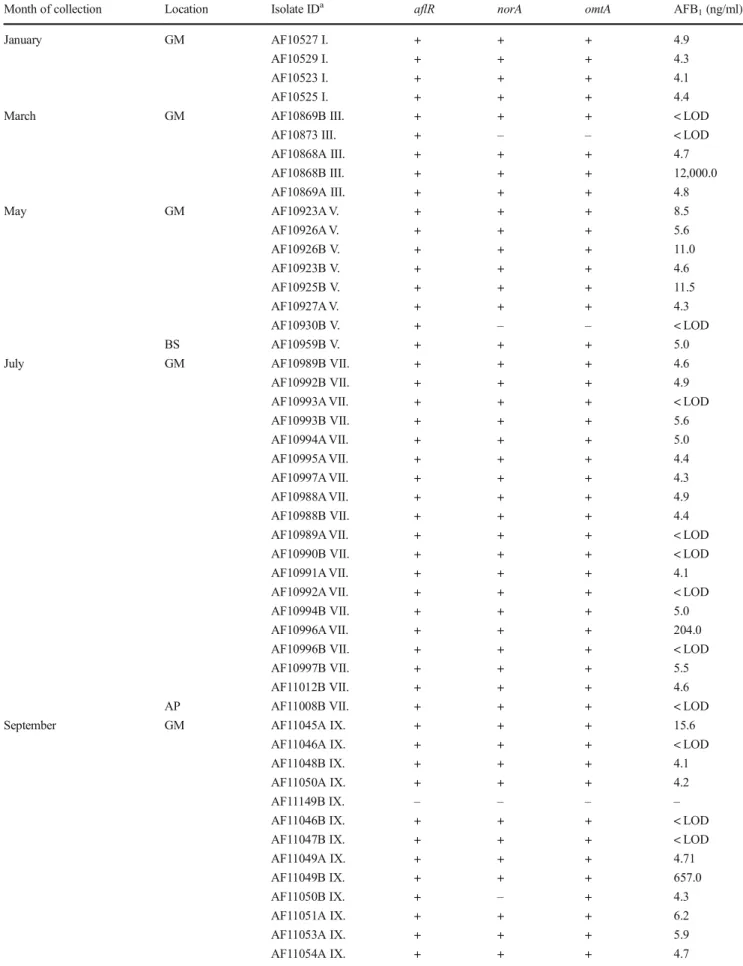
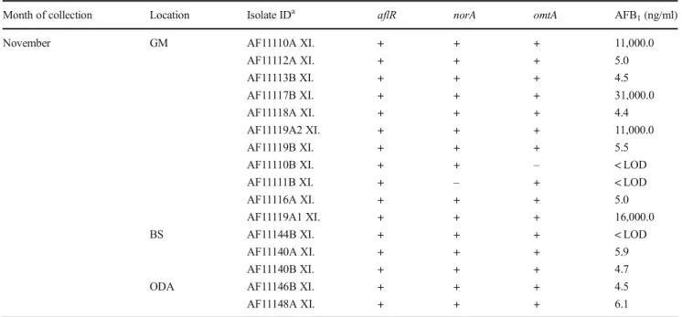
Based onCaMgene region sequencing, among a total of 67 Aspergilli section Flavi isolates, 65 were identified as A. flavus and 2 as A. parasiticus (Table 3) (data on A. parasiticus are not shown; results were published in Baranyi et al.2015). The majority ofA. flavusstrains (89%) were recovered from the GM. Among these strains, 76% pro- duced AFB1; the highest amounts of AFB1(10–30 μg/ml) were produced by strains isolated in March and November.
For five strains ofA. flavus, isolated from the GM environ- ment, production of low amounts of AFB2 (0.03 ± 0.02 μg/ml) was detected, in addition to AFB1 (13.4 ± 9.8 μg/ml). These five strains represented 10% of all A. flavusisolates from the GM environment. The production of AFB1and AFB2correlated with genes detected for the transcriptional regulator of aflatoxin biosynthesis (aflR), norsolorinic acid reductase (norA), and O-methyltransferase gene (omtA); 15 strains were positive for two or all three genes for aflatoxin biosynthesis, but the amounts of the toxin were below the LOD. Several earlier studies have suggested the significance of respiratory exposure toA. flavusin biowaste- handling facilities (up to 105cfu/m3) and AFB1in grain dust ranging from 0.04 to 107 ng/m3 (Fischer et al. 2000a,b;
Fischer and Dott 2003). Hardin et al. (2009) reviewed the significance of exposure to airborne fungi and mycotoxins in light of concentration of no toxicologic concern-CoNTC (30 ng/m3), which showed that in occupational environments, such as grain, handling the concentration of AFB1in the fa- cilities can exceed the CoNTC. Recently, in poultry farms in Portugal,A. flavuswas found in three of seven poultry units, and this Aspergilli was the third most frequently found fungal species among indoor airborne fungi (Viegas et al.2012). In the same study, the presence of aflatoxigenic strains was only confirmed in an outdoor air sample from one of the poultry units but direct evidence of the poultry workers’professional exposure to AFB1 was found in 58% of positive serum Table2AbundanceofAspergillifromthesectionFlaviinindoorandoutdoorairforeachsamplingperiod Grainmill(GM)Apartment(AP)Basement(BS)Outdoorair(ODA) cfu/m3cfu/m3cfu/m3cfu/m3 AbsoluteEstimated*AbsoluteEstimated*%Mean±SDRange%Mean±SDRange%Mean±SDRange% Mean±SDRange January290±2181743±15760–6000–39421.06––––––––– March2035±248613,370±16,3300–50000–32,8505.57–––1±4.50–200.13––– May405±3692579±24950–10000–65701.51–––1±4.50–200.09––– July1335±7518771±4932100–3000657–19,7103.342±60–200.20––2±60–200.10 September1839±44468091±29,1290–20,0000–131,40012.851±4.50–200.221±4.50–200.10––– November150±176167±1990–6000–6902.39–––1±4.50–200.086±100–202.5 *TheprobablenumberofviableparticlescalculatedfromFeller’sformula(Pr=N1/N+1/N-1+1/N-2+1/N-r+1),givenbythemanufacturer(MerckKgaA,Darmstadt,Germany;Pr=probablestatistical total;r=numberofcfucounted;N=totalnumberofholesinthesamplinghead) %,–MeanconcentrationofAspergillisectionFlavi/meanconcentrationoftotalairbornefungi×100.(MeanconcentrationoftotalairbornefungiadoptedfromJakšićDespotandŠegvićKlarić(2014))
Table 3 AFB1-producing abilities of indoorA. flavusisolates from different sampling periods and locations
Month of collection Location Isolate IDa aflR norA omtA AFB1(ng/ml)
January GM AF10527 I. + + + 4.9
AF10529 I. + + + 4.3
AF10523 I. + + + 4.1
AF10525 I. + + + 4.4
March GM AF10869B III. + + + < LOD
AF10873 III. + – – < LOD
AF10868A III. + + + 4.7
AF10868B III. + + + 12,000.0
AF10869A III. + + + 4.8
May GM AF10923A V. + + + 8.5
AF10926A V. + + + 5.6
AF10926B V. + + + 11.0
AF10923B V. + + + 4.6
AF10925B V. + + + 11.5
AF10927A V. + + + 4.3
AF10930B V. + – – < LOD
BS AF10959B V. + + + 5.0
July GM AF10989B VII. + + + 4.6
AF10992B VII. + + + 4.9
AF10993A VII. + + + < LOD
AF10993B VII. + + + 5.6
AF10994A VII. + + + 5.0
AF10995A VII. + + + 4.4
AF10997A VII. + + + 4.3
AF10988A VII. + + + 4.9
AF10988B VII. + + + 4.4
AF10989A VII. + + + < LOD
AF10990B VII. + + + < LOD
AF10991A VII. + + + 4.1
AF10992A VII. + + + < LOD
AF10994B VII. + + + 5.0
AF10996A VII. + + + 204.0
AF10996B VII. + + + < LOD
AF10997B VII. + + + 5.5
AF11012B VII. + + + 4.6
AP AF11008B VII. + + + < LOD
September GM AF11045A IX. + + + 15.6
AF11046A IX. + + + < LOD
AF11048B IX. + + + 4.1
AF11050A IX. + + + 4.2
AF11149B IX. – – – –
AF11046B IX. + + + < LOD
AF11047B IX. + + + < LOD
AF11049A IX. + + + 4.71
AF11049B IX. + + + 657.0
AF11050B IX. + – + 4.3
AF11051A IX. + + + 6.2
AF11053A IX. + + + 5.9
AF11054A IX. + + + 4.7
samples (mean 2 ng/ml) in contrast to the AFB1-negative se- rum sampled from control individuals. On the other hand, in living environments, AFB1does not seem to be relevant since its formation on building materials during fungal growth has not been detected (Rao2016; Ren1999). Our results suggest that a serious threat to human health due to exposure to air- borneA. flavus could come only in occupational environ- ments, such as grain mill facilities, but not from exposure in urban apartments and basements.
Cytotoxicity of AFB1vsA. flavusextracts
The cytotoxic potential of Aspergilli extracts and AFB1ap- plied alone on A549 cells and THP-1 macrophage-like cells is presented in Table4and Fig.1. Although AFB1alone signif- icantly reduced the viability of both A549 and THP-1 macro- phage-like cells at concentrations up to 100μmol/l, the reduc- tion did not fall below 50% and the IC50was not calculated.
Our results are consistent with previous studies on A549 cells and human bronchiolar epithelium BEAS-2B cells (Palanee et al.2001; Vleet et al.2002; McKean et al.2006). Van Veelt
et al. (2002) showed that CYP 1A2 and 3A4 enzymes are responsible for the bioactivation of AFB1 into metabolites (AFB18,9-epoxide and aflatoxin Q1) that evoked cytotoxicity in CYP-transfected cells of the bronchiolar epithelium (B- CMV1A2 and B3A4). On the contrary, BEAS-2B and A549 have limited CYP activity, which was in agreement with their CYP gene expression profile (Garcia-Canton et al.2013) and may explain the weak cytotoxicity of AFB1obtained in both A549 and THP-1 macrophage-like cells.
The extract of AFB1-producingA. flavusinduced a signif- icant drop in the viability of A549 cells, while the non- producing strain was not cytotoxic to them. THP-1 macro- phage-like cells were more sensitive to both extracts, but the AFB1-producing strain exerted stronger cytotoxicity with IC50
(0.37 ± 0.024 mg/ml; the corresponding AFB1concentration is 2.78 ± 0.18μmol/l AFB1). Thus, we may suggest that AFB1
contributed to the A. flavusextract cytotoxicity but only in THP-1 macrophage-like cells. Piecková and Wilkins (2004) showed that endo- and exometabolite extracts of dust-borne A. flavuswere able to stop chicken tracheal cilia beating after the first 24 h of activity. Recently, it was shown that exposure Table 3 (continued)
Month of collection Location Isolate IDa aflR norA omtA AFB1(ng/ml)
November GM AF11110A XI. + + + 11,000.0
AF11112A XI. + + + 5.0
AF11113B XI. + + + 4.5
AF11117B XI. + + + 31,000.0
AF11118A XI. + + + 4.4
AF11119A2 XI. + + + 11,000.0
AF11119B XI. + + + 5.5
AF11110B XI. + + – < LOD
AF11111B XI. + – + < LOD
AF11116A XI. + + + 5.0
AF11119A1 XI. + + + 16,000.0
BS AF11144B XI. + + + < LOD
AF11140A XI. + + + 5.9
AF11140B XI. + + + 4.7
ODA AF11146B XI. + + + 4.5
AF11148A XI. + + + 6.1
aIsolate ID, isolates ofA. flavusare deposited under their designated ID in the microbial collection (MFBF) at the Department of Microbiology Faculty of Pharmacy and Biochemistry, University of Zagreb
GM, grain mill; AP, apartment; BS, basement; ODA, outdoor air
Table 4 The calculated IC50of AFB1negative and positive A. flavusextracts (0.05– 0.6 mg/ml) in A549 and THP-1 macrophage-like cell lines using non-linear curve fitting (0.9914)
Treatment IC50(A549) IC50(THP-1)
(x̅± SEM) (x̅± SEM)
A. flavus(AFB1-negative) > 0.6 mg/ml > 0.4 mg/ml
A. flavus(AFB1-positive) > 0.4 mg/ml 0.37 ± 0.024 mg/ml (AFB12.78μM)
AFB1 > 100μM > 100μM
of primary human sinonasal and bronchial cell cultures to aflatoxins (0.1 to 10μmol/l) as well as to conditioned media fromA. flavusreduced the ciliary beat frequency. The effect was blocked by an anti-aflatoxin antibody suggesting that the aflatoxin was responsible for the reduction of ciliary beat fre- quency (Lee et al.2016). The same study revealed that AFB2
activates protein kinase C in A549 cells, which has been linked to inflammation and apoptosis (Diaz-Meco and Moscat2012; Zhao et al.2012). Taken together, our results suggest that although AFB1 alone exerted weak cytotoxic properties in THP-1-like macrophages and A549 lung cells, it significantly contributes to the cytotoxicity ofA. flavusex- tracts. Therefore, chronic exposure to aflatoxins in the mixture ofA. flavusmetabolites in an occupational environment may stimulate epithelial damage in airways accompanied by lowered macrophage viability contributing to the pathogene- sis of respiratory diseases.
Genotoxicity of AFB1vsA. flavusextracts
Genotoxic effects were evaluated in A549 cells using the al- kaline comet assay (Fig.2). AFB1alone evoked significant concentration-dependent DNA damage measured as tail inten- sity but not in terms of tail length as compared to control.
Contrary to AFB1, Aspergilli extracts provoked a significant concentration-dependent increase of tail length but not tail intensity. The difference in the responses at DNA level ob- served after the treatments could be explained by the intrinsic differences in the mechanisms behind DNA lesions. Without conducting additional experiments, we cannot establish the exact mechanism(s) responsible for the observed effects.
However, as a comet’s tail length is proportional to the number of relaxed DNA loops (Collins et al. 2008), it seems that treatment with Aspergilli extracts led to an increased number of relaxed DNA loops, but without a concomitant increase in the amount of DNA breaks. This could be, at least in part, associated with the chemical composition of the extract, which—besides AFB1—contains other active compounds as well, which possess different potentials to damage DNA or even modulate the DNA damaging effects of AFB1itself.
When discussing the significance of the comet assay pa- rameters evaluated in this study, it has to be stressed that tail intensity has been deemed the most useful because as the level of damage increases so does the relative intensity of DNA staining in the tail, rather than tail length (Collins et al.
2008). Therefore, the significantly increased tail intensity we recorded at both of the tested concentrations represents an important piece of information regarding AFB1genotoxicity, Fig. 1 Survival of THP-1 macrophage-like cells and A549 cells after 24 h
of the treatment with the extract prepared from AFB1-negative (a) and AFB1-positive (b) isolate ofA. flavusand single AFB1(c). Mass concentrations of fungal extracts prepared from AFB1-negative isolate ofA. flavuswere I 0.05, II 0.1, III 0.25, and IV 0.4 mg/ml (a), in the extract of toxin-producingA. flavusI 0.1, II 0.2, III 0.4, and IV 0.6 mg/ml,
while the corresponding content of AFB1was 0.75, 1.5, 3.0, and 4.5μmol/l, 1–4 respectively (b). Each data point represents the mean ± SEM of cell viability (% of control, control = 100% of cell viability). *,
**Significantly different as compared to control in A549 and THP-1 macrophage like cells, respectively (P< 0.05)
which will be useful for planning future experiments with other cell types and exposure scenarios.
The underlying mechanism of AFB1genotoxicity was pre- viously studied in the liver in vivo as well as in liver cells and several human bronchial cell lines with good expression of CYP 1A2 and 3A4 enzymes responsible for AFB1biotrans- formation into reactive AFB18,9-epoxide that binds to DNA (reviewed in Marchese et al.2018). An association between lung cancer development following AFB1exposure was de- scribed in workers occupationally exposed to grain dust con- taminated with this compound (Hayes et al.1984). It was demonstrated that AFB1exposure induced the production of a DNA binding metabolite (epoxide) by lung cytosols which was correlated with lipoxygenase and prostaglandin H syn- thase and increased human pulmonary susceptibility to AFB1(Massey et al.2000). Also, oxidative DNA damages in mouse lung cells were correlated to AFB1genotoxicity, owing to the induction of 8-hydroxy-20-deoxyguanosine (8- OHdG) formation (Guindon et al.2008). Although A549 cells possess a limited expression of CYP enzymes (Garcia-Canton et al.2013), our results suggest that it was sufficient to induce DNA damage upon exposure to low concentrations of AFB1. In our previous study, AFB1(at 5μmol/l) also evoked signif- icant DNA damage and mutagenic activity, as revealed by the alkaline comet assay, and led to micronuclei formation (Jakšić et al.2012). Since both the AFB1-positive and the AFB1-neg- ative Aspergilli extracts exerted similar genotoxic action in A549 cells, we may hypothesize that other metabolites present
in the extract might antagonise AFB1’s genotoxic action in AFB1-producing strains.
Immunomodulatory effects of AFB1vsA. flavus extracts
Many macrophage and epithelial cells produce cytokines and chemokines after challenges from various inflammatory stim- uli. Thus, we explored the differences in the secretion of pro- inflammatory cytokines (TNF-α, IL-1β, IL-6, IL-8, IL-17) by THP-1 macrophage-like cells upon exposure to single AFB1
or Aspergilli extracts (Fig.3). TNF-αis an endogenous pyro- gen and immunoregulatory cytokine responsible for the pro- duction of IL-1, IL-6, and IL-8. Deficiency of TNF-αin ex- perimental animals promoted cancer (Suganuma et al.1999).
TNF-αstimulates polymorphonuclear leukocytes to damage Aspergillus hyphae enhancing phagocytosis (Roilides et al.
2002). IL-1βis a potent pro-inflammatory cytokine secreted by macrophages and monocytes which has a stimulatory ef- fect on CD4+ T cells promoting differentiation into the T helper cell. It has a beneficial role of mediating an immune response against pathogenic infiltration, but it can also pro- mote the pathogenesis of tissue damage that leads to chronic inflammation (Eder 2009; Turner et al.2014). Similarly to TNF, IL-6 is a pleiotropic cytokine showing both pro- inflammatory and anti-inflammatory activities, but, in chronic inflammation, it is rather pro-inflammatory. The synthesis of this cytokine is induced by IL-1 and TNF-α (Duque and Fig. 2 Genotoxicity of AFB1-negative and AFB1-positive extracts of
A. flavus isolates as well as AFB1 applied in subcytotoxic concentrations determined by alkaline comet assay as tail length and tail intensity in A549 cells. C control (0.1% DMSO), I AFB1-negative extract ofA. flavus0.05 mg/ml, II AFB1-negative extract ofA. flavus
0.1 mg/ml, III AFB1-positive extract ofA. flavus0.1 mg/ml, IV AFB1- positive extract ofA. flavus0.2 mg/ml, VAFB11 μmol/l, VI AFB1
10μmol/l. The statistical significance of the treatment compared to control is marked with an asterisk (*,P< 0.05) above each histogram
Descoteaux2014). IL-8, also known as chemokine CXCL8, is a monocyte- and macrophage-derived cytokine that serves as a chemoattractant of neutrophils to the site of infection or injury and whose secretion from macrophages can be
stimulated with TNF-α, IL-1β, or a lipopolysaccharide (Carré et al.1991). IL-17 is a pro-inflammatory cytokine that plays an essential role in the host’s defense against microbial infections and is implicated in various inflammatory Fig. 3 Relative concentration of cytokines measured in the supernatant of
THP-1 macrophage-like cells upon treatment: I AFB1-negative extract of A. flavus0.05 mg/ml, II AFB1-negative extract ofA. flavus0.1 mg/ml, III AFB1-positive extract ofA. flavus0.1 mg/ml, IVAFB1-positive extract of
A. flavus0.2 mg/ml, VAFB11μmol/l, VI AFB110μmol/l. The statistical significance of the treatment compared to control (0.1% DMSO) is marked with an asterisk (*,P< 0.05) above each histogram
conditions, such as autoimmune diseases, metabolic disorders, and cancer (Gu et al.2013).
AFB1alone vs Aspergilli extracts showed differences in the immunomodulatory pattern. AFB1 has the most pro- nounced concentration-dependent effect on IL-6 and IL-8 ex- cretion. Although the release of TNF-αand IL-17 was also increased by AFB1in a concentration-dependent manner, it was not significantly different from controls. Additionally, a difference in IL-1βrelease by THP-1 macrophage-like cells upon treatment with single AFB1vs Aspergilli extract was also observed; AFB1decreased IL-1β levels, while both Aspergilli extracts increased the release of this cytokine in a concentration-dependent manner. Several in vivo studies have also shown that AFB1 stimulates the expression of IL-6 (Hinton et al.2003; Meissonnier et al.2008; Qian et al.
2014; Abbès et al.2016). The same in vivo studies suggested that secretion of TNF-αsignificantly depends on the admin- istered dose of AFB1; e.g., a dose of 1.8 mg/kg AFB1during four weeks of administration resulted in an increased expres- sion of TNF-α(Meissonnier et al.2008), while at a signifi- cantly lower dose (80μg/kg), AFB1decreased TNF-α(Abbès et al.2016). In primary alveolar pig macrophages, AFB1(1– 100 ng/ml) did not have an effect on the expression of both TNF-αand IL-1βbut decreased phagocytosis efficiency (Liu et al.2002). The inhibition of macrophage phagocytic activity was also established in vivo in a study on pigs which found that the inhalation of 16.8μg/kg of AFB1inhibited the phago- cytic activity of alveolar macrophages without recovery of function two weeks after inhalation (Jakab et al. 1994).
Similarly to our study, AFB1evoked an increased expression of IL-8 mRNA in human lymphoblastoid Jurkat T-cells. The activation of an inflammatory response and over-active IL-8- induced recruitment of neutrophils to specific tissues can re- sult in extensive tissue damage (Luongo et al.2014). As in our study, IL-17 levels increased as the dose increase of AFB1in mice liver did, but no differences were detected between the treated and control groups (Ishikawa et al.2017). Opposite to AFB1, both Aspergilli extracts provoked a significant increase of TNF-α, IL-1β, and IL-17 in THP-1 macrophage-like cells, while not inducing a release of IL-6 and IL-8. The excretion of TNF-α, IL-1β, and IL-17 was probably affected by the other metabolites in the extracts ofA. flavusthat could have acted in synergy with AFB1. At the same time, the metabolite mixtures in the extract could have had an antagonising activity on the induction of IL-6 and IL-8. Additionally, IL-17 appeared to play a central role in eosinophil extravasation from the blood into the lungs of mice upon intranasal exposure to A. fumigatusconidia (Murdock et al.2012), which also con- tains metabolite mixture. Taken together, AFB1elevated IL-6 and IL-8 while Aspergilli extracts increased IL-1b, TNF-a, and IL-17 release in THP-1 macrophage-like cells suggesting that AFB1alone and both AFB1-positive and AFB1-negative Aspergilli extracts could potentially impart adverse effects on
innate immunity but with different mechanisms that could be influenced by the metabolite mixture composition in the extracts.
Considering the limitations of in vitro experiments, we may only suggest that chronic inhalatory exposure to AFB1and/or otherA. flavusmetabolites in occupational environments can stimulate epithelial damage of airways accompanied by lowered macrophage viability but with a different underlying mechanism depending on the extrolite profile of the airborne A. flavus.
Acknowledgments This work was financially supported by the University of Zagreb (Grant No. 1126). This study forms part of the project GINOP-2.3.2-15-2016-00012, supported by the European Social Fund. This work was also supported by OTKA grant Nos.
K115690 and K8407 as well as through András Szekeres, who received support through the new national excellence program of the Hungarian Ministry of Human Capacities.
Compliance with ethical standards
Conflicts of interest The authors declare that they have no conflict of interest.
Publisher’s noteSpringer Nature remains neutral with regard to jurisdic- tional claims in published maps and institutional affiliations.
References
Abbès S, Ben Salah-Abbès J, Jebali R, Younes RB, Oueslati R (2016) Interaction of aflatoxin B1and fumonisin B1in mice causes immunotoxicity and oxidative stress: possible protective role using lactic acid bacteria. J Immunotoxicol 13:46–54.https://doi.org/10.
3109/1547691X.2014.997905
Abdel Hameed AA, Ayesh AM, Abdel Razik Mohamed M, Abdel Mawla H (2012) Fungi and some mycotoxins producing species in the air of soybean and cotton mills: a case study. Atmos Pollut Res 3:
126–131.https://doi.org/10.5094/APR.2012.012
Altschul S, Gish W, Miller W, Myers EW, Lipman DJ (1990) Basic local alignment search tool. J Mol Biol 215:403–410
Baranyi N, JakšićDespot D, Palágyi A, Kiss N, Kocsubé S, Szekeres A, Kecskeméti A, Bencsik O, Vágvölgyi C,ŠegvićKlarićM, Varga J (2015) Identification ofAspergillusspecies in Central Europe able to produce G-type aflatoxins. Acta Biol Hung 66:339–347.https://doi.
org/10.1556/018.66.2015.3.9
Bennett JW, Klich M, Mycotoxins M (2003) Mycotoxins. Clin Microbiol Rev 16:497–516.https://doi.org/10.1128/CMR.16.3.497
Cafarchia C, Camarda A, Iatta R, Danesi P, Favuzzi V, Di Paola G, Pugliese N, Caroli A, Montagna MT, Otranto D (2014) Environmental contamination byAspergillusspp. in laying hen farms and associated health risks for farm workers. J Med Microbiol 63:464–470.https://doi.org/10.1099/jmm.0.065946-0 Campbell-Platt G (1994) Fermented foods—a world perspective. Food Res
Int 27:253–257.https://doi.org/10.1016/0963-9969(94)90093-0 Carré PC, Mortenson RL, King TE, Noble PW, Sable CL, Riches DW
(1991) Increased expression of the interleukin-8 gene by alveolar macrophages in idiopathic pulmonary fibrosis. A potential mecha- nism for the recruitment and activation of neutrophils in lung fibro- sis. J Clin Invest 88:1802–1810.https://doi.org/10.1172/JCI115501