C E L L B I O L O G Y
The chromatin remodeler ALC1 underlies resistance to PARP inhibitor treatment
Szilvia Juhász1*, Rebecca Smith2*, Tamás Schauer3, Dóra Spekhardt1, Hasan Mamar1, Siham Zentout2, Catherine Chapuis2, Sébastien Huet2,4†, Gyula Timinszky1†
Poly(ADP-ribose) polymerase (PARP) inhibitors are used in the treatment of BRCA-deficient cancers, with treat- ments currently extending toward other homologous recombination defective tumors. In a genome-wide CRISPR knockout screen with olaparib, we identify ALC1 (Amplified in Liver Cancer 1)—a cancer-relevant poly(ADP-ribose)- regulated chromatin remodeling enzyme—as a key modulator of sensitivity to PARP inhibitor. We found that ALC1 can remove inactive PARP1 indirectly through binding to PARylated chromatin. Consequently, ALC1 deficiency enhances trapping of inhibited PARP1, which then impairs the binding of both nonhomologous end-joining and homologous recombination repair factors to DNA lesions. We also establish that ALC1 overexpression, a common feature in multiple tumor types, reduces the sensitivity of BRCA-deficient cells to PARP inhibitors. Together, we conclude that ALC1-dependent PARP1 mobilization is a key step underlying PARP inhibitor resistance.
INTRODUCTION
Poly(ADP-ribose) polymerase 1 (PARP1) is a DNA damage sensor important for maintaining genomic integrity. PARP1 recognizes and binds both single-strand breaks (SSBs) and double-strand breaks (DSBs), triggering its ADP-ribose polymerase activity (1).
Upon DNA damage, PARP1 poly(ADP-ribosyl)ates (PARylates) several DNA damage repair–associated proteins and chromatin components that are crucial for efficient DNA damage repair. As expected, the loss of PARP1 or the inhibition of PARP activity sensitizes cells to DNA-damaging agents (2–5).
PARP inhibitors (PARPis) were shown to be particularly toxic for cells deficient in homologous recombination (HR) repair factors BRCA1 (Breast cancer type 1 susceptibility protein) and BRCA2 (Breast cancer type 2 susceptibility protein) even in the absence of exogenous DNA damage, a pheno menon with great therapeutic potential because of the high prevalence of BRCA deficiency in tu- mor cells (6–8). The observation that the synthetic lethality between BRCA deficiency and PARPi treatment is abrogated by the loss of PARP1 revealed that the inhibited DNA-bound PARP1 is the toxic product—the phenomenon called PARP trapping—rather than deficient DNA damage signaling in the absence of PARylation (4).
Olaparib was the first PARPi to be approved for the treatment of BRCA-deficient breast and ovarian cancers (9, 10). The therapeutic use of PARPis brought increased interest in elucidating genetic alterations that lead to sensitivity or resistance to PARP inhibition. The loss of sev- eral HR and interstrand cross-link repair components leads to PARPi sensitivity, signifying that HR is essential for the faithful repair of the increased level of DNA lesions induced by PARP1 trapping (7, 8, 11). In HR-deficient cells, these lesions are handled by the error-prone nonho- mologous end-joining (NHEJ) pathway that ultimately leads to chro-
mosome aberrations and cell death. The synthetic lethality between PARPis and BRCA1 deficiency can be reversed by the loss of the NHEJ factor 53BP1 (Tumor Protein P53 Binding Protein 1), which reactivates HR in a PALB2 (Partner And Localizer Of BRCA2)–dependent manner (12). Recently, it has become apparent that PARPi sensitivity can be attributed to malfunction of pathways other than defective HR. For ex- ample, defective ribonucleotide excision repair due to the loss of ribonu- clease H2 activity was recently identified as a source of DNA lesions that can cause PARP1 trapping (13). Moreover, loss of PARylation factors such as histone PARylation factor 1 (HPF1) and poly(ADP-ribose) glycohydrolase (PARG) can sensitize cells or promote resistance to PARPi, respectively (14, 15). In the current study, we used a genome- wide CRISPR knockout screen with olaparib to identify other molecular mechanisms that could modulate sensitivity to PARPi treatment to which the therapeutic spectrum could potentially be extended.
RESULTS
CRISPR-based knockout screen identifies ALC1 deficiency as a source PARPi sensitivity
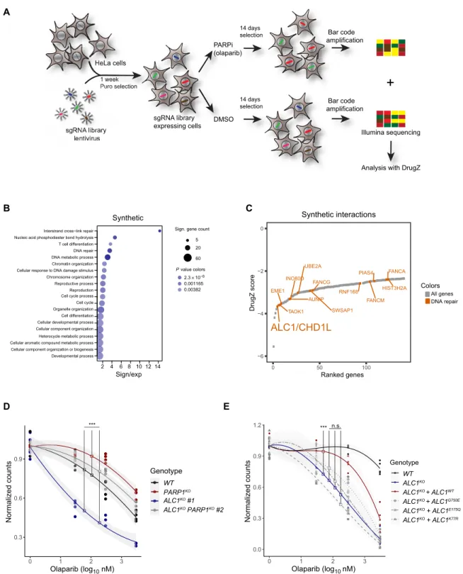
To identify previously unknown factors that modulate cell sensitivity to the clinically approved PARPi, olaparib, we infected wild-type (WT) HeLa cells with the GeCKOv2 whole-genome CRISPR-based knockout pooled library targeting each gene with six single-guide RNAs (sgRNAs) (16, 17). Knockout cells were subjected to olaparib at a concentration yielding approximately 40% survival, enriching the proportion of cells resistant to the drug treatment while depleting knockout cells with increased sensitivity to treatment. Genomic DNA was collected after 14 days of treatment, and sgRNA cassettes were amplified and deep-sequenced. Sequence output from dimethyl sulf- oxide (DMSO) control and olaparib-treated samples were analyzed using the DrugZ algorithm (18) to identify candidate genes influenc- ing sensitivity to olaparib (Fig. 1, A to C; fig. S1, A and B; and table S1).
Examination of Gene Ontology terms of genes whose loss led to PARPi resistance revealed a number of cell cycle– and mTOR (mammalian target of rapamycin)–associated processes. Deficiency in PARP1 or PARG also conveyed resistance to PARPi (fig. S1, A and B), in agreement with previous reports (4, 15). Conversely, we found that loss of several genes belonging to DNA repair processes, in particular
1MTA SZBK Lendület DNA Damage and Nuclear Dynamics Research Group, Institute of Genetics, Biological Research Centre, 6276 Szeged, Hungary. 2Univ Rennes, CNRS, IGDR (Institut de Génétique et Développement de Rennes), UMR 6290, BIOSIT, UMS 3480, F-35000 Rennes, France. 3Biomedical Center, Bioinformatics Unit, Ludwig Maximilian University of Munich, 82152 Planegg-Martinsried, Germany. 4Institut Universitaire de France, Paris France.
*These authors contributed equally to this work.
†Corresponding author. Email: sebastien.huet@univ-rennes1.fr (S.H.); timinszky.
gyula@brc.hu (G.T.)
Copyright © 2020 The Authors, some rights reserved;
exclusive licensee American Association for the Advancement of Science. No claim to original U.S. Government Works. Distributed under a Creative Commons Attribution License 4.0 (CC BY).
on January 21, 2021http://advances.sciencemag.org/Downloaded from
sgRNA library expressing cells HeLa cells
library 1 week Puro selection
14 days selection 14 days selection PARPi (olaparib)
DMSO )
Illumina sequencing
+
Analysis with DrugZ NA library A
i llll sgRNNAA librarylibra ke
ke ke es es eeo eo wero 1 wr 111 w 1 11 1uu P Puu P P P Puuurorooos P P P oouuuror P P P P P P
yyyyyyyyy ry aryry ar braa br b ill
HeLa cells H H HeLa ce H
n
Bar code amplification
Bar code amplification
2 4 6 8 10 12 14
Synthetic
Sign/exp
Developmental process Cellular component organization or biogenesis Cellular aromatic compound metabolic process Heterocycle metabolic process Cellular component organization Cellular developmental process Cell differentiation Organelle organization Cell cycle Cell cycle processReproduction Reproductive process Chromosome organization Cellular response to DNA damage stimulus Chromatin organization DNA metabolic process DNA repair T cell differentiation Nucleic acid phosphodiester bond hydrolysis
Interstrand cross−link repair Sign. gene count
5 20 60 Pvalue colors
2.3 × 10⁻⁶ 0.001165 0.00382
ALC1/CHD1L
TAOK1 EME1
AUNIP INO80D
UBE2A
SWSAP1 FANCG
RNF168 FANCM PIAS4 FANCA
HIST3H2A
−6
−4
−2 0
0 50 100
Ranked genes
DrugZ score
Colors All genes DNA repair
Synthetic interactions B
A
C
D E
Genotype WT ALC1KO ALC1KO + ALC1WT ALC1KO + ALC1G750E ALC1KO + ALC1E175Q ALC1KO + ALC1K77R
0.0 0.3 0.6 0.9 1.2
0 1 2 3
Olaparib (log10 nM)
Normalized counts
*** n.s.
0.3 0.6 0.9
0 1 2 3
Olaparib (log10 nM)
Normalized counts
Genotype WT ALC1KO #1 ALC1KO PARP1KO #2 PARP1KO
***
Fig. 1. A genome-wide CRISPR knockout screen reveals ALC1 as a gene conveying PARP inhibitor resistance. (A) Schematic of the CRISPR screen. (B) Dot plot showing the enrichment of 20 Gene Ontology processes. The size of the dots represents the number of significant genes associated with the Gene Ontology term, and the color of the dots represents the P value. (C) Scatterplot of DrugZ analysis result of the genes showing synthetic interactions with olaparib. Genes annotated with functions in DNA repair are colored red. (D) Clonogenic cell survival assay of U2OS WT, ALC1KO, PARP1KO, and ALC1KO PARP1KO double knockout cells after a 24-hour treatment with olaparib. (E) Clonogenic cell survival assay of U2OS WT and ALC1KO expressing mCherry-ALC1 variants after a 24-hour treatment with olaparib. Graphs in (D) and (E) include all data points (n = 3 to 5) and fitted curves with 95% confidence intervals (gray shading). Asterisks indicate P values obtained by polynomial regression (n.s., not significant; ***P < 0.001). Model summary is provided in table S2.
on January 21, 2021http://advances.sciencemag.org/Downloaded from
cross-link repair, was associated with increased sensitivity to PARPi including previously identified genes involved in HR such as FANCM (Fanconi anemia complementation group M) (fig. 1, B and C) (11).
BRCA1 and BRCA2, whose loss was previously shown to provide PARPi sensitivity (7, 8), failed to score in our screen due to reduced cell fitness upon loss of each of these two genes (fig. S1C and table S1).
One of the strongest candidate genes whose loss increased sensitivity to PARPi was the poly(ADP-ribose) (PAR)–dependent chromatin remodeler ALC1 (Amplified in Liver Cancer 1)/CHD1L (Chromo- domain Helicase DNA Binding Protein 1 Like)..
ALC1 is a member of the SNF2 superfamily of chromatin re- modelers. It is unique among the hits for synthetic lethality as a protein that directly binds PAR, the product of PARP1 activity, promoting its activation (19, 20). ALC1 is amplified in many solid tumors and is associated with tumor progression (21). Previous studies demon- strated the correlation between ALC1 overexpression and poor patient survival in non–small cell lung cancer (22) as well as patient chemo- therapy resistance in human hepatocellular carcinoma (23). Studies based on DT40 model cell line investigated the role of ALC1 in base excision repair with PARP1 cooperation and showed that both PARP1KO and ALC1KO, PARP1KO double knockout cells had similarly impaired SSB repair. Moreover, cells expressing ATPase-dead ALC1 dis- played hypersensitivity to various DNA- damaging agents (24), while the depletion of ALC1 increased cell sensitivity to various DNA- damaging agents [H2O2, ultraviolet (UV), and phleomycin] (19, 25). To validate the impact of ALC1 loss on PARPi sensitivity, which was also found in another screen (13), we generated ALC1KO in U2OS cell lines using CRISPR-Cas9–based gene editing and studied clono- genic cell survival in the presence of varying olaparib concentrations.
All three independent ALC1KO cell lines tested showed sensitivity to olaparib even at low concentrations, confirming their synthetic lethality with PARPi (Fig. 1D and fig. S1D). To address whether the synthetic lethality between ALC1KO and PARPi required the PARP1 protein, we generated ALC1KO PARP1KO double knockout cell lines.
PARP1KO showed resistance to olaparib treatment in agreement with previous reports, and we also observed that the loss of PARP1 in ALC1KO cells decreased sensitivity to PARPi (Fig. 1D and fig. S1D).
These results are consistent with PARPi toxicity requiring the presence of PARP1 (4). This observation was also confirmed in ALC1KO cells by RNA interference (RNAi)–mediated down- regulation of PARP1 (fig.
S1E). Furthermore, ALC1KO cells showed sensitivity to the PARPis veliparib and niraparib, which have lower and higher trapping potential, respectively, as compared to olaparib (fig. S1, F and G) (26). The differences in inhibitor concentrations at which the hy- persensitivity of the ALC1KO cells can be detected mirror the rela- tive PARP1 trapping potential of the three inhibitors (27). Last, the sensitivity of ALC1KO cells to PARPi treatment could be par- tially rescued when complemented with ALC1-WT (Fig. 1E and fig. S1H) but not with a PAR-binding mutant of ALC1 that cannot recruit to sites of DNA damage (fig. S1I) or with ALC1 ATPase- deficient mutants that recruit to sites of DNA damage as efficiently as ALC1-WT (fig. S1I) but are unable to remodel the chromatin (5).
Together, these results identify ALC1 as a crucial component pro- viding resistance to PARPi. Failure of cells to form Rad51 foci has been used as a predictor of PARPi sensitivity. HR-deficient cells such as BRCA1 and BRCA2 null cells do not form foci irrespective of PARPi treatment, while their WT counterparts show robust Rad51 foci formation in the presence of PARPi (7, 8, 28). We saw an increase in Rad51 foci formation in WT
cells treated with PARPi as well as increased Rad51 foci levels in PARP1KO cells irrespective of PARPi treatment, as previously de- scribed (fig. S1, J and K) (29). In ALC1KO cells, we observed elevated levels of Rad51 foci compared to WT cells in the absence of PARPi and that this did not increase upon the addition of PARPi (fig. S1, J and K). This result indicates that PARPi sensitivity of ALC1KO cells may differ from HR deficiency, a common cause of PARPi sen- sitivity.
The observation that a PAR-responsive enzyme is important for cell survival in the presence of PARPi where PARylation is blocked seems counterintuitive. Nevertheless, we observed that olaparib concentrations that were toxic for ALC1KO cells did not fully sup- press ALC1 recruitment to DNA lesions in WT cells likely due to residual PARP1 catalytic activity (fig. S1, L and M). This incomplete inhibition of PARylation signaling upon PARPi treatment may also explain why the deletion of PARG causes PARPi resistance, while PARG degrades PAR, the formation of which should be blocked by PARPis (15).
PARP inhibition induces DNA damage and cell cycle arrest in ALC1 knockouts
In the absence of genomic stress, most nuclear PARylation is limited to S phase where unligated Okazaki fragments provide the trigger for PARP1 binding and activation (30). In addition to this role in DNA replication, PARP1 activation has been reported to be im- portant for the regulation of HR components and replication fork reinitiation after replication-induced fork stalling and subsequent DNA damage (31, 32). Conversely, PARP inhibition was reported to induce abnormal acceleration of replication fork elongation, leading to DNA damage accumulation (33). Together, these data prompted us to assess the impact of ALC1 and PARP inhibition in S-phase progression.
To assess replication fork dynamics, we looked for colocalization of EdU (5-ethynyl-2′-deoxyuridine) and IdU (5-iodo-2′-deoxyuridine) after an initial pulse labeling of active replication foci using the nu- cleotide analog EdU and a second pulse labeling of ongoing replication with IdU (fig. S2A). While we were able to see DNA damage–induced replication fork arrest after high-dose x-ray [8 gray (Gy)] treatment, shown as a lack of IdU incorporation after EdU pulse labeling (fig. S2B), we found that ALC1KO, PARP1KO, and ALC1KO PARP1KO double knockout cells showed IdU incorporation at EdU signals after olaparib treatment, indicating that PARP inhibition does not impair S-phase progression in any of the tested cell lines (fig. S2C).
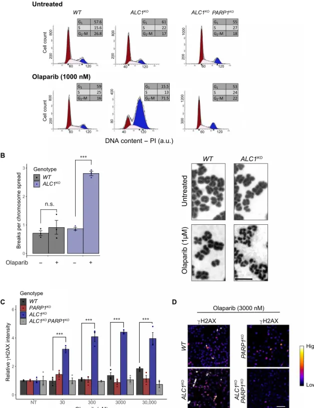
Next, we analyzed the ALC1KO cells by flow cytometry to further characterize the consequence of PARPi treatment on cell cycle pro- gression. We find that a 24-hour olaparib treatment leads to the accumulation of ALC1KO cells arrested in the G2-M phase. In con- trast, the cell cycle distribution of the olaparib-resistant ALC1KO PARP1KO double knockouts was not affected by PARPi treatment similar to WT cells (Fig. 2A). In addition, in ALC1KO cells, PARPi treatment resulted in an increase of both chromosome breaks, as shown by chromosome spread (Fig. 2B and fig. S2D), and DNA breaks as mea- sured by alkaline comet tail moment length (fig. S2E). Consistent with this increase in DNA lesions, ALC1KO cells treated with PARPi also displayed an increase in the phosphorylated fraction of histone H2AX indicative of elevated DNA damage signaling (Fig. 2, C and D).
These accumulating DNA lesions are feasibly the cause of the cell cycle arrest at G2-M, ultimately leading to the synthetic lethality ob- served between PARPi treatment and ALC1 deficiency.
on January 21, 2021http://advances.sciencemag.org/Downloaded from
A
UntreatedOlaparib (1µM)
WT ALC1KO
γH2AX
Olaparib (3000 nM) γH2AX
WTALC1KO PARP1KO PARP1 KO ALC1KO
C D
WT ALC1KO ALC1KOPARP1KO
Untreated
Cell count 800200 800200 2001000120030080400
800200Cell count
DNA content - PI (a.u.)
60 120 40 120 60 120
60 120
120
60 120
Olaparib (1000 nM)
40
0 2 4 6
NT 300
Olaparib (nM)
Relative γH2AX intensity
Genotype WT PARP1KO ALC1KO ALC1KO PARP1KO
30 3000 30,000
KO
High
Low
B
0 1 2 3
Breaks per chromosome spread
Genotype WT ALC1KO
Olaparib - + - +
***
n.s.
***
*** *** ***
Fig. 2. PARP1 inhibition induces chromosome aberrations and cell cycle arrest in ALC1KO cells. (A) Representative flow cytometry profiles of cells with the indicated genotypes with or without 1 M olaparib treatment for 24 hours. The distribution of cells in G1, S, or G2-M is indicated in the inserted boxes. PI, propidium iodide; a.u., arbitrary units. (B) Cells were grown for 24 hours in the presence or absence of 1 M olaparib. After 24 hours, cells were treated with colchicine to arrest the cells in M phase and collected after 6 hours. Left: Chromosome aberrations were counted in 40 chromosome spreads per sample. Right: Representative images of chromosome spreads of WT and ALC1KO cells with or without olaparib treatment. Scale bar, 2 m. (C) Relative H2AX intensity in ALC1KO and/or PARP1KO U2OS cells. The intensity of H2AX signal was measured in untreated (NT) or olaparib-treated cells after 12 hours. (D) Representative images of the level of H2AX in ALC1KO and/or PARP1KO U2OS cells after olaparib treatment. Scale bar, 30 m. Graphs in (B) and (C) include all data points and mean ± SEM (n = 3). Asterisks indicate P values obtained by linear regression (***P < 0.001). Models in (C) were fitted independently for each concentration. Model summary is provided in table S2.
on January 21, 2021http://advances.sciencemag.org/Downloaded from
B
D C
A
0.4 0.6 0.8 1.0
0.0 × 100 2.5 × 10⁻⁴ 5.0 × 10⁻⁴ 7.5 × 10⁻⁴ 1.0 × 10⁻³
0.0 × 100 2.5 × 10⁻⁴ 5.0 × 10⁻⁴ 7.5 × 10⁻⁴ 1.0 × 10⁻³ 0.0 × 100 2.5 × 10⁻⁴ 5.0 × 10⁻⁴ 7.5 × 10⁻⁴ 1.0 × 10⁻³
0.0 × 100 2.5 × 10⁻⁴ 5.0 × 10⁻⁴ 7.5 × 10⁻⁴ 1.0 × 10⁻³
0.0 × 100 2.5 × 10⁻⁴ 5.0 × 10⁻⁴ 7.5 × 10⁻⁴ 1.0 × 10⁻³
MMS (%)
Normalized counts
Genotype WT ALC1KO ALC1KO PARP1KO PARP1KO
0 20 40 60
NT 2 hours 4 hours 6 hours 2 hours 4 hours 6 hours
Time after 2 Gy irradiation (hours)
γH2AX foci per cell
Genotype WT PARP1KO ALC1KO ALC1KO PARP1KO
0 20 40 60
NTTime after 2 Gy irradiation (hours)
γH2AX foci per cell
Genotype WT PARP1KO ALC1KO ALC1KO PARP1KO ALC1KO ALC1KO PARP1KO PARP1KO
WT
0.0 0.3 0.6 0.9
MMS (%)
Normalized counts
Treatment NTOP 30 nM OP 300 nM OP 3000 nM
NT Olaparib
***
* *** ***
*
***
***
*
***
ns ns
****
** **
*******
** **
***
n.s.
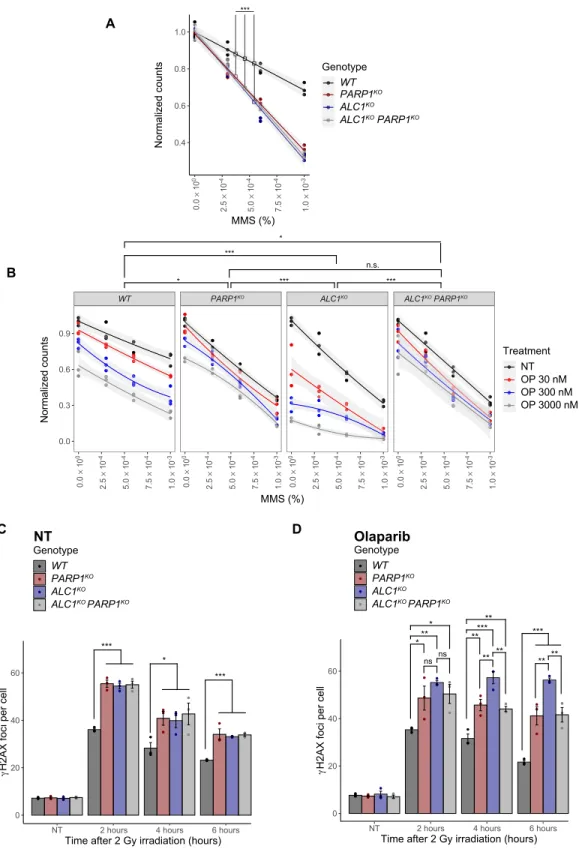
Fig. 3. The hypersensitivity of ALC1-deficient cells to DNA-damaging agents is enhanced by PARP1 inhibition. (A) Clonogenic cell survival assay of ALC1KO and/or PARP1KO U2OS cells. DNA damage was induced by MMS for 1 hour. (B) Clonogenic cell survival assay of ALC1KO and/or PARP1KO U2OS cells. PARP1 inhibition was induced by olaparib (OP) treatment for 24 hours, and then DNA damage was induced by MMS for 1 hour. NT, non-treated. (C and D) Quantification of H2AX foci in ALC1KO and/or PARP1KO U2OS cells. Where indicated, cells were treated with 1 M olaparib for 1 hour before DNA damage induction with x-ray irradiation (2 Gy). H2AX foci were count- ed at different time points after irradiation. NT, non-treated. Graphs in (A) and (B) include all data points (n = 3) and fitted curves with 95% confidence intervals (gray shading). Asterisks indicate P values obtained by linear or polynomial regression, respectively (*P < 0.05, **P < 0.01, and ***P < 0.001). P values in (B) correspond to three- way interaction terms comparing genotypes. Graphs in (C) and (D) include all data points and mean ± SEM (n = 3). Asterisks indicate P values obtained by linear regression fitted independently for each time point. Statistical summary is provided in table S2.
on January 21, 2021http://advances.sciencemag.org/Downloaded from
Sensitivity of ALC1-deficient cells to genomic stress is increased in the presence of PARPis
In a context of active PARylation signaling, both PARP1 and ALC1 are important for efficient DNA repair because cells lacking PARP1 or ALC1 both display similar hypersensitivity to various DNA damage stress (Fig. 3A and fig. S3, A to C). Noteworthy, knocking down or knocking out PARP1 in ALC1-deficient cells did not increase sensi- tivity to genotoxic stress, suggesting that PARP1 function in DNA repair occurs mainly via ALC1 (Fig. 3A and fig. S3, A to C). These findings are in contrast to cell sensitivity obtained in the presence of PARPi. ALC1KO cells show marked hypersensitivity to olaparib and were further sensitized by methyl methanesulfonate (MMS) treat- ment. WT, PARP1KO, and ALC1KO PARP1KO double knockout cells have similar sensitivity to MMS in response to olaparib (Fig. 3B).
We also examined H2AX foci formation after x-ray irradiation as a measure of DNA damage repair efficacy. In the absence of PARPi treatment, PARP1KO, ALC1KO, and ALC1KO PARP1KO double knock- out cells all showed similarly elevated H2AX foci formed at each time point examined after x-ray irradiation, consistent with compa- rable defective repair in these knockout lines (Fig. 3C). Treatment with olaparib resulted in an elevated number of H2AX foci in ALC1KO cells for prolonged periods of time after x-ray irradiation as compared to WT, PARP1KO and ALC1KO PARP1KO double knock- out cells (Fig. 3D). Consistent with the increase in H2AX signal, ALC1KO treated with PARPi also displayed increased comet tail mo- ment length (fig. S3D). These different results—showing that the acute repair defects displayed by ALC1KO cells in the presence of PARPi require the presence of PARP1—suggest a role of ALC1 in the regulation of PARP1 trapping at DNA lesions, which was shown to be the major source of PARPi-dependent cytotoxicity (4).
ALC1 deficiency increases olaparib-induced PARP1 trapping To gain insight into the molecular mechanisms underlying the ALC1-dependent modulation of PARPi sensitivity, we analyzed the characteristics of PARP1 binding to chromatin. First, we quantified the fraction of chromatin-bound mCherry-PARP1 after detergent preextraction in the absence of exogenous damage. In comparison to controls, cells depleted of ALC1 displayed an elevated fraction of chromatin-bound PARP1 upon olaparib treatment (fig. S4A). Similar behavior was observed upon depletion of BRCA1, which also displays PARP-dependent hypersensitivity to PARPi treatment (fig. S4A).
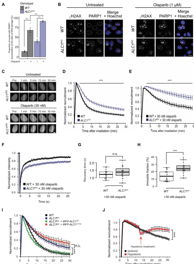
To assess whether the increase in chromatin-bound PARP1 in ALC1KO cells is related to PARP1 trapping at DNA lesions, we mea- sured the fraction of endogenous PARP1 bound to sites of DNA damage 30 min after UV micropore irradiation (Fig. 4, A and B) or to sites of x-ray–induced DNA damage (fig. S4B). While, as expect- ed, PARP inhibition increased PARP1 trapped at DNA lesions, we found more PARP1 trapped in ALC1KO cells as compared to WT controls regardless of the presence of PARPis (Fig. 4, A and B, and fig. S4B). We measured similar trapped PARP1 fraction in the BRCA2-deficient cell line after olaparib treatment upon DNA damage induction (fig. S4C).
Next, to test whether these differences in PARP trapping are consecutive to a role of ALC1 in the mobilization of PARP1 from the sites of DNA damage, we examined in living cells the kinetics of accumulation to sites of laser microirradiation of GFP-PARP1, whose efficient recruitment to damage relies on intact DNA bind- ing domain [fig. S4D and (34)]. In the presence of active PAR sig- naling, the progressive release of PARP1 from DNA lesions was
slower in ALC1KO compared to control cells (Fig. 4, C and D).
Upon olaparib treatment, PARP1 release was markedly slowed in WT cells, and this release was again slower in ALC1-deficient cells (Fig. 4, C and E). In addition, we analyzed the dynamics of PARP1 turnover at sites of laser microirradiation in PARPi-treated cells by fluorescence recovery after photobleaching (FRAP) (Fig. 4F).
The recovery curves revealed the presence of two populations of PARP1 molecules: a fast-exchanging population characterized by a recovery time of about 1 s and a more stably bound fraction that can be considered as immobile within the analyzed time frame. While the recovery time of the mobile PARP1 population was comparable in WT and ALC1KO cells (Fig. 4G), the immobile fraction, which likely represents trapped PARP1 molecules, was higher in ALC1-defi- cient cells (Fig. 4H). In addition, we measured the levels of PAR at sites of laser microirradiation by following the recruitment of the PAR-binding macrodomain of macroH2A1.1 and found that, while the peak PARylation levels in WT and ALC1KO cells are similar, PAR levels remain elevated for a longer time in ALC1KO cells. Moreover, ALC1KO cells show slightly elevated levels of PARylation in the pres- ence of PARPi. Both situations are consistent with a slower release of PARylated PARP1 from sites of DNA lesions (fig. S4E).
ALC1 is activated when it binds PAR with its macrodomain (19, 35, 36), these PAR chains being present on PARP1 itself but also on histones located nearby the damage sites (37, 38). Conse- quently, the contribution of ALC1 to PARP1 mobilization could be due to either a remodeling of the autoPARylated PARP1 proteins by the ALC1 remodelers that are directly bound it or a more distal impact of the activity of ALC1 molecules recruited along the chromatin fiber.
To investigate these two hypotheses, we analyzed the mobilization of the PARP1-E988K mutant, which is only capable of producing mono–ADP-ribose on which ALC1 is unable to bind (36), from DNA lesions in WT and ALC1KO cells. To make such experiment conclusive, we first had to test whether PARP1-E988K could be PARylated in trans by WT PARP1, which could provide a binding site for ALC1. We used the PAR3H assay we previously developed (39) to assess the PARylated PARP1 binding to the LacI-anchored macrodomain of macroH2A1.1 enriched at a genome-integrated LacO-array. While WT PARP1 released from the DNA lesions readily enriches at the anchored macrodomain upon DNA damage (fig. S4, F and G), the E988K mutant PARP1, which shows dynamic turnover at the break (fig. S4H), does not accumulate at the anchored macrodomain, indicating that PARP1 E988K is not PARylated in trans by WT PARP1. Consequently, our observation that the PARP1 E988K mutant is mobilized slower in the absence of ALC1 (Fig. 4I) reveals that ALC1 can participate in the removal of PARP1 without directly binding to it. Moreover, this mobilization of PARP1 E988K requires the ATPase activity of ALC1 because the expression of WT ALC1, but not that of an ATPase-deficient mutant, in ALC1KO cells efficiently mobilized PARP1 E988K from the lesions.
One hypothesis that could explain how ALC1 indirectly mobilizes non-PARylated PARP1 is that the rapid chromatin relaxation induced by ALC1 activity at DNA lesions (5) facilitates PARP1 release. To investigate this possibility, we analyzed the impact on PARP1 mobi- lization of rescuing the defect in chromatin relaxation observed in the absence of ALC1 by bathing the cells with hypotonic medium, which is known to induce global chromatin opening (40), after damage induction. However, relaxing chromatin by hypotonic treat- ment did not accelerate PARP1 release but instead increased the amount of PARP1 accumulating at the break sites (Fig. 4J). This
on January 21, 2021http://advances.sciencemag.org/Downloaded from
B
Merge + Hoechst PARP1
WTWTALC1KO ALC1KO
Untreated Olaparib (1 µM)
γH2AX Merge
+ Hoechst PARP1
γH2AX A
Pre 1 min 5 min 15 min 30 min
0 5 10 15 20 25 30
WT + 30 nM olaparib ALC1KO + 30 nM olaparib
Normalized recruitment
Time after irradiation (min) 0.0
0.2 0.4 0.6 0.8 1.0
0 5 10 15 20 25 30
0.0 0.2 0.4 0.6 0.8 1.0
Normalized recruitment
Time after irradiation (min) WT
ALC1KO
1.0 0.8 0.6 0.4 0.2 0.0
0 5 10 15 20 25
WT + 30 nM olaparib ALC1KO + 30 nM olaparib
Time (s)
Normalized intensity
Pre 1 min 5 min 15 min 30 min
0 5 10 15 20 25 30
0 0.2 0.4 0.6 0.8 1.0
Time after irradiation (min)
Normalized recruitment
Hypotonic treatment Isotonic
Hypotonic
C D E
F G
I J
Olaparib (30 nM) Untreated
ALC1KOWTALC1KOWT
ALC1KO WT ALC1KO ALC1KO
+ iRFP-ALC1E175Q + iRFP-ALC1WT
0 5 10 15 20 25 30
0 0.2 0.4 0.6 0.8 1.0
Time after irradiation (min)
Normalized recruitment
H
Immobile fraction (%) 0 10 20 30 40
ALC1KO WT
+30 nM olaparib 1.0
1.5 2.0
ALC1KO WT
+30 nM olaparib
Recovery time (s) ***
***
n.s.
*** ***
n.s. ***
Genotype WT ALC1KO
Proportion of nuclei with PARP1 accumulation at the sites of DNA damage (%)
Olaparib - + - +
100 75 50 25 0
*** ******
Fig. 4. ALC1 deficiency increases olaparib-induced PARP1 trapping. (A) PARP1 at UV-induced DNA damage sites was quantified in WT and ALC1KO cells treated or not with 1 M olaparib. (B) Representative images from (A). Scale bar, 10 m. (C) Representative images showing GFP-PARP1 accumulation at sites of laser-induced DNA damage in WT and ALC1KO cells treated or not with 30 nM olaparib. Scale bar, 5 m. (D and E) Quantified accumulation of GFP-PARP1 at DNA damage in WT or ALC1KO cells untreated (D) or treated with 30 nM olaparib (E). (F) Normalized FRAP curves of GFP-PARP1 at sites of DNA damage 30 min after irradiation in WT and ALC1KO cells.
(G) Recovery time and (H) the immobile fraction of GFP-PARP1 at the break were calculated from the FRAP curves. (I) Quantified accumulation of GFP-PARP1 E988K at DNA damage in U2OS WT or ALC1KO cells expressing iRFP670-ALC1 variants. (J) Quantified accumulation of mCherry-PARP1 at DNA damage in ALC1KO cells ± hypotonic treatment. Graphs include all data points and mean ± SEM. Asterisks in (A) indicate P values obtained by linear regression (***P < 0.001). Model summary is provided in table S2. P values for (D) to (J) were obtained using an unpaired Student’s t test with Bonferroni correction (***P < 0.001).
on January 21, 2021http://advances.sciencemag.org/Downloaded from
result, which is consistent with our previous observation that chro- matin relaxation facilitates the binding of DNA binding proteins to DNA lesions (41), makes our initial hypothesis regarding the con- tribution of chromatin relaxation to PARP1 release unlikely and, thus, rather call for an alternative model in which PARP1 is “peeled off” from DNA through nucleosome sliding by ALC1 anchored on PARylated nucleosomes. Together, our results suggest that ALC1 indirectly promotes the removal of even inactive, non-PARylated PARP1 from DNA lesions through anchoring to PARylated chro- matin, to avoid deleterious consequences associated with PARP trapping.
In the absence of ALC1, neither HR nor NHEJ is able to efficiently resolve PARP1-DNA adducts
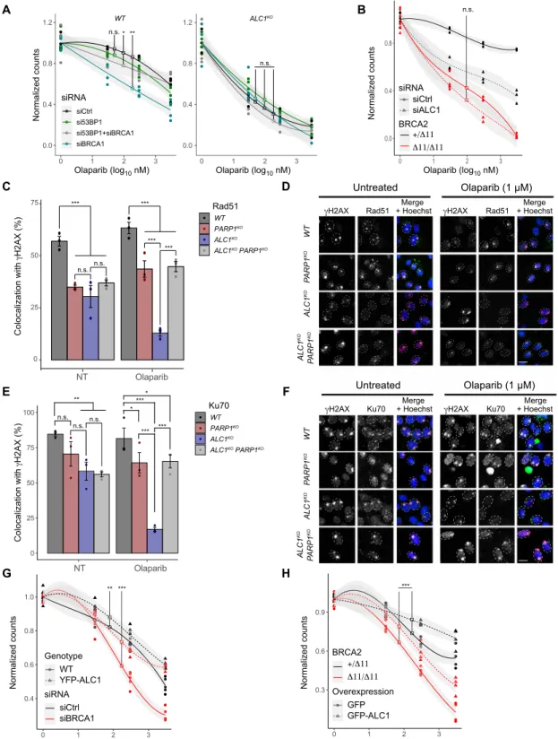
Next, we sought to determine how PARP trapping in ALC1KO cells affected DNA repair pathways. In many cases, sensitivity to PARPi arises from an imbalanced DSB pathway choice. In HR-deficient cells, PARPi toxicity is due to the activation of the error-prone NHEJ pathway for resolving PARP1-DNA adducts (42). Conversely, PARPi resistance upon loss of 53BP1 in BRCA1-deficient cells is the consequence of the reactivation of HR, which is able to faithfully repair the DNA lesions associated with PARP1-trapping (12). To gain more insight into the mechanism of the ALC1-synthetic lethal phenotype, we compared the impact of RNAi-mediated BRCA1 and 53BP1 depletion on olaparib treatment in WT and ALC1KO cells. In line with previous reports, olaparib is toxic in BRCA1-depleted WT cells, and the codepletion of 53BP1 and BRCA1 rescues the synthetic lethality with olaparib treatment (43). In contrast, the synthetic lethal phenotype seen in ALC1KO cells upon PARP1 inhibition re- mained unchanged after down-regulation of BRCA1 and/or 53BP1 (Fig. 5A and fig. S5A). In addition, we found that ALC1 depletion does not increase the sensitivity of cells suffering from BRCA2 defi- ciency to olaparib treatment (Fig 5B and fig. S5B). These observations suggest that ALC1 has to handle the PARP1-DNA adducts before the DSB repair pathway choice to allow the resolution of these lesions by either HR or NHEJ.
To more directly assess the impact of ALC1 on HR and NHEJ, we used two previously described reporter systems (fig. S5, C and D) (44). First, I-SceI–induced breaks repaired by HR in pGC-HeLa cells will restore a GFP cassette, allowing GFP fluorescence to act as a readout for HR efficiency (fig. S5C). Second, in the pEJ-HeLa reporter assay, I-SceI–induced breaks repaired by NHEJ will allow expression of a GFP cassette, providing a measure for NHEJ effi- ciency (fig. S5D). In agreement with previous reports, we verified that down-regulation of the HR factor BRCA1 decreased while down-regulation of the NHEJ factor Ku70 increased HR efficiency in the pGC-HeLa cells and that they had the opposite effect in the pEJ-HeLa reporter line (fig. S5, E to G). The down-regulation of ALC1 resulted in reduced efficiency of HR and NHEJ; however, the latter was not statistically significant, albeit in the absence of PARPi (fig. S5, E to G).
To study the effect of ALC1 deficiency in combination with PARPi on HR and NHEJ, we studied the recruitment of Ku70, an actor of NHEJ, as well as Mre11, Rad51, and phosphorylated replication protein A (pRPA), which are specific to HR, to sites of DNA damage induced by micropore UV irradiation. In the absence of PARPi, Rad51, Ku70, Mre11, and pRPA showed reduced accumulation to the H2AX-labeled DNA damage sites in both ALC1KO and PARP1KO as well as the double knockout cells as compared to WT cells
(Fig. 5, C to F, and fig. S5, H to K). Upon olaparib treatment, the accumulation of Rad51, Ku70, pRPA, and Mre11 was reduced even further at the sites of DNA damage in ALC1KO as compared to other cell lines, including the ALC1KO PARP1KO double knockout cells.
Together, these results show that ALC1 is important for mobilizing PARP1 and that, in its absence, trapped inhibited PARP1 interferes with the binding of downstream HR and NHEJ repair factors to the sites of DNA damage.
ALC1 overexpression reduces the PARPi sensitivity of BRCA-deficient cells
ALC1 is an oncogene frequently overexpressed in cancer correlating with poor prognosis for patient survival (21–23). While the loss of ALC1 sensitizes cells to DNA-damaging agents and PARPi treat- ment (Figs. 1D and 3 and fig. S1, F and G), we aimed to address whether ALC1 overexpression would have the opposite effect. We found that ALC1 overexpression reduced sensitivity to various DNA-damaging agents (fig. S5, L to N) in agreement with previous studies (22, 23) and increased the frequency of both HR and NHEJ in the reporter cell lines (fig. S5, O and P). Last, overexpression of ALC1 reduced the sensitivity to olaparib of WT and BRCA-deficient cells (Fig. 5, G to H, and fig. S5, Q and R). Notably, BRCA-deficient cells overexpressing ALC1 are almost as sensitive as WT cells to olaparib. This suggests that an increase of ALC1 expression in BRCA-deficient tumors would tend to reduce or even suppress the therapeutic window in which PARPi could be used to efficiently kill BRCA-deficient tumor cells while sparing their healthy counterpart.
DISCUSSION
Synthetic lethal interactions between PARPis and BRCA-deficient tumors have been a focus for cancer therapies for a number of years (6–8, 45, 46). In addition to specific mutations in the BRCA1/2 genes, this therapeutic strategy has been extended to tumors show- ing defects in the HR pathway that phenocopy the loss of BRCA, a phenotype often referred to as BRCAness (47). In the current study, we identify the PAR-dependent chromatin remodeler ALC1 as a key modulator of sensitivity to the PARPi olaparib and show that loss of ALC1 impairs both the HR and NHEJ repair pathways. Furthermore, the hypersensitivity to PARPi treatment observed in ALC1KO cells is not rescued by down-regulating 53BP1, in contrast to what is re- ported for BRCA-deficient cells (43). These findings show that ALC1 deficiency does not display the classical BRCAness signature and that the sensitivity of ALC1KO cells to PARPi arises from defects at the very early stage of the DNA damage response, before the pathway choice between HR and NHEJ.
Our results demonstrate that the chromatin remodeler ALC1 is involved in the timely mobilization of PARP1 from the DNA lesions.
In the context of active PARylation signaling, the loss of ALC1 already affects PARP1 removal from the lesions, but alternative mechanisms such as PARP1 autoPARylation are sufficient to com- pensate for this defect, allowing the cell to proceed to DNA repair.
In contrast, as soon as PARylation is impaired, ALC1 becomes essential for the mobilization of PARP1 from the sites of damage.
Failure to achieve efficient PARP1 removal interferes with down- stream steps in the DNA repair pathway, ultimately leading to cell death (Fig. 6). Our results reveal that the processivity of ALC1 along the chromatin fiber is able to peel PARP1 off the sites of damage. It should be noted that HPF1 deficiency—which abolishes the PARyl ation
on January 21, 2021http://advances.sciencemag.org/Downloaded from
C
ALC1KOWTPARP1KO
Ku70 Merge + Hoechst
γH2AX Ku70 Merge
+ Hoechst γH2AX
E
H F
G A
D
0.3 0.6 0.9
0 1 2 3
Olaparib (log10 nM)
Normalized counts
BRCA2
Overexpression GFPGFP-ALC1 0.4
0.6 0.8 1.0
0 1 2 3
Olaparib (log10 nM)
Normalized counts
siRNA siCtrl siBRCA1 Genotype
WTYFP-ALC1 0
25 50 75 100
NT
Colocalization with γH2AX (%)
Ku70 WT PARP1KO ALC1KO ALC1KO PARP1KO
Olaparib
0 1 2 3 0 1 2 3
0.0 0.4 0.8 1.2
Olaparib (log10 nM)
Normalized counts
siRNA siCtrl si53BP1 si53BP1+siBRCA1
siBRCA1 0.0
0.4 0.8
0 1 2 3
Olaparib (log10 nM)
Normalized counts
siRNA siCtrl siALC1 BRCA2
+/∆11
∆11/∆11
+/∆11
∆11/∆11
B
NT
Colocalization with γH2AX (%)
Rad51 WT PARP1KO ALC1KO ALC1KO PARP1KO
Olaparib
Rad51 Merge + Hoechst
γH2AX Rad51 Merge
+ Hoechst γH2AX
PARP1KOALC1KOWTPARP1KOALC1KO
Untreated Olaparib (1 µM)
Untreated Olaparib (1 µM)
PARP1KOALC1KO
25 50 75
0
n.s. n.s.
*** ***
*** ***
ALC1KO WT
0.0 0.4 0.8 1.2 n.s.***
n.s.
Olaparib (log10 nM)
n.s. n.s.
**
n.s.
n.s.
*** ***
*** ***
***
*
*
**
Fig. 5. ALC1 acts upstream of DSB repair pathway choice. (A) Clonogenic cell survival assay of WT and ALC1KO U2OS cells transfected with siCtrl or siBRCA1 and/or si53BP1 treated or not with olaparib for 24 hours. (B) Clonogenic cell survival assay of DLD1-BRCA2+/11 (BRCA2+/−) and DLD1-BRCA211/11 (BRCA2−/−) cells transfected with siCtrl or siALC1 and treated or not with olaparib for 24 hours. (C to F) Quantification and representative images of Rad51 or Ku70 localization to UV-induced DNA damage sites in WT, PARP1KO, ALC1KO, and PARP1KO ALC1KO double knockout cells, treated or not with olaparib (1 M). Scale bar, 10 m. (G) Clonogenic cell survival assay of WT and YFP-ALC1 overexpressing U2OS cells transfected with siCtrl or siBRCA1 and treated or not with olaparib for 24 hours. (H) Clonogenic cell survival assay of DLD1- BRCA2+/11 (BRCA2+/−) and DLD1-BRCA211/11 (BRCA2−/−) cells transfected with GFP-ALC1 and treated or not with olaparib for 24 hours. Graphs in (A), (B), (G), and (H) include all data points (n = 3) and fitted curves with 95% confidence intervals (gray shading). Graphs in (C) and (E) include all data points and mean ± SEM (n = 3). Asterisks indicate P values obtained by polynomial (A, B, G, and H) or linear regression (C and E) (*P < 0.05, **P < 0.01, and ***P < 0.001). Model summary is provided in table S2.
on January 21, 2021http://advances.sciencemag.org/Downloaded from
of histones—also leads to PARPi sensitivity as well as increased PARP1 trapping (14, 38, 48), which is consistent with ALC1 acting indirectly on PARP1 mobility through chromatin PARylation and subsequent nucleosome sliding.
ALC1 overexpression is a common trait of multiple tumors, often associated with poor prognosis (23). According to our findings, the risks are high that such tumors would display low responsiveness to PARPis, thus strongly arguing for a systematic analysis of the ALC1 expression level before the use of PARPi-driven cancer therapies.
Noteworthy, the region 1q21 within chromosome 1, where the ALC1 gene is located, is found amplified in many cancers including breast tumors (49) and is associated with resistance to chemotherapy treatment in ovarian cancers (50). Moreover, ALC1 was found overexpressed in ovarian carcinoma metastasis (51), a feature that is associated with shorter patient survival. This, together with our results, suggests that, in addition to a potential role as a predictive biomarker, there is also a call for the development of new therapeu- tic agents against ALC1. A first compound targeting this remodeler and showing some therapeutic potential against colorectal cancer has been developed very recently (52). On the basis of our data, this new agent should synergize the cytotoxic potential of the currently available PARPis.
MATERIALS AND METHODS Cell lines and cell culture
All cells used here were cultured in Dulbecco’s modified Eagle’s medium (DMEM; Sigma-Aldrich) or RPMI supplemented with
10% fetal bovine serum (FBS), penicillin (100 g/ml), streptomycin (100 U/ml), and 1% nonessential amino acid and maintained at 37°C in a 5% CO2 incubator unless otherwise stated. DLD1-BRCA2+/11 (BRCA2+/−), DLD1-BRCA211/11 (BRCA2−/−) (53), U2OS YFP- ALC1, U2OS PARP1KO, and U2OS ALC1KO #1 were generated pre- viously (5). Additional U2OS ALC1KO cells were generated using CRISPR-Cas9 technology as previously described (5, 54) using WT U2OS cells as the parental cell line. U2OS ALC1KO PARP1KO double knockout cells were generated by knocking out ALC1 in PARP1KO U2OS cells. The sgRNA sequences used for targeting ALC1 (table S3) were designed using an online tool (55). HeLa PARP1-mCherry stable cells were generated by transfecting HeLa cells with PARP1- mCherry (56) and growing them in culture media supplemented with Geneticin (500 g/ml) for 2 weeks. HeLa pGC and HeLa pEJ reporter cells were a gift from W. Mansour (44) and were cultured in DMEM with 10% FBS, penicillin (100 g/ml), streptomycin (100 U/ml), puromycin (600 g/ml), and G418 sulfate (800 g/ml).
RNAi and plasmid transfection
pYFP-macrodomain of macroH2A1.1 (56); pLacI-GFP trap, piRFP670- ALC1, and piRFP670-ALC1 E175Q (39); pPARP1-GFP, pPARP1- R34E-GFP, and pPARP1-R138E-GFP (34); pmEGFP-ALC1 and pmCherry-ALC1 G750E (36); and pmCherry-ALC1, pmCherry- ALC1 K77R, pmCherry-ALC1 E175Q, photoactivatable GFP (PAGFP)–H2B, and photoactivatable TagRFP (PATagRFP)–H2B (5) were previously described. PARP1 complementary DNA was ampli- fied from PARP1-mCherry and PARP1 E988K-mcherry (primers in table S3) (56) and ligated into pmEGFP-C1, pDendra2-C1, or pmCherry-C1 between Bgl II and Xma I. Cells were transfected with plasmids using X-tremeGENE HP (Roche) or Xfect (Takara Bio) according to the manufacturer’s instructions.
Transfection of cell lines with specific small interfering RNAs (siRNAs) (table S4) was carried out using DharmaFECT (Dharmacon) transfection reagent according to the manufacturer’s instructions.
Experiments were performed 48 hours after siRNA transfection.
The down-regulation of the indicated genes was verified by Western blot using specific antibodies, which are detailed in table S5.
Genome-wide CRISPR screen
A CRISPR-Cas9 genome-wide knockout screen was performed using the GeCKOv2 system as previously described (16, 17). GeCKOv2 human CRISPR knockout library was amplified as described using New England Biolabs 5-alpha Electrocompetent Escherichia coli.
For lentiviral production, 293T cells were transfected with amplified GeCKOv2 plasmid DNA, psPAX2 (a gift from D. Trono; Addgene, plasmid #12260), and pLP-eco env, a mouse Lentivirus envelope packaging vector (a gift from G. Schotta) using Xfect transfection reagent (Takara) according to the manufacturer’s instructions.
Supernatant was collected 48 hours after transfection, filtered through a 0.45-m Steriflip filter unit, and stored at −80°C. Virus titer [multi- plicity of infection (MOI)] was calculated as previously described (16).
For the screen, HeLa cells stably expressing mCAT1 (mouse High affinity cationic amino acid transporter 1) (57) were transduced with the GeCKOv2 lentiviral library at an MOI of 0.3 and selected with puromycin (0.3 g/ml) for 7 days as previously described (16). After puromycin selection, cells were split into five replicates of 2 × 10⁷ cells. Two replicates were cultured with the addition of DMSO, and two replicates were cultured in the presence of 3 M olaparib for 14 days. One replicate was immediately collected as an
PARP1
Ku70
DNA repair
PARP1
PARPi
PARP1
Ku70 Rad51
Rad51 PARP1
ALC1
PARP1 PARP1
ALC1 ALC1
ALC1
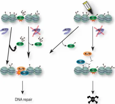
Fig. 6. A model for the role of ALC1 in olaparib-mediated synthetic lethality.
PARP1 is recruited to sites of DNA damage where it PARylates proteins in and around the break site, including itself. PARP1 removal from sites of damage in- volves a combination of autoPARylation and ALC1-dependent mobilization, which allows the recruitment of essential subsequent repair actors such as Ku70 or Rad51.
While the impairment of either of the two modes of PARP1 mobilization does not have major deleterious consequences, inhibiting autoPARylation in ALC1-defi- cient cells fully blocks PARP1 release from DNA lesions, thus preventing the recruit- ment of downstream repair factors and ultimately leading to cell death.
on January 21, 2021http://advances.sciencemag.org/Downloaded from