Contents lists available atScienceDirect
Redox Biology
journal homepage:www.elsevier.com/locate/redox
Research paper
Peroxidasin-mediated crosslinking of collagen IV is independent of NADPH oxidases
Gábor Sirokmány
a,b, Hajnal A. Kovács
a,b, Enik ő Lázár
a,b, Krisztina Kónya
a,b, Ágnes Donkó
a,b, Balázs Enyedi
a, Helmut Grasberger
c, Miklós Geiszt
a,b,⁎aDepartment of Physiology, Faculty of Medicine, Semmelweis University, PO Box 259, H-1444 Budapest, Hungary
b“Momentum”Peroxidase Enzyme Research Group of the Semmelweis University and the Hungarian Academy of Sciences, Budapest, Hungary
cDivision of Gastroenterology, Department of Internal Medicine, University of Michigan Medical School, Ann Arbor, MI, USA
A R T I C L E I N F O Keywords:
Peroxidasin NADPH oxidase Hydrogen peroxide Collagen IV Sulfilimine
A B S T R A C T
Collagen IV is a major component of the basement membrane in epithelial tissues. The NC1 domains of collagen IV protomers are covalently linked together through sulfilimine bonds, the formation of which is catalyzed by peroxidasin. Although hydrogen peroxide is essential for this reaction, the exact source of the oxidant remains elusive. Members of the NOX/DUOX NADPH oxidase family are specifically devoted to the production of su- peroxide and hydrogen peroxide. Our aim in this study was tofind out if NADPH oxidases contributein vivoto the formation of collagen IV sulfilimine crosslinks. We used multiple genetically modifiedin vivomodel systems to provide a detailed assessment of this question. Our data indicate that in various peroxidasin-expressing tissues sulfilimine crosslinks between the NC1 domains of collagen IV can be readily detected in the absence of func- tioning NADPH oxidases. We also analyzed how subatmospheric oxygen levels influence the collagen IV network in collagen-producing cultured cells with rapid matrix turnover. We showed that collagen IV crosslinks remain intact even under strongly hypoxic conditions. Our hypothesis is that during collagen IV network formation PXDN cooperates with a NOX/DUOX-independent H2O2source that is functional also at very low ambient oxygen levels.
1. Introduction
Basement membranes are extracellular structures that are essential in organizing individual cells into tissues and organs [39]. Besides providing mechanical support for different cell types, including epi- thelial and endothelial cells, basement membranes are also important in cellular signaling. An essential core component of basement membranes is collagen IV, which is organized into a sheet-like network through covalent binding between individual collagen IV triplexes [14]. Sulfi- limine bond is a unique type of chemical link which is so far only found in collagen IV molecules in living organisms[34]. The formation of this recently identified chemical bond is catalyzed by peroxidasin (PXDN), a member of the animal heme peroxidase family[4,26].
First, in the presence of hydrogen peroxide (H2O2), PXDN turns bromide into hypobromous acid (HOBr), which then reacts with a methionine in the NC1 domain of collagen IV forming a methionine halosulfonium intermediate[4,22,28]. Finally, this intermediate reacts with a hydroxylysine on the opposing protomers’NC1 domain to create
the sulfilimine crosslink between neighboring protomers.
While bromine is a ubiquitously accessible trace element in animal tissues, the exact source of H2O2has not been identified yet. There are numerous metabolic and signaling pathways the activity of which are accompanied by the generation of reactive oxygen species (ROS) [35,37]. There are also some extracellular enzymes which are also known to produce H2O2while accomplishing their catalytic functions (e.g. lysyl oxidases and their homologs). The family of NADPH oxidases comprises enzymes that are specifically devoted to the generation of superoxide anion and H2O2 [2,31]. There are seven members of the NOX/DUOX family of NADPH oxidases, including NOX1–5, DUOX1, and DUOX2. NOX/DUOX enzymes show unique expression pattern and biochemical regulation[2,6]. When exerting its pleiotropic effects H2O2
can directly react with and modify target molecules. Some actions of H2O2, however, are mediated through animal heme peroxidases, which use H2O2to oxidize substrates in a diverse array of reactions[9]. The group of mammalian heme peroxidases includes myeloperoxidase (MPO), eosinophil peroxidase (EPX), lactoperoxidase (LPO),
https://doi.org/10.1016/j.redox.2018.03.009 Received 7 March 2018; Accepted 13 March 2018
⁎Corresponding author at: Department of Physiology, Semmelweis University, Faculty of Medicine, PO Box 259, H-1444 Budapest, Hungary.
E-mail address:geiszt.miklos@med.semmelweis-univ.hu(M. Geiszt).
Abbreviations:col4a, collagen type IV alpha; DUOX, Dual oxidase; DUOXA, Dual oxidase Activator; NC1, non-collagenous; HIF-1alpha, Hypoxia-inducible Factor 1-alpha, Nox: NADPH oxidase; PHG, phloroglucinol; PXDN, peroxidasin
Available online 14 March 2018
2213-2317/ © 2018 Published by Elsevier B.V. This is an open access article under the CC BY-NC-ND license (http://creativecommons.org/licenses/BY-NC-ND/4.0/).
T
thyroperoxidase (TPO) and peroxidasin (PXDN)[9,32]. Peroxidases are often expressed in cells and tissues, where NOX/DUOX NADPH oxidases can also be found and there are a number of examples for the co- operation between NADPH oxidases and heme peroxidases. For ex- ample, in neutrophil granulocytes, MPO uses H2O2 produced by the NOX2 complex to oxidize chloride into hypochlorous acid, which is a potent antimicrobial compound[15]. NOX2 is also highly expressed in eosinophils, where EPX utilizes its product for the oxidation of bromide and thiocyanate [36]. The oxidation of iodide in the thyroid is also dependent on the cooperation between a peroxidase and an NADPH oxidase, namely TPO and DUOX2. While DUOXes were originally considered to be specific for the thyroid, subsequent research has un- covered they role in antimicrobial host defense where they supply H2O2
for lactoperoxidase, an enzyme expressed in different exocrine glands, including the salivary gland and submucosal glands of large airways [12].
For a complete understanding of collagen IV role in basement membrane biosynthesis, it would be crucial to determine the bio- chemical and mechanical importance of sulfilimine crosslinks. An early step towards this goal could be the characterization of the H2O2source for PXDN-mediated crosslinks. Our primary goal in this study was to find out if members of the NOX/DUOX family participate in the for- mation of collagen IV sulfilimine crosslinks in mammalian tissues. Our results suggest that NADPH oxidases do not support collagen IV cross- linking and the molecular identity of the H2O2source in collagen IV synthesis remains to be identified.
2. Materials and methods 2.1. Reagents
Collagenase type I was ordered from Worthington Biochemical Corporation. Isoform-specific rat monoclonal col4a1, col4a5 NC1 anti- bodies were purchased from Chondrex Inc. We used rabbit HIF1alpha antibody from Abcam (ab179483). The anti-actin antibody, phlor- oglucinol, diphenyliodonium were all purchased from Sigma. PFHR-9, a mouse embryonic carcinoma cell line was obtained from ATCC. For transfecting PFHR-9 cells, we used TurboFect from Thermo Scientific.
AlamarBlue and calcein-AM were ordered from ThermoFisher Scientific.
Rabbit polyclonal anti-Nox4 antibody was a generous gift from Professor Ajay Shah (King's College, London) and polyclonal anti-Duox1 antibody was a kind gift from Dr. Xavier De Deken (Université Libre de Bruxelles).
2.2. Cell culture
PFHR-9 cells (obtained from ATCC) were grown in Dulbecco's Modified Eagles Medium with glutamine and 4,5 g/L glucose, supple- mented with 50 U/ml penicillin and 50 µg/ml streptomycin (Lonza Group Ltd., Basel, Switzerland) and 10% fetal calf serum (Biowest SAS, France, S182P-500, French origin). Cells were grown in a humidified incubator with 5% CO2in the air, at 37 °C.
2.3. PFHR-9 peroxidasin CRISPR
PFHR-9 cells were genetically mutated for peroxidasin, using a pSpCas9(BB)−2A-GFP (PX458) vector. The vector contained the 5′- caggcccgcttctctga-3′guide sequence which is identical to the 408–424 bases of the 17th exon of the mousePxdngene. The cells were trans- fected with Turbofect and GFP positive cells were sorted onto 96 well plates. Cell clones were screened by PCR of genomic DNA using 5′- acaactgctcagacatgtgc-3′sense and 5′-acacattggaggcatcgatg-3′antisense oligos. PCR products were analyzed by Surveyor mismatch analysis, then subcloned into a pcDNA cloning vector and sent for sequencing.
One cell line was selected which contained frameshift mutations on
both alleles. We confirmed by Western blot analysis that the selected cell line does not express PXDN and is not able to crosslink collagen IV NC1 domains.
2.4. Mouse strains used in the study
PXDN-deficient mice were generated by SAGE Laboratories using Compo Zr zinc finger endonuclease technology [18]. Detailed char- acterization of PXDN-deficient animals is subject to a different paper.
The knockout line contains a 2 bp deletion at the beginning of the peroxidase domain coding region.
Duox1 knockout mice were purchased from Lexicon Pharmaceuticals, Inc. (TheWoodlands, TX, USA) and described earlier by Donkóet al.[11].
Thep22phoxmutantnmf333mouse strain[23]was obtained from The Jackson Laboratory.
TheDuoxaknockout mice were developed by Grasberger et al.[13].
TheNox4knock-out animals were generated by the Transcription Activator-Like Effector Nuclease (TALEN) technique using plasmids ordered from Addgene (TALE Toolbox Kit # 1000000019 deposited by Feng Zhang). MouseNox4specific TALEN recognition sites were de- signed for the genomic sequence prior to and to thefirst exon using the 5`-TN19 N14–20 N19A-3` formula, with the following sequences: left TALEN recognition site: 5′TCCCCGCGCCGGCGGCATGG3′, right TALEN recognition site: 5′GCTGGCCAACGAAGGGGTTA3′, and a 19 bp (5′ CGGTGTCCTGGAGGAGCTG3′) FokI nuclease dimerization and cutting sequence in between. MouseNox4 recognizing TALEN plasmids were assembled according to the protocol of Sanjana et al. [29] TALEN mRNA was injected into the pronuclei of one-cell embryos of FVB/NJ mice. Pups were analyzed with Surveyor assay plus sequencing and a founder bearing a 518 bp deletion including the START and 18 bp ad- ditional nucleotide was chosen to establish colony.
Nox4 and Duox1 specific Western blot data of knockout animals are shown inSupplementary Fig. 2. Western blots for p22phoxand Duox1 were already shown in[16]and (11) respectively.
All animal experiments were approved by the Animal Experimentation Review Board of the Semmelweis University.
2.5. Western blots
Laemmli sample buffer was added to the cell lysate samples and these were run on 8–10% SDS polyacrylamide gels and blotted onto nitrocellulose membranes. Membranes were blocked in phosphate buffered saline containing 0,1% Tween- 20% and 5% dry milk. Thefirst antibodies were diluted in phosphate buffered saline containing 0,1%
Tween- 20% and 5% bovine serum albumin and used either for 2 h at room temperature or overnight at 4 °C. After several washing steps in PBS-Tween-20, membranes were incubated with HRP-linked secondary antibodies (Santa Cruz Biotechnology, Inc., USA) diluted in blocking buffer. Antibody binding was detected using enhanced chemilumines- cence and Fuji Super RX medical X-rayfilms.
For the detection of collagen IV NC1 monomers and dimers, cells or small tissue pieces were harvested in hypotonic lysis buffer (10 mM CaCl2, 50 mM Hepes, pH 7.4) completed with 0.1 mM benzamidine hydrochloride, 25 mM 6-aminocaproic acid, 1 mM Phenylmethylsulfonyl fluoride and 0.5 mg/ml type I collagenase at 37 °C for 12–18 h. For the lysis of hypoxia/anoxia treated and control cells, the lysis buffer was supplemented also with 150 µM phlor- oglucinol to inhibit PXDN activity during the lysis process.
2.6. PXDN antibody
Polyclonal mouse anti-PXDN antibody was raised against the full- length mouse PXDN inPxdnknockout mice. The recombinant mouse PXDN-V5 antigen was produced in HEK293-FS cells and purified using monoclonal anti-V5 (Clone V5–10) antibody coupled to agarose beads
(Sigma).Pxdnknockout animals (see above) were injected with Freund adjuvant containing the purified PXDN-V5 antigen.
2.7. Hypoxic treatments
Cells grown on 6-well plates were kept at 37 °C in a Billups- Rothenberg modular hypoxia chamber (Billups-Rothenberg, Inc., Del Mar CA, USA) which was previously ventilated with the appropriate gas mixture (anoxic mixture: 95% N2, 5% CO2;1% O2mixture: 1% O2, 5%
CO2, 94% N2;5% O2mixture: 5% O2, 5% CO2, 90% N2). After 24 h cells from 1 well were lysed in ice-cold RIPA lysis buffer in the presence of protease inhibitors (for HIF1alpha detection) and content of another well was lysed in hypotonic lysis buffer containing collagenase I (see above) for collagen IV NC1 crosslink analysis.
2.8. Cell viability assays
A two-fold serial dilution of PFHR9 cells was plated on black 96- well plates and grown at normoxic or anoxic conditions. After 24 h cell culture medium was replaced with extracellular “H-medium” (con- taining 145 mmol/L NaCl, 5 mmol/L KCl, 1 mmol/L MgCl2, 0.8 mmol/L CaCl2, 5 mmol/L glucose, and 10 mmol/L HEPES). Alamar Blue (1:10 of stock solution) and calcein-AM (2μM) was added to the cells and fluorescent intensities were measured every third minute for 15 min in a POLARstar OPTIMAfluorometer (BMG Labtech) at 37 °C. Excitation/
Emissionfilters were 500/530 and 580/610 for calcein and alamarBlue respectively. The slope of fluorescence increase over time was de- termined for each cell dilution and displayed as the function of relative cell number.
2.9. Catalase constructs
Catalase sequence was cloned into the pmRFP-N1 vector to express RFP-catalase fusion protein in a cytosolic localization. As control, the F153V mutation was introduced to destroy catalase activity of the protein. For targeting catalase to the mitochondrial matrix, we used duplicate of the N-terminal localization signal of human COX8a[27].
3. Results
3.1. Disruption of the Pxdn gene inhibits collagen IV crosslinking in mice
To investigate the enzymes which participate in collagen IV cross- linkingin vivo, wefirst analyzed mice where thePxdngene was mutated by zinc finger endonucleases (ZFN) [18]. Pxdn knockout animals showed the ocular developmental defect and white belly spot, which were earlier described by Yan X.et al. in a different PXDN mutant strain [38]. The detailed characterization of the PXDN-deficient animals is subject of a separate study. Previously, we reported deficient collagen crosslinks in cultured embryonicfibroblasts of these ZFN-modified mice [18]. To analyze the effect of PXDN deficiency onin vivocollagen IV coupling, we prepared collagenase digests from different organs, in- cluding the kidneys and the lungs. There are six different collagen IV isoforms, designated as alpha1 to alpha6[14]. In our experiments, we analyzed the crosslink formation between the col4a1 and col4a5 NC1 domains. The isoform-specific monoclonal antibodies consequently detected the̴54 kD dimeric and̴ ̴28 kD monomeric form of NC1 do- mains in the collagenase-digested tissue lysates (Fig. 1). Importantly, a dramatic difference was observed between the tissue lysates of wild- type andPxdnknockout mice. In the PXDN-deficient samples the ratio of NC1 dimers and monomers was substantially reduced (Fig. 1, Supplementary Fig. 2A). These observations suggested that mammalian PXDN has an indispensable,in vivorole in the generation of collagen IV crosslinks.
3.2. Assessment of the role of NADPH oxidases in collagen IV crosslinking
Animal heme peroxidases and members of the NOX/DUOX family of NADPH oxidases often cooperate in reactions where peroxidases use NOX-derived H2O2to oxidize different substances. Halides and pseu- dohalides are well-recognized substrates of peroxidases [9], thus we wanted to know whether NOX/DUOX enzymes provide H2O2for the reaction where bromide is oxidized into hypobromite by PXDN.
Knockout models are now available for almost all NOX/DUOX isoforms (except NOX5, which is not present in mice)[31], therefore we decided to analyze the collagen IV crosslinking reaction in different NOX/DUOX knockout models. First, we tested whether NOX4 is involved in collagen IV crosslinking. This particular NOX isoform was especially interesting for us because NOX4 is expressed in cell types (including endothelial, epithelial cells and cardiomyocytes) which are in close contact with underlying or surrounding basement membranes[2,5]. To investigate the role of NOX4, we created Nox4knockout animals using TALEN- mediated gene modification. A detailed description of the knockout strategy is described in Materials & Methods.Fig. 2(A-C, second lanes) show that the western blot picture of Nox4-deficient aorta and kidney samples is indistinguishable from that of wild-type collagenase digests (Fig. 2,Supplementary Fig. 2B). In these and subsequent Western blot experiments, we always included PXDN-deficient samples, where di- minished collagen IV crosslinking was detected (Fig. 2A-C, fourth lanes). An important shared feature of NOX1, NOX2, NOX3, and NOX4 is that their activity is dependent on the co-expression of the p22phox protein [1,21]. The nmf333 mouse strain carries a mutation in the p22phoxgene, which leads to the destabilization of the protein with a consequent loss of Nox2 and Nox4 activity[23,40]. Testing collagen IV crosslinking in this strain offered us the possibility to study the possible involvement of four different NOX isoforms simultaneously, although the uniquely limited expression pattern of NOX3 made unlikely that this particular isoform contributes to the formation of sulfilimine lin- kages. As shown inFig. 2(A-C, third lanes) the crosslinking of collagen IV was perfectly normal when tested in different organs ofnmf333 animals. These experiments suggested that a p22phox-dependent NOX isoform is unlikely to provide H2O2for the PXDN-catalyzed reaction.
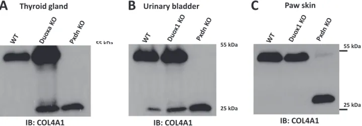
Dual oxidases, DUOX1 and DUOX2 are NADPH oxidase isoforms which operate independently of p22phoxand show prominent expres- sion in epithelial cells [10,20]. We aimed to examine the possible contribution of DUOXes in collagen IV crosslinking. To this end, we analyzed thyroid tissue isolated from mice which are deficient for dual oxidase maturation factor 1 and dual oxidase maturation factor 2 (DUOXA1 and DUOXA2 respectively) [13]. In the absence of DUOX activators, DUOX1 and DUOX2 are not functional and mice without DUOXA proteins show the symptoms of severe hypothyroidism[13].
Collagen IV crosslinking, however, was undisturbed inDuoxaknockouts (Fig. 3A,Supplementary Fig. 2C) suggesting that Duox enzymes do not serve as an H2O2source in the crosslinking reaction. Interestingly, the amount of NC1 monomers and dimers were consequently higher in the double-knockout thyroid samples, possibly explained by a more active basement membrane formation in the TSH-stimulated thyroid glands of knockout animals (Fig. 3A). Corroborating these results, we found that Duox1 knockout animals [11]also showed normal collagen IV NC1 crosslinks in the skin and urinary bladder samples where DUOX1 shows high expression levels in wild-type animals (Fig. 3A, B).
3.3. Determination of the oxygen-dependence of collagen IV crosslinking
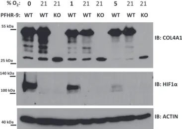
Animal heme peroxidases use H2O2 to oxidize a variety of sub- strates, which in the case of PXDN appears to be bromide. Although H2O2 can be produced through divergent biochemical pathways, oxygen is required for all of these biosynthetic mechanisms. We, therefore, wanted to determine the dependency of collagen IV cross- linking on oxygen concentration. We chose an embryonic cancer cell line, PFHR-9 cells as a model system in these experiments. Using this
particular cell line was justified by previousfindings describing intense collagen IV production and crosslinking in PFHR-9 cells[4]. We also established PXDN-deficient PFHR-9 cells making use of the CRISPR gene editing technique. To determine the crosslinking of collagen IV in wild-type and PXDN-deficient PFHR-9 cells we digested the cells with collagenase I and the amount of NC1 dimers and monomers were
analyzed by western blot using isoform-specific anti-collagen IV anti- bodies (Fig. 4). Next, we measured the formation of NC1 dimers at four different oxygen concentrations, anoxia (0%), 1%, 5% and 21%, re- spectively.
We wanted to reduce the potential toxicity of anoxia, therefore we determined the minimal time which was required to detect the effect of
A B
IB: COL4A1
55 kDa
25 kDa
NC1 dimer
NC1 monomer
kidney lung
55 kDa
25 kDa
kidney lung
IB: COL4A5
Fig. 1.Collagen IV crosslinking in wild type andPxdnknockout mouse kidney and lung tissues. Small tissue pieces of wild type (WT) andPxdnknockout (PXDN KO) adult littermate animals were lysed in collagenase I containing hypotonic lysis buffer and digested O/N at 37 °C before processing for collagen IV NC1 western blot. Isoform specific monoclonal col4a1 (A) and col4a5 (B) antibodies were used to detect the crosslinked NC1 domain dimers and non-crosslinked NC1 monomers.
Aorta
IB: COL4A1 IB: COL4A1
Kidney
IB: COL4A5 Kidney
55 kDa
25 kDa
55 kDa
25 kDa
55 kDa
25 kDa
A B C
Fig. 2.Collagen IV crosslinking is not alteredin vivoin the lack of functional NOX1, NOX2 or NOX4 NADPH oxidase complexes. Small pieces of the thoracic aorta (A) and of the kidney (B,C) from wild type (WT),NOX4-deficient (Nox4 KO),p22phoxmutant (P22 KO) andPxdnknock-out (Pxdn KO) adult animals were lysed in collagenase I containing hypotonic lysis buffer and digested O/N at 37 °C before processing for collagen IV NC1 western blot. Isoform specific monoclonal col4a1 (inAandB) and col4a5 (inC) antibodies were used to detect the crosslinked NC1 domain dimers and non-crosslinked NC1 monomers. For each genotype the tissues of two animals were analyzed with similar results.
C
IB: COL4A1
55 kDa
25 kDa
Thyroid gland
IB: COL4A1 Urinary bladder
B
IB: COL4A1 Paw skin
A
55 kDa
25 kDa
55 kDa
25 kDa
Fig. 3.In the absence of DUOX1 and DUOX2 activity collagen IV crosslinking is not inhibitedin vivo.Thyroid gland (A) fromDuoxaknockout (Duoxa KO), wild type (WT) andPxdn knockout (Pxdn KO) mice, paw skin (B), urinary bladder (C) fromDuox1knockout (Duox1 KO), wild type (WT) andPxdnknockout (Pxdn KO) animals were lysed in collagenase I containing hypotonic lysis buffer and digested O/N at 37 °C. Isoform specific monoclonal col4a1 antibody was used to detect the crosslinked NC1 domain dimers and non-crosslinked NC1 monomers. The thyroid gland ofDuoxaknockout animals were significantly enlarged in accordance with the chronic TSH stimulus of the hypothyroid tissue. Note that the proliferative effect of TSH might explain the different Col4a1 picture compared to WT animals. For each genotype the tissues of two animals were analyzed with similar results.
inhibiting the PXDN-catalyzed crosslinking pathway in PFHR-9 cells. To achieve this, we treated the cells with the drug phloroglucinol (PHG) for different times. Phloroglucinol was previously shown to effectively inhibit PXDN activity in PFHR-9 cells[4]. Interestingly, PHG treatment for as little as 6 h was enough to reduce the amount of NC1 dimers and parallelly increase the amount of NC1 monomers, indicating the rapid turnover of collagen IV matrix (Fig. 5). After 24 h of PHG treatment, we observed marked changes in the NC1 dimer/monomer ratio, therefore, we chose 24 h incubation times for the hypoxic treatments. The sub- atmospheric oxygen levels were applied in a sealed Billups-Rothenberg chamber. As displayed inFig. 6at different levels of hypoxia collagen IV crosslinking was essentially undisturbed. To test that oxygen levels were indeed reduced within the cells, we detected the accumulation of HIF-1alpha by western blot (Fig. 6).
To assess the viability of cells after 24 h long anoxic conditions, we applied a fluorescent cell viability assay based on the parallel mea- surement of calcein-AM cleavage and AlamarBlue reduction. Although we observed reduced viability values with AlamarBlue under anoxic
conditions, the result of the calcein-AM assay did not suggest significant toxicity. This indicates that the cells were still biochemically active and survived the 24 h long anoxic treatment (Supplementary Fig. 2).
3.4. Exploring the potential role of mitochondria-derived ROS in collagen IV crosslinking
The previous experiments have essentially excluded NOX/DUOX NADPH oxidases as possible sources of H2O2in collagen IV synthesis. In the following experiments, we tested the potential contribution of other H2O2sources to NC1 crosslinking. During mitochondrial respiration, ROS are produced as byproducts by complexes I. and III.[17]. Diphe- nyliodonium (DPI), a potent inhibitor of numerousflavoproteins in- hibits the mitochondrial respiratory chain [19]. However, DPI was unable to inhibit the collagen IV crosslinking activity of PXDN (Fig. 7,
IB: ACTIN
40 kDa
IB: COL4A1 NC1 dimer
NC1 monomer
55 kDa
25 kDa
IB: PXDN PXDN
300 kDa
140 kDa
100 kDa
PFHR9 WT KO
170 kDa 250 kDa
Fig. 4.Characterization of CRISPR modified PFHR-9 cells. Wild type (WT) or CRISPR modified PXDN knockout (KO) PFHR9 cells were grown on 6-well plates and confluent cultures were harvested in either a RIPA lysis buffer (for PXDN and actin western blots) or in a collagenase I containing hypotonic lysis buffer (for col4a1 NC1 western blot). The two bands around 170 kDa most probably represent the full length and the proprotein convertase- (e.g., furin) cleaved form of PXDN both recognized by our polyclonal anti- PXDN serum.
6 hrs 8 hrs 24 hrs 48 hrs
IB: COL4A1
55 kDa
25 kDa
PHG: - + - + - + - + Pxdn KO
IB: acn
40 kDa
Fig. 5.Analysis of the turnover rate of crosslinked collagen IV in the extracellular matrix of PFHR-9 cells. One day after seeding the cells on culture plates 50 µM phloroglucinol (PHG) was added to the cells for the indicated times. Parallel to each PHG-treated well there was one non-treated control well (indicated by“-”). Cells were lysed in collagenase I containing hypotonic lysis buffer and digested O/N at 37 °C before processing for collagen IV NC1 western blot. This western blot is representative of three experiments with similar results.
55 kDa
25 kDa
140 kDa
100 kDa
40 kDa
IB: COL4A1
IB: HIF1α
IB: ACTIN WT WT KO WT WT KO WT WT KO
% O2: 0 21 21 1 21 21 5 21 21 PFHR-9:
Fig. 6.Various levels of hypoxia do not disturb the collagen IV NC1 crosslinking in cultured PFHR-9 cells. Cells were plated on 6-well culture plates and one day later, nearly confluent cells were placed in a Billups-Rothenberg hypoxia chamber which was venti- lated with a gas mixture containing the indicated oxygen levels for 40 min before closing the chamber. A control plate was incubated in a standard cell culture incubator at at- mospheric oxygen concentrations. 24 h later cells were lysed either in RIPA buffer (for HIF1-alpha and actin western blot) or lysed in collagenase I containing hypotonic lysis buffer and digested O/N at 37 °C before processing for collagen IV NC1 western blot. The hypotonic lysis buffer was supplemented also with 150 µM phloroglucinol to inhibit PXDN activity during the lysis process. The anoxic treatment (0% O2) was repeated in 4, the 5% and 1% hypoxia in 3 separate experiments with similar results.
IB: COL4A1
55 kDa
25 kDa
Fig. 7.Analysis of the role of mitochondria as potential ROS sources for PXDN-mediated collagen IV crosslinking. PFHR-9 cells were plated on 6-well plates and one day later 50 µM phloroglucinol (phg) or 10 µM DPI was added to the cells for 24 h. Cells were lysed in collagenase I containing hypotonic lysis buffer and digested O/N at 37 °C before pro- cessing for collagen IV NC1 western blot. This western blot is representative of 5 similar experiments.
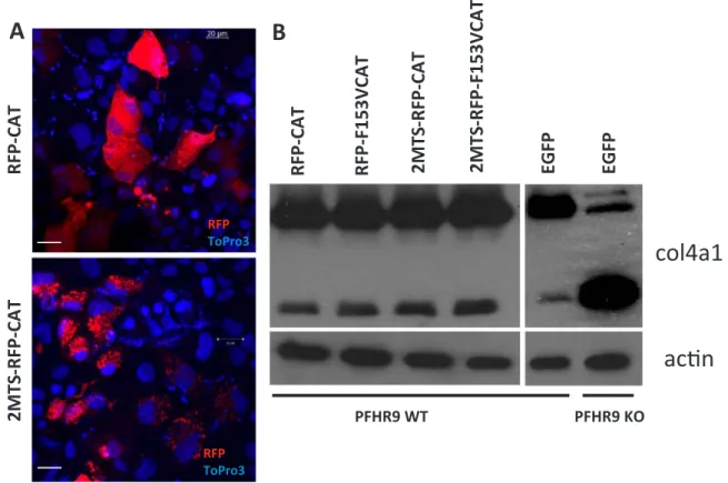
third lane). Next, we tried an approach where we targeted hetero- logously expressed catalase to different subcellular locations in PFHR-9 cells (cytosol, and mitochondrial matrix) and checked if this causes any changes in NC1 crosslinking.Fig. 8A shows the intracellular localiza- tion of the different catalase constructs. Catalase conjugated to RFP was evenly distributed in the cytosol (upper panel) and RFP-catalase fusion proteins targeted to the mitochondrial matrix (middle panel) showed characteristic mitochondrial localization. In previous experiments, we confirmed that fusing catalase to fluorescent proteins and different targeting sequences did not affect the catalytic activity of the enzyme (data not shown). When we analyzed collagen IV crosslinking in G418 selected, stably transfected cells we also included controls, where cat- alytically-inactive catalase proteins were introduced into the cells. As shown in Fig. 8B, the tested catalase constructs did not affect the amount of NC1 dimers synthesized by PFHR-9 cells.
4. Discussion
A common feature of reactions catalyzed by animal heme perox- idases is that halides or pseudohalides are oxidized by the enzymes. A recent addition to this list is the reaction catalyzed by PXDN, which involves the oxidation of bromide and the subsequent hypobromite- mediated crosslinking of collagen IV NC1 domains via methionine and hydroxylysine residues [4,22,28]. The presence of bromide and per- oxidasin is indispensable for the collagen IV NC1 crosslink formation [4,22], however, the source of H2O2in the PXDN-catalyzed reaction has not yet been identified. It is also an important question and subject of current research whether HOBr attacks other matrix components as well or it creates only the specific halosulfonium intermediate on Met93 of collagen IV NC1.
PXDN is a highly conserved protein that appears early in evolution
and it can be found across the animal kingdom[32]. Based on the ex- amples where NOX/DUOX enzymes act as a source of H2O2 in the peroxidase-catalyzed reactions we hypothesized that the activity of PXDN might be also dependent on NOX/DUOX isoforms. We tested this idea by assessing collagen IV crosslinking in mice which are deficient for either p22phoxor for certain NOX/DUOX isoforms. We found col- lagen IV crosslinking to be normal in all tested Nox/Duoxknockout models, indicating that PXDN uses H2O2 produced in a NOX-in- dependent manner. By measuring collagen IV crosslinking in multiple organs of PXDN-deficient animals we demonstrated the essential role of PXDN in this biochemical pathway. These results extend earlier ob- servations, where deficient collagen IV crosslinking was described in PXDN knockout embryonic fibroblasts and kidney [3,18]. Although PXDN belongs to the family of heme peroxidases, several features make this protein unique. Most importantly, besides its peroxidase part, PXDN also contains domains, which are characteristic of protein com- ponents of the extracellular matrix[24]. This raises the possibility that extracellular enzymes might act as an H2O2source for PXDN. However, in previous studies, lysyl oxidases, which are ROS-producing and ex- tracellular collagen- modifying enzymes, were also excluded as a po- tential H2O2source for NC1 crosslinking[4].
Another distinct feature of PXDN is that its expression is more widespread than that of other members of the animal heme peroxidase family since PXDN is expressed in virtually all tissues, where basement membranes are formed[7,25]. The expression pattern of PXDN is ac- tually consistent with the idea that H2O2is supplied for the enzyme in a NOX/DUOX independent manner because no single NOX/DUOX en- zyme shows similarly widespread expression. It is also important to note that currently no data obtained fromin vivoexperiments prove the essential role of H2O2in the crosslinking reaction, thus the contribution of alternative oxidants cannot be excluded. For example, protein
RFP-CAT 2MTS -RFP-CAT
col4a1 acn
RFP-CAT RFP-F153VCAT 2MTS -RFP-CAT 2MTS -RFP-F153VCAT
RFP ToPro3
RFP ToPro3
EGFP EGFP
PFHR9 WT PFHR9 KO
A B
Fig. 8.Analysis of the effect of recombinant catalase-mediated decomposition of intracellular hydrogen peroxide on collagen IV crosslinking. Catalase-RFP fusion proteins were targeted to the cytosol (RFP-CAT) or to the mitochondrial matrix (2MTS-RFP-CAT). Confocal microscopic images of transiently transfected PFHR9 cell. Topro3 positive nuclei are displayed in blue (A). Transfected PFHR9 cells were selected with 250μg/ml G418 for 7 days. During this time all non-transfected cells died and G418 cells reached confluency. Wild type andPxdnKO cells were also transfected with an EGFP expressing plasmid and went through the same 7 day long G418 selection. Cells were lysed in collagenase I containing hypotonic lysis buffer and digested O/N at 37 °C before processing for collagen IV NC1 western blot. Before adding collagenase I to the lysis buffer a small fraction of the lysates were processed for actin western blot.
peroxides, which are formed in a wide variety of reactions between proteins and free radicals, may act as oxidants during extracellular matrix formation[8].
Since we know so little about the source of ROS in collagen IV crosslinking, we were interested in characterizing the oxygen depen- dence of the process. Interestingly, we found that the coupling of col- lagen IV is undisturbed even at very low oxygen levels, indicating that a ROS-producing pathway with low Kd for oxygen participates in the process. Another intriguing implication of the above observations might be that even relatively long hypoxia,per se,will not disturb the integrity of collagen IV crosslinks in the basal membrane of vessel walls in hy- poxic tissues.
In polarized epithelial cells, mitochondria are often located and enriched at the basolateral side of the cells, in relative proximity to the underlying basement membrane. Moreover, the mitochondrial re- spiratory chain has a very high affinity for oxygen. It is not easy to assess the possible role of mitochondrial ROS in the crosslinking reac- tion. The flavoprotein inhibitor, DPI, a non-specific inhibitor of mi- tochondrial respiration and NADPH oxidases, did not affect NC1 cou- pling. However, it is possible, that inhibition of the respiratory chain by DPI or other inhibitors may actually increase the production of super- oxide by the organelles[33].
To explore the role of mitochondrial ROS in a more specific way, we expressed catalase constructs in PFHR9 cells and checked NC1 dimer formation. However, targeting catalase to the cytosol or to the mi- tochondrial matrix did not cause any decrease in collagen IV cross- linking. Schrineret al.created a transgenic mouse model which over- expressed mitochondrially targeted catalase[30]. These mice showed delayed aging, had a longer lifespan but did not display any gross de- velopmental abnormalities. This is in contrast with the eye develop- ment and skin pigmentation phenotype of peroxidasin mutant mice. As the affinity of catalase towards H2O2can be significantly lower than that of animal heme peroxidases we have to admit that our approach to testing the involvement of mitochondria can be considered only pre- liminary and further experiments are needed to explore the potential contribution of mitochondria-produced H2O2to the process.
In summary, our observations suggest that in collagen IV synthesis PXDN cooperates with a NOX/DUOX-independent H2O2source that is functional also at low oxygen levels.
Acknowledgments
We are grateful to Beáta Molnár and Barbara Bodor-Kis for technical assistance. This work was supported by grants from the National Research, Development and Innovation Office (K106138, K119955, NVKP_16-1-2016-0039) and by “Lendület” grant from the Hungarian Academy of Sciences (LP2011-001/2011).
Author disclosure statements
The authors declare no competingfinancial interests.
Appendix A. Supplementary material
Supplementary data associated with this article can be found in the online version athttp://dx.doi.org/10.1016/j.redox.2018.03.009.
References
[1] R.K. Ambasta, P. Kumar, K.K. Griendling, H.H. Schmidt, R. Busse, R.P. Brandes, Direct interaction of the novel Nox proteins with p22phox is required for the for- mation of a functionally active NADPH oxidase, J. Biol. Chem. 279 (2004) 45935–45941.
[2] K. Bedard, K.H. Krause, The NOX family of ROS-generating NADPH oxidases:
physiology and pathophysiology, Physiol. Rev. 87 (2007) 245–313.
[3] G. Bhave, S. Colon, N. Ferrell, The sulfilimine cross-link of collagen IV contributes to kidney tubular basement membrane stiffness, Am. J. Physiol. Ren. Physiol. 313
(2017) 596–602.
[4] G. Bhave, C.F. Cummings, R.M. Vanacore, C. Kumagai-Cresse, I.A. Ero-Tolliver, M. Rafi, J.S. Kang, V. Pedchenko, L.I. Fessler, J.H. Fessler, B.G. Hudson, Peroxidasin forms sulfilimine chemical bonds using hypohalous acids in tissue genesis, Nat.
Chem. Biol. 8 (2012) 784–790.
[5] R.P. Brandes, K. Schroder, Composition and functions of vascular nicotinamide adenine dinucleotide phosphate oxidases, Trends Cardiovasc. Med. 18 (2008) 15–19.
[6] R.P. Brandes, N. Weissmann, K. Schroder, Nox family NADPH oxidases: molecular mechanisms of activation, Free Radic. Biol. Med. 76C (2014) 208–226.
[7] G. Cheng, J.C. Salerno, Z. Cao, P.J. Pagano, J.D. Lambeth, Identification and characterization of VPO1, a new animal heme-containing peroxidase, Free Radic.
Biol. Med. 45 (2008) 1682–1694.
[8] M.J. Davies, Protein oxidation and peroxidation, Biochem. J. 473 (2016) 805–825.
[9] M.J. Davies, C.L. Hawkins, D.I. Pattison, M.D. Rees, Mammalian heme peroxidases:
from molecular mechanisms to health implications, Antioxid. Redox Signal. 10 (2008) 1199–1234.
[10] A. Donko, Z. Peterfi, A. Sum, T. Leto, M. Geiszt, Dual oxidases, Philos. Trans. R. Soc.
Lond. B Biol. Sci. 360 (2005) 2301–2308.
[11] A. Donko, E. Ruisanchez, A. Orient, B. Enyedi, R. Kapui, Z. Peterfi, D., X. De, Z. Benyo, M. Geiszt, Urothelial cells produce hydrogen peroxide through the acti- vation of Duox1, Free Radic. Biol. Med. 49 (2010) 2040–2048.
[12] M. Geiszt, J. Witta, J. Baffi, K. Lekstrom, T.L. Leto, Dual oxidases represent novel hydrogen peroxide sources supporting mucosal surface host defense, FASEB J. 17 (2003) 1502–1504.
[13] H. Grasberger, D., X. De, O.B. Mayo, H. Raad, M. Weiss, X.H. Liao, S. Refetoff, Mice deficient in dual oxidase maturation factors are severely hypothyroid, Mol.
Endocrinol. 26 (2012) 481–492.
[14] J. Khoshnoodi, V. Pedchenko, B.G. Hudson, Mammalian collagen IV, Microsc. Res.
Technol. 71 (2008) 357–370.
[15] S.J. Klebanoff, A.J. Kettle, H. Rosen, C.C. Winterbourn, W.M. Nauseef, Myeloperoxidase: a front-line defender against phagocytosed microorganisms, J.
Leukoc. Biol. 93 (2013) 185–198.
[16] I. Kovacs, M. Horvath, A. Lanyi, G.L. Petheo, M. Geiszt, Reactive oxygen species- mediated bacterial killing by B lymphocytes, J. Leukoc. Biol. 97 (2015) 1133–1137.
[17] A.J. Kowaltowski, N.C. Souza-Pinto, R.F. Castilho, A.E. Vercesi, Mitochondria and reactive oxygen species, Free Radic. Biol. Med. 47 (2009) 333–343.
[18] E. Lazar, Z. Peterfi, G. Sirokmany, H.A. Kovacs, E. Klement, K.F. Medzihradszky, M. Geiszt, Structure-function analysis of peroxidasin provides insight into the me- chanism of collagen IV crosslinking, Free Radic. Biol. Med. 83 (2015) 273–282.
[19] Y. Li, M.A. Trush, Diphenyleneiodonium, an NAD(P)H oxidase inhibitor, also po- tently inhibits mitochondrial reactive oxygen species production, Biochem.
Biophys. Res. Commun. 253 (1998) 295–299.
[20] A.C. Little, A. Sulovari, K. Danyal, D.E. Heppner, D.J. Seward, D. Van, V Paradoxical roles of dual oxidases in cancer biology, Free Radic. Biol. Med. 110 (2017) 117–132.
[21] K.D. Martyn, L.M. Frederick, K. von Loehneysen, M.C. Dinauer, U.G. Knaus, Functional analysis of Nox4 reveals unique characteristics compared to other NADPH oxidases, Cell Signal. 18 (2006) 69–82.
[22] A.S. McCall, C.F. Cummings, G. Bhave, R. Vanacore, A. Page-McCaw, B.G. Hudson, Bromine is an essential trace element for assembly of collagen IV scaffolds in tissue development and architecture, Cell157 (2014) 1380–1392.
[23] Y. Nakano, C.M. Longo-Guess, D.E. Bergstrom, W.M. Nauseef, S.M. Jones, B. Banfi, Mutation of the Cyba gene encoding p22phox causes vestibular and immune defects in mice, J. Clin. Invest 118 (2008) 1176–1185.
[24] R.E. Nelson, L.I. Fessler, Y. Takagi, B. Blumberg, D.R. Keene, P.F. Olson, C.G. Parker, J.H. Fessler, Peroxidasin: a novel enzyme-matrix protein of Drosophila development, EMBO J. 13 (1994) 3438–3447.
[25] Z. Peterfi, A. Donko, A. Orient, A. Sum, A. Prokai, B. Molnar, Z. Vereb, E. Rajnavolgyi, K.J. Kovacs, V. Muller, A.J. Szabo, M. Geiszt, Peroxidasin is secreted and incorporated into the extracellular matrix of myofibroblasts andfibrotic kidney, Am. J. Pathol. 175 (2009) 725–735.
[26] Z. Peterfi, M. Geiszt, Peroxidasins: novel players in tissue genesis, Trends Biochem.
Sci. 39 (2014) 305–307.
[27] R. Rizzuto, M. Brini, P. Pizzo, M. Murgia, T. Pozzan, Chimeric greenfluorescent protein as a tool for visualizing subcellular organelles in living cells, Curr. Biol. 5 (1995) 635–642.
[28] G.E. Ronsein, C.C. Winterbourn, P. Di Mascio, A.J. Kettle, Cross-linking methionine and amine residues with reactive halogen species, Free Radic. Biol. Med. 70 (2014) 278–287.
[29] N.E. Sanjana, L. Cong, Y. Zhou, M.M. Cunniff, G. Feng, F. Zhang, A transcription activator-like effector toolbox for genome engineering, Nat. Protoc. 7 (2012) 171–192.
[30] S.E. Schriner, N.J. Linford, G.M. Martin, P. Treuting, C.E. Ogburn, M. Emond, P.E. Coskun, W. Ladiges, N. Wolf, H. Van Remmen, D.C. Wallace, P.S. Rabinovitch, Extension of murine life span by overexpression of catalase targeted to mitochon- dria, Science 308 (2005) 1909–1911.
[31] G. Sirokmany, A. Donko, M. Geiszt, Nox/duox family of NADPH oxidases: lessons from knockout mouse models, Trends Pharmacol. Sci. (2016).
[32] M. Soudi, M. Zamocky, C. Jakopitsch, P.G. Furtmuller, C. Obinger, Molecular evolution, structure, and function of peroxidasins, Chem. Biodivers. 9 (2012) 1776–1793.
[33] J.F. Turrens, Superoxide production by the mitochondrial respiratory chain, Biosci.
Rep. 17 (1997) 3–8.
[34] R. Vanacore, A.J. Ham, M. Voehler, C.R. Sanders, T.P. Conrads, T.D. Veenstra, K.B. Sharpless, P.E. Dawson, B.G. Hudson, A sulfilimine bond identified in collagen
IV, Science 325 (2009) 1230–1234.
[35] E. Veal, A. Day, Hydrogen peroxide as a signaling molecule, Antioxid. Redox Signal.
15 (2011) 147–151.
[36] J. Wang, A. Slungaard, Role of eosinophil peroxidase in host defense and disease pathology, Arch. Biochem. Biophys. 445 (2006) 256–260.
[37] C.C. Winterbourn, The biological chemistry of hydrogen peroxide, Methods Enzymol. 528 (2013) 3–25.
[38] X. Yan, S. Sabrautzki, M. Horsch, H. Fuchs, V. Gailus-Durner, J. Beckers, d.
A. Hrabe, J. Graw, Peroxidasin is essential for eye development in the mouse, Hum.
Mol. Genet. 23 (2014) 5597–5614.
[39] P.D. Yurchenco, Basement membranes: cell scaffoldings and signaling platforms, Cold Spring Harb. Perspect. Biol. 3 (2011) a004911.
[40] M. Zana, Z. Peterfi, H.A. Kovacs, Z.E. Toth, B. Enyedi, F. Morel, M.H. Paclet, A. Donko, S. Morand, T.L. Leto, M. Geiszt, Interaction betweenp22(phox) and Nox4 in the endoplasmic reticulum suggests a unique mechanism of NADPH oxidase complex formation, Free Radic. Biol. Med. 116 (2018) 41–49.