Contents lists available atScienceDirect
Plant Physiology and Biochemistry
journal homepage:www.elsevier.com/locate/plaphy
Research article
H
2O
2homeostasis in wild-type and ethylene-insensitive Never ripe tomato in response to salicylic acid treatment in normal photoperiod and in prolonged darkness
Zoltán Takács
a,1, Péter Poór
a, Péter Borbély
a, Zalán Czékus
a, Gabriella Szalai
b, Irma Tari
a,∗aDepartment of Plant Biology, University of Szeged, H-6726 Szeged, Közép fasor 52., Hungary
bDepartment of Plant Physiology, Centre for Agricultural Research, Hungarian Academy of Sciences, H-2462 Martonvásár, Brunszvik u. 2., Hungary
A R T I C L E I N F O
Keywords:
Antioxidant enzymes Prolonged dark treatment Ethylene signalling
Hydrogen-peroxide homeostasis Never ripemutant
Salicylic acid Tomato
A B S T R A C T
Ethylene proved to be an important modulator of salicylic acid (SA) signalling pathway. Since SA may regulate both the production and scavenging of hydrogen peroxide (H2O2), which show light-dependency, the aim of this study was to compare H2O2metabolism in the leaves of SA-treated wild-type (WT) tomato (Solanum lycopersicum L. cv. Ailsa Craig) and in ethylene receptorNever-ripe(Nr) mutants grown in normal photoperiod or in prolonged darkness. H2O2accumulation was higher in the WT than in the mutants in normal photoperiod after 1 mM SA treatment, whileNrleaves contained more H2O2after light deprivation. The expression of certain superoxide dismutase (SOD) genes and activity of the enzyme followed the same tendency as H2O2, which was scavenged by different enzymes in the two genotypes. Catalase (CAT, EC 1.11.1.6) activity was inhibited by SA in WT, while the mutants maintained enhanced enzyme activity in the dark. Thus, in WT, CAT inhibition was the major component of the H2O2accumulation elicited by 1 mM SA in a normal photoperiod, since the expression and/or activity of ascorbate (APX, EC 1.11.1.11) and guaiacol peroxidases (POD, EC 1.11.1.7) were induced in the leaves. The absence of APX and POD activation in mutant plants suggests that the regulation of these enzymes by SA needs functional ethylene signalling. While the block of ethylene perception inNrmutants was overwritten in the transcription and activity of certain SOD and CAT isoenzymes during prolonged darkness, the low APX and POD activities led to H2O2accumulation in these tissues.
1. Introduction
The plant hormone, salicylic acid (SA) plays an important role in the signal transduction pathways of abiotic and biotic stress responses (Vlot et al., 2009). During this process SA interacts with other principal de- fence hormones such as jasmonic acid (JA) and ethylene (ET) making the hormonal homeostasis and signalling cross-talk crucial for thefine regulation of defence mechanisms (Koornneef and Pieterse, 2008). In the case of biotic stress SA is generally implicated in the defence against biotrophic pathogens, whereas JA/ET mediate plant responses to in- sects or necrotrophic pathogens (Glazebrook, 2005).
SA and ET are involved in both synergistic and antagonistic inter- actions in several physiological processes. SA was found to inhibit the ET synthesis in tomato (Li et al., 1992) but to promote ET accumulation
inArabidopsis(Rao et al., 2002) and inSolanum chilense(Gharbi et al., 2016) in response to O3exposure and salt stress, respectively. More- over, ET induced the accumulation of SA in tomato plant infected with the necrotrophic pathogenXanthomonas campestris (O'Donnell et al., 2001).
The rapid production and accumulation of reactive oxygen species (ROS), such as singlet oxygen, superoxide anion radical, hydroxyl ra- dical and hydrogen peroxide, are generated by SA and ET in response to stress. ROS, especially H2O2, can act together with SA or ET in a self- amplifying feedback loop where SA and/or ET induce H2O2accumu- lation, which then enhances the accumulation of these hormones (Wi et al., 2010;Xia et al., 2015). The biotic and abiotic stress responses of plants are often accompanied by biphasic ROS (Apel and Hirt, 2004;Wi et al., 2010), H2O2(Takács et al., 2017) or ET production (Wi et al.,
https://doi.org/10.1016/j.plaphy.2018.02.026
Received 14 November 2017; Received in revised form 22 January 2018; Accepted 23 February 2018
∗Corresponding author.
1Present address: Bioresources Center for Health & Bioresources, Austrian Institute of Technology GmbH, Konrad-Lorenz-Straße 24, 3430 Tulln, Austria.
E-mail addresses:takacszoltan8923@gmail.com(Z. Takács),poorpeti@bio.u-szeged.hu(P. Poór),borbely.peter01@gmail.com(P. Borbély),czekuszalan@gmail.com(Z. Czékus), szalai.gabriella@agrar.mta.hu(G. Szalai),tari@bio.u-szeged.hu(I. Tari).
Abbreviations:AC, Ailsa Craig; ACC, 1-aminocyclopropane-1-carboxylic acid; APX, ascorbate peroxidase; CAT, catalase; EL, electrolyte leakage; ET, ethylene; HR, hypersensitive response; JA, jasmonic acid;Nr,Never ripe; POD, guaiacol dependent peroxidase; ROS, reactive oxygen species; SA, salicylic acid; SAR, systemic acquired resistance; SOD, superoxide dismutase
Available online 02 March 2018
0981-9428/ © 2018 Elsevier Masson SAS. All rights reserved.
T
2010). In the case of defence responses, it was found that increases in SA levels are preceded by apoplastic H2O2bursts mediated by plasma membrane-localized NADPH oxidases and by extracellular peroxidases (PODs) (Mammarella et al., 2015) but both abiotic and biotic stress factors can also generate ROS in other compartments such as chlor- oplasts and peroxisomes (Apel and Hirt, 2004).
A number of reports indicated that SA not only plays a pro-oxidant role, but also has an antioxidant role in concert with glutathione (GSH), and controls cellular redox homeostasis through the regulation of an- tioxidant enzyme activities under stress conditions (Herrera-Vásquez et al., 2015;Xia et al., 2015). SA may increase the activity of superoxide dismutase (SOD, EC 1.15.1.1), the main scavenger of superoxide radi- cals, which catalyses the conversion of superoxide to H2O2in different cell compartments (Rao et al., 1997). Among the isoforms of SOD, Mn- SOD is localized in the mitochondria and peroxisomes, Fe-SOD in the chloroplast and Cu/Zn-SOD in the chloroplasts, peroxisomes, cytosol and apoplast. SA inhibits the activity of certain catalase isoenzymes (CAT, EC 1.11.1.6), cytosolic ascorbate peroxidase (APX, EC 1.11.1.11) and guaiacol peroxidases (POD, EC 1.11.1.7), the main scavengers of H2O2in plant cells (reviewed byHorváth et al., 2007;Rivas-San Vicente and Plasencia, 2011 Herrera-Vásquez et al., 2015).
Biphasic ROS production is also accompanied by a biphasic shift in cell redox potential in tissues treated with SA. An initial oxidative phase, characterized by a transient increase in ROS levels and a decline in GSH reducing power, is followed by a reductive phase characterized by increased GSH levels and reducing power. These temporal phases determine the sequential activation of redox-regulated processes, among them the transcription of defence genes (Herrera-Vásquez et al., 2015). SA-promotes the redox modification of specific Cys residues and the monomerisation of the oligomeric form of the NPR1 (NON-EXPR- ESSOR OF PR GENES 1) transcription coactivator, a central compound of SA signalling, which is transported to the nucleus and interacts with certain TGA transcription factors (TGA2 and TGA3), the regulators of SA-dependent late genes (e.g.PATHOGENESIS RELATED 1,PR1gene) (Pape et al., 2010). Class II TGA transcription factors (TGA2/5/6) are also essential regulators in SA-induced gene expression, but they mediate the NPR1-independent induction of early defence genes in- cluding antioxidant enzymes (Blanco et al., 2009).
It was found that ET could also regulate the ROS metabolism by modulating antioxidant enzymes. Pre-treatment with the ET precursor 1-aminocyclopropane-1-carboxylic acid (ACC) enhanced SOD, APX and POD activities inAgrostis stoloniferaunder heat stress (Larkindale and Huang, 2004). Moreover, treatment with 1-methylcyclopropene, which blocks the effects of ET by inhibiting the ethylene receptor, decreased the POD and CAT activities in plum (Kan et al., 2011).
ET is sensed by multiple receptors (e.g. ETR1, ERS1 inArabidopsis) localized to the endoplasmic reticulum and Golgi membranes. Genetic analysis has shown that these receptors function as negative regulators of the ET signalling pathway and actively suppress ET responses in the absence of ET. CTR1, a Ser/Thre protein kinase, acts downstream of the receptors and is also a negative regulator of ET signalling. Once ET is bound, suppression is cancelled, permitting the response to occur through the activation of positive regulators, including ETHYLENE INSENSITIVE2 (EIN2), a member of the NRAMP metal-ion transporter family, and the transcription factors ETHYLENE INSENSITIVE3 (EIN3), EIN3-like (EIL) and ERFs, which directly regulate the primary and secondary ethylene response genes, respectively. EIN3 directly up-reg- ulates ERF1expression by binding to the primary ethylene response elements (PERE) of theERF1promoter (Cheng et al., 2013).
ET receptor and signalling mutants make it possible to analyse not only the molecular steps of signalling but also the physiological func- tions of ET (Poór et al., 2015). In tomato there are seven ET receptors, LeETR 1, 2, 4 to 7, andNever ripe(Nr),five of which were shown to bind ET with high affinity (Kamiyoshihara et al., 2012). LeETR3 of tomato is referred to asNrfor historical reasons, and shows high homology with theArabidopsisET receptor ETR1 (Zhong et al., 2008). TheNrmutant
was described as a dominant mutation due to a single base substitution in the N terminal coding region. It shows insensitivity to ET not only during fruit ripening but in vegetative tissues as well, in the triple re- sponse, in leaf petiole epinasty, in senescence of petals andflower ab- scission. Nevertheless,Nrplants maintained high ET production after pathogen infection, indicating that the mutation did not affect ET biosynthesis (Lanahan et al., 1994).
The illumination of plant tissues has been shown to have a strong influence on hormone-regulated defences. Plants infected in the dark showed reduced systemic acquired resistance (SAR) (Karpiński et al., 2003) and lesion formation in response to avirulent pathogens (Zeier et al., 2004). The accumulation of SA, SA-mediated oxidative stress (Chandra-Shekara et al., 2006; Poór et al., 2017), ET biosynthesis (Rodrigues et al., 2014) and signalling are also dependent on the pre- sence of light (Liebsch and Keech, 2016).
Light may either stimulate or inhibit ET production, depending on the plant species, tissue and developmental phase (Bassi and Spencer, 1983). It is also well known that keeping leaf tissues in the dark induced the accumulation of ET, the breakdown of chlorophyll and carbohy- drates or the recycling of chloroplast proteins and activated dark-in- duced senescence (Lim et al., 2007). Since the light-driven electron transport chain in the chloroplasts also generates ROS, the production and physiological role of various forms of ROS may differ in illuminated or dark environments (Xing et al., 2013).
The allocation of resources between growth and defence under stress conditions is critical for the survival of photosynthesizing plants.
Thus the activation of growth or plant defence against pathogens and insects needsfine interaction between SA and ET signalling (Ballaré, 2014). These processes are controlled by light, sensed by red light re- ceptors, phytochromes, which interact with both SA- and ET-induced signalling and oxidative stress.
The aim of this study was to elucidate how the SA-dependent oxi- dative stress is modulated in WT and, in the absence of ET signalling, in ET receptor mutantNrtomato plants grown under a 12/12 h light/dark photoperiod or in prolonged darkness. Since SA proved to have dif- ferent effects on H2O2homeostasis and on the activation of antioxidant enzymes in the presence or absence of light in WT tomato plants (Poór et al., 2017), the question arose of whether the SA-dependent H2O2
metabolism was controlled by the same or by different series of anti- oxidant isoenzymes located in different compartments in WT plants and Nrmutants under the two types of light conditions.
2. Materials and methods
2.1. Plant material and growth conditions
Wild-type and ET receptor mutantNrtomato plants (Solanum lyco- persicumL. cv. Ailsa Craig) were grown in a controlled environment with 24 °C/22 °C day/night temperature, 55–60% relative humidity, 12/12 h day/night cycle, and 200μmol m−2s−1photon flux density (F36W/GRO lamps, Sylvania, Germany) in plastic boxes (40 cm length, 30 cm width, 20 cm depth, 12 seedlings per box) filled with 20 L of nutrient solution containing 2 mM Ca(NO3)2, 1 mM MgSO4, 0.5 mM KCl, 0.5 mM KH2PO4and 0.5 mM Na2HPO4at pH 6.2. The concentra- tions of micronutrients were 0.001 mM MnSO4, 0.005 mM ZnSO4, 0.0001 mM (NH4)6Mo7O24, 0.01 mM H3BO4and 0.02 mM Fe(III)-EDTA.
The culture medium was changed twice a week. Eight-week-old plants were treated by adding 0.1 mM or 1 mM SA to the nutrient solution for 24 h, half of the plants remaining under the same 12/12 h light/dark period and conditions and half of them being placed in a darkroom at the same temperature and humidity for the next 24 h. Samples were prepared in three replicates from the second, fully expanded young leaves after 24 h. The experiments were repeated 3–4 times.
Z. Takács et al. Plant Physiology and Biochemistry 126 (2018) 74–85
2.2. Determination of salicylic acid content
The extraction and determination of methanol-soluble free and bound SA were performed on 0.5 g of leaf tissue as described byPoór et al. (2017)following the procedure ofPál et al. (2005). The organic phases containing free and bound SA were evaporated to dryness under a vacuum and resuspended in 1 mL of the HPLC initial mobile phase.
Free and bound SA were separated using high-performance liquid chromatography (HPLC) on a reverse-phase column (Supelcosil ABZ Plus, 5μm 5 μ; 150 mm × 4.6 mm) at 25 °C using a 2690 separation module equipped with a W474 scanning fluorescence detector (WA- TERS, Milford, MA, USA) and monitored with excitation at 305 nm and emission at 407 nm.
2.3. Measurement of ethylene production
Samples of 0.5 g leaf tissue were taken after SA treatment and were incubated in gas-tight flasks fitted with rubber stoppers. After 1 h 2.5 mL of the gas was removed from the tubes with a gas-tight syringe and injected into the gas chromatograph. The production of ET was measured with a Hewlett-Packard 5890 Series II gas chromatograph equipped with aflame ionization detector and a column packed with activated alumina. as described byPoór et al. (2015).
2.4. Determination of H2O2levels and the integrity of the plasma membrane
The H2O2level was measured spectrophotometrically as described byTakács et al. (2016). After the homogenization of 0.2 g of leaf tissue with 1 mL of ice-cold, 0.1% trichloroacetic acid, the samples were centrifuged at 10 000 g for 20 min at 4 °C. The reaction mixture con- tained 0.25 mL of 10 mM phosphate buffer (pH 7.0), 0.5 mL of 1 M potassium iodide (KI) and 0.25 mL of the supernatant. The absorbance of the samples was measured after 10 min at 390 nm. The amount of H2O2was calculated using a standard curve prepared from H2O2stock solution. 3,3′-diaminobenzidine (DAB) staining was used to detect the production of H2O2in situ,which was carried out according toThordal- Christensen et al. (1997). Detached leaves from SA-treated tomato plants were incubated for 2 h in darkness in 2 mg/mL DAB solution at room temperature. After incubation the leaves were immersed in 96%
(v/v) ethanol to eliminate the chlorophyll content completely and then they were incubated in 50% (v/v) glycerol solution. To detect the in- tensity of the dye color digital camera (Sony Cyber-shot DSC-H9, Sony Co., Tokyo, Japan) was used.
Disintegration of cell membranes was determined by the measure- ment of electrolyte leakage. 0.1 g of fresh leaf tissues was incubated in 25 mL of double distilled water at 25 °C for 2 h. The conductivity in the bathing solution was determined (C1) with conductivity meter (Radelkis, Hungary). Then, the samples were heated at 95 °C for 40 min and the total conductivity (C2) of the cooled samples was measured.
Relative EL was expressed as EL (%)=(C1/C2)×100 (Kovács et al., 2016).
2.5. RNA extraction and expression analysis with real-time RT-PCR
Gene expression analysis was performed following the description of Takács et al. (2016)using quantitative real-time reverse transcrip- tion-PCR (qRT-PCR; Piko Real-Time qPCR System, Thermo Scientific).
Tomato genes were mined from the Sol Genomics Network (SGN;
http://solgenomics.net/, accessed 17 January 2018) and National Center for Biotechnology Information (NCBI;http://www.ncbi.nlm.nih.
gov/, accessed 17 January 2018) databases. Primers were designed using NCBI and Primer 3 softwares (http://frodo.wi.mit.edu/, accessed 17 January 2018) and are listed in Supplementary Table 1. The PCR reaction consisted of 10 ng cDNA template, 400 nM each of forward and reverse primers, 5μL of Maxima SYBR Green qPCR Master Mix (2X) (Thermo Scientific) and nuclease-free water in a total volume of 10μL.
The qRT-PCR was conducted at 95 °C for 7 min, followed by 40 cycles at 95 °C for 15 s and annealing extension at 60 °C for 60 s. A melting curve analysis of the product was performed (increasing the temperature from 55 to 90 °C, 0.2 °C s−1) to determine the specificity of the reaction. Data analysis was made using PikoReal Software 2.2 (Thermo Scientific).
Tomato 18S rRNA and elongation factor-1αsubunit genes were used as the reference genes and the 2(-ΔΔCt)formula was used to calculate data from the qRT-PCR. Each reaction was repeated at least three times.
2.6. Extraction and analysis of antioxidant enzymes
To analyse the enzyme activities, 0.5 g leaf tissue was homogenised on ice in 1 mL of 100 mM phosphate buffer (pH 7.0) containing 1 mM phenylmethylsulfonylfluoride and 1% (w:v) polyvinyl-polypirrolidone and the extracts were centrifuged at 12 000 g for 20 min at 4 °C. APX was extracted in the presence of 1 mM ascorbate (AsA). The supernatant was used for measuring enzyme activity with a dual-beam spectro- photometer (KONTRON, Milano, Italy) as described by Poór et al.
(2017). The enzyme activities were expressed as U mg−1protein. SOD (EC 1.15.1.1) activity was determined by measuring the ability of the enzyme to inhibit the photochemical reduction of nitroblue tetrazolium (NBT) in the presence of riboflavin in the light. One enzyme unit (U) of SOD corresponds to the amount of enzyme causing 50% inhibition of NBT reduction in the light. CAT (EC 1.11.1.6) activity was determined from the absorbance change at 240 nm based on the amount of de- composed H2O2(ε240= 39.4 mM−1cm−1). One unit of CAT is defined as the amount of enzyme necessary to decompose 1μmol min−1H2O2. APX (EC 1.11.1.11) activity was measured by monitoring the decrease in AsA content at 290 nm (ε290= 2.8 mM−1cm−1). One unit of APX activity is defined as the amount of enzyme necessary to oxidize 1μmol min−1AsA. POD (EC 1.11.1.7) activity was measured at 470 nm (ε 470= 26.6 mM−1cm−1). The amount of enzyme producing 1μmol min−1of oxidised guaiacol was defined as one U. The soluble protein concentration was determined according toBradford (1976) using bovine serum albumin as a standard. All chemicals were pur- chased from Sigma-Aldrich, Darmstadt, Germany.
2.7. Native PAGE and enzyme activity staining
Leaf samples weighing 0.5 g were crushed to afine powder in a mortar under liquid N2,after which soluble proteins were extracted by resuspending the powder in two volumes of 50 mM potassium phos- phate buffer (pH 7.0) containing 1 mM phenylmethylsulfonylfluoride.
For APX, 2 mM AsA was added to the enzyme extraction buffer. The homogenate was centrifuged at 12 000 g for 20 min at 4 °C.
The electrophoretic separation of SOD, APX and POD isoenzymes was performed with native polyacrylamide gel electrophoresis (PAGE) using an omniPAGE electrophoresis system (Cleaver Scientific Ltd., Rugby, Warwickshire, UK). Electrophoresis was performed at 4 °C for 1–3 h at a constant voltage of 120 V using 25 mM Tris and 192 mM glycine solution (pH 8.3) as a running buffer. For SOD and APX, 4%
stacking and 10% resolving polyacrylamide gels were used, whereas for POD, 3% and 6% stacking and resolving gels were used, respectively.
Samples were mixed with 62.5 mM Tris-HCl buffer (pH 6.8), containing 10% (v/v) glycerol and 0.025% (w/v) bromophenol blue before loading onto the gels. Equal amounts (30μg) of protein were loaded for the SOD, APX and POD determinations and after electrophoresis the gels were stained for enzyme activities according to well-established pro- tocols, as indicated.
SOD was stained according to the method described byMonteiro et al. (2011). The APX and POD activities were detected according to Kim et al. (2015)andGarcía-Limones et al. (2002), respectively.
2.8. Statistical analysis
The results are expressed as means ± S.E. After analyses of
variance (ANOVA) a multiple comparison followed by Tukey's test was performed with SigmaPlot version 12 software (SYSTAT Software Inc.
SPSS). The means were treated as significant ifP≤0.05. All the ex- periments were carried out at least three times. In each treatment at least three samples were measured.
3. Results
3.1. Effects of SA treatment on endogenous SA content and ET production
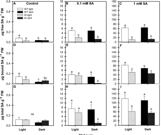
To detect the exogenous SA-induced physiological responses in the leaves of WT andNrmutants, the accumulation of free and bound (as well as total) methanol-soluble SA was measured in tomato plants treated with 0.1 or 1 mM exogenous SA in an illuminated or dark en- vironment. Under control growth conditions, the free SA content was about 0.1μg g−1fresh mass in illuminated WT leaves and less than 0.05μg g−1fresh mass in prolonged darkness (Fig. 1A). While the free and bound SA contents were lower inNrleaves in the light, they were equal to those of WT samples in the dark (Fig. 1A, D). Free SA contents were significantly increased to 50-fold by 0.1 mM SA and to 600-fold by 1 mM SA treatment compared with the control in WT under both en- vironmental conditions (Fig. 1B and C). However, the free SA content increased only slightly in Nr leaves after SA treatment and, un- expectedly, was always lower than in the corresponding WT leaves
(Fig. 1B and C). In accordance with our earlierfindings, free SA con- tents were not significantly different in the WT (Poór et al., 2017) or in the mutant plants after SA treatments in normal photoperiod or in prolonged darkness. Unlike free SA, the amount of bound and total SA inNrmutants after 0.1 mM SA treatment was similar to that in WT in the light, but these fractions were significantly lower in mutant plants in prolonged darkness (Fig. 1E, H). In the presence of 1 mM SA, the fractions of bound and total SA inNrleaves were much lower than in WT under the same conditions, but there were no significant differences in the total SA contents between light and dark conditions in either WT orNrleaves (Fig. 1F, I).
The control leaves ofNrplants exhibited higher ET production than WT in both environments (Supplementary Fig. 1A). In the presence of 0.1 mM SA, ET production did not change significantly in the two genotypes under a normal photoperiod, but in the dark the WT leaves emitted more ET and theNrleaves less ET than their respective controls (Supplementary Fig. 1B). In the case of treatment with 1 mM SA, there was no significant change in ET production of WT plants in the light but a significant decrease was observed inNrleaves. In prolonged darkness this resulted in similar tendencies in ET production after 1 mM SA treatment as under control conditions (Supplementary Fig. 1).
Fig. 1.Changes in free, bound and total methanol-soluble salicylic acid (SA) content in the leaves of wild type (WT, open columns) and ethylene receptor mutantNever ripe(Nr, latticed columns) tomato plants treated with 0.1 mM or 1 mM SA for 24 h in the presence or absence of light (□Light,■Dark). Control: A, D, G; 0.1 mM SA: B, E, H; 1 mM SA: C, F, I. Results are means ± SE from at least three independent experiments. Data with different letters indicate significant differences (P≤0.05, n = 3).
Z. Takács et al. Plant Physiology and Biochemistry 126 (2018) 74–85
3.2. Effect of SA treatment on hydrogen peroxide content and electrolyte leakage
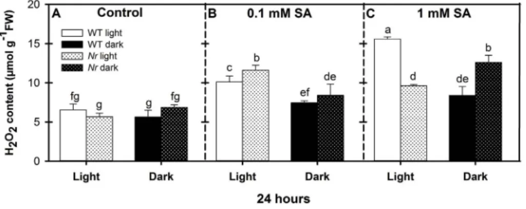
To investigate the effect of ET signalling on SA-induced H2O2ac- cumulation, the H2O2content in the leaves was recorded. There were no significant differences between the control of WT and Nrmutants under either light or dark conditions (Fig. 2A). However, H2O2accu- mulated in response to both SA treatments in the light, in both geno- types increasing with the SA concentration in WT plants, but reaching a maximum at 0.1 mM SA inNrplants. In prolonged darkness there was a gradual increase in the H2O2content of the tissues as a function of SA concentration in both genotypes. After 1 mM SA treatment Nrleaves contained less than 62% of the H2O2content of WT in a normal pho- toperiod, but much higher amount, 150% of the respective WT in prolonged darkness (Fig. 2B and C andSupplementary Fig. 2). Since H2O2is a major component of oxidative stress caused by SA, electrolyte leakage (EL), which indicates the loss of plasma membrane integrity, was determined. It was found that EL followed the tendency obtained for H2O2 accumulation. In the leaves of illuminated WT 1 mM SA caused significant, 40% increase in EL compared to untreated control, whileNrmutants displayed only 17% increment (Fig. 3). Conversely, in the absence of light, EL was significantly lower in WT than inNr, which suggested thatNrplants were more sensitive to SA under prolonged darkness than the WT, while WT plants were more sensitive thanNrin normal photoperiod.
3.3. Effect of SA treatment on antioxidant enzyme activities and on the expression of their coding genes
The SA-induced H2O2 metabolism is mediated by various anti- oxidant enzymes. In order to determine whether antioxidant enzyme activity and gene expression were differentially regulated by 0.1 mM or 1 mM SA in the ET receptor mutantNrand WT in the presence or ab- sence of light, the activities and transcript levels of genes encoding SOD (SlMnSOD,SlFeSOD,SlCu/ZnSOD), CAT (SlCAT1,SlCAT2,SlCAT3) and APX (SlAPX1, SlAPX2) together with the activity of POD were
determined.
3.3.1. Superoxide dismutase
Three SOD genes from the tomato genome, the mitochondrial MnSOD, the chloroplastic FeSOD and Cu/ZnSOD, were used in the experiments. The transcripts levels of these genes were not significantly different in WT andNrplants under a normal photoperiod. However, SlMnSODwas down-regulated, the expression ofSlCu/ZnSODwas in- duced and the expression of theSlFeSODgene remained at a constant level in the leaves of WT controls after 24 h of darkness. Surprisingly, all the SOD coding genes were suppressed inNrmutants in the dark (Fig. 4A, D, G). No significant differences were observed in the presence of 0.1 mM SA compared to the control in either WT orNrplants, except thatSlFeSOD andSlCu/ZnSOD showed down-regulation in prolonged darkness in WT (Fig. 4B, E, H). In contrast, a very strong induction of SlMnSODandSlCu/ZnSODtranscription was observed after 1 mM SA treatment in illuminated WT leaves, while the transcription ofSlFeSOD remained constant (Fig. 4C, F, I). Unexpectedly, one chloroplastic en- zyme,SlCu/ZnSODwas up-regulated in prolonged darkness to a greater extent inNrleaves than in WT after 1 mM SA treatment (Fig. 4I).
In spite of the differences in gene expression, SOD activity did not differ in WT andNrplants under control conditions in the presence or absence of light (Fig. 5A). The activity of SOD remained constant inNr leaves under a normal photoperiod after 0.1 mM SA treatment, but decreased in WT leaves in both environments (Fig. 5B). However, SOD activity increased significantly after 1 mM SA treatment in illuminated WT leaves, but was significantly higher inNr than in WT leaves in prolonged darkness (Fig. 5C). The changes followed similar pattern as H2O2in leaf tissues, the specific activity inNrextracts was 66% of WT in normal photoperiod and 140% in prolonged darkness.
Native PAGE analysis of SOD indicated the presence offive bands in both WT andNrleaf extracts, although thefirst, less mobile band was hardly detectable (Fig. 5D). The first band could be identified as MnSOD, since it was not inhibited by either KCN or H2O2, the second band was characterized as FeSOD since it was negatively affected by H2O2,and the third, fourth and fifth bands were identified as Cu/
Fig. 2.Changes in H2O2content in the leaves of wild type (WT, open columns) and ethylene receptor mutantNever ripe (Nr, latticed columns) tomato plants treated with 0.1 mM or 1 mM SA for 24 h in the presence or absence of light (□Light,■Dark). Control: A; 0.1 mM SA: B; 1 mM SA: C. Results are means ± SE from at least three in- dependent experiments. Data with different letters indicate significant differences (P≤0.05, n = 3).
Fig. 3.Changes in the electrolyte leakage from the leaves of wild type (WT, open columns) and ethylene receptor mu- tantNever ripe(Nr, latticed columns) tomato plants treated with 0.1 mM or 1 mM SA for 24 h in the presence or absence of light (□Light,■Dark). Control: A; 0.1 mM SA: B; 1 mM SA: C. Results are means ± SE from at least three in- dependent experiments. Data with different letters indicate significant differences (P≤0.05, n = 5).
ZnSODs, since they were inhibited by both inhibitors (data not shown).
Based on the native PAGE, the Cu/ZnSODs (I, II, III) were the main SOD isoforms in WT andNrleaves showing results similar to those observed for total SOD activity (Fig. 5A, B, C). Under dark conditions, the activity of SOD isoforms in Nrleaves was higher than in WT after 1 mM SA treatment (Fig. 5D).
3.3.2. Catalase
All theSlCATgenes were less strongly expressed in theNrleaves than in WT control plants in a normal photoperiod (Fig. 6A, D, G). In addition, SlCAT2expression decreased significantly in the dark com- pared to illuminated samples in the case of WT (Fig. 6D). In the pre- sence of SA, the transcript levels ofSlCAT1were suppressed, but the expression ofSlCAT2andSlCAT3exhibited constant expression in WT leaves under a normal photoperiod. At the same time, the expression of CAT isoenzymes was low inNrleaves and displayed only small changes after SA treatments in the light (Fig. 6). In darkness, SA inhibited the expression of the CAT genes or caused moderate changes in their transcript levels in WT, while inNrleaves a significant induction was observed in the relative transcript levels of SlCAT1 at both
concentrations of SA and of SlCAT3 after treatment with 1 mM SA (Fig. 6B, C, I).
In accordance with previous findings SA caused a concentration- dependent, about 68% inhibition in CAT activity in the light, which was not so pronounced (48%) inNrleaves, while the inhibiting effect of SA could only be observed in WT plants in prolonged darkness.
Unexpectedly, SA inhibition was not manifested inNr tissues in the dark, since the enzyme activity increased as a function of SA con- centration (Fig. 7).
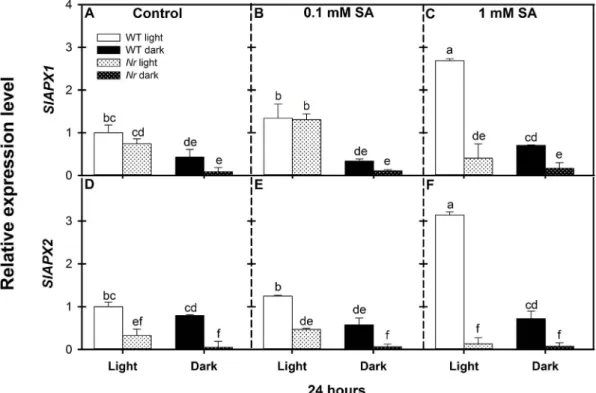
3.3.3. Ascorbate peroxidase
Besides CAT and POD, APX is the most important H2O2scavenging enzyme in plant cells. In the dark, the levels ofSlAPX1 andSlAPX2 transcripts in control leaves were suppressed both in WT andNrcom- pared to those in illuminated plants (Fig. 8A, D). SA up-regulated the APX genes and caused 2.6- and 3.1-fold higher expression ofSlAPX1 andSlAPX2transcripts, respectively, in WT leaves at the 1 mM con- centration under a normal photoperiod, while the expression ofSlAPX genes did not change at this SA concentration in illuminatedNrleaves compared to the control. In addition, the transcript levels of SlAPX Fig. 4.Changes in relative expression levels ofSlMnSOD,SlFeSODandSlCu/ZnSODgenes in the leaves of wild type (WT, open columns) and ethylene receptor mutantNever ripe(Nr, latticed columns) tomato plants treated with 0.1 mM or 1 mM SA for 24 h in the presence or absence of light (□Light,■Dark). Control: A, D, G; 0.1 mM SA: B, E, H; 1 mM SA: C, F, I.
Results are means ± SE from at least three independent experiments. Data with different letters indicate significant differences (P≤0.05, n = 3).
Z. Takács et al. Plant Physiology and Biochemistry 126 (2018) 74–85
genes remained at the control level in the dark in both genotypes (Fig. 8).
APX activity, in correlation with the expression of APX genes, was generally higher in WT than inNr leaves under both environmental conditions (Fig. 9A). Both SA treatments induced APX activity in WT and more moderately in Nrleaves in the light, while the enzyme ac- tivities remained at the control level in the dark (Fig. 9B and C).
Native PAGE analysis of APX indicated two isoforms in both WT and Nrleaves (Fig. 9D). The intensity of the bands was, in general, well correlated with the total enzyme activity of APX, assayed in the same samples (Fig. 9A, B, C). Increased APX bands were detected after SA treatment in both WT andNrleaves in the light, but much lower ac- tivities were found in the dark (Fig. 9D).
3.3.4. Guaiacol dependent peroxidase
POD, the other important H2O2 metabolizing enzyme, was also determined (Fig. 10) and the results clearly showed that SA caused a concentration-dependent increase in POD activity in illuminated WT leaves, which was significantly lower in a prolonged dark period (Fig. 10B and C). On the other hand, the POD activity in the leaves ofNr mutants was substantially lower than in WT plants and the enzyme activity changed only slightly after SA treatment (Fig. 10B and C).
As far as the POD isoenzyme profile was concerned (Fig. 10D), two distinct isoforms were observed on the gels. The intensity of the POD
bands in different samples generally correlated well with the enzyme activities assayed previously in the same samples (Fig. 10A, B, C). The POD isoforms exhibited higher activity in WT leaves as a function of SA concentration, whereas the activity was always lower inNrleaves.
4. Discussion
ET receptorNrmutants of tomato make it possible to analyse the role of ET signalling and the functions of ET in SA-induced H2O2pro- duction and metabolism. In previous work it was found that treating WT tomato (Solanum lycopersicumcv. Ailsa Craig) with 0.1 or 1 mM SA increased the free SA content in the leaves to 5–6μg g−1 and
∼60μg g−1fresh mass, respectively, which corresponds to the SA le- vels in the tissues in the systemic leaves or in tissues surrounding pe- netrating pathogens during hypersensitive response (HR) (Poór et al., 2017). Thus, the lower concentration of SA could lead to abiotic stress acclimation or SAR during biotic stress, while the higher concentrations can initiate cell death and necrotic lesions in leaf tissues. Unexpectedly, Nrplants showed lower basic SA contents, and exogenous SA treatment resulted in much lower free SA accumulation in the mutant leaves than in WT. These differences were not dependent on the presence of light.
In spite of the low endogenous SA content inNrleaves, ROS production and scavenging in the two genotypes were comparable, suggesting that SA may induce oxidative stress by diverse mechanisms.
Fig. 5.Specific activity of superoxide dismutase (SOD) in the leaves of wild type (WT, open columns) and ethylene receptor mutantNever ripe(Nr, latticed columns) tomato plants treated with 0.1 mM or 1 mM SA for 24 h in the presence or absence of light (□Light,■Dark). Control: A; 0.1 mM SA: B; 1 mM SA: C. Results are means ± SE from at least three independent experiments. Data with different letters indicate significant differences (P≤0.05, n = 3). (D) Native PAGE analysis of SOD activity in the leaves of WT andNrmutants. Separation of isoenzymes and activity staining were performed as described byMonteiro et al. (2011)using 30μg of protein per lane.
In accordance with the results ofLanahan et al. (1994)the present data confirmed that ET biosynthesis was not impaired inNrplants. ET may either inhibit or promote its own biosynthesis. The control of ET synthesis by ET in vegetative tissues occurs via autoinhibition, while autocatalytic ET production can be observed during certain stress
conditions, senescence and fruit ripening (Rodrigues et al., 2014). Not surprisingly, in the absence of negative feedback due to the mutation of the most effective ET receptor, ET accumulation was significantly higher inNrplants under both environmental conditions. Since in the present work dark treatment reduced the ET emanation from the tissues Fig. 6.Changes in relative expression levels ofSlCAT1,SlCAT2andSlCAT3genes in the leaves of wild type (WT, open columns) and ethylene receptor mutantNever ripe(Nr, latticed columns) tomato plants treated with 0.1 mM or 1 mM SA for 24 h in the presence or absence of light (□Light,■Dark). Control: A, D, G; 0.1 mM SA: B, E, H; 1 mM SA: C, F, I. Results are means ± SE from at least three independent experiments. Data with different letters indicate significant differences (P≤0.05, n = 3).
Fig. 7.Specific activity of catalase (CAT) in the leaves of wild type (WT, open columns) and ethylene receptor mu- tantNever ripe(Nr, latticed columns) tomato plants treated with 0.1 mM or 1 mM SA for 24 h in the presence or absence of light (□Light,■Dark). Control: A; 0.1 mM SA: B; 1 mM SA: C. Results are means ± SE from at least three in- dependent experiments. Data with different letters indicate significant differences (P≤0.05, n = 3).
Z. Takács et al. Plant Physiology and Biochemistry 126 (2018) 74–85
Fig. 8.Changes in relative expression levels ofSlAPX1andSlAPX2genes in the leaves of wild type (WT, open columns) and ethylene receptor mutantNever ripe(Nr, latticed columns) tomato plants treated with 0.1 mM or 1 mM SA for 24 h in the presence or absence of light (□Light,■Dark). Control: A, D; 0.1 mM SA: B, E; 1 mM SA: C, F. Results are means ± SE from at least three independent experiments. Data with different letters indicate significant differences (P≤0.05, n = 3).
Fig. 9.Specific activity of ascorbate peroxidase (APX) in the leaves of wild type (WT, open columns) and ethylene receptor mutantNever ripe(Nr, latticed columns) tomato plants treated with 0.1 mM or 1 mM SA for 24 h in the presence or absence of light (□Light,■Dark). Control: A; 0.1 mM SA: B; 1 mM SA: C. Results are means ± SE from at least three independent experiments. Data with different letters indicate significant differences (P≤0.05, n = 3). (D) Native PAGE analysis of APX activity in leaves of WT andNrtomato plants. Separation of isoenzymes and activity staining were performed as described byKim et al. (2015)using 30μg of protein per lane.
compared to corresponding light samples, it can be concluded that the lack of ET-induced negative feedback inNrplants can be modulated by the illumination conditions and SA.
Thefirst 24 h after stress stimuli is particularly important in plant responses where H2O2 plays a central role in the action of various hormones, such as SA and ET (Van Aken and Van Breusegem, 2015). In a previous study on tomato it was demonstrated that a sublethal con- centration of SA increased the H2O2 accumulation under a normal photoperiod while high concentrations caused biphasic H2O2accumu- lation, which was much more moderate during prolonged dark period.
The H2O2metabolism was also controlled by different antioxidant en- zymes in WT plants in the presence or absence of light (Poór et al., 2017).
Although the function of ET in ROS production during stress reac- tions is well known, the SA-induced antioxidant mechanisms in con- junction with ET signalling and plant illumination have not yet been fully elucidated. It was revealed that the tomato ERF transcription factor, TERF1, is the key factor in the activation of ROS scavenging in ET signalling. The transcription of TERF1 is regulated by the upstream, EIN3-like transcription factors, LeEIL3 and LeEIL4 and can also be in- duced by reagents that generate ROS. The ectopic expression of TERF1 in tobacco promoted the expression of CAT and glutathione peroxidase, reduced ROS accumulation and increased the oxidative stress tolerance of plants (Zhang et al., 2016). The EIN3 (Mur et al., 2013) and ERFs may be also targets of the crosstalk between ET/SA signalling pathways (Broekgaarden et al., 2015).
In the present experiments H2O2accumulation was not significantly different in the two genotypes under control conditions. SA stimulated H2O2accumulation in WT tissues as a function of concentration, but much more so in the light, than in the dark. SA-induced increases in H2O2were also observed inNrplants. The most interesting tendencies
could be detected at 1 mM SA concentration. While the inhibition of ET signalling by theNrmutation decreased the SA-induced H2O2content in the light compared to WT, it resulted in pronounced H2O2accumu- lation in prolonged darkness. This suggests that not only the SA and ET levels but also their signalling pathways are dependent on the presence or absence of light (Chandra-Shekara et al., 2006;Fukao et al., 2012) and also raises the possibility that H2O2-producing and -scavenging processes and enzymes are regulated differently in WT andNrplants.
H2O2accumulation exhibited strong correlation with the electrolyte leakage, a good marker of plasma membrane damage. These results demonstrated that theNrmutants were more tolerant to SA-induced oxidative stress than WT in normal photoperiod but they proved to be more sensitive during prolonged darkness.
The elevated H2O2level caused by exogenous SA treatment may act not only as a toxic compound inducing oxidative stress and HR, but also as a signalling molecule regulating antioxidant enzyme activity and gene expression (Van Aken and Van Breusegem, 2015). Five distinct SOD isoforms, one MnSOD (I), one FeSOD (II) and three Cu-ZnSODs (III-IV), were detected in the leaf tissues of both genotypes. This cor- responds with the results ofMonteiro et al. (2011), who characterized alsofive SOD isoforms in the leaves of WT andNrmutants in a Micro- Tom background, though these authors found an extra MnSOD band in the root extract and demonstrated organ- and time-dependent altera- tions in enzyme activities in extracts prepared from salt- and Cd- stressed plants. In the present experiments 1 mM SA increased the ac- tivity of SOD in WT plants in both environments, but the changes were more pronounced under a normal photoperiod. The concentration-, time- and organ-specific activation of SOD by SA was observed earlier in tomato (Tari et al., 2015) and tobacco (Horváth et al., 2007), and it was also demonstrated that darkness had an inhibitory effect on SOD activity in WT plants (Camejo et al., 2007).
Fig. 10.Specific activity of guaiacol peroxidase (POD) in the leaves of wild type (WT, open columns) and ethylene receptor mutantNever ripe(Nr, latticed columns) tomato plants treated with 0.1 mM or 1 mM SA for 24 h in the presence or absence of light (□Light,■Dark). Control: A; 0.1 mM SA: B; 1 mM SA: C. Results are means ± SE from at least three independent experiments. Data with different letters indicate significant differences (P≤0.05, n = 3). (D) Native PAGE analysis for POD activity from leaves of wild type and ethylene receptor mutantNever ripetomato plants. Separation of isoenzymes and activity staining were performed as described byGarcía-Limones et al. (2002)using 30μg of protein per lane.
Z. Takács et al. Plant Physiology and Biochemistry 126 (2018) 74–85
At the same time, the present results confirmed that ET signalling has a great influence on the SA-induced activation of SOD. While SA treatment did not increase the activity of SOD in illuminatedNrleaves, the total activity, the activity of SlCu/ZnSODs and the expression of chloroplastic SlCu/ZnSOD were significantly induced by 1 mM SA during prolonged dark period in the mutants. Considering that SlCu/
ZnSODs are the predominant isoforms, the total SOD activity showed a good correlation with these bands. Furthermore, the increased SOD activity of WT leaves in the light and that ofNrin the dark could ex- plain the increased H2O2content in the leaves. It seems reasonable that active ET signalling is necessary for the SA-induced expression ofSlCu/
ZnSOD, but the prolonged dark treatment may partially substitute for the activation of ET-induced transcription factors without the activation of the upstream components in ET signalling.
The high H2O2levels induced by SA treatments may be partially generated by SOD, but the equilibrium between H2O2production and scavenging may be perturbed in the absence of H2O2-inactivating en- zymes such as CAT, APX and POX. It is well known that CAT activity is inhibited by SA at both the gene expression and protein level (Agarwal et al., 2005;Horváth et al., 2007;Tari et al., 2015). The present results demonstrated that both concentrations of SA inhibited the CAT activity in WT leaves under both environmental conditions and in illuminated Nrleaves. However, in prolonged darkness SA still increased the CAT enzyme activity inNrleaves, suggesting that some SOD and CAT iso- enzymes are controlled in a similar way in the dark in mutant plants. In parallel with enzyme activities, the expression of two peroxisomal CAT genes,SlCAT1andSlCAT3, were also up-regulated in the mutants by 1 mM SA in darkness. It was found by other authors that the expression of wheatCATisoenzymes was regulated by the circadian rhythm and modulated by darkness (Luna et al., 2005).Han et al. (2015)observed that the use of 1-methylcyclopropene, which interacts with ET receptors and thereby inhibits ET action, increased CAT activity after 24 h in bitter melon in the dark, which was also supported by our experiments.
In agreement with the results ofKanazawa et al. (2000), who found that APX activity decreased during dark-induced senescence, the two important H2O2scavenging enzymes, APX and POD, exhibited strong light dependence and parallel changes after SA treatment in both gen- otypes. APX activity was basically lower in theNrmutant and the ac- tivity of the enzyme increased as a function of SA concentration in il- luminated WT andNrleaves. However, the enzyme activity remained constant in the dark in both genotypes. The expression of theSlAPX1 andSlAPX2genes displayed tendencies similar to those observed for enzyme activities: both APX genes were highly expressed after 1 m SA treatment in the presence of light, but their expression was down- regulated during a prolonged dark period. Irrespective of the illumi- nation, the transcription of the SlAPX1 and SlAPX2 genes was sig- nificantly lower in Nrleaves, suggesting that active ET signalling is necessary for APX expression.
It was found earlier that the POD activity in the cytosol and cell wall increased in a dose-dependent manner after pre-treatment with SA (Rao et al., 1997;Ananieva et al., 2004). The present results confirmed that SA caused a concentration-dependent activation of POD in illuminated WT leaves. Moreover, the partial activation of PODs could contribute to the decomposition of H2O2in the dark in WT, which is particularly important in the absence of CAT and APX induction in the SA-mediated protective actions in WT plants. Interestingly, the effect of SA on POD induction was significantly less pronounced inNrplants.Monteiro et al.
(2011)also observed lower POD activities inNrleaves compared to WT plants, suggesting that ET has significant control over the activity of PODs. In the present work, the activity of POD was very low in Nr mutants and was only slightly elevated after SA treatment in the dark.
5. Conclusion
It can be concluded that H2O2accumulation in WT leaves in re- sponse to 1 mM SA treatment is mainly determined by increased SOD
and decreased CAT activities in the light. In these plants APX and POD proved to be main participants of H2O2scavenging and as a net result, H2O2content was much higher in the WT than inNrleaves. In a pro- longed dark period the SOD and CAT activities and the expression of SlCu/ZnSODandSlCAT3genes were activated inNrmutants exposed to 1 mM SA but the absence of APX and POD activation led to a significant accumulation of H2O2in these samples. These results suggest that the SA-induced H2O2metabolism is regulated by different enzymes in WT and ET signalling mutant plants, which is modified by the presence or absence of light. Thus,Nr mutants seem to be more tolerant to SA- induced oxidative stress than WT plants in normal photoperiod, but they are more sensitive in prolonged darkness.
Contributions
ZT, PP, PB and CZ performed and analysed the experiments, GSz was responsible for the analysis of salicylic acid and its derivatives. PP, ZT and IT designed the experiments, wrote and provided critical review of the manuscript.
Acknowledgements
The authors thank Mrs. A. Bécs for her excellent technical assis- tance. This work was funded by grants from the National Research, Development and Innovation Office (OTKA K101243 and OTKA PD112855) and by the Hungary-Serbia IPA Cross-border Co-operation Programme [HUSRB/1203/221/173]. There is no conflict of interest to declare.
Appendix A. Supplementary data
Supplementary data related to this article can be found athttp://dx.
doi.org/10.1016/j.plaphy.2018.02.026.
References
Agarwal, S., Sairam, R.K., Srivastava, G.C., Tyagi, A., Meena, R.C., 2005. Role of ABA, salicylic acid, calcium and hydrogen peroxide on antioxidant enzymes induction in wheat seedlings. Plant Sci. 169, 559–570.
Ananieva, E.A., Christov, K.N., Popova, L.P., 2004. Exogenous treatment with salicylic acid leads to increased antioxidant capacity in leaves of barley plants exposed to paraquat. J. Plant Physiol. 161, 319–328.
Apel, K., Hirt, H., 2004. Reactive oxygen species: metabolism, oxidative stress, and signal transduction. Ann. Rev. Plant Biol. 55, 373–399.
Ballaré, C.L., 2014. Light regulation of plant defense. Ann. Rev. Plant Biol. 65, 335–363.
Bassi, P.K., Spencer, M.S., 1983. Does light inhibit ethylene production in leaves? Plant Physiol. 73, 758–760.
Blanco, F., Salinas, P., Cecchini, N.M., Jordana, X., Van Hummelen, P., Alvarez, M.E., Holuigue, L., 2009. Early genomic responses to salicylic acid inArabidopsis. Plant Mol. Biol. 70, 79–102.http://dx.doi.org/10.1007/s11103-009-9458-9451.
Bradford, M.M., 1976. A rapid and sensitive method for the quantitation of microgram quantities of protein utilizing the principle of protein-dye binding. Anal. Biochem. 72, 248–254.
Broekgaarden, C., Caarls, L., Vos, I.A., Pieterse, C.M.J., Van Wees, S.C.M., 2015. Ethylene:
traffic controller on hormonal crossroads to defense. Plant Physiol. 169, 2371–2379.
Camejo, D., Martí, M.D.C., Nicolás, E., Alarcón, J.J., Jiménez, A., Sevilla, F., 2007.
Response of superoxide dismutase isoenzymes in tomato plants (Lycopersicon escu- lentum) during thermo-acclimation of the photosynthetic apparatus. Physiol. Plant.
131, 367–377.
Chandra-Shekara, A.C., Gupta, M., Navarre, D., Raina, S., Raina, R., Klessig, D., Kachroo, P., 2006. Light dependent hypersensitive response and resistance signaling against Turnip Crinkle Virus inArabidopsis. Plant J. 45, 320–334.
Cheng, M.C., Liao, P.M., Kuo, W.W., Lin, T.P., 2013. TheArabidopsisETHYLENE RESPONSE FACTOR1 regulates abiotic stress-responsive gene expression by binding to different cis-acting elements in response to different stress signals. Plant Physiol.
162, 1566–1582.
Fukao, T., Yeung, E., Bailey-Serres, J., 2012. The submergence tolerance geneSUB1A delays leaf senescence under prolonged darkness through hormonal regulation in rice. Plant Physiol. 160, 1795–1807.
García-Limones, C., Hervás, A., Navas-Cortés, J.A., Jiménez-Díaz, R.M., Tena, M., 2002.
Induction of an antioxidant enzyme system and other oxidative stress markers as- sociated with compatible and incompatible interactions between chickpea (Cicer arietinumL.) andFusarium oxysporumf. sp. ciceris. Physiol. Mol. Plant Pathol. 61, 325–337.
Gharbi, E., Martínez, J.P., Benahmed, H., Fauconnier, M.L., Lutts, S., Quinet, M., 2016.
Salicylic acid differently impacts ethylene and polyamine synthesis in the glycophyte Solanum lycopersicumand the wild-related halophyteSolanum chilenseexposed to mild salt stress. Physiol. Plant. 158, 152–167.
Glazebrook, J., 2005. Contrasting mechanisms of defense against biotrophic and necro- trophic pathogens. Ann. Rev. Phytopathol. 43, 205–227.
Han, C., Zuo, J., Wang, Q., Xu, L., Wang, Z., Dong, H., Gao, L., 2015. Effects of 1-MCP on postharvest physiology and quality of bitter melon (Momordica charantiaL.). Sci.
Hortic. 182, 86–91.
Herrera-Vásquez, A., Salinas, P., Holuigue, L., 2015. Salicylic acid and reactive oxygen species interplay in the transcriptional control of defense genes expression. Front.
Plant Sci. 6, 171.http://dx.doi.org/10.3389/fpls.2015.00171.
Horváth, E., Szalai, G., Janda, T., 2007. Induction of abiotic stress tolerance by salicylic acid signaling. J. Plant Growth Regul. 26, 290–300.
Kamiyoshihara, Y., Tieman, D.M., Huber, D.J., Klee, H.J., 2012. Ligand-induced altera- tions in the phosphorylation state of ethylene receptors in tomato fruit. Plant Physiol.
160, 488–497.
Kan, J., Che, J., Xie, H.Y., Jin, C.H., 2011. Effect of 1-methylcyclopropene on postharvest physiological changes of‘Zaohong’plum. Acta Physiol. Plant. 33, 1669–1677.
Kanazawa, S., San, S., Koshiba, T., Ushimaru, T., 2000. Changes in antioxidative enzymes in cucumber cotyledons during natural senescence: comparison with those during dark-induced senescence. Physiol. Plant. 109, 211–216.
Karpiński, S., Gabtys, H., Mateo, A., Karpinska, B., Mullineaux, P.M., 2003. Light per- ception in plant disease defence signalling. Curr. Opin. Plant Biol. 6, 390–396.
Kim, Y.H., Park, C.S., Ji, Y.C., Lee, J.J., Jeong, C.J., Lee, S.H., Kwak, S.S., 2015. Diverse antioxidant enzyme levels in different sweet potato root types during storage root formation. Plant Growth Regul. 75, 155–164.
Koornneef, A., Pieterse, C.M., 2008. Cross talk in defense signaling. Plant Physiol. 146, 839–844.
Kovács, J., Poór, P., Szepesi, Á., Tari, I., 2016. Salicylic acid induced cysteine protease activity during programmed cell death in tomato plants. Acta Biol. Hung. 67, 148–158.
Lanahan, M.B., Yen, H.C., Giovannoni, J.J., Klee, H.J., 1994. TheNever ripemutation blocks ethylene perception in tomato. Plant Cell 6, 521–530.
Larkindale, J., Huang, B., 2004. Thermotolerance and antioxidant systems inAgrostis stolonifera: involvement of salicylic acid, abscisic acid, calcium, hydrogen peroxide, and ethylene. J. Plant Physiol. 161, 405–413.
Li, N., Parsons, B.L., Liu, D., Mattoo, A.K., 1992. Accumulation of wound-inducible ACC synthase transcript in tomato fruit is inhibited by salicylic acid and polyamines. Plant Mol. Biol. 18, 477–487.http://dx.doi.org/10.1007/BF00040664.
Liebsch, D., Keech, O., 2016. Dark-induced leaf senescence: new insights into a complex light-dependent regulatory pathway. New Phytol. 212, 563–570.
Lim, P.O., Kim, H.J., Gil, Nam, H., 2007. Leaf senescence. Ann. Rev. Plant Biol. 58, 115–136.
Luna, C.M., Pastori, G.M., Driscoll, S., Groten, K., Bernard, S., Foyer, C.H., 2005. Drought controls on H2O2accumulation, catalase (CAT) activity andCATgene expression in wheat. J. Exp. Bot. 56, 417–423.
Mammarella, N.D., Cheng, Z., Fu, Z.Q., Daudi, A., Bolwell, G.P., Dong, X., Ausubel, F.M., 2015. Apoplastic peroxidases are required for salicylic acid-mediated defense against Pseudomonas syringae. Phytochemistry 112, 110–121.http://dx.doi.org/10.1016/j.
phytochem.2014.07.010.
Monteiro, C.C., Carvalho, R.F., Gratão, P.L., Carvalho, G., Tezotto, T., Medici, L.O., Peres, L.E.P., Azevedo, R.A., 2011. Biochemical responses of the ethylene-insensitiveNever ripetomato mutant subjected to cadmium and sodium stresses. Environ. Exp. Bot. 71, 306–320.
Mur, L.A.J., Prats, E., Pierre, S., Hall, M.A., Hebelstrup, K.H., 2013. Integrating nitric oxide into salicylic acid and jasmonic acid/ethylene plant defense pathways. Front.
Plant Sci. 4 (215). http://doi.org/10.3389/fpls.2013.00215.
O'Donnell, P.J., Jones, J.B., Antoine, F.R., Ciardi, J., Klee, H.J., 2001. Ethylene-dependent salicylic acid regulates an expanded cell death response to a plant pathogen. Plant J.
25, 315–323.
Pál, M., Horváth, E., Janda, T., Páldi, E., Szalai, G., 2005. Cadmium stimulates the ac- cumulation of salicylic acid and its putative precursors in maize (Zea maysL.) plants.
Physiol. Plant. 125, 356–364.
Pape, S., Thurow, C., Gatz, C., 2010. TheArabidopsisPR-1 promoter contains multiple integration sites for the coactivator NPR1 and the repressor SNI1. Plant Physiol. 154, 1805–1818.
Poór, P., Kovács, J., Borbély, P., Takács, Z., Szepesi, Á., Tari, I., 2015. Salt stress-induced production of reactive oxygen and nitrogen species and cell death in the ethylene receptor mutantNever ripeand wild type tomato roots. Plant Physiol. Biochem. 97, 313–322.
Poór, P., Takács, Z., Bela, K., Czékus, Z., Szalai, G., Tari, I., 2017. Prolonged dark period modulates the oxidative burst and enzymatic antioxidant systems in the leaves of salicylic acid-treated tomato. J. Plant Physiol. 213, 216–226.
Rao, M.V., Paliyath, G., Ormrod, D.P., Murr, D.P., Watkins, C.B., 1997. Influence of sal- icylic acid on H2O2production, oxidative stress, and H2O2-metabolizing enzymes (salicylic acid-mediated oxidative damage requires H2O2). Plant Physiol. 115, 137–149.
Rao, M.V., Lee, H.I., Davis, K.R., 2002. Ozone-induced ethylene production is dependent on salicylic acid, and both salicylic acid and ethylene act in concert to regulate ozone- induced cell death. Plant J. 32, 447–456.
Rivas-San Vicente, M., Plasencia, J., 2011. Salicylic acid beyond defence: its role in plant growth and development. J. Exp. Bot. 62, 3321–3338.
Rodrigues, M.A., Bianchetti, R.E., Freschi, L., 2014. Shedding light on ethylene metabo- lism in higher plants. Front. Plant Sci. 5 (665).http://dx.doi.org/10.3389/fpls.2014.
00665.
Takács, Z., Poór, P., Tari, I., 2016. Comparison of polyamine metabolism in tomato plants exposed to different concentrations of salicylic acid under light or dark conditions.
Plant Physiol. Biochem. 108, 266–278.
Takács, Z., Poór, P., Szepesi, Á., Tari, I., 2017. In vivo inhibition of polyamine oxidase by a spermine analogue, MDL-72527, in tomato exposed to sublethal and lethal salt stress. Funct. Plant Biol. 44, 480–492.
Tari, I., Csiszár, J., Horváth, E., Poór, P., Takács, Z., Szepesi, Á., 2015. The alleviation of the adverse effects of salt stress in the tomato plant by salicylic acid shows a time- and organ-specific antioxidant response. Acta Biol. Cracov. Ser. Bot. 57, 21–30.
Thordal-Christensen, H., Zhang, Z., Wei, Y., Collinge, D.B., 1997. Subcellular localization of H2O2in plants. H2O2accumulation in papillae and hypersensitive response during the barley-powdery mildew interaction. Plant J. 11, 1187–1194.
Van Aken, O., Van Breusegem, F., 2015. Licensed to kill: mitochondria, chloroplasts, and cell death. Trends Plant Sci. 20, 754–766.
Vlot, A.C., Dempsey, D.M.A., Klessig, D.F., 2009. Salicylic acid, a multifaceted hormone to combat disease. Ann. Rev. Phytopathol. 47, 177–206.
Wi, S.J., Jang, S.J., Park, K.Y., 2010. Inhibition of biphasic ethylene production enhances tolerance to abiotic stress by reducing the accumulation of reactive oxygen species in Nicotiana tabacum. Mol. Cell. 30, 37–49.
Xia, X.J., Zhou, Y.H., Shi, K., Zhou, J., Foyer, C.H., Yu, J.Q., 2015. Interplay between reactive oxygen species and hormones in the control of plant development and stress tolerance. J. Exp. Bot. 66, 2839–2856.
Xing, F., Li, Z., Sun, A., Xing, D., 2013. Reactive oxygen species promote chloroplast dysfunction and salicylic acid accumulation in fumonisin B1-induced cell death. FEBS Lett. 587, 2164–2172.
Zeier, J., Pink, B., Mueller, M.J., Berger, S., 2004. Light conditions influence specific defence responses in incompatible plant–pathogen interactions: uncoupling systemic resistance from salicylic acid and PR-1 accumulation. Planta 219, 673–683.
Zhang, H., Li, A., Zhang, Z., Huang, Z., Lu, P., Zhang, D., Liu, X., Zhang, Z.-F., Huang, R., 2016. Ethylene response factor TERF1, regulated by ETHYLENE-INSENSITIVE3-like factors, functions in reactive oxygen species (ROS) scavenging in tobacco (Nicotiana tabacumL.). Sci. Rep. 6 (29948). http://dx.doi.org/10.1038/srep29948.
Zhong, S., Lin, Z., Grierson, D., 2008. Tomato ethylene receptor–CTR interactions: vi- sualization of NEVER-RIPE interactions with multiple CTRs at the endoplasmic re- ticulum. J. Exp. Bot. 59, 965–972.
Z. Takács et al. Plant Physiology and Biochemistry 126 (2018) 74–85