OBTAINING AND CULTURING HUMAN MONOCYTES Steven D. Douglas
Steven H. Zuckerman and
Samuel K. Ackerman
I. GENERAL INTRODUCTION
Since tissue sources are not routinely available, the blood monocyte is an obvious choice for studying human mono- nuclear phagocytes. Peripheral blood is easily obtained in sufficient quantities for investigations. There are, however, numerous technical difficulties that complicate study of the human monocyte. It is the purpose of this chapter to describe methods for the isolation (1) and in vitro maintenance
(2) of human blood monocytes, which have been reliable and reproducible in our laboratory. Sanderson et al. (3) have reported à method for preparation of monocytes in suspension using countercurrent centrifugation that is not universally available. The methods have been designed to produce a pure population of blood monocytes in suspension form derived from between 10 and 100 ml of blood and to allow culture of the cells for periods up to 4 months. This isolation technique is valuable for the investigation of the blood monocyte per se,
METHODS FOR STUDYING Copyright © 1981 by Academic Press, Inc.
MONONUCLEAR PHAGOCYTES 3 3 All rights of reproduction in any form reserved.
ISBN 0-12-044220-5
and the culture technique should allow in depth analysis of the changes that occur during in vitro maturation of monocytes to macrophages.
II. METHOD FOR MONOCYTE ISOLATION ON MICROEXUDATE-COATED SURFACES
A. Introduction
Our method for purification of human monocytes relies on the fact that these cells adhere to, and are easily releasable from, plastic surfaces that have been previously coated with the microexudate of an adherent fibroblast cell line. That mononuclear phagocytes adhere to many surfaces has been known for decades, and in fact this property serves as a characteris- tic and identifying feature of these cells. However, unlike other adherent cells, mononuclear phagocytes are extremely difficult to detach from the substratum once they have at- tached. Consequently, purification techniques based on ad- herence have not been extensively employed for preparation of monocytes if the cells are required to be in suspension. Al- though chelators of divalent cations tend to detach them from glass or plastic surfaces, this effect is neither complete nor reproducible enough for use as a preparative method. In our technique, interface cells from Ficoll-Hypaque gradients
(a mixture of lymphocytes, monocytes, and platelets) are applied to a plastic surface from which a confluent growth of fibroblasts has been removed. Monocytes adhere to this sur- face and, in contrast to their behavior on uncoated surfaces, can be easily and completely removed with EDTA after the non- adherent cells have been poured from the flask. Cells thus obtained are in suspension and easily adaptable to a variety of experimental protocols. An added advantage of using the
"coated" flasks is that platelets do not adhere to them, where- as platelets do adhere to uncoated surfaces, and thus platelet contamination is much reduced.
B. Reagents
Eagle's minimum essential medium, or other standard tissue culture medium (MEM)
MEM containing 10% horse, human, or fetal calf serum (Mem/HS)
EDTA solution, 0.01 M in phosphate-buffered saline, pH 7.4 (check pH after EDTA is added)
Ficoll-hypaque solution: 24 parts of 9% Ficoll to 10 parts 33% hypaque
Microexudate-coated flasks (BHK flasks), prepared as described below
C. METHOD
1 Preparation of "BHK" Flasks. For preparation of microexudate-coated flasks, we utilize BHK-21 fibroblasts
(clone 13) obtained from the American-type culture collection.
These are grown to confluence in Falcon plastic T-75 flasks.
We have used basal minimal Eagle's medium with 10% fetal calf serum, 10% tryptose phosphate broth, and antibiotics, although other media to which the cells were accommodated would be satisfactory. After the cells reach confluence, incubation is continued 2 days more, with daily changes into fresh medium, to allow the cells to reach "superconfluence." Medium is then poured off and EDTA [0.01 M in phosphate-buffered saline (PBS)]
is added. Flasks are left at room temperature for 10-15 min, by which time the fibroblasts should be completely detached.
Fibroblasts are removed from the flasks and set aside to be passed. Ten milliliters of fresh EDTA solution are added to the "empty" flask and shaken vigorously to remove all frag- ments of fibroblasts but leaving the microexudate. Shaking is repeated with 10 ml of fresh EDTA solution, and the flask rinsed with 5 ml MEM to remove EDTA. If the flask is not to be used immediately, it may be stored at 4°C with 5 ml of the EDTA solution and then rinsed with MEM immediately prior to use.
2 Preparation of Monocytes. Twenty milliliters of heparinized blood is mixed with 15 ml of MEM, underlaid with 15 ml of Ficoll-hypaque, and centrifuged at 400 g for 45 min.
The interface cells are removed in a total volume of 7-8 ml and diluted to 36 ml with MEM with fetal calf serum (FCS).
(If it is desired to remove cells immediately from the re- maining Ficoll-hypaque, they may be spun at 400 g for 15 min after dilution to 36 ml and then resuspended to 36 ml. Cells should be spun at 4°C to minimize clumping of monocytes. How- ever, some compromise of yield usually accompanies any extra manipulation of monocytes.) Each of three BHK flasks receives 12 ml of the cell suspension and are incubated for 45 min at 37°C in a C02 incubator. The flasks are gently agitated after 20 min to promote maximal exposure of all monocytes to the surface of the flasks. Precise control of pH during this step is critical for good yield of monocytes (see earlier). Follow- ing incubation and agitation, the nonadherent cells and plate- lets are decanted. Each flask is then rinsed with two to three changes of MEM (prewarmed to 37°C each time) with gentle
agitation. Prewarming the MEM is necessary to maximize mono- cyte yields. At this point, the flask should be examined with an inverted microscope: Only monocytes should be adherent and no round cells or platelets should be seen between the mono- cytes. If platelets or lymphocytes are adherent, either the flask had not been grown to full confluence or BHK cell frag- ments remain. A few platelets may, however, remain attached to the adherent monocytes as a rosette. If necessary these may be eliminated as indicated below. Following the final rinse with warm MEM, monocytes are detached by introducing into the flasks 5 ml of a 1:1 mixture of PBS-EDTA and MEM-10%
FCS. This mixture was chosen for convenience, and other com- binations may be used that contain EDTA (in excess of divalent cations) and 10% serum. Serum is not required for detachment, and is included to prevent sticking of the monocytes to the side of the tube during subsequent centrifugation.
Flasks containing the EDTA-serum mixture are incubated at 37°C for 15 min and then examined with the inverted microscope to verify complete detachment. The 5 ml of fluid is removed from the flask, placed in a 12 x 75 mm plastic tube and centri- fugea at 200 g for 15 min at 4° or 22°C. Both EDTA and serum are required during centrifugation to prevent irreversible sticking of the monocytes to each other and to the side of the tube. Following centrifugation, the monocytes should be in a loose button at the bottom of the tube and may be resuspended in the medium of choice. However, as soon as the EDTA is re- moved, the cells become extremely sticky. We therefore make every attempt to keep them at 4°C and use them as rapidly as possible. Also, centrifugation following removal of EDTA may clump the cells, and we are therefore careful to resuspend the cells in a smaller volume than will be needed. Following counting, the cells may then be diluted (not spun) to the correct final concentration. If extensive washing of the cells is necessary, this can ordinarily be easily done while the cells remain attached to the BHK flasks.
D. Critical Comments
We have had only limited experience with fibroblasts other than BHK cells, but we believe that other lines would also be satisfactory, including normal fibroblasts from explanted tissue. We believe that the most important aspect of the preparation of the flask is growth of the cells to excessive confluence with frequent changes of medium.
Adequate control of pH is critical during both attachment and detachment. In general, low pH favors attachment. We keep the pH at 7.3-7.5 throughout the entire procedure, which seems to give optimal yields. Incomplete detachment of mono-
cytes may result from too low a pH during either step of the procedure.
If many platelets rosette around the monocytes during attachment, these may be eliminated from the final monocyte preparation by a 30 sec exposure at 22 °C of the rinsed mono- cyte monolayer to the EDTA-serum mixture. The attachment of platelets to the monocytes is reversed virtually instantaneous- ly upon exposure to the EDTA, and the platelets can be poured from the flask before the monocytes have detached. Five mil- liliters of fresh EDTA-serum are then added to the flask and monocyte detachment continued at 37°C as usual.
Normal yields of monocytes are 2-7 x 1θ6 monocytes from 20 ml of blood, but there is considerable donor-to-donor variability. Purity is generally 95% as judged by staining
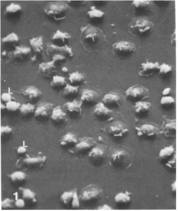
for nonspecific esterase, phagocytosis of antibody-coated red cells, and cytoplasmic spreading (1) (Figs. 1 and 2). Poor purity usually reflects inadequate rinsing of the flasks fol- lowing adherence.
Fig. 1. Scanning electron micrograph of freshly isolated monocyte undergoing cytoplasmic spreading. Most cells are monocytes, although two lymphocytes (L) are also seen, x 625.
isolated monocytes. x 6000.
III. CULTURE OF ISOLATED HUMAN BLOOD MONOCYTES A. Introduction
We have recently developed a method for prolonged in vitro culture of the purified human monocytes. Although reports of techniques for culturing monocytes have appeared in the lit- erature (4-10), none of these procedures in our hands has been consistently capable of establishing successful cultures. In- terestingly, each of the published methods are significantly different. Hence, we believe that small variations in tissue culture technique from laboratory to laboratory may have dra- matic effects on the success of culturing these notoriously
fastidious cells. We have developed a technique that results in successful establishment of primary monocyte cultures with virtually every attempt. In addition, the technique has been utilized in other institutions with comparable results.
B. Materials
Blood monocytes (purified as above)
Monocyte culture medium: Dulbecco's modified Eagle's medium with 10% fetal calf serum, 10% horse serum,
4 mAf glutamine, lx nonessential amino acids and anti- biotics
C. Procedures
1. Method. Monocytes purified by the above technique are resuspended in monocyte culture media at 0.5-1.0 x 10^ cells/
ml, and 1-2 ml are introduced into 16 millimeter Linbro wells.
Alternatively, 0.1-0.2 ml are added to microtiter wells.
These are placed in a humidified CO2 incubator at 37°C. Sev- eral aspects of this rather straightforward procedure require emphasis. First, strict pH control is required, particularly during the first 24 hr of culture. Variations from a range of 7.3 to 7.5 may seriously impair monocyte viability, es- pecially when more alkaline. Therefore, we recommend that the monocyte medium be preequilibrated in the C02 incubator prior to use. Also, we ensure that medium left overnight in the incubator is in fact at pH 7.3-7.5 (i.e., that the C02
flow and distribution are adequate). Alternatively, a non- bicarbonate buffer such as 20 mM HEPES may be used in a non- 002 incubator. Second, complete humidification of the incu- bator is mandatory, as monocytes are very sensitive to slight evaporation of the media. Third, serum type is critical. A mixture of horse serum and fetal calf serum is superior to either alone, and, in our experience, only very occasional batches of either are unsatisfactory. Fourth, the presence of contaminating lymphocytes or platelets has an unpredictable effect on the monocytes. Therefore, consistency of success depends on use of a purified cell population from the begin- ning of culture.
2. Long-term Culture. Under these culture conditions monocytes attach firmly to the substratum and begin to flatten onto it within 2 hr after introduction into tissue culture vessels. They begin to locomote slowly and randomly over the surface. When one cell meets another, they contact and often remain closely associated. By several days, some of the mono- cytes are grouped into small clumps adherent to the substra- tum while others remain as single cells. Simultaneously and beginning within 12 hr of the initiation of culture, the monocytes visibly begin to enlarge and to display phase-dense granules. After 4 to 5 days of culture, monocytes are ap- proximately two to three times their original size and begin
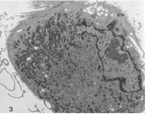
to show either a round or a fusiform shape. During the next week of culture, groups of monocytes begin to disperse so that by 2 weeks few if any clumps remain. The monocytes re- main tenaciously adherent and are evenly distributed across the substratum. As culture continues past 2 weeks, the mono- cytes continue to enlarge and many spontaneously fuse into giant multinucleated cells. Both round and fusiform shapes continue to be present, although round forms increase in num- ber with increasing duration of culture (see Fig. 3). Medium is not changed for 10 days, and thereafter cultures are fed weekly by replacement of half of the medium with fresh com- plete medium. Monocyte cultures generally remain apparently healthy for about 3 months, at which time the cells detach from the substratum and die despite continued feeding. We have not observed monocytes to divide at any time during rou- tine culture, either by morphologic or biochemical criteria (2).
Fig. 3. Transmission electron micrograph of human mono- cyte cultured 8 weeks, same magnification as Fig. 2. There is a large Golgi zone, numerous mitochondria, lysosomes, and extensive cytoskeleton. x 6000.
D. Critical Comments
We are presently evaluating several of the biochemical changes that occur during the first 2 weeks of monocyte cul- ture, as we believe that the cultured monocyte will prove to be an informative model of cellular differentiation. The re- lationship between in vitro alterations of monocyte morphology and biochemistry, as seen during culture, and true differenti- ation in vivo of blood monocytes to tissue macrophages, how- ever, is not known. Besides study of the process of differen- tiation, there are numerous other aspects of the monocyte cul- ture system that could be profitably examined. These include the process of cellular senescense, the changes in immunologie function that accompany in vitro maturation, and perhaps ulti- mately an alternative to cultured fibroblasts (11) for the study of metabolic processes and defects.
ACKNOWLEDGMENTS
Studies in the author's laboratory are supported by
grants from the United States Public Health Service, NIH 12478, March of Dimes-Birth Defects Foundation (6-246), the Kroc
Foundation and the
Steven H. Zuckerman is a fellow of the American Lung Associa- tion and Samuel K. Ackerman is an NIH Clinical Investigator, AM00642-01.
REFERENCES
1. S. K. Ackerman and S. D. Douglas. Purification of human monocytes of microexudate coated surfaces. J. Immunol.
120: 1372-1374, 1978.
2. S. H. Zuckerman, S. K. Ackerman, and S. D. Douglas.
Long-term peripheral blood monocyte cultures: Establish- ment, metabolism, and morphology of primary human mono- cyte-macrophage cell cultures. Immunology 38: 401-411, 1979.
3. R. J. Sanderson, F. T. Shepperdson, A. E. Vatter, and D. W. Talmadge. Isolation and enumeration of peripheral blood monocytes. J. Immunol. 118: 1409-1414, 1977.
4. W. D. Johnson, B. Mei, and Z. A. Cohn. The separation long-term cultivation and maturation of the human mono- cyte. J. Exp. Med. 146: 1613-1626, 1977.
5. Y. Rabinowitz. Separation of lymphocyte, polynophonuclear leukocytes, and monocytes on glass columns, including tis- sue culture observations. Blood 23: 811-826, 1964.
M. R. Lewis. The formation of macrophages, epithelial cells, and giant cells from leukocytes in incubated blood. Am. J. Pathol. 1: 91-99, 1925.
M. N. Goldstein. Formation of giant cells from human monocytes cultivated on cellophane. Anat. Rec. 118:
577-591, 1954.
P. Parakkal, J. Pinto, and J. M. Hanifin. Surface morphology of human mononuclear phagocytes during maturation and phagocytosis. J. Ultrastruct. Res. 48:
216-226, 1974.
L. P. Einstein, E. E. Schneeberger, and H. R. Colten.
Synthesis of the second component of complement by long term primary cultures of human monocytes. J. Exp. Med.
143: 114-126, 1976.
T. Hovi, D. Mosher, and A. Vaheri. Cultured human mono- cytes synthesize and secrete 2~macr°gl°t>ulin· J· Exp.
Med. 145: 1580-1589, 1977.
S. Yatziv, L. B. Epstein, and C. T. Epstein. Monocyte- derived macrophages: An in vitro system for studying hereditary lysosomal storage diseases. Pediatr. Res.
12: 939-944, 1978.