ICANCERRESEARCH56, 1902-1908.Ap@il15.19961
cell-platelet, tumor cell-endothelial cell, and tumor cell-ECM4 inter actions (9—13).The integrin aIIbj33 is expressed in human colon adenocarcinoma (14), prostate, melanoma, and breast cancer cell lines,5 suggesting that a11bf33may play an important role in tumor progression and metastasis.
Given the importance of integrins in various steps of the metastatic cascade, it is logical that factors that regulate integrin expression and function may have a significant impact on the metastatic process.
Integrin expression is controlled at the transcriptional level as well as the posttranscriptional levels. Several transcriptional regulators of integrin gene expression have been reported in the literature. Regu lators such as transforming growth factor @3(15), interleukin I (16), IFN--y (17), tumor necrosis factor-a (17), basic fibroblast growth factor ( 18), and platelet-derived growth factor ( 19) positively or negatively regulate integrin gene expression. However, these regula tors require treatmentof severalhours to days to alter integrin gene expression. A more rapid form of integrin regulation is at the post transcriptional level. This change in integrin expression is qualitative because it involves receptor translocation from an intracellular pool to the plasma membrane and vice versa. Mechanisms involved in post transcriptional regulation of integrins are not as well characterized as the transcriptional regulators. Previous data demonstrate that treat ment of tumor cells with the 12-LOX metabolite of arachidonic acid, 12(S)-HETh, induces posttranscriptional surface expression of inte grins within 15 mm (9, 10, 20—22).This effect is achieved primarily by increased translocation of the integrin from the cytoplasm to the cell surface (22). In addition, 12(S)-HETh is also capable of activat ing transcription of integrin genes as reported for av gene expression in microvascular endothelial cells (23).
Cumulative reports demonstrate that 12(S)-HETh is involved in all of the three major aspects of tumor cell invasion, i.e., adhesion, release of matrix degradative proteinases, and motility (2, 9, 10, 22).
Exogenous 12(S)-HETh stimulates tumor cell motility (24), whereas endogenously generated 12(S)-HETh mediates motility in responseto AMF (25). AMF is a member of a family of tumor cell cytokines that stimulates motility (26) via a receptor-mediated signaling pathway (27—30).Signal transduction following AMF binding to its cell sur face receptor, gp78 (AMFR), is mediated by a pertussis toxin-sensi tive G protein (31), phosphorylation of gp78 itself (28), inositolphos phate production (32), and activation of 12-LOX-mediated signaling, which involves activation of PKC (24, 25). Since AMF activates the
12-LOX signaling pathway, we questioned whether it could also regulate integrin expression and function. In this report, we provide evidence that AMF not only regulates motility, but it also modulates integrin-mediated tumor cell adhesion, spreading, and invasion.
4 The abbreviations used are: ECM, extracellular matrix; AMF, autocrine motility factor; COX, cyclooxygenase; 12-LOX, 12-lipoxygenase; PFA, paraformaldehyde; mAb, monoclonal antibody; PKA, cyclic AMP-dependent kinase; PKC, protein kinase C;
I2(S)-HETE, l2-(S)-hydroxyeicosatetraenoic acid; BHPP, N-benzyl-N-hydroxy-5- phenylpentanamide;SFM, serom-freemedia;ASA, acetylsalicylicacid.
5 Trikha, M., Chen, Y. Q., Timar, J., Szekeres, C., Bazaz, R., and Horns, K. V.,
unpublished observations.
1902
Autocrine Motility Factor Signals Integrin-mediated Metastatic Melanoma Cell Adhesion and Invasion1
Jozsef Timar,2 Mohit Trikha,2 Karoly Szekeres, Rajesh Bazaz, Jozsef Tovari, Steve Sifietti, Avraham Raz, and Kenneth V. Honn3
First Institute of Pathology and Experimental Cancer Research, Sensmelweis University of Medicine, Budapest, H-JOBS, Hungary (J. Ti., J. To.J: Departments of Radiation Oncology (M. T.. K. S., R. B., K. V. H.J and Pathology and Chemistry (K. V. HI, Wayne State University, Detroit, Michigan 48202; and Metastasis Research Program, Karmanos Cancer Institute, Detroit, Michigan 48201 (S. S.. A. R.J
ABSTRACT
The binding of autocrine motility factor (AMF) to its cell surface receptor, gp78, stimulates tumor cell motility. In this report, we provide evidence that stimulation of gp78 by either AMF or a monoclonal anti body to gp78 (3F3A) increases adhesion and spreading of metastatic murine melanoma (B16a) cells on fibronectin. This gp7S-regulated in crease is mediated by up-regulation of surface aIIbfi3 and a5@31integrin receptors. In addition, AMF treatment of B16a cells increased transloca tion of allbfI3 and a5fil from the cytoplasm to the cell surface. However, aIIbI@3and a5@1 demonstrate separate and unique staining patterns at the surface of B16a cells in response to stimulation of gp7S. Furthermore, stimulation of B16a cells with AMF increased their invasion through MatrigeL This stimulated invasion was inhibited by antibodies to a1Th133 but not by antibodies to aSfil. The increased integrin surface expression and function in response to AMF was blocked by N.benzyl-N.hydroxy-5- phenylpentanamide, an inhibitor of 12-lipoxygenase, and calphostin C, an Inhibitor of protein kinase C. The results demonstrate that AMF stimu lates integrin-mediated B16a cell adhesion, spreading, and invasion, and these events are regulated by a signaling pathway involving 12-lipoxyge nase and protein kinase C.
INTRODUCTION
Metastasis is a complex, yet well co-ordinated, process that can be defined by a series of integrated events ( 1): (a) tumor cells must adhere and spread on the extracellular matrix; (b) proteinases are necessary to degrade the matrix; and (c) tumor cells must migrate through the degraded matrix. Adhesion and spreading of tumor cells on the matrix is primarily mediated by integrins (1—4).These recep tom also play a pivotal role in initiating the proteolytic degradation of the matrix (5), thereby facilitating tumor cell invasion.
The @land (33 subfamilies of integrins play an important role in tumor invasion and dissemination (6). Qualitative and quantitative changes in expression of the fibronectin receptor (a5/31) are involved in the proliferative response of quiescent human melanoma cells (5, 7), whereas overexpression of /33 integrins directly correlates with metastatic potential in melanoma (2). The integrin aIIb(33 initially was believed to be expressed only on platelets (3). However, by using reverse transcription-PCR, Northern and Southern blotting, and im munoprecipitation, we demonstrated that this integrin receptor is expressedon metastatic B 16a melanoma cells (8). Furthermore, it was demonstrated that this receptor plays an important role in tumor
Received9/I 3/95; accepted2/I 5/96.
The costs of publication of this article were defrayed in part by the payment of page charges.This article mustthereforebe herebymarkedadvertisementin accordancewith 18 U.S.C. Section 1734 solely to indicate this fact.
I This work was supported by NIH Department of Health and Human Services Grants
CA-471 15-04 (to K. V. H.), CA-29997-08 (to K. V. H.), TW-285-02 (to K. V. H. and J. T.), and CA-5l7l4 (to A. R.); Thc @aulZuckermann Support for Cancer Research (to A. R.); NATO LG923 I I (K. V. H. and J. T.); and the Hungarian National Science Fund. T6336 (toJ.T.).
2 The first two authors contributed equally to this work.
3 To whom requests for reprints should be addressed, at Department of Pathology and
Chemistry,431 Chemistry,Wayne StateUniversity. Detroit, MI 48202. Phone:(313) 577-1018;Fax:(313)577-0798.
AMF REGULATES INTEGRIN EXPRESSION AND FUNCI'ION
mouse IgG conjugated to biotin (1:100 dilution) and streptavidin-Texas Red.
Stained cells were visualized through a Nikon epifluorescence microscope at x 100 magnification and photographed on KOdak Tmax 400 black and white
film. Direct labeling of B16a cells with secondary antibody-biotin conjugate
and streptavidin-Texas Red exhibited no significant background staining.
B16a MelanomaCell InvasionthroughMatrigel. This assaywasper
formed in Matrigel-coated Biocoat polycarbonate filter inserts (4 @.tm;Collab orative Research, Bedford, MA). These inserts were placed on top of 500 @d
of AMF containing SFM in a 24-well plate. B16a cells (5 X l0@'/200Ml) in SFM were added to the upper chamber of the insert. Tumor cells were
incubated in a tissue culture incubator for 12 h. Following incubation, tumor cells in the upper chamber were scraped off, and the bottom of the polycar bonate filter was stained with Diffquick solution (American Scientific Prod
ucts, McGraw Park, IL). The filter was removed from the insert and mounted on glass coverslides. Tumor cells that migrated through the insert were counted undera light microscope.Eachdatapoint wasperformedin triplicate.
Drug Treatment of B16a Cells. H8 (Seigaku America, Rockville, MD) was used to inhibit PKA activity at a concentration of 50 @LM(10). Calphostin
C (Calbiochem,La Jolla, CA) at a concentrationof 5 @Mwas used to inhibit PKCactivity(24). ASA (300 ELM;Ref. 10) andBHPP(I pM;Refs. 10 and34) were used to inhibit COX and 12-LOX activity, respectively. Optimal inhibitor
concentrations were determined in previous experiments reported in the above cited references.
RESULTS
Effect of AMF on B16a Cell Adhesion and Spreading on Im mobilized Fibronectin. B16a cells (2 X l0@/ml) after treatment with various concentrationsof AMF were added to fibronectin-coated wells, and the cell adhesion assay was performed as described in “Materialsand Methods.―The number of adherent cells per unit area was counted through a phase contrast microscope. AMF in a dose dependent manner stimulated adhesion of B16a cells to immobilized fibronectin (Fig. 1A). Maximum AMF-stimulated adhesion to fi bronectin was observed at a concentration range of 0.5—2.5ng/ml.
Since AMF stimulated B16a cell adhesion to fibronectin, we ques tioned whether it also could influence cell spreading. AMF in a dose-dependent manner increased spreading of B 16a cells on fi bronectin (Fig. 1B). At dosesofO.25—2.5ng/ml, AMF induced greater than an 8-fold increase in cell spreading as compared to spreading in the absence of AMF.
aIIb@33 andaSfll AreInvolvedin AMF-stimulated B16aCell
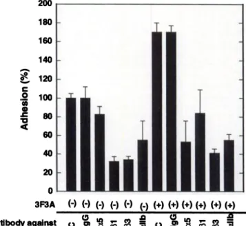
Adhesion to Immobilized Fibronectin. To determine which inte grins are involved in AMF-stimulated B16a cell adhesion to fibronec tin, we used adhesion blocking mAbs to the @31and J33subfamilies of integrins. Both mAbs to the @land to the (33integrins inhibited basal cell adhesion to fibronectin (in the absence of gp78 stimulation) as well as gp78-stimulated cell adhesion (Fig. 2). These findings suggest that AMF-stimulated B16a cell adhesion to fibronectin may involve both the f31 and f33 subfamily of integrins. To further determine which integrin receptor within the /33 subfamily is involved in AMF-stim ulated cell adhesion, we used adhesion blocking antibodies to aIIbf33.
mAbs to alib (AP-4; Fig. 2) and aIIb(33 (AP-2; data not shown) block AMF-stimulated cell adhesion to fibronectin. Furthermore, antibodies to the fibronectin receptor (polyclonal antibody to a5@l ; data not shown) and mAb 1 (anti-aS) also inhibit AMF-stimulated B16a cell adhesion to fibronectin (Fig. 2). Adhesion blocking antibody to avf33 was not used becausewe have determined previously that these cells lack av message(8). These results, collectively, suggestthat B16a cell adhesion to fibronectin in the presence or absence of AMF involves at least the a5@1 and aIIbj33 integrin receptors.
AMF Treatment of B16a Cells IncreasesSurface Expression of aIIbj33 and aSØl. Flow cytometry was usedto determinewhether the AMF-induced increase in adhesion and spreading was mediated by an up-regulation of aSf3l and/or aIIb@33.Tumor cells were treated
1903
MATERIALS AND METHODS
Murine Melanoma Cell Culture. Munne B 16a melanoma cells were obtained from the Animal and Human Tumor Bank National Cancer Institute (Frederick, MD). Tumor cells were cultured in RPMI containing 5% fetal
bovine serum (Life Technologies, Inc., Bethesda, MD) and antibiotics (22).
Confluent cells were harvested with 0.45 M EDTA and HBSS (EDTAIHBSS).
AMF Purification. AMF was purifiedfrom HT1O8Ohumanfibrosarcoma conditioned media as described previously (24). Briefly, semiconfluent HT1O8Ocells were washedwith PBS and cultured in SFM for 24 h. This
medium was replaced with fresh SFM, which was collected after 24 h.
Conditionedmedium was pooled, centrifuged,concentrated,and purifiedby molecular sieve chromatography as described (24).
Antibodies. Rat monoclonal 1gM antibody (3F3A), which stimulates AMF receptor (gp78) signaling, was prepared as described previously (27). Adhe sion blocking mAb to the fibronectin receptor (anti-a5) was purchased from
Oncogene Science (Uniondale, NY). A polyclonal antibody to the fibronectin
receptor a5f3l and mAb to the (31 integrin subunit were purchased from Life
Technologies, Inc. Nonimmunerat serumor IgG (MOPC21) was purchased from Sigma Chemical Co. (St. Louis, MO). AP-4 (m.Ab to olIb; Ref. 8), OPG-2 (mAb to @3;Ref. 8), and AP-2 (mAb to aI1b@3complex; Refs. 8 and 33) were generousgifts from Dr. ThomasKunicki (The ScrippsResearch Institute, La Jolla, CA).
Tumor Cell Adhesion and Spreading Assay. Bl6a cells in exponential growth phase were harvested with EDTA/HBSS and washed extensively with SFM. After washing, tumor cells were incubated at 37°Cfor 1 h prior to platingon fibronectin(5 g.@g/well)-coated24-wellplates.Fibronectincoatingof
24-well plates was performed by air drying wells containing 5 @gof protein in
PBS. Following incubation,Bl6a cells (l0@cells/500 ,.d SFM) were incubated on the fibronectin-coated well for 60 mm at 37°C.After incubation, wells were
washed three times with PBS to remove nonadherent cells. The remaining
adherent cells were fixed with 1% PFAIPBS for 10 mm. Two approaches were used to quantitate the number of adherent cells: (a) adherent cells per unit area were counted using a phase contrast microscope (X200); (b) adherent cells were stainedwith Giemsastainfor 5 mm, afterwhichexcess stainwas washed off, and absorbance at 600 nm was recorded by a Bio-Rad plate reader (Model 3550; Bio-Rad, Richmond, CA).
Cell spreading was determined as described previously (10). Briefly, ad herent cells were examined under X400 magnification, and the cell population was divided into round, intermediate, and fully spread groups. The spreading
factor was calculated by obtaining a ratio of the number of spread to corn pletely round cells. Three randomly selected areas per well were counted. For
every data point, at least 100 cells were counted, and each data point was performed in triplicate.
Flow Cytometry of B16a Cells. Bl6a cells were detachedwith EDTA/
HBSS, washedthreetimes with SFM,andallowedto recoverfor 1 h in a tissue culture incubator. Bl6a cells (I X 106)were treated with 3F3A (20 @.tg/ml)or
nonimmune rat serum (I :25 dilution) for 15 mm at 37°C and subsequently
washed with SFM and fixed with 1% PFA/HBSS for 10 mm. After fixation, cells were blocked with normal goat serum (1:2 dilution) for 30 mm and then incubated with AP-2 (20 @g/ml)or mAbl (20 @g/ml)for 1 h. Bound primary antibodywas detectedwith goat antimouseIgG conjugatedto biotin (1:200
dilution; Amersham, Arlington Heights, IL) and streptavidin-FITC (1:200 dilution; Amersham). Fluorescence of labeled B16a cells (1 X l0@) was
measured in a Coulter Epic flow cytometer (Coulter, Hialeah, FL), and the mean fluorescence was determined. Background fluorescence of labeled B 16a
cells was determined by incubating tumor cells with either nonimmune rat serum (1:25 dilution) or secondary antibody in the absence of primary antibody.
Immunofluorescence. B16a cells were plated on a fibronectin-coated glass
surface for 60 mm in the presence or absence of AMF (1.25 ng/ml) or 3F3A (20 @sg/ml).Intracellular proteins were detected by fixing the cells with a methanol/acetonesolution at —20°C(25). Plasma membraneproteins were detected by fixing tumor cells with 1% PFAIPBS solution for 10 mm. After fixation, cells were permeabilizedwith 0.5% TritonX-100 for 3 mm at room
temperature. Nonspecific binding was blocked by incubating immobilized cells
with normal goat serum diluted 1:2 in PBS for 30 mm. aIIb@33integrin was detectedby AP-2 (20 @g/ml),andfibronectinreceptor(a5f31)wasdetectedby mAb 1 (20 pg/mI). Bound primary antibodies were detected with goat anti
AMF REGULATES INTEGRIN EXPRESSION AND FUNC1'ION
presence or absence of 3F3A (Figs. 4, a and b). 3F3A treatment dramatically altered the cytoplasmic localization of aS(31. In the absence of 3F3A treatment, B16a cells demonstrated a distinct pe rinuclear staining pattern for the fibronectin receptor (Fig. 4a). How ever, after the addition of 3F3A, the fibronectin receptor stained with a diffuse cytoplasmic pattern (Fig. 4b). A similar response was observed for an intracellular aHb@3 pool when adherent B16a cells were labeled with AP-2 (data not shown). These morphological ob servations were quantitated by evaluating the ratio of dispersed versus perinuclear integrin staining in the tumor cell population. Table 1 indicates that treatment of B16a cells with 3F3A increased by 4-fold
3F3A (—(—(—(—(—(—) Antibody agalnat
@
@
@
@ @.
@ c.@
@
@ @.
@
Fig. 2. Inhibition of AMF-stimulated Bl6a cell adhesion to fibronectin by anti-integrin antibodies. B16a cells were preincubated in the presence or absence of nonimmune antibody, MOPC21 (20 @sg/ml),or the following adhesion blocking antibodies: anti fibronectin receptor antibody (anti-aS; mAb 1, 20 @ig/ml),anti-el (20 @g/ml),anti-f33 (OPG-2, 20 @sg/m1),or anti-aIIb (AP-4, 20 @g/ml)in the presence or absenceof 3F3A (20
@g/m1).A cell adhesion assay was performed as described in “Materialsand Methods.―
Adherent cells were stained with Giemsa, and absorbanceat 600 nm was recorded. Results are expressed as the change in the percentage of adhesion in the presence of various antibodies with respect to adhesion in the absenceof antibody. Each data point represents the mean of triplicate determinations; bars. SD.
3F3A (-) (+) (-) (+)
Antibody AP-2 mAbi
Fig. 3. gp78-stimulated increase in integrin surface expression on B16a cells. Bl6a cells (1 X 10⁶)were treated with normal rat serum (1:25 dilution) or anti-AMF receptor antibody(3F3A,20 @g/m1)for 15mm.After incubation,surfaceexpressionof allb(33and a5@l was detected by AP-2 and mAb 1, respectively. Flow cytometry was performed as describedin “Materialsand Methods.―Resultsare expressedas the meanof triplicate determinations of relative fluorescence; bars, SD.
200 180 160 140 120 100 80 60 40 20 0
C0
In
4@0
A
350300 250
@‘200 I 150
< 100 50 0
B
12
5..
0 0
@ U. 8
0)C 6
‘O 0
U)0. 2 0
(+)(+)(+)(+) (+)(+)
Fig. 1. Dose-dependent stimulation by AMF of B16a cell adhesion and spreading on fibronectin.Bl6a cells (1 X l0@/500p1) were suspendedin SFM or varying concentra tionsof AMF and addedin triplicate to 24-well platesprecoatedwith 5 @sgof fibronectin.
A, dose-dependent stimulation of adhesion of B16a cells to fibronectin. A cell adhesion assaywas performedas describedin “Materialsand Methods.―Resultsare expressedas the percentage increase in adhesion in the presence of various concentrations of AMF with respect to adhesion in the absence of AMF. B. dose-dependent stimulation by AMF of Bl6a cell spreading on fibronectin. Bl6a cells were treated with varying concentrations of AMF as in A, and a cell spreading assay was performed as described in “Materialsand Methods.―Results are expressed as the increase in spreading factor, which was deter minedasa ratio of spreadto roundcells. Threerandomareaswerecountedfor adhesion and spreading, and each data point is expressed as the mean of triplicate determinations;
bars, SD.
00
0U
0
0 U.
0 0
1904 0 0.25 0.5 1.25 2.5
AMF (ng/ml)
0 0.25 0.5 1.25 2.5
AMF (ng/ml)
with normal rat serum (1:25 dilution) or 3F3A (an AMF receptor stimulating antibody) for 15 mm and fixed with 1% PFAIPBS. Sur face expression of aHbf33 and a5@1 was detected by AP-2 and mAb 1, respectively. 3F3A treatment of B16a cells increased surface cx pression of both integrin receptors (Fig. 3). These findings suggest that AMF-increased adhesion and spreading of B16a cells on fi bronectin may correlate with increased surface expression of aIIb(33 and a5@3l.
Activation of gp78 Differentially Regulates Cell Surface Local ization of aS@31and allb@33. Flow cytometric analysis indicated that stimulation of gp78 receptor results in increased surface expression of a5J31and allb@33.Therefore, we used immunofluorescent staining to investigate whether this increase in surface expression of a5@l and aHbj33 was due to receptor translocation from the cytoplasm to the cell surface. To visualize the intracellular a5@l integrin pool, B16a cells were fixed with methanol/acetone and labeled with mAb 1 in the
AMF REGULATESINTEGRINEXPRESSIONAND FUNCTION
Fig. 4. Effect of gp78 ligation on cellular local ization of aHb@33and aSfil on B16a cells. Tumor cells were incubated on fibronectin-coated cover slips and stimulated with either nonimmune rat serum(1:25 dilution), 3F3A (20 @g/ml),or AMF (1.25 ng/ml) for 60 mm at 37'C. Integrins in the cytoplasm or at the plasma membrane were de tected by fixing adherent tumor cells with metha nol/acetone (a and b) or PFAiTriton (c-f), respec tively. Following fixation, fibronectin receptor and aHb@3 were detected by mAb 1 and AP-2, respec tively. Immunofluorescencewas performedas de scribed in “Materialsand Methods.―Detection of a cytoplasmic pool of a5@l receptor in untreated cells (a) and in 3F3A-treated cells (b). Detectionof a5@l receptorat the plasmamembranein untreated cells (c) and in AMF-treated cells (d). Detectionof a11b133at the plasmamembranein untreatedcells (e) and in AMF-treated cells (f). Single arrowhead, integrin localization at the cell periphery; double arrowhead, integrin localization at areas resem bling adhesioncontacts.N. nucleus;bar, 10 @.tm.
4
the number of cells with a dispersed a5@l pattern and by 7-fold in cells with a dispersed aIIb(33 pattern, suggesting that stimulation of gp78 induces translocation of integrins to the cell surface. Another interpretation of this finding is that AMF treatment induces a change in cell shape(Fig. 1B) that alters staining of integrin receptors without inducing translocation of integrins from an intracellular pool to the cell surface. However, flow cytometry (Fig. 3) indicates that 3F3A treatment of tumor cells increased surface expression of a5f31 and aIIbj33, and in addition, it has been reported that membrane translo cation of integrins can occur during cell spreading (10, 22). These findings, collectively, suggest that stimulation of gp78 in B16a cells results in increased cell spreading and translocation of integrins from the perinuclear zone to the cell periphery.
Since stimulation of gp78 resulted in an increased surface expres sion of aSfJl and aIIb$33, we determined subcellular staining for these integrins. Fibronectin-adherent Bl6a cells were permeabilized with
Triton X-l00 and labeled with antibodies to the fibronectin receptor (mAb 1) or to aIIbj33 (AP-2). B l6a cells in the presence or absence of AMF displayed no significant change in subcellular localization of a5@3l (Figs. 4, c and d). In contrast, labeling of aIIb(33 in untreated Bl6a cells was dramatically different when compared to AMF-treated cells. In the absenceof AMF stimulation, staining of aIIbf33 appeared to be uniformly distributed along the apical cell membrane (Fig. 4e).
In contrast, staining for aIIbf33 in the AMF-treated cells was concen trated to the peripheral regions of the cell membrane (Fig. 4f). These results demonstrate that AMF differentially regulates the localization of a5@3land cxllb(33.
aIIbfi3 Is Involved in AMF-stimulated B16a Invasion through Matrigel. Since antibodies to a11bf33 and to a5@l inhibited gp78- stimulated tumor cell adhesion (Fig. 2), we characterized their effect on tumor cell invasion through Matrigel. AMF (1.25 nglml) stimu lated Bl6a cell invasion through Matrigel, and antibodies to calib, (33,
1905
;-@@
IntegrinDispersion
ratiohControl3F3A-treateda5@l0.20
±0.020.93 ±
0.35aIIb@30.30
±0.092.09 ±0.40
600 400 200
2.0
0 1.5 UC 0U 0
g
U.
0
@ 0.5
0.0
DrugAdhesion (%)aSpreading factor@@Adhesion (%)Spreading factorEthanol100
±91.23± 0.31190± 125.26±
0.41ASAND'@ND154±
154.81±
0.13BHPPNDND82±
172.26± 0.18
AMF REGULATES INTEGRIN EXPRESSION AND FUNCTION
Table I Morphometric analysis of intracellular distribution of aJlb@33and a5j31 in
B/ba cells― tively, the data suggest that gp78 signaling is coupled to 12-LOX
activity but not to COX activity.
Effect of PKA and PKC Inhibitors on AMF-stimulated B16a Cell Adhesion and Spreading. It has been reported that AMF induced tumor cell motility involves PKC, but not PKA, stimulation (24, 25); therefore, we questioned whether AMF-stimulated cell ad hesion was mediated via the PKC or the PKA signaling pathway. To address this question, H8 was used to inhibit PKA activity, and calphostin C was used to inhibit PKC activity. Incubation of B16a cells with calphostin C (5 p.M) resulted in a complete inhibition of 3F3A-induced adhesion of tumor cells to fibronectin, whereas treat ment of tumor cells with H8 (50 pM) partially blocked 3F3A-stimu lated Bl6a cell adhesion to fibronectin (Table 3). In addition, cal phostin C, but not H8, blocked the spreading of Bl6a cells on fibronectin (Table 3). These findings, in combination with previous reports (24, 25), suggest that gp78-dependent adhesion, spreading, and motility are primarily coupled to PKC.
DISCUSSION
Integrins play a critical role in tumor cell growth and metastasis as a result of their involvement in tumor cell adhesion, spreading, and
3F3A (.) (+) (+) (+)
Drug (.) (.) BHPP ASA
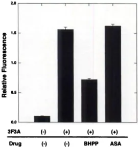
Fig. 6. Inhibition of gp78-induced increase in aIIbf33 integrin expression on B16a cells by 12-LOX and COX inhibitors. B16a cells (1 X l0@)were incubated with 3F3A (20
@.&g/ml)in the presence of BHPP (1 @sM)or ASA (300 @sM)for 15 mm. Following incubation, flow cytometry was performed on tumor cells as described in “Materialsand Methods.―Surface expression of aIIb@33was detected by AP-2 (20 @sWml).Results are expressedas the meanof triplicate determinationsof relative meanfluorescence;bars, SD.
Table 2 Role of 12-LOX and COX inhibitors in 3F3A-stimulated tumor cell adhesion and spreading
B16a cells, in the presence of nonimmune rat serum (1 :25 dilution. control) or 3F3A (20 giglml), were treated with BHPP (I MM), ASA (300 SM), or diluent (ethanol), and the cells were allowed to adhere to fibronectin. The cell adhesion and spreading assay was performedas describedin “Materialsand Methods.―The resultsare expressedas the means of triplicate determinations ±SD.
Control 3F3A
a B16a cells that had adhered to fibronectin were stimulated with 3F3A (20 @sg/ml) or
nonimmunerat serom(1:25 dilution) for 60 mm at 37'C, fixed with methanol/acetone, and stainedfor the cytoplasmicpool of a5f3l or aI1b@3as describedin “Materialsand Methods.―
b Dispersion ratio. the intracellular distribution of aIIb@3 and a5@l, was evaluated in 100 cells, and a dispersion ratio was determined. The dispersion ratio was calculated by obtaining a ratio of the number of cells demonstrating a dispersed intracellular integrin stainingpattern:thenumberof cellsdemonstratinga perinuclearintegrinstainingpattern.
Data are the means of triplicate determinations ±SD.
5.
0 U.
0 C.)0
0
.@
z
E 1200 1000 800AMF (@) (@) (@) (@) (+) (+)
a Adhesion was obtained by calculating the percentage of change in cell adhesion in
the presence of drug with respect to adhesion in the presence of the control diluent (ethanol).
b Calculated by obtaining a ratio of the number of spread to round cells that were adhered to fibronectin.
C ND, not determined.
1906 Antibody against 0 0 ‘°‘ .@ C')
Fig. 5. Inhibition of AMF-stimulated invasionby anti-integrin antibodies.B16a cells (5 X l0'@)were addedto Matrigel-coated filters in the absenceor presenceof AMF (1.25 ng/ml) and the following antibodies: nonimmune IgG (MOPC21, 20 @.&g/ml),anti-fl bronectin receptor (anti-aS; mAb 1, 20 @sg/ml),anti-@3l(20 @gIml),anti-alIb (AP-4, 20
@sg/ml),or anti-f33 (OPG-2, 20 @g/ml).Tumor cell invasion assay was performed as described in “Materialsand Methods.―Results are expressed as the number of cells that migrated through the Matrigel-coated filter. Each data point is the mean of triplicate determinations;bars, SD.
and aIIbj33 inhibited this invasion (Fig. 5). However, adhesion blocking antibodies to the fibronectin receptor (mAb 1) or anti-(31 did not block AMF-stimulated tumor cell invasion (Fig. 5). The number of B16a cells that invaded through Matrigel in the absence of AMF was so small that the effect of anti-integrin antibodies on basal invasion could not be determined accurately. These findings suggest that aIIbf33, but not a5(31, may be involved in AMF-stimulated tumor cell invasion through Matrigel.
Role of Arachidonic Acid Metabolites in gp78 Signaling. Pre vious studies demonstrated that AMF stimulates endogenous 12- LOX protein expression and 12(S)-HETE production (25), sug gesting that 12-LOX inhibitors may suppress gp78-mediated signaling. Therefore, events believed to be downstream of gp78- mediated signaling were classified into the following three cate gories: integrin expression; adhesion; and spreading. To investi gate the role of arachidonic acid metabolism in these events, BHPP (1 @.LM)and ASA (300 p.M) were used as 12-LOX and COX inhibitors, respectively. Pretreatment of B16a cells (15 mm) with BHPP inhibited gp78-mediated increase in aIIb(33 surface expres sion by approximately 50% (Fig. 6). In contrast, ASA (300 @.tM) treatment of B 16a cells did not inhibit the gp78-mediated increase in surface expression of aIIb/33 (Fig. 6). In addition, BHPP (1 @.tM), but not ASA (300 @.LM),blocked gp78-stimulated B16a cell adhe sion and spreading to immobilized fibronectin (Table 2). Collec
Table3 Roleofprotein kinaseinhibitors in 3F3A-stimulatedB16acelladhesionand spreadingB
16a cells, in the presence of nonimmune rat serum (1 :25 dilution. control) or 3F3A(20
@sg/ml),were treated with H8 (50 ,.LM),caiphostin C (5 @sM),or diluent (ethanol), andthe
cells were allowed to adhere to fibronectin. The cell adhesion and spreading assay wasperformed
as describedin “Materialsand Methods.―The resultsare expressedas themeans
of triplicate determinations ±SD.Control 3F3ADrug
Adhesion(%)“Spreadingfactor―Adhesion(%) Spreading factorEthanol
100 ±9 0.25 ±0.06 191 ±14 5.21 ±
1.29H8
ND' ND 132 ±10 6.05 ±
1.09Calphostin
C ND ND 60 ±15 0.33 ±0.09
CellMembrane
AMF REGULATESINTEGRINEXPRESSIONAND FUNCTION
adhesion and spreading is a consequence of surface up-regulation of aHbf33 and a5f3l. Furthermore,pretreatmentof Bl6a cells with antibod ies to a5/3l and aIIb[33 abrogateAMF-stimulated tumor cell adhesion, thereby demonstrating that AMF-stimulated tumor cell adhesion and spreading involve a5f31 and allb(33. This is in accordance with our
previous study reporting that l2(S)-HETE-stimulated B16a cell adhesion
and spreadingalso useallb(33 (10), implying that AMF and l2(S)-HETE participate in the same intracellular signaling pathway.
Results from flow cytometry and immunofluorescence indicate that stimulation of gp78 can induce translocation of both a5j31 and aIIb(33 receptors from the cytoplasm to the cell surface. Interestingly, it appears that stimulation of gp78 by either AMF or 3F3A, similar to the addition of exogenous 12(S)-HETh ( 10), differentially regulates subcellular localization of a5(3l and aIIb(33. Immunofluorescent staining indicates that AMF treatment of B 16a cells results in no significant change in subcellular localization of aS(3l . In contrast, membrane labeling of aIIbf33 after AMF treatment is markedly dif ferent from the staining of raIIbf33in untreated cells. In the absenceof AMF, aIIbf33 is uniformly distributed at the apical cell surface, whereas after AMF treatment, aIIb/33 distinctly localizes to the pe ripheral region of the cell membrane. Why a5(3l and aIIb(33 localize differently at the cell surface in responseto AMF is presently unclear.
We speculate that other signaling pathways that do not involve gp78, 12-LOX, and/or PKC may regulate surface distribution of a5/31.
Both AMF and 3F3A stimulate the motility of several metastatic tumor cells, including Bl6a cells (24, 25, 27—29).In this study, we demonstrate for the first time that AMF stimulates invasion of B16a cells through Matrigel and that antibodies to aIIbf33, but not to a5(3l, inhibit this AMF-induced invasion. Seftor el a!. (36) reported that invasion of human melanoma cells through Matrigel involves av(33 because antibodies to this receptor stimulated invasion. B l6a cells do not express message for av but instead express the aIIb/33 integrin receptor (8). In contrast with the findings of Seftor et a!. (36), we observed that antibodies to cxIIbf33 inhibited AMF-stimulated inva sion of B16a cells through Matrigel. Currently, the role of av(33 in AMF-stimulated melanoma cell invasion is under investigation in our
a Adhesion was obtained by calculating the percentage of change in cell adhesion in
the presence of drug with respect to adhesion in the presence of the control diluent (ethanol).
b Calculated by obtaining a ratio of the number of spread to round cells that were
adheredto fibronectin.
C ND. not determined.
migration (1, 2, 7, 35). Therefore, the discovery of signal transduction pathways that regulate integrin expression and function may have a major impact on tumor progression and invasion. The 12-LOX me tabolite of arachidonic acid, 12(S)-HETh, enhancesintegrin-mediated tumor cell adhesion to endothelial cells (2, 35) and to the ECM (2, 10, 35). Previously, we demonstrated that Bl6a cells possess the integrin aIIbj33, and they use this receptor to adhere to endothelial cells and to the subendothelial matrix (9—13).This adhesion could be enhanced with 12(S)-HETh treatment due to the increased translocation of aIIbj33 from a cytoplasmic pool to the cell surface (9, 22).
The literature indicates that AMF-stimulated tumor cell motility also can be induced by 12(S)-HETh, and this response is coupled to 12-LOX and PKC signaling events (24, 25). Since this signaling pathway is involved in integrin expression and function (9, 10, 20—
22), we hypothesized that AMF should also enhance integrin expres sion and function on tumor cells. In this study, we report for the first time that AMF mimics the 12(S)-HETE-mediated response in tumor cells in terms of stimulating tumor cell adhesion, spreading, invasion, and up-regulation of surface integrins. Results from flow cytometry suggest that the AMF receptor (gp78)-mediated increase in tumor cell
(d)
r_@—
@
I lntegrin
t'1 Pool
OUT gp78 )\ )\ lntegnns
(a)@
AMFcUoj
Fig. 7. Schematic representation of the proposed “autocrinemotility signaling pathway―that results in integrin-mediated tumor cell adhesion, spreading, and invasion. Signal transductionbeginswhenAMF bindsto its receptorgp78(a), therebyactivating 12-LOX (b), which resultsin the productionof 12(S)-HETE,andit activatesPKC (c). Activation of PKC results in translocation of the cytoplasmic pool of integrins (d) and gp78 (e) to the cell surface. Increased integrin expression at the cell surface regulates adhesion, spreading, and invasion.Dotted lines indicatethat thesemoleculesare not directly connectedto eachother.
1907
@HI
(e)
ArachidonicAcid I
(C) ,“
I
12(S)-HETE
_, (b)
I'
AMF REGULATEStN'rEGRINEXPRESSIONANDFUNCTION laboratory. Nonetheless, these observations imply that av(33 and
a11b133may transmit different intracellular signals during the process of tumor cell invasion. The finding that antibodies to cs5(3l did not block AMF-stimulated invasion could be attributed to at least two possibilities: (a) the concentration of fibronectin in Matrigel may be too insignificant to support binding of a5(3l on B16a cells; and (b) as mentioned above, surface localization of a5(31 may not be coupled to the gp78 signaling cascade.
gp78-stimulated tumor cell adhesion,spreading,and invasion are de pendent on 12-LOX activity. This is supported by the fact that a selective 12-LOX inhibitor BHPP (10, 34) blocked gp78-mediated increase in surfaceexpressionof allb(33. Also, BHPP inhibited adhesionand spread ing of B16a cells on fibronectin. Furthermore, BHPP inhibited AMF stimulated B16a invasion through Matrigel (data not shown). In contrast, a COX inhibitor, ASA, had no effect on AMF-stimulated surface up regulation of allb(33, adhesion,or spreading.These results in combina tion with previous reports (24, 25) demonstratethat 12-LOX is part of the gp78 signaling pathway, which is involved in AMF-stimulated tumor cell adhesion,spreading,and invasion.
Our previous reports demonstratedthat 12-LOX is upstream of PKC because12(S)-HFFE activates PKC (10, 21, 24, 34). Since gp78-medi ated signaling activates 12-LOX (24, 25), we predicted that ligation of this receptor should also activate PKC. A PKC inhibitor, calphostin C, blocks gp78-stimulated tumor cell adhesion and spreading. Furthermore, AMF treatment of B16a cells induces a translocation of PKC from the cytoplasm to the peripheral membranes(data not shown). Thesefindings suggestthat gp78-mediatedsignalingis coupledto 12-LOX, which in turn activates PKC in B16a cells. Based on data from this study in combination with previous reports, we propose the existence of an “autocrinemotility signaling pathway―involving (Fig. 7): (a) AMF binding to its receptor gp78; (b) signaling of 12-LOX to synthesize 12(S)-HErE (Fig. 6; Table 2; Refs. 24 and 25); (c) 12(S)-HETE-induced activation of PKC (10, 21, 24, 34); (d) translocation of PKC from subcellular structuresto lipid membranes(34); (e) PKC-dependenttrans location of allb(33, a5(31, and gp78 from intracellular pools to the cell surface (Fig. 4; Tables 1 and 3; Refs. 22 and 25); and (J) differential surface localization of a5(3l and allb(33 (Fig. 4) and increasedsurface expression of gp78 (24), which contributes to enhanced tumor cell adhesion,spreading,and invasion.
Tumor cell invasion is a complex multistep process that requires regulated expression of cellular adhesion molecules such as integrins.
Our data demonstrate that in B16a cells, AMF regulates integrin dependent tumor cell adhesion, spreading, and invasion by using the 12-LOX-PKC signaling pathway. Detailed analysis of this signaling pathway will further enhance our understanding of tumor cell invasion.
REFERENCES
1. Liotta, L. A. Tumor invasion and metastasis: role of the extracellular matrix. Cancer Res.,46: 1—7,1986.
2. Honn, K. V., and Tang, D. G. Adhesion molecules and tumor cell interaction with the endothelium and subendothelial matrix. Cancer Metastasis Rev., 11: 353-375, 1992.
3. Hynes, R. 0. Integrins: versatility, modulation, and signaling in cell adhesion. Cell, 69: 11—25,1992.
4. Smyth, S. S., Joneckis, C. C., and Parise, L. V. Regulation of vascular integrins.
Blood, 81: 2827-2843, 1993.
5. Seftor,R. E. B., Seftor,E. A., Stetler-Stevenson,W. 0., and Hendrix,M. J. C. The 72-Wa type IV collagenase is modulated via differential expression of av@3 and a5@l integrin expression during human melanoma cell invasion. Cancer Res., 53:
3411—3415,1993.
6. Kramer, R. H., Vu, M., Cheng, Y. F., and Ramos, D. M. Integrin expressionin malignant melanoma. Cancer Metastasis Rev., 10: 49—59,1991.
7. Ruoslahti, E. The Walter Herbert lecture: control of cell motility and tumor invasion by extracellular matrix mtegrins. Br. J. Cancer, 66: 239—242,1992.
8. Chen, Y. Q., Gao, X., Timar, 3., Tang, D. G., Grossi, I. M., Chelladurai, M., Kunicki, T. J., Fligiel, S. E. G., Taylor, J. D., and Horns, K. V. Identification of the ctllb@3 integrin in munne melanoma tumor cells. J. Biol. Chem., 267: 17314—17320,1992.
9. Chopra,H., Timar, J., Chen,Y. Q., Rong,X., Grossi,I. M., Fitzgerald,L. A., Taylor, J. D., and Honn, K. V. The lipoxygenase metabolite 12(S)-HETE induces a cytoskel eton-dependentincreasein surfaceexpressionof integrin allb@3on melanomacells.
Int. J. Cancer, 49: 774—786,1991.
10. Timar, J., Chen, Y. Q., Liu, B., Bazaz, R., Taylor, J. D., and Honn, K. V. The lipoxygenasemetabolite12(S)-HETEpromotesallb@3integrin-mediatedtumor cell spreadingon fibronectin.mt. j. Cancer,52: 594—603,1992.
11. Horns,K. V., c.hen,Y. Q., Timar, J., Onoda,J. M., Hatfield, J. S., Fligiel, S. E., Steinert, B. W., Diglio, C. A., Grossi, I. M., Nelson, K. K., and Taylor, J. D. allb@3 integrin expression and function in subpopulations of murine tumors. Exp. Cell Res., 201: 23-32, 1992.
12. Chang, Y. S., Chen, Y. Q., Timar, J., Nelson, K. K., Grossi, I. M., Fitzgerald, L A., Diglio, C. A., and Honn K. V. Increased expression of aHb@3 integrin in subpopu lationsof murinemelanomacells with high lung-colonizingability. mt. i. Cancer,51:
445—451,1992.
13. Tang,D. 0., Onoda,J. M., Steinert,B. W., Grossi,I. M., Nelson,K. K., Umbarger, L., Diglio, C. A., Taylor, J. D., and Honn, K. V. Phenotypicpropertiesof cultured tumor cells: integrin allb@3 expression, tumor-cell-induced platelet aggregation, and tumor-cell adhesion to endothelium as important parameters of experimental metes tasis. list. J. Cancer, 54: 338—347,1993.
14. Chang,S. Y., Chen,Y. Q., Fitzgerald.L. A., and Bonn, K. V. Analysisof integrin mRNA in human and rodent tumor cells. Biochem. Biophys. Res. Commun., 176:
108—113,1991.
15. Heino, J., and Massagu@,J. Transfonninggrowth factor
@ switchesthe patternof
integrinsexpressedin MG-63 humanosteosarcomacells andcausesa selectiveloss of cell adhesionto laminin. J. Biol. Chem.,264: 21806—21811, 1989.
16. Burrow, F. J., Haskard,D. 0., Hart, I. R., Marshall,1. F., Selkirk, S., Poole,S., and Thorpe,P. E. Influenceof tumor-derivedinterleukin-l on melanoma-endothelialcell interactionsin vitro. CancerRes.,51: 4768—4775,1991.
17. Defilippi, P., Truffa, G., Stefanuto, G., AltrUda, F., Silengo, L., and Tarone, G. Tumor necrosis factor a and interferon ‘ymodulate the expression of the vitronectin receptor (av@3) in human endothelialcells. J. Biol. Chem., 266: 7638—7645,1991.
18. Klein, S., Giancotti, F. 0., Presta, M., Albelda, S. M., Buck, C. A., and Rilkin, D. B.
Basicfibroblastgrowth factor modulatesintegrin expressionin microvascularendo thelial cells. Mol. Biol. Cell, 4: 973—982,1993.
19. Alden, K., and Rubin, K. Platelet derived growth factor-BB sthnulates synthesis of the integrin a2 subunit in human diploid fibroblasts. Exp. Cell Res., 215: 347-353, 1994.
20. Tang, D. G., Orossi, I. M., Chen, Y. Q., Diglio, C. A., and Bonn, K. V. 12(S)-HETE promotes tumor-cell adhesion by increasing surface expression of av@3 integrins on endothelial cells. mt. j. Cancer, 54: 102—111, 1993.
21. Tang, D. G., (len, Y. Q., Diglio, C. A., and Honn, K. V. Protein kinase C-dependent effects of 12(S)-HETE on endothelial cell vitronectin receptor and fibronectin recep tor. J. Cell Biol., 121: 689—704,1993.
22. Timar, J., Bazaz, R., Kinder, V. A., Haddad, M. M., Tang, D. 0., Robertson, D., Taylor, J. D., and Honn, K. V. Immunomorphological characterization and effects of 12(S)-HETEon a dynamic intracellular pool of the aHb@3-integrinin melanoma cells. J. Cell Sci., 108: 2175—2186,1995.
23. Tang, D. 0., Diglio, C. A., Bazaz, R., and Horns, K. V. Transcriptional activation of endothelial cell integrin av by protein kinase C activator 12(S)-HETE. J. Cell Sci., 108: 2629—2644, 1995.
24. Timar, 1., Silletti, S., Bazaz,R., Raz,A., and Horns,K. V. Regulationof melanoma cell motility by the lipoxygenase metabolite 12(S)-HETE. mt. j. Cancer, 55: 1003—
1010, 1993.
25. Silletti, S., Timar, J., Honn, K. V., and Raz, A. Autocrine motility factor induces differential 12-lipoxygenaseexpressionand activity in high- and low-metastatic Kl735 melanomacell variants.Cancer Res., 54: 5752—5756,1994.
26. Stocker,M., andGherardi,E. Regulationof cell movement:themotogeniccytokines.
Biochini. Biophys.Acta, 1072: 81—102,1991.
27. Nabi, I. R., Watanabe, H., and Raz, A. Identification of Bl6a-Fl melanoma autocrine motility-like factor receptor.CancerRes.,50: 409—414,1990.
28. Watanabe, H., Carmi, P., Hogan, V., Raz, T., Silletti, S., Nabi, I. R., and Raz, A.
Purificationof humantumor cell autocrinemotility factor and molecularcloning of its receptor. J. Biol. Chem., 266: 13442—13448,1991.
29. Watanabe,H., Nabi, I. R., and Raz, A. The relationshipbetweenmotility factor receptor internalization and the lung colonization capacity of murine melanoma cells.
Cancer Res., 51: 2699—2705,1991.
30. Silletti, S., and Raz, A. Autocrine motility factor is a growth factor. Biochem.
Biophys. Res. Commun., 194: 446—457,1993.
31. Stracke, M. L., Guirguis, R., Liotta, L. A., and Schiffman, E. Pertussis toxin inhibits stimulatedmotility independentlyof the adenylatecyclasepathwayin humanmela nomacells. Biochem.Biophys.Res.Commun.,146: 339—345,1987.
32. Kohn, E. C., Liotta, L. A., and Schiffman, E. Autocrine motility factor stimulates a threefold increase in inositol phosphate in human melanoma cells. Biochem. Biophys.
Res.Commun.,166: 757—764,1990.
33. Pidard, D., Montgomery, R. R., Bennett, J. S., and Kunicki, T. J. Interaction of AP-2, a monoclonalantibodyspecificfor the humanplateletglycoproteinlIb-ilIa complex, with intact platelets.J. Biol. Chem., 258: 12582—12586,1983.
34. Liu, B., Khan, W. A., Hannun, Y. A., Bazaz, R., Renaud,C., Stojakovic, S., Timar, J., Taylor, 3., and Honn, K. V. l2(S)-HETE and 13(S)-HODE regulation of protein kinase C a in melanoma cells: role of receptor mediated hydrolysis of inositol phospholipids. Proc. NatI. Acad. Sci. USA, 92: 9323-9327, 1995.
35. Tang, D. 0., and Honn, K. V. Adhesion molecules and tumor metastasis: an update.
Invasion Metastasis,14: 109—122,1995.
36. Seftor,R. E. B., Seftor,E. A., Gehlsen,K. R., Stetler-StevensonW. 0., Brown, P.D., Ruoslahti, E., and Hendrix, M. J. C. Role of the av@3 integrin in human melanoma invasion. Proc. Natl. Acad. Sci. USA, 89: 1557—1561,1992.
1908