OBTAINING AND CULTURING MURINE MONOCYTES
Carleton C. Stewart
INTRODUCTION
Murine monocytes, like other sources of mononuclear phago- cytes, will proliferate and form colonies when they are grown in medium containing fetal calf serum and L-cell conditioned medium (1,2), a potent source of macrophage growth factor (3).
Monocytes are probably the immediate precursor to the tissue macrophage (4). The colony-forming cells found in the tissues
are also the most likely descendants of monocytes.
Due to the small quantity of blood that can de derived from mice, we have tried to optimize our procedures for the greatest yield of mononuclear cells. We shall describe the methodology for isolating and culturing murine blood monocytes.
METHODS FOR STUDYING Copyright © 1981 by Academic Press, Inc.
MONONUCLEAR PHAGOCYTES 21 AU rights of reproduction in any form reserved.
ISBN 0-12-044220-5
I I . REAGENTS
All the reagents used to culture bone marrow cells will be used to culture monocytes. Refer to Chapter 2 for their pre- paration. In addition to those reagents it is necessary to prepare other solutions for separating peripheral blood mono- nuclear cells.
A. Ficol1-Hypaque
Prepare 100 ml of stock Ficoll solution by dissolving 9 gm Ficoll (Sigma Chemical Co., St. Louis, Missouri) in 100 ml dis- tilled water. Heat to 40°C with stirring to dissolve. Steri- lize by passing through 0.45 ym micropore filter. To prepare the gradient, mix together 7 ml of Ficoll, 2 ml of 50% sodium hypaque (Winthrop Laboratories, New York) and 1 ml of distilled water. Shake the solution vigorously to make sure the ingre- dients are well mixed.
B. OLMOPS Preparation Medium
The organic buffer, 2-iV-morpholinopropane sulfonic acid (MOPS), is used for the manipulative procedures so that the pH of the medium remains stable in an air atmosphere. This medium must not be used in a C02~containing atmosphere. It is our ex- perience that cell survival is exquisitely dependent on a
stable pH within the physiologic range of 7.0-7.4.
Mix 1 gm aMEM powder without sodium bicarbonate with 0.4 gm MOPS, 0.04 gm NaCl, 0.27 ml of 5 N NaOH, 2 ml 100* penicillin-
streptomycin solution (GIBCO) and 98 ml distilled water.
Filter and sterilize the medium by passing it through a 0.22 ym micropore filter. The additional sodium chloride is required to produce a final osmolality of 300±5 Mosm and the NaOH is necessary to produce a pH 7.2-7.4.
C. Removing Adherent Monocytes (a) Lidocaine solution (12 mM)
Mix together 1.7 ml of 2% lidocaine-HCl (Astra Pharmaceutical Products, Inc., Framingham, Massachusetts), 1 ml fetal or newborn bovine serum and 7.3 ml otMOPS.
(b) Ethylenediaminetetraacetic acid solution (mO mM) Mix together 0.37 gm EDTA (disodium), 0.85 gm NaCl, and 100 ml distilled H20. Adjust the pH to 7.2 with 1 N NaOH.
(c) Sodium Pyrophosphate (10 ταΜ)
Mix together 0.45 gm tetrasodium pyrophosphate (Sigma T-6379) and dissolve in 100 ml phosphate-buffered saline. Adjust pH to 7.2 with about 1 ml of 1 N HCl.
Filter sterilize all solutions by passage through a 0.22 ym micropore filter.
III. PROCEDURES
A. Humidity Chambers: See Chapter 2.
B. Obtaining Blood Mononuclear Cells
First read all the procedures listed below. Assuming a recovery of 0.5 ml of whole blood per mouse, add a volume of OMOPS, containing 20 U heparin/ml, equal to the amount of blood desired. We usually put no more than 7 ml heparinized otMOPS and 7 ml blood per 15 ml conical centrifuge tube. For example, if 8 ml of blood are required from 16 mice, we would add 4 ml heparinized aMOPS to two tubes and put 4 ml blood in each.
Anesthetize the mouse using ether, pin it supine to a board, and wash thoroughly with 70% EtOH, maintain anesthesia by placing an etherized gauze pad in a 50-ml beaker over its nose. Cut a midline incision in the chest wall and expose the heart and lungs. Insert a 26-gauge needle on a 1-ml syringe into the left ventricle and aspirate the blood. When aspira- ting the blood from the heart, use gentle suction so as not to collapse the heart. Put blood in the heparinized OtMOPS (at room temperature).
In these separation procedures we shall account for all cells to determine the actual recovery of monocytes. After collecting all the blood, pool it and obtain a cell count (use the pronase cetrimide procedure outlined in Chapter 2 for counting cells). Also prepare two or more slides (using a cytocentrifuge, if possible. Shandon Southern Instruments, Inc., 515 Broad St., Bewickley, Pennsylvania 15143). The cells will be stained later for esterase activity. Overlay up to 10 ml of the cell suspension over 5 ml Ficoll-Hypaque gradient solution in a 15-ml conical centrifuge tube. Make sure all solutions remain at room temperature. Centrifuge cells at 1200 g for 10 min. This procedure is different than the stan- dard procedure originally described by Boyum (5), but we have found it satisfactory for separation of murine mononuclear cells. If difficulty is encountered, more or less, water may be added to the 7 parts of Ficoll and 2 parts of Hypaque to
change the density of the solution. After centrifugation, a band of mononuclear cells and platelets should be found with little erythrocyte contamination. Withdraw the medium/plasma layer to within 1 cm of the gradient mixture and discard.
Withdraw the gradient solution containing the mononuclear cells to within 1 cm of the erythrocyte layer and put it in a 50-ml conical centrifuge tube (Falcon No. 2070). Pool up to four gradients into one centrifuge tube and then bring the volume to 50 ml with aMOPS. Mix thoroughly to eliminate any possi- bility of reforming a gradient. Centrifuge cells at 200 g for 10 min at 4°C. Withdraw supernatant to 0.1 cm of the pellet.
Resuspend pellets and pool all samples into one, bring to 50 ml with aMOPS and centrifuge again. Now resuspend the pellet in aMOPS using a volume of 1 ml times the number of mice used.
At this point obtain another cell count and prepare two more slides using the cytocentrifuge. All slides should be stained using the esterase staining procedure outlined in Morahan's Chapter.
C. Colony Formation of Monocytes in Agar Culture
The method for separating monocytes from lymphocytes will be described later. Prepare a 24-ml stock solution of 2 x 10⁵ mononuclear cells/ml in Agar Growth Medium. Add 3 ml of this
suspension to 3 ml of Agar Medium, mix and plate 1 ml into 4-35 mm culture dishes. Place in two humidity chambers and incubate. On days 21 and 28, count the number of colonies that have formed using a dissecting microscope. Since the lymphocytes do not proliferate in the growth medium, their presence can be ignored and the total mononuclear cell fraction can be used.
Colonies may be fixed in situ from two cultures each time by adding 1 ml of 3% glutaraldehyde. Colonies may also be re- moved using a Pasteur pipette and, after preparing a cytocentri-
fuge preparation, cells may be processed for cytological iden- tification.
D. Colony Formation in Liquid Culture
Colony formation by the total mononuclear cell population or by the adherent and nonadherent fractions may be obtained as follows: Make a 1/100 dilution of the suspension prepared in Section III. C by adding 0.15 ml of cells to 15 ml liquid growth medium. Mix thoroughly and plate 3 ml of cells into 4-35 mm culture dishes, label two dishes "TOTAL" and place them in a humidity chamber. Incubate all dishes at 37°C for 1 hr. Remove the two unlabeled dishes, resuspend the cells in
the overlying medium and transfer them to new 35-mm dishes labeled "nonadherent cells," Immediately add 3 ml liquid growth medium to the empty dish. Label it "adherent cells."
Keep dishes paired by designating them A and B. Put each pair in a humidity chamber and incubate them all for 14 days.
Plates may be stained directly with méthylène blue, Wrights blood stain, or giemsa stain. The phagocytic activity of the adherent cells can also be determined prior to staining. First, rinse the cultures to remove nonadherent cells. Cytocentrifuge preparations may be made of these nonadherent cells. Add 1 ml medium containing 10% fetal or newborn bovine serum and 1%
freshly reconstituted (with distilled water, do not use re- storing solution as it contains sodium azide) guinea pig com- plement to the adherent cells. Add 50 yl of autoclaved bakers
(Chapter 2) yeast and mix. Incubate the cells for 30 min at 37°C. Remove the cultures, rinse them three times with 3 ml saline each time and add 2 ml méthylène blue solution, Chap- ter 2. After 20 min, discard stain, rinse with running tap water, and air dry.
E. Growth of Mononuclear Cells in Liquid Culture
Dilute the cells prepared in Section III.C by adding 18 ml of a-0 to 18 ml of the cell suspension in a 50-ml conical cen- trifuge tube; these cells are now at a concentration of 105 cells/ml. Then add 3 ml of this suspension to 27 ml of liquid growth medium; these cells are now at a concentration of 104 cells/ml.
Pipette 3 ml of cells at 10 cells/ml into 6-35 mm culture dishes labeled 10 ; pipette 3 ml of cells at 104 cells/ml into 12-35 mm dishes labeled 10 . Place cultures into humidity chambers and incubate them. The adherent and nonadherent cell count can be made every two days using two cultures each day.
For days 0, 2, 4, 6, and 8 use dishes plated with 3 x 10⁵ cells (labeled 10^) and, for days 8, 10, 12, 14, use dishes plated with the lower cell concentration. Since both concentrations are counted on day 8, a comparison of the growth kinetics of the two groups can be made. To count cells, resuspend the non- adherent cells in the growth medium and transfer them to a 7-dram vial. Rinse the plates with 2 ml of aMOPS and pool it with the nonadherent cells. Immediately add (so it will not dry) to the culture dish containing the adherent cells 1 ml pronase diluted 1 to 10 with aMOPS. Then add 0.5 ml undiluted pronase to the vial containing the 5 ml of nonadherent cells.
Incubate vials and dishes at 37°C for 15 min. Add 10 ml cetrimide to the vial of nonadherent cells and count them (or count directly with hemocytometer). Rinse off adherent cells as follows: Transfer diluted pronase in culture dish to a
7-dram counting vial. Add 2 ml of cetrimide solution and sys- tematically rinse the plate using a transfer pipette. We generally rinse the dish in a clockwise fashion beginning at 1:00 and proceed around the plate to 12:00 and then end up in the center of the dish. Be careful not to create air bubbles by always keeping the pipette tip submerged. This systematic rinse is important because macrophages are extremely adherent and they will not be quantitatively removed unless this pro- cedure is strictly followed. Do not scrape bottom of dish with pipette tip to avoid plastic particles. This procedure takes about 1 min/plate, therefore, if a large number of plates are going to be treated, they should be processed in batches of ten so that none of the plates are incubated longer than 25 min with pronase. For electronic particle counting, transfer the 2 ml of nuclei to 8 ml of cetrimide. For hemo- cytometer counting, count directly without further dilution.
On days 1-6 counts will be low due to cell death. Extra cul- tures can be prepared for specific mononuclear phagocyte mar- ker or functional studies.
F. Separation of Monocytes from Lymphocytes
The procedure we have developed for isolating monocytes from lymphocytes utilizes the adherence properties of both cell types. It has the advantage that all cells can be ac- counted for even if they are lost. The procedure has the dis- advantage that monocytes, once adherent, must be removed and most methods for removing them do not always give good viable cell recoveries.
There are two populations of adherent monocytes: One population will adhere in the presence of serum and the second population will adhere only in the absence of serum. Lympho- cytes will adhere under either of these conditions and the monocyte preparations are contaminated by them. In the follow-
ing procedure it is possible to obtain a highly enriched popu- lation of monocytes.
After counting the cells in Section III.B, adjust them in OMOPS, so that NO MORE than 105 cells/cm2 will be plated.
This is important because a subconfluent monolayer of cells is required if all monocytes are to be recovered. The wells of a 24-well cluster plate (Costar No. 3524) have a surface area of 2 cm2 (diameter = 16 mm). This plate will be used to obtain the monocytes. The procedure outlined is designed to account for all the mononuclear cells and their purity as well as to obtain monocytes for the desired experiments.
Place 12-mm round coverslips (Belco Glass Co., Vineland, New Jersey) into the four wells Al through Dl of the cluster plate. Coverslips may be sterilized by quickly passing them through a flame.
Prepare a 50 ml mononuclear cell suspension at 10^/ml and plate 2 ml in each well (105 cells/cm ). Use about eight mice for this number of mononuclear cells.
Incubate the cells for 1 hr at 37°C. Rinse the nonadherent cells from each well and pool them as follows: Resuspend the nonadherent cells in overlying medium doing one well at a time;
after removing cells, immediately add 1 ml OMOPS containing 10%
newborn bovine serum to each well so the adherent cells do not become dry. Pool all nonadherent cells. At this point, most monocytes have adhered but so have a significant number of lymphocytes.
Remove the coverslips from wells Al and Bl. Mount the coverslips cell side up on the end of a microscope slide using permount and fix them. Also remove the adherent cells from wells A2 and B2 using cetrimide (Section III.E) to determine the actual number of cells that have adhered. Incubate the plate at 37°C in humidified air overnight. Do not use a C02
incubator.
Prepare two cytocentrifuge preparations of the nonadherent cells and count 2 ml of the suspension (Section III.E) to de- termine the actual number that were removed. These nonadherent cells can be used to determine if any will form colonies (see Section III.D for details). To do this, centrifuge 200 g for 10 min and resuspend pellet in liquid growth medium to give 7 ml at 104 and 7 ml at 10 cells/ml. Plate 3 ml into two 35-mm dishes each, place them in humidity chambers and incubate for 14 days. Stain and count colonies. Correlate esterase positive cells in the original sample of nonadherent cells with the number of colonies formed.
Stain all slides and mounted coverslips for nonspecific esterase using the procedures outlined in Morahans Chapter.
Determine the number of esterase positive cells in each frac- tion: Number of esterase positive cells = fraction positive x number of cells/well.
After overnight incubation, rerinse each well with aMOPS pooling the nonadherent cells as before. Add OMOPS immediately after rinsing so the monolayer does not dry out. Remove cover- slips from wells Cl and Dl mount and fix them and count the number of adherent cells in wells C2 and D2 as before. Prepare cytocentrifuge preparations and count the cells in the nonad- herent fraction. Determine nonadherent cells that will form colonies as described above. The remaining 16 wells containing monocytes can be used for functional studies.
We have found no procedure for removing all monocytes in a viable state. We have, however, used lidocaine, EDTA, or so- dium pyrophosphate with varying degrees of success using the following procedure:
Remove the medium overlying the adherent cells, discard, and add 0.5 ml of either lidocaine, EDTA, or sodium pyrophos- phate. Do not treat any more wells than can be processed in 5 min. Incubate these cells for 10 min and then add 1.0 ml OMOPS containing 10% newborn bovine serum (alOMOPS) and rinse the surface of the wells systematically and pool them. After removal of cells, put 0.5 ml of the suspension in 10 ml cetri- mide for a cell count and then dilute the rest of the cells threefold in alOMOPS to further reduce the drug concentration.
Centrifuge the cells for 10 min at 150 g at 40°C.
Rinse two of the treated wells with cetrimide and, along with 0.5 ml of the sample of removed cells, count them to de- termine the recovery: Fraction recovery = cells removed/(cells removed + cells still adherent).
After centrifuging the cells, resuspend them to the desired concentration. We usually replate two wells with 105 cells to determine the number that will readhere after overnight culture.
The number that will form colonies can also be determined using 10 , 10J, and 10 cells/ml and plating 3 ml into 35-mm culture dishes. These two parameters, readherence and colony formation, provide stringent assessments of the recovered cell viability.
IV. CONCLUDING REMARKS
We have described how to isolate monocytes, how to account for all the cells, and how to culture them. A typical account- ability chart is shown in Table I for C^H mice. The actual
TABLE I. Isolation of Mononuclear Cells from Murine Blood Cells/ml blooda
Cell fraction (millions) Peripheral blood 9.75 ± 0.88
Mononuclear cells 2.87 ± 0.82 Erythrocyte pellet 1.90 ± 0.32 Total cells 4.77 ± 11.13 Recovery (%) 49.0 ± 16.0
a Number of cells per milliliter of the original blood that were recovered in each fraction (± standard error, four experi- ments) .
number of cells recovered after each step from the original number of cells per milliliter of blood is shown. The cumula- tive recovery is shown by the last number. In our experience about half the cells are lost during isolation.
The distribution of adherent cells is shown in Table II.
When serum was absent, 41% of the mononuclear cells adhered and 38% of the adherent cells were monocytes (esterase posi- tive) . This represents virtually all the monocytes as none were found in the nonadherent fraction. Thus 15% of the mono- nuclear cells in the suspension were monocytes (41% x 0.38).
It should be pointed out, however, that if the number of cells plated greatly exceeds 105 cells/cm2, an increasing number of monocytes will be found in the nonadherent fraction because there are too many cells plated for the amount of surface area available for them to adhere. Nonadherent cells as well as platelets in the mononuclear cell fraction will compete for space interfering with monocyte adherence.
When serum is present, adherence falls to about 9% of the mononuclear cell fraction. While two-thirds of the adherent cells are monocytes, only about 40% of the monocytes were ad- herent, i.e., 60% of them were found in the nonadherent frac- tion. Because fewer lymphocytes also adhered, the monocyte purity was much better.
It is possible to remove the adherent lymphocytes. By adding medium with serum to the cultures after the initial 1-hr incubation in serum-free medium, culturing overnight and rerinsing, virtually all lymphocytes can be removed leaving a highly purified monocyte preparation.
With regard to adherence properties, it follows that there are two monocyte populations, those which adhere in serum and those which do not. These two populations can be separated from one another by first adhering the cells in serum-contain- ing medium to give the serum-adherent monocyte population.
The nonadherent cells derived from the cultures are then washed, resuspended, and replated in serum-free medium. After an addi-
TABLE JJ. Adherence of Mononuclear Cells
Percentage of monocytes Serum Adherent Monocytes Adherent Nonadherent
(V (V (%)
0 41 38 >99 <1 1 9 69 40 60 10 9 58 33 67
tional hour of incubation, the serum-nonadherent monocyte popu- lation can be obtained. To remove adherent lymphocytes, both adherent cell populations are incubated overnight with aMOPS containing 3% serum, the plates are rerinsed to remove lympho- cytes, and fresh medium is added.
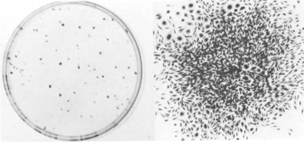
The formation of colonies in the different fractions are shown in Table III for C3H mouse cells. The appearance of colonies is shown in Fig. 1. Colony formation by monocytes is a very sensitive indicator of monocyte contamination because they do form colonies. For the experiment shown, 104 mononu- clear cells produced one colony in the nonadherent and 72 in the adherent fraction. However, monocytes clearly went through the Ficoll-Hypaque into the erythrocyte fraction during separa- tion as 45 colonies/104 nucleated cells were found in that fraction.
We attempt to plate the density of mononuclear cells that will give the desired number of monocytes so they will not have to be removed. This is because the recovery of viable monocytes, once they have attached, is not predictable and usually only about half the cells recovered are viable. While the viability of a freshly isolated preparation is often great- er than 90% initially, it has been our experience that by the next day many of the previously viable cells have died; this is likely due to the fact that it takes time for cells to die once they have acquired lethal damage.
TABLE III. Colony Formation by Murine Monocytes
Colonies per 10 cells 1 Hour
Nonadherent mononuclear cells 1 Adherent mononuclear cells 72
Erythrocyte fraction 45 24 Hours
Nonadherent mononuclear cells 2 Adherent mononuclear cells 22
Fig. 1. Colony formation by murine peripheral blood mono- nuclear cells. 104 Peripheral blood mononuclear cells were cultured 14 days in 3-ml liquid growth medium. Colonies as they appear on the culture dish after staining with méthylène blue are shown on the left and a photomicrograph of one of the colonies is shown on the right. Note the larger polyhedral like cells particularly in the upper left corner. The majori- ty of cells, however, have an ameboid like morphology.
REFERENCES
1. H. Lin. Colony formation in vitro by mouse blood mono- cytes. Blood 49:593-598, 1977.
2. C. C. Stewart. Formation of colonies by mononuclear phagocytes outside the bone marrow. In "Mononuclear Phagocytes: Functional Aspects" (R. van Furthf ed.), pp. 377-413. Martinus Nijhoff, Boston, 1980.
3. C. C. Stewart and H. -S. Lin. Macrophage growth factor and its relationship to colony-stimulating factor.
J. Reticuloendothel. Soc. 23.-269-285, 1978.
4. R. van Furth. Cells of mononuclear phagocyte system.
Nomenclature in terms of sites and conditions. In "Mono-
nuclear Phagocytes: Functional Aspects" (R. van Furth, ed.), pp. 1-31. Martinus Nijhoff, Boston, 1980.
5 . A. Boyum. I s o l a t i o n o f mononuclear c e l l s and g r a n u l o c y t e s from human b l o o d . Scand. J. Clin. Lab. Invest. 21 (Suppl. 97)ill, 1 9 6 8 .