REVIEW
Advancing brain barriers RNA sequencing:
guidelines from experimental design to publication
David M. F. Francisco1†, Luca Marchetti2†, Sabela Rodríguez‑Lorenzo3†, Eduardo Frías‑Anaya4, Ricardo M. Figueiredo5,6, BtRAIN Network, Peter Winter5, Ignacio Andres Romero4, Helga E. de Vries3, Britta Engelhardt2* and Rémy Bruggmann1*
Abstract
Background: RNA sequencing (RNA‑Seq) in its varied forms has become an indispensable tool for analyzing differential gene expression and thus characterization of specific tissues. Aiming to understand the brain barriers genetic signature, RNA seq has also been introduced in brain barriers research. This has led to availability of both, bulk and single‑cell RNA‑Seq datasets over the last few years. If appropriately performed, the RNA‑Seq studies provide powerful datasets that allow for significant deepen‑
ing of knowledge on the molecular mechanisms that establish the brain barriers. However, RNA‑Seq studies comprise complex workflows that require to consider many options and variables before, during and after the proper sequencing process.
Main body: In the current manuscript, we build on the interdisciplinary experience of the European PhD Training Network BtRAIN (https ://www.btrai n‑2020.eu/) where bioinformaticians and brain barriers researchers collaborated to analyze and establish RNA‑Seq datasets on vertebrate brain barriers. The obstacles BtRAIN has identified in this process have been integrated into the present manuscript. It provides guidelines along the entire workflow of brain barriers RNA‑Seq studies starting from the overall experimental design to interpretation of results. Focusing on the vertebrate endothelial blood–brain barrier (BBB) and epithelial blood‑cerebrospinal‑fluid barrier (BCSFB) of the choroid plexus, we provide a step‑by‑step description of the workflow, highlighting the decisions to be made at each step of the workflow and explaining the strengths and weaknesses of individual choices made. Finally, we propose recommendations for accurate data interpretation and on the information to be included into a publication to ensure appropriate accessibility of the data and reproducibility of the observations by the scientific community.
Conclusion: Next generation transcriptomic profiling of the brain barriers provides a novel resource for understand‑
ing the development, function and pathology of these barrier cells, which is essential for understanding CNS homeo‑
stasis and disease. Continuous advancement and sophistication of RNA‑Seq will require interdisciplinary approaches between brain barrier researchers and bioinformaticians as successfully performed in BtRAIN. The present guidelines are built on the BtRAIN interdisciplinary experience and aim to facilitate collaboration of brain barriers researchers with bioinformaticians to advance RNA‑Seq study design in the brain barriers community.
© The Author(s) 2020. This article is licensed under a Creative Commons Attribution 4.0 International License, which permits use, sharing, adaptation, distribution and reproduction in any medium or format, as long as you give appropriate credit to the original author(s) and the source, provide a link to the Creative Commons licence, and indicate if changes were made. The images or other third party material in this article are included in the article’s Creative Commons licence, unless indicated otherwise in a credit line to the material. If material is not included in the article’s Creative Commons licence and your intended use is not permitted by statutory regulation or exceeds the permitted use, you will need to obtain permission directly from the copyright holder. To view a copy of this licence, visit http://creat iveco mmons .org/licen ses/by/4.0/. The Creative Commons Public Domain Dedication waiver (http://creat iveco mmons .org/publi cdoma in/
zero/1.0/) applies to the data made available in this article, unless otherwise stated in a credit line to the data.
Open Access
*Correspondence: bengel@tki.unibe.ch; remy.bruggmann@bioinformatics.
unibe.ch
†David M. F. Francisco, Luca Marchetti and Sabela Rodríguez‑Lorenzo contributed equally to this work
1 Interfaculty Bioinformatics Unit and Swiss Institute of Bioinformatics, University of Bern, Bern, Switzerland
2 Theodor Kocher Institute, University of Bern, Bern, Switzerland Full list of author information is available at the end of the article The members of the BtRain Network are listed in the Acknowledgements.
Background
Brain barriers: terms and definitions
Central nervous system (CNS) homeostasis is ensured by endothelial, epithelial, mesothelial and glial brain bar- riers that divide the CNS into compartments [1]. CNS barriers allow undisturbed neuronal function within the parenchyma while ensuring immune surveillance at the borders of the CNS.
For the purpose of clarity, we here define some general terms, as they lack a cohesive reference within the brain barriers community. For the purposes of this manuscript:
The blood–brain barrier (BBB) is localized at the level of endothelial cells of the CNS microvasculature, which includes capillaries, pre-capillary arterioles and post- capillaries venules. BBB characteristics are not intrinsic to CNS microvascular endothelial cells but rather rely on the continuous crosstalk of cellular and acellular ele- ments around CNS microvessels, which are referred to as the neurovascular unit (NVU). The NVU contains BBB endothelial cells, the endothelial basement membrane with a high number of embedded pericytes and the glia limitans composed of the parenchymal basement mem- brane and astrocytic endfeet [2]. The blood-cerebrospi- nal fluid barrier (BCSFB) is composed of epithelial cells surrounding the choroid plexuses (ChP), which extend into the cerebrospinal fluid (CSF) filled brain ventricles (Fig. 1).
The known functions of the BBB and BCSFB include inhibition of free diffusion of molecules from the blood to the CNS while ensuring rapid efflux of toxic metabolites out of the CNS [3]. In addition, both the BBB and BCSFB control immune cell entry into the CNS [4–6]. The pre- sent study does not include references to the following CNS barriers: The arachnoid mater, which establishes a BCSFB between the dura mater lacking a BBB and the CSF filled subarachnoid space [7]. The pia mater, which is localized at the surface of the brain and spinal cord and embraces the subarachnoid arteries [4]. The glia limitans, which ensheaths the entire CNS parenchyma [4].
Endothelial cells of the BBB are biochemically unique The BBB endothelial cells are characterized by the pres- ence of molecularly unique, complex and continuous tight junctions, in addition to adherens junctions, lack of fenestrations and a low rate of pinocytotic activity [8, 9].
Moreover, BBB endothelial cells express specific enzymes and transporters that allow an efficient transport of nutrients into the CNS and efflux of toxic metabolites out of the CNS [10, 11]. Despite these unique biochemical characteristics, endothelial cells of the BBB share some properties with endothelial cells in peripheral micro- vascular beds. For example, all endothelial cells develop adherens junctions and may express tight junction
proteins, but at the BBB, adherens junctions are accom- panied by complex and continuous tight junction strands surrounding the entire circumference of the brain micro- vascular endothelial cells [1]. A better understanding of the unique structural and functional characteristics of the BBB endothelium would significantly improve our understanding of the contribution of BBB impairment in neurological disorders.
Phenotypic characteristics of the brain barriers are ulti- mately regulated at the transcriptional level. In fact, the analysis of transcription profiles has been a useful tool in biomedical research and has had an increasing impact in the last few decades. Therefore, different research groups have begun to investigate the BBB transcriptome employ- ing different methodologies. For instance, the first RNA- Seq study of CNS cells, including the BBB, was published 10 years ago [12] while a recent study made use of clau- din-5-GFP reporter mice to sort GFP+ endothelial cells from the brain of mice for subsequent single cell RNAseq (scRNA-Seq) [13]. This study identified zonated tran- scriptional profiles of brain endothelial cells along the arteriovenous axis [13]. Other studies have employed bulk RNA-Seq of endothelial cells isolated from the brain and peripheral tissues of VE-Cadherin-CreERT2-Rosa- tdTomato mice to isolate endothelial cells from the brain and peripheral tissues in health and disease, including mouse models of stroke, multiple sclerosis, traumatic brain injury and seizures [14]. This molecular profiling has defined core BBB genes expressed by brain endothe- lial cells that become deregulated in pathology, suggest- ing potential therapeutic targets common to multiple neurological disorders [14]. These approaches under- score the relevance of transcriptomic profiling of the brain barrier cells to advance our understanding of the molecular pathways underlying brain barriers function and dysfunction.
Progression of transcriptional analysis techniques:
from Sanger to next generation sequencing
Throughout the 1980s, Sanger sequencing was used to identify transcripts within tissues and cells, while quantitative methodologies, such as quantitative real- time polymerase chain reaction (qRT-PCR), came into prominence in the 1990s. These methods, usu- ally referred to as low-throughput or ‘first-generation’
sequencing, are still being used to this day for specific purposes, although they are laborious, costly (for out- put level) and therefore, not suitable for establish- ing full transcriptomes of an entire tissue. The new millennium brought high-throughput techniques for transcriptomics analysis, with microarrays followed by next-generation sequencing (NGS) technologies.
The most relevant in the present context and the most
commonly used NGS technique is RNA-Seq, which allows advances in the characterization and quantifica- tion of transcriptomes, including whole transcriptome sequencing in a much less laborious and time-consum- ing fashion compared to previous methods [15–17].
New technologies bring along opportunities that allow for a more in depth understanding of known mechanisms and the discovery of novel pathways.
The new challenges and problems must be addressed, and so, the field of bioinformatics with its associated researchers who are highly specialized data analysts, started. In their short history, RNA-Seq methods have seen a sharp decline in costs coupled with the improve- ment of the underlying technology. This translated to an exponential increase of studies and groups taking
advantage of this technology and of the amount of large datasets produced and published (Fig. 2).
Transcriptome analysis and brain barriers: challenges and manuscript objectives
A major challenge when aiming to compare transcrip- tome profiles from a given cell is understanding the source of the cell and how it was isolated. In the present context it is important to highlight that we found discrep- ancies in the protocols used for isolating brain endothe- lial cells to be common but remarkably underreported.
The inaccuracy begins with a lack of consensus in the nomenclature for the different CNS vascular segments isolated and analyzed, with some laboratories referring to brain microvessels when isolating pure capillary fractions Fig. 1 The blood–brain barrier in the context of the neurovascular unit and the blood‑CSF barrier. The blood–brain barrier (BBB) is located
within the neurovascular unit (NVU, left scheme) at the level of the brain parenchymal microvasculature and composed of endothelial cells tightly connected by unique tight junctions. It separates brain parenchyma from the peripheral blood. Endothelial cells produce a basement membrane in which pericytes are embedded. Astrocyte endfeet closely contact the microvessels and astrocytes lay down the parenchymal basement membrane. The choroid plexus (ChP) stroma is separated from the CSF space by the blood‑CSF barrier (BCSFB, right scheme), which is composed of ChP epithelial cells tightly connected by apical tight junctions. The apical side of the epithelium faces the CSF, while the basolateral side resting on an epithelial basement membrane faces the ChP stroma. The ChP stroma is highly vascularized with blood vessels lacking a BBB and populated by immune cells. The endothelial cells produce their own endothelial basement membrane
and others referring to capillaries when in fact the iso- lated microvessels are comprised of a mixture of arteri- oles, venules and capillaries. Considering the reported zonated gene expression of endothelial cells along the CNS vascular tree [13], transcriptome profiling studies performed on the BBB can hardly be compared, as most of the published studies lack an in depth description of the CNS endothelial isolation procedures.
To unveil the full power of transcriptome profiling it is, thus, essential to have a solid intersection in the fields of transcriptome profiling, bioinformatic analysis and clas- sical brain barriers research. In this manuscript we high- light the intersection of transcriptomic profiling (with an emphasis on RNA-Seq) and the field of studying the brain barriers (with an emphasis on the endothelial BBB and the epithelial BCSFB). We start by addressing consid- erations to be taken into account for the overall experi- mental design, and then elaborate on the multiple and essential intermediate steps throughout the workflow, including comparing different BBB isolation method- ologies for RNA-Seq, data analysis and publishing rec- ommendations. It is not our intention to establish rigid rules on how to perform an RNA-seq study in the field of brain barriers. Rather, our aim, based on our collabora- tive approaches in BtRAIN, is to raise awareness of the relevance of each experimental step and to highlight the relative strengths and weaknesses of the available alter- natives. We then summarize what we consider essen- tial information to be included in original manuscripts describing RNA-seq to define BBB signature genes in
health and disease. We are convinced that appropri- ate availability of information will improve comparabil- ity and reproducibility of the different studies and thus advance quality and cost-efficiency of these studies in the field. By setting the stage for datasets that allow for meta- analysis-based research, our suggestions will furthermore allow for the implementation of the 3R rules of experi- mental animal research by reducing and refining animal experimentation.
Main body
Considerations for the experimental design of a BBB RNA‑Seq study
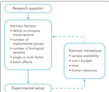
Experimental design is possibly the most important step of any transcriptomic experiment as the success of the project heavily depends on the choices made at this early stage. The first step is to have clear and defined goals. Questions that should be addressed before start- ing an experiment include: (i) Is the intent of the experi- ment to specifically define the transcriptome of the brain endothelial cells along the entire vascular tree or rather solely of BBB endothelial cells in CNS microvessels or even capillaries? (ii) Is the aim to compare the transcrip- tome of brain endothelial cells at different stages of e.g.
development or under specific pathological conditions?
(iii) Is the intent to define the transcriptome of a specific tissue (i.e. ChP epithelial cells vs kidney epithelial cells), a specific time point (i.e. embryonic vs post-natal BBB development) or a specific pathological condition (Mul- tiple Sclerosis vs Alzheimer’s Disease)? (iv) What are the Fig. 2 RNA‑Seq is an increasingly popular tool for transcriptional analysis. Number of searchable publications in Pubmed per year by the terms specified in the legend. Data source: Pubmed. Search date 31/12/2019. Y‑axis shows number of publications, X‑axis shows years
possible batch effects, such as the sacrificing groups of experimental animals on different days? These, and other similar questions, are known as intrinsic factors and their impact on the experimental design is direct, as they are defined by the question and objectives of the study.
There are also extrinsic factors that influence experi- mental design in the form of practical limitations. They are (i) biological sample availability, e.g.: human CNS tissue is sparse and may not be obtained in the required quality to allow for RNAseq analysis. (ii) costs, e.g.:
pre-sequencing optimization costs as well as sufficient sequencing of samples per group to the required depth.
(iii) time, e.g. time required for breeding experimental animals to obtain the required brain barriers genotype, time for protocol optimization of the tissue of interest (be it BBB, NVU, whole cortex or others) isolation protocol and validation of the results and iv) human resources, as a transcriptomics project might involve several scientists, from the principal investigator to wet lab researchers and technicians, sequencing facility technicians and bioinfor- maticians (Fig. 3).
Guidelines
• Define a clear goal for the transcriptomics study:
What is the specific intention for the project.
• Consider both extrinsic and intrinsic factors when designing an experiment.
• Plan the experiment including the advice from the experts involved in the different steps, such as sequencing facility staff and bioinformaticians.
Vascular heterogeneity in the CNS to be considered when characterizing the BBB transcriptome
The vasculature is heterogeneous throughout the CNS [18] (Fig. 4). This heterogeneity is reflected in the tran- scriptome, and therefore should be considered prior to isolating CNS microvessels for a transcriptomic study of the BBB. Two main factors are the capillary density and the BBB properties, which may in addition be affected by age, sex or the pathological conditions investigated.
Capillary density is related to the metabolic demands, and thus neuronal activity, of the respective CNS regions [19]. The gray matter (GM) of the cerebral cortex harbors many neuronal cell bodies and is therefore more meta- bolically active than the white matter (WM), where the myelinated axonal fibers run. Thus, GM harbors a higher density of capillaries when compared to WM [20]. In addition, there are specific regions in the CNS, such as the hippocampus, characterized by a remarkable hetero- geneity in capillary density [21].
The cellular and molecular characteristics of the NVU components are also heterogeneous throughout the CNS. Endothelial cells of the BBB present some of the highest regional differences. Indeed, expression of endothelial junction proteins (occludin, claudin-5 and a-catenin) is higher in the WM compared to the GM [22].
In the blood spinal cord barrier, the endothelium is less tight and is characterized by a lower pericyte coverage [18]. Astrocytes also show heterogeneity along the brain vasculature, including higher expression of glial fibril- lary acidic protein (GFAP), an intermediate filament, in WM relative to GM [22, 23]. In contrast, the expres- sion of aquaporin-4 (AQP4), a water channel localized at astrocyte endfeet, is more homogenous throughout the perivascular glia limitans [22, 24]. Moreover, endothe- lial cells do not form a BBB throughout the whole CNS.
Particularly microvessels within the circumventricular organs (CVOs) lack BBB properties. CVOs are localized around the brain ventricles and fulfill neurosecretory and neurosensory functions. The CVOs include the subfor- nical organ, the vascular organ of the lamina terminalis, the area postrema, the median eminence, the neurohypo- physis, the pineal gland, and also the ChPs (Fig. 4). CVOs contain fenestrated microvessels that allow for the free diffusion of blood components into the CVO stroma.
Thus, co-isolation of microvessels from the CVOs should be avoided when aiming to specifically analyze the BBB transcriptome [25].
The vascular tree presents gradual phenotypic hetero- geneity, a phenomenon known as zonation, accompanied
Fig. 3 Workflow for the experimental design of an RNA‑Seq study.
The research question will guide the initial experimental setup based on intrinsic factors. Then, extrinsic limitations should be taken into account to adjust and refine the overall design
by transcriptional differences [13]. Organization of TJs, rate of pinocytosis, expression of enzymes such as alka- line phosphatase, Na+/K+ ATPase, expression of trans- porters or efflux pumps or of adhesion molecules are not the same in endothelial cells of brain arterioles, capillar- ies and venules (Table 1) [26], in line with the different functions of these vascular segments. In addition, the mural cell subsets in these microvascular segments differ, with smooth muscle cells embracing arterioles and, to a lesser degree, venules, while pericytes are highly concen- trated at the level of the capillaries [27, 28] (Fig. 5).
Vascular heterogeneity can also be induced by CNS pathology, e.g. neuroinflammation, neurodegeneration or brain tumors, which may in fact lead to focal alterations in the NVU associated with the CNS pathology. This may range from changes in cellular composition of the NVU, e.g. pericyte drop during stroke, perivascular accumula- tion of inflammatory cells in multiple sclerosis or to alter- ations in vessel diameters as observed in brain tumors.
All these alterations will affect the outcome of estab- lished brain barrier cell isolation protocols with respect to purity of the brain barrier cells as well as RNA stability.
Guidelines
• Consider CNS capillary density of the region of inter- est to obtain enough RNA yield: capillary density is higher in GM compared to WM.
• Consider regional differences in BBB properties.
Clearly indicate from which CNS region microves- sels, capillaries or endothelial cells were isolated.
• Consider if you wish to analyze the transcriptome of endothelial cells from a specific vascular segment or a specific region of the CNS.
• Consider general or regional alterations in BBB prop- erties when studying CNS pathologies. Clearly indi- cate from which CNS region and at what disease stage the microvessels, capillaries or endothelial cells were isolated.
Regional heterogeneity among the four choroid plexus to consider for analyzing the BCSFB transcriptome
The choroid plexus (ChPs) protrude into each of the brain ventricles, and thus there are two lateral (telencephalic) ChPs, one in the third ventricle (diencephalic) and one in the fourth ventricle (hindbrain or myelencephalic). There is increasing evidence that the gene expression profile of each ChP reflects their positional identities. The mouse lateral and fourth ventricle whole ChPs present a differ- ential transcriptome and secretome, as assessed by RNA- seq and mass spectrometry [41]. A recent single nucleus RNA-seq study revealed a unique cellular composition in each of the mouse ChPs [42]. Of note, the more regional- ized cell types were epithelial cells and fibroblasts, while ChP endothelial cells were found to be more homogene- ous across the ventricles.
Although most transcriptomic studies have focused on the lateral ChPs, the choice of ChP will influence the sequencing results and the comparison with avail- able datasets. The heterogeneity of the ChP among Fig. 4 Regional differences in the brain microvasculature. Schematic representation of a brain sagittal section (left) and a spinal cord transverse section (right). Capillary density is higher in the CNS gray matter than in the white matter, according to their differential metabolic activity. The white matter of the corpus callosum is highlighted. The microvessels in the circumventricular organs (CVOs, highlighted in blue) lack BBB characteristics, rather they are fenestrated and thus permeable to blood components. CVOs include the subfornical organ, the vascular organ of the lamina terminalis, the area postrema, the median eminence, the neurohypophysis, the pineal gland, and the choroid plexus
the four ventricles in the human brain remains to be characterized.
Guidelines
• Consider regional transcriptional heterogeneity among the four ChPs. The choice of ChP should be stated in the methods.
Brief recommendations on how to select a reporter mouse Many genetically modified mouse models have been developed for studying brain barriers development and function. For a general overview on the available genetic tools that are used to study the BBB function, we recom- mend the review of Sohet et al. [43], while for a study involving a solid report in the BCSFB we recommend the reading of Johnson et al. [44]. In the context of this man- uscript the brain barriers reporter mouse lines that allow to distinguish brain barriers endothelial or epithelial cells from other cells of the CNS by means of expression of a fluorescent reporter are of specific interest. While many of those mouse lines have been developed for imaging purposes they also allow for sorting of the cells of interest from the CNS based on expression of their fluorescent reporter [12, 13, 45]. For the purpose of this manuscript, we will simply mention some recommendations and
possible pitfalls while using genetic mouse models in RNA-Seq studies in the brain barriers field.
Before any experimental approach, a deep understand- ing of the genetic mouse model that is used is needed.
To this end, online tools such as http://www.infor matic s.jax.org/ can be used to have a detailed overview of the mouse line of interest. Original literature on how the mouse line has been created and the expression pattern of the respective reporter needs to be carefully evalu- ated. This includes consideration of the promoter used for driving the expression of the fluorescent reporter with respect to cellular specificity or efficiency of expres- sion, which could depend also on the age or disease state investigated. Furthermore, inducible expression systems, e.g. based on Cre-recombinase or TET-ON or TET-OFF- regulation need to be tested for their specificity, leakiness and completeness of driving reporter expression [46, 47].
Specificity and intensity of the brain barriers reporter expression should always be tested in house prior to using the respective mouse model for sorting of brain barriers cells.
Considerations for protocols on the isolation of CNS microvessels, capillaries or single endothelial cells
A RNA-Seq study of CNS microvessels, capillaries or sin- gle BBB endothelial cells necessarily relies on the protocol used to isolate the target tissue or cell. Due to the highly Table 1 Segmental heterogeneity in the brain microvasculature endothelial cells (arterioles, capillaries and venules).
Adapted from [29] and [30]
Presence of perivascular cells is indicated by Yes/No. Presence of BBB features is indicated by Yes/No/non determined (n.d.). Relative expression of transporters/
enzymes is indicated by +, ++ or +++ from lowest to highest expression
Arterioles Capillaries References
Cells
Smooth muscle cells Yes No [13, 30]
Pericytes Yes/No Yes [13, 31]
Perivascular macrophages Yes Yes [13, 30]
BBB features
Tight junctions (TJs) Continuous and elaborate TJs/? Highly expressed TJs [29, 32, 33]
Permeability for BBB markers ? No [30, 34]
Astrocytic end‑foot shealth No Yes [29, 30]
Transporters/enzymes/receptors
P‑glycoprotein ? +++ [35, 36]
Na+/K+‑ATPase +++ + [29]
Transferrin receptor (TFRC) – +++ [13, 37]
Alkaline phosphatase +++ +++ [29]
Mg2+‑ATPase +++ + [29]
5′‑nucleotidase +++ + [29]
γ‑Glutamyl transpeptidase (GGTP) ? +++ [38, 39]
Bidirectiona/l vesicular horsedish peroxidase transport +++ + [29, 40]
a
b
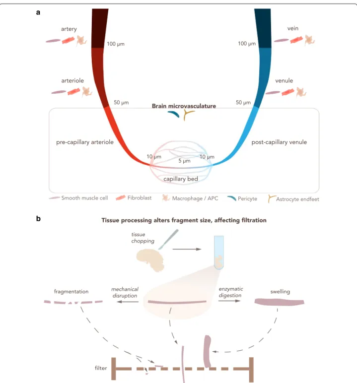
Fig. 5 Heterogeneity in microvasculature diameter determines the outcome of filtration steps for BBB enrichment. a The CNS vascular tree from arteries to veins. An indicative range of vessel diameter for each vascular section is provided, along with other cell types that may be co‑isolated.
Arteries and veins have a diameter > 100 μm, arterioles and venules from 100 μm to 50 μm. The brain microvasculature has a diameter smaller than 50 μm and consists of pre‑capillary arterioles, capillaries and post‑capillary venules. Capillaries are generally considered those with a diameter < 10 μm and often show diameters of about 5 μm. APC: Antigen‑presenting cell. b Enzymatic digestion and mechanical disruption during CNS vascular isolation protocols alter the physical properties of the microvasculature fragments, influencing the downstream steps of the isolation protocol, particularly size dependent filtration across nylon membranes. Small vascular fragments obtained by mechanical disruption can allow for undesired vessels to pass through the filter. Enzymatic digestion leads to swelling of the blood vessels, prohibiting their elution through the filter
complex structure of the NVU and the aforementioned heterogeneity of NVU components throughout the CNS vasculature, a detailed description of the protocol used to isolate the material for RNA-seq analysis is mandatory.
Each step of the isolation protocol is critical and has a direct impact on the sample purity as well as on the yield of the material obtained for sequencing. To date, many different protocols for the isolation of endothelial cells of the BBB, or CNS microvessels, or brain endothelial cells have been published. Most classical isolation pro- tocols consist of a combination of mechanical disruption and enzymatic digestion and a subsequent size selection of the isolated vascular segments by filtration through nylon membranes with different pore sizes (Fig. 5). Addi- tional enrichment of the respective vascular segment is usually achieved by density gradient separation [48–50].
More refined techniques can be incorporated to increase purity, such as selection with antibody-coupled mag- netic beads or FACS, or taking advantage of fluorescent reporter mice [51], as discussed above.
BBB microvessel or capillary isolation Tissue disaggregation: enzymatic digestion versus mechanical disruption
The first step for the isolation of CNS microvessels or endothelial cells of the BBB is to properly slice or cut the tissue into small pieces in order to facilitate enzyme- based digestion or the following Dounce homogenization steps [52]. Typical techniques of brain tissue disaggre- gation are either enzymatic or mechanical. Enzymatic digestion is often performed using a combination of enzymes such as collagenase and dispase as well as DNAse. Mechanical disruption, on the other hand, is usually performed using Dounce homogenizers with dif- ferent loose pestle sizes depending on the amount of tis- sue and the selected species [53, 54]. This ensures tissue loosening by shear forces without affecting cell viability.
Mechanical techniques may prove to be more effective while at the same time may be too harsh depending on certain factors, such as CNS region or age of the indi- vidual (i.e. aged tissue is more susceptible to damage).
Therefore, the choice of technique for tissue disaggrega- tion can influence the rest of the protocol and should be described in detail in the methods of the study.
Microvessel hierarchy selection depending on size:
filtration steps
Filtration of the dissociated tissue or CNS vasculature fragments enriches for a specific component from the brain homogenate. One of the most common methods for the isolation of CNS microvessels or specifically cap- illaries is performing either single or a series of filtra- tions through nylon or polyester membranes followed
by gradient centrifugation steps with Percoll™, Dextran or serum albumin, in order to separate microvessel frag- ments from cellular debris, myelin and other non-desired cell types [55–58].
The enrichment of a certain type of CNS microvessel (i.e. capillaries) over the others depends on their diam- eter and can be achieved by using different filter pore sizes. Therefore, the combination of larger (~ 100 μm) to smaller (~ 20 μm) filter pore sizes, in addition to using one or several filters in sequence, can determine the final vascular segment that is isolated [59]. Indeed, the choice of meshes should take into account the different vessel caliber of the CNS vasculature tree (Fig. 5a). Generally speaking, arteries have a diameter ≥ 100 μm, arterioles and venules between 100 and 50 μm, post-capillary ven- ules and pre-capillary arterioles between 50 and 10 μm, and capillaries are considered to have a diameter ≤ 10 μm [60–62]. The diameter of the arteries decreases from the surface of the brain towards the deeper regions [63].
Moreover, variability in brain artery diameter between different mouse strains has been observed [64], under- scoring the necessity of a detailed description of the source of sequencing material. In humans, CNS vessel diameters are affected by the health status of the donor [65] while in rat age was shown to contribute to reduced capillary diameter in the brain stem [66].
Although size selection represents a possible choice for defining the CNS microvascular segment that is isolated, some technical details have to be kept in mind. Other steps of the protocol can highly influence the physi- cal properties of the isolated microvessels or capillaries (Fig. 5b). For instance, mechanical disruption reduces the length of the vasculature fragments and therefore might impact size selection. Enzymatic digestion causes swell- ing of the microvasculature fragments, increasing their diameter. Due to these technical aspects, the researcher should use the size filtration as a guideline, and empiri- cally determine the exact fraction of the vascular tree that has been purified at the end of the isolation proto- col (visually analyzing the vascular fragment). Alternate tools that help achieve this validation include publicly available scRNA-Seq datasets of different BBB cell types [13]. These datasets collect information about specific cell markers that can be used to complement the isola- tion. However, it should be clear that expression of a few chosen markers might not successfully identify specific microvascular segments and that gene expression differ- ences along brain endothelial cells conform to a gradient rather than discrete segments.
In sum, size selection is a critical step in the iso- lation protocol of microvessels, capillaries or indi- vidual endothelial cells of the BBB, and ambiguity in
terminology should be avoided by accompanying qualita- tive and quantitative information.
Guidelines
• Tissue processing methods can affect size selection.
Indicate the method used and, if possible, the state of the tissue after dissociation.
• Indicate the size-dependent filtration steps, if used, including pore size of the filter mesh and the combi- nation of more than one filter.
• For animal studies, indicate the strain, age and sex of the animals used. For reporter mouse studies indicate the precise mouse line used as indicated on the MGI homepage (http://www.infor matic s.jax.org).
• For human studies, provide age, sex and relevant clinical information of the individuals.
Isolation methods of microvessel fragments or single endothelial cells from the BBB Strategies for BBB purity refinement: selection with antibody‑coupled magnetic beads
Selection via magnetic beads coated with specific anti- bodies represents a useful and precise method to iso- late microvessels or endothelial cells of the BBB. This technique can be used for positive selection of the material of interest or for negative selection of possible contaminants, alone or combined. For example, to purify endothelial cells, positive selection can be achieved by using beads coated with anti-CD31 and/or VE-cadherin antibodies [67, 68]. On the other hand, beads coated with antibodies against CD68, PDGFRβ, NG2 or GLAST might be used to specifically select macrophages, peri- cytes and astrocytes respectively [69–73], or to deplete these cells in those cases where a pure endothelial frac- tion is required. Positive and negative selection may be combined in order to improve the specificity of the technique.
Despite the high selectivity and improved final purity that the bead-mediated selection offers by targeting specific cell types, some disadvantages need to be con- sidered. First, the state of the vessel suspension and the physical interaction between different BBB components is a critical factor. During the isolation protocol, the brain vessels are not revealed in a single cell suspension but rather as vascular fragments that consist of tightly con- nected cell types, such as endothelial cells, pericytes and astrocyte endfeet [59]. Prior to bead selection, additional disaggregation steps, including enzymatic or mechani- cal disruption, might aid in obtaining a higher fraction of single cells versus microvessel fragments by weakening
the interaction between different cell types, which may enhance the disassociation and determine the outcome of the isolation [54]. The close interaction between the different components of the BBB [74, 75], makes a total single cell suspension from CNS microvessels a chal- lenging task, ultimately limiting the availability of bind- ing sites for the antibodies and influencing the isolation efficiency if not performed properly. An additional aspect to consider is that the enzymatic digestion may influence the surface expression of receptors which could be inter- nalized or lost by shedding or affect presence of surface epitopes and thus ultimately cell surface expression of potential antigens chosen for positive selection. There- fore, if the digestion and disaggregation processes are not fully achieved, this may lead to low yields despite the high purity, which must be taken into account when perform- ing sequencing analysis since it may influence the down- stream procedure. Also, extended purification protocols aiming to reach single brain endothelial cell suspensions bear the risk of inducing changes in gene expression in the endothelial cells due to loss of the tight junction interactions, as cross-talk between mature cell–cell junc- tions and the nucleus are well established.
Another important factor is that the selection is based on generally accepted markers for the cell population of interest, with the above-mentioned limitations. In addi- tion, heterogeneity in marker expression along the brain microvasculature might influence targeting efficiency.
Therefore, it is good practice to refer to the most recent studies that better define different cell populations of the brain vasculature [13, 76], in order to improve the target- ing strategy and the selectivity of the technique.
When using magnetic beads in a positive selection it is also important to know how, or if, to separate cells from the beads afterwards. A step that might become essen- tial is when the isolated fraction is intended to be used in cell culture. According to the first protocols using this method of isolation, incubation of the cells in trypsin/
EDTA at 37 °C releases the beads once the selection is achieved [77]. Improvements in this technique allow for the establishment of procedures which require a less aggressive approach or even do not require detachment of the beads following isolation [78], since they do not affect growth nor survival of isolated cells. Therefore, also depending on the brand, some beads have been shown to detach spontaneously from the cultured cells after several days, whereas other beads might need a DNAse treatment to break the DNA chain that attaches the microbead to the antibody [52, 70]. In any case, most of the currently used magnetic beads are completely suit- able for subsequent analysis.
Guidelines
• Clearly indicate the antibody used to coat the beads and the rationale behind the choice; refer to recent publications (if possible) to define the cell population that will ideally be targeted.
• Clearly indicate the amount of material obtained after bead selection, including number of isolated cells and amount of RNA extracted from them; this is useful information for the study itself and for future reference.
• Clearly indicate the necessity of separating beads from cells or not, if choosing positive selection of the target cell.
Specific separation of microvessel cell populations:
fluorescence‑activated cell sorting (FACS)
FACS is a powerful technique that uses flow cytometry to selectively separate cell populations from complex pools of different cells. FACS-based selection has been used in transcriptome profiling studies of the brain barriers [13, 14, 79]. Interestingly, these techniques include high puri- fication of cells expressing fluorescent reporters in trans- genic mice, however, any cell that expresses the construct will be selected, introducing possible contamination in the downstream analysis. For example, FACS has been successfully used to enrich brain endothelial cells iso- lated from claudin-5-GFP mice [13] or tamoxifen-treated Rosa-tdTomato; VE-Cadherin-CreERT2 mice [14]. Alter- natively to fluorescent reporter mice, other studies have achieved isolation of brain endothelial cells via FACS by antibody staining prior to sorting, using fluorochrome- conjugated antibodies against CD31 [80, 81] or by a com- bination of antibodies against CD31 and CD13 to isolate endothelial cells and pericytes, respectively, from differ- ent microregions in the mouse brain [82].
In any case, the final sample after FACS consists of a highly enriched fraction of the cell type of interest. How- ever, a high amount of starting material is often necessary to obtain a sufficient yield after the sorting, although it will also depend on the needs of the downstream appli- cation and/or analysis [83, 84]. Also, FACS sorting may induce an oxidative stress response in the endothelial cells that needs to be considered.
As already mentioned in other sections of this paper (e.g.: beads selection), obtaining a viable single cell suspension is also crucial when isolating cell popula- tions of the brain barriers prior to FACS. Similar to the beads selection, a combination of mechanical and enzy- matic digestion prior to FACS is often used to improve cell–cell dissociation. Indeed, a good single cell suspen- sion reduces the amount of false positives/negatives
produced by antibody staining (when not relying on reporter mice) or reporter proteins prior to FACS. In line with the necessity of a single cell suspension, duplet exclusion should be tightly controlled to ensure the best purity and reliability of the sorted material, as well as a strict gating strategy according to the experimental needs [85]. In addition, abundance of the population of inter- est is also critical, e.g.: if the level of endothelial cells is lower than 15% of the total, the sorted cells might not be viable. A density gradient enrichment before performing FACS could potentially solve this issue [51]. In general, FACS poses some technical challenges and a fine balance between time needed for the sorting, quality of the start- ing material and viability of the sorted cells needs to be experimentally tested.
Guidelines
• The flow conditions and the instrument used should be indicated.
• The precise scatter and fluorescence gating strat- egy used for the FACS of the target cell should be included in the supplement of the research article, as well as a detailed description of the isolation protocol and potential staining steps performed prior to the sorting and their duration.
• Duration of sorting itself and yield of cells received should be described.
• Time of RNA extraction following the FACS should be clearly stated, as different experimental designs implement differences in time points, e.g.: extraction of RNA right after FACS of after several hours due to travel from the sorting facility back to the laboratory.
Laser capture microdissection (LCM)
Laser capture microdissection (LCM) allows for the dis- section of CNS microvessels from a CNS tissue section with the help of a microscope and a laser. Dissected CNS microvascular endothelial cells can be later captured by adsorption, ejection, gravity or aspiration. LCM permits to take a snapshot of the transcriptomic profile of the BBB, in opposition to methodologies that require long incubation times.
One of the main limitations of LCM is the low yield of this laborious technique, which can be circumvented by using kits designed to isolate RNA from small amounts of cells [86] or by performing rounds of RNA amplifica- tion prior to downstream analysis [87]. However, using LCM to capture small cells, such as BBB endothelial cells, may be challenging, and contamination from astrocytic endfeet and/or pericytes is a major concern. In order to improve the cellular purity of the preparation, thinner
sections can be used, thus decreasing the chances of including cells above or below the plane of interest. Alter- natively, LCM on cross sections of vessels also provides better purity than longitudinal sections, although lower yield [88]. To aid the visualization of endothelial cells, rapid immunohistochemistry may be coupled to LCM [89], in a technique known as immuno-LCM. The repro- ducibility of immuno-LCM to study BBB gene expression in mice has been demonstrated [90]. This technique was further validated in human postmortem frozen [87] and Formalin-Fixed Paraffin-Embedded (FFPE) brain sections [89].
Guidelines
• Consider the balance between yield and purity of the isolated CNS microvascular endothelial cells when deciding to use cross sections or longitudinal sec- tions.
• It is recommended to use RNA isolation kits specifi- cally designed for small amounts of cells, or perform- ing RNA amplification prior to sequencing.
• Test for and consider the possible cellular contami- nants co-isolated with the BBB endothelial cells.
BBB in vitro models: cultured primary brain endothelial cells and brain endothelial cell lines
Isolated brain microvascular fragments or single cells can be cultured and used as a BBB endothelium in vitro model. Most of the in vivo BBB characteristics are main- tained by primary cultures of brain endothelial cells, hence representing powerful tools to study various aspects of BBB properties. However, often these cul- tures offer restricted capacity of genetic manipulation (e.g. transfection) and can be maintained in culture for a limited amount of time and/or passages. Nevertheless, numerous primary cultures of brain endothelial cells have been established from both mouse [58], rat [91] and human [92, 93] brain.
On the other hand, BBB in vitro model established by immortalized cell lines allow for much easier handling, as in many cases the cells can be cultured and passed as needed, in addition to much better tolerance to genetic manipulations. This makes cell lines a very suitable tool for high-throughput screening purposes, as they are also a much more homogenous cell population compared to primary cell cultures, where often contaminants are found in the culture. As a major drawback, cell lines of the BBB endothelium do not strictly retain BBB charac- teristics such as high tightness and very low permeabil- ity to the same degree as primary brain endothelial cells, therefore careful selection of the best suited BBB cell line
is needed according to the specific scientific question being answered.
In both cases, the presence of additional BBB cell types found in vivo is not always modeled in the in vitro sys- tems, such as for example the presence and anatomical disposition of the astrocytic endfeet found in the NVU in vivo. To overcome these limitations, sophisticated co- or tri-cultures of brain endothelial cells together with pericytes or astrocytes from different sources have been established and, to a certain degree, mimic the in vivo NVU structure [94–96].
In the context of transcriptomic studies, both BBB cell lines and primary brain endothelial cells cultures have been successfully used in transcriptomic approaches.
For example bulk RNA-seq has been performed on the human cerebrovascular endothelial cells (hCMEC/D3) [97] and on primary mouse brain microvascular endothe- lial cells (pMBMECs) [79]. Interestingly, a comparative microarray analysis between freshly isolated or cultured pMBMECs with the endothelioma cell line bEnd.5 has highlighted important changes in the mRNA levels of genes associated with BBB characteristics [69].
Recent advancements in stem cell technology have furthermore allowed derivation of human in vitro mod- els of the BBB from stem cell sources including human cord blood-derived stem cells of circulating endothelial progenitors [98] and human induced pluripotent stem cells (hiPSCs; summarized in [99]). hiPSCs derived from one individual opens the entirely novel opportunity to study BBB dysfunction from individual patients as their hiPSCs provide a scalable and renewable source for establishing brain microvascular endothelial cells. The presently available hiPSC derived in vitro BBB models are very well characterized with respect to their barrier properties and expression of BBB specific transporters and efflux pumps [100–102]. At the same time RNA-Seq analysis has shown that hiPSC derived brain microvascu- lar endothelial cells, as most hiPSCE—derived cells, do not fully recapitulate all aspects of the BBB [98]. Present hiPSC derived in vitro BBB models, e.g. still lack expres- sion of the full array of trafficking molecules required for immune cell interaction with the BBB.
Guidelines
• Consider the effect of culture-induced mRNA expression changes in the in vitro BBB models due to medium composition.
• Use RNA-Seq profiling of in vitro BBB models and especially of hiPSC- derived in vitro BBB models to benchmark them against the BBB in vivo.
Considerations for isolating the entire choroid plexus vs choroid plexus epithelium
The ChP consists of a highly vascularized stroma popu- lated by immune cells and is surrounded by a layer of highly specialized epithelial cells which form the BCSFB.
Contrary to the BBB, the ChP endothelium is fenestrated and does not form a BBB [103].
ChP transcriptomic studies of the entire ChP tissue will include the transcriptome of the epithelial cells forming the BCSFB but also from the endothelial cells, stromal fibroblasts and immune cells of the ChP. Alternatively, the ChP epithelial cells can be isolated to focus on the transcriptome of the BCSFB.
Using the whole ChP greatly simplifies the protocol for tissue isolation, but the cellular heterogeneity within the ChP will complicate subsequent analysis and inter- pretation of the results especially when performing bulk RNAseq studies. However, these studies will provide additional information on the other components of the ChP, such as the vasculature or immune populations.
Many RNA-seq studies have taken this approach, par- ticularly those focused on humans [104, 105].
Alternatively, if the barrier component of the ChP is the focus of the study, the epithelial cells can be iso- lated. While this results in cleaner data, dissociating the ChP may be challenging (see below). To overcome these difficulties, the novel single-nucleus RNAseq method emerges as an option for tissues that are hard to dissoci- ate, such as the ChP [42].
The research question and the technical limitations will determine whether the whole ChP or the isolated epithe- lium will be sequenced.
Methods for isolating the choroid plexus epithelium The techniques for isolating the ChP epithelial cells are similar to those for the BBB. Of note, the ChP epithelium is composed of large cuboidal cells, which are easier to dissect microscopically with LCM compared to the thin BBB endothelium. Indeed, LCM has been used to isolate the human ChP epithelium for microarray studies of the BCSFB [106–108]. In animal models, mechanical disrup- tion of the entire ChP surgically removed from the brain ventricles is typically combined with enzymatic digestion.
In order to release epithelial adherens and tight junctions as well integrin mediated adhesive contacts to the epi- thelial basement membrane, calcium removal is recom- mended, for example by using the chelator EDTA [109, 110] or calcium free medium [111], but is not an essential requirement [112]. Further purification can be achieved by FACS using an epithelial marker such as TTR [41].
However, expression of TTR has been recently identi- fied in ChP macrophages [113], and the choice of mark- ers should be done with awareness of their limitations. To
our knowledge, the only human primary epithelial cells are those commercially available (ScienCell), and no iso- lation protocol has been published to date.
Guidelines
• The research question and the technical limitations will determine whether the whole ChP or the iso- lated epithelial cells will be sequenced. This should be specified in the methods.
• The ChP can be particularly challenging to dissoci- ate. The techniques for isolating the ChP epithelium include LCM, mechanical and enzymatic digestion.
Resulting purity should be assessed and reported.
Pre‑sequencing tissue or cell purity assessment
Before performing RNA-seq, it is good practice to ensure that the chosen isolation strategy results in the desired brain barrier cell purity. Indeed, knowledge about the degree of brain barrier cell purity is essential as it dra- matically reduces possible biases in the downstream analysis, overall improving the biological meaningful- ness of any RNA-seq study. Pre-sequencing purity assess- ments can be achieved by different techniques, often used in combination. Common contaminants when iso- lating endothelial cells of the BBB are pericytes or peri- cyte fragments and astrocytic endfeet, which can hardly be avoided (Fig. 5, Table 1). These contain RNA and are thus readily detectable by assessing expression by qPCR of specific markers such as platelet-derived growth factor receptor beta (PDGFR-β) or GFAP, respectively. Immu- nostaining provides information about the location of the probed proteins while flow cytometry allows for quanti- tative detection of the contaminants with higher sensitiv- ity, although it requires a significantly higher number of cells compared to immunofluorescence imaging. There- fore, qPCR can be used in combination with immunoflu- orescence imaging in order to estimate the purity at both the RNA and protein level. All of the techniques depend, to differing degrees, on described cell markers (Table 2).
Another potential source of contaminants when iso- lating endothelial cells of the BBB are ChP cells, in par- ticular the ChP epithelium. Indeed, in the vast majority of the preparation the ChP is not removed from the processed material, therefore potentially accounting for contamination. Performing qPCR for choroid plexus specific markers such as transthyretin or keratin-8 will allow to determine the presence of ChP mRNA in the brain endothelial preparation. For example, low expres- sion of claudin-3 mRNA has been reported in freshly isolated brain microvessels, despite recent evidence proving lack of claudin-3 expression in the mouse brain
microvasculature. This could be due to contamination of the isolated brain microvessels with ChP epithelial cells, which express claudin-3 [79].
Isolation and purification of RNA from BBB endothelial cells or microvessels
RNA isolation methods have to be chosen depending on the type and availability of starting material, on the one Table 2 Selected molecular markers of relevant CNS cell types with species and methodological information
Italic represents evidence in the mouse, bold italic represents evidence in humans, underline represents evidence in the mouse and humans
IHC immunohistochemistry, RM reporter mice, IF immunofluorescence, PCR polymerase-chain reaction, QPCR quantitative polymerase-chain reaction, RNASeq next generation RNA sequencing, scRNAseq single-cell RNA sequencing, KO knock-out, WB Western-blot, ICC immunocytochemical staining, ELISA enzyme-linked immunosorbent assay
a A study showing contrary evidence (absence) Endothelial
cells of the BBB
Pericytes Astrocytes Smooth
muscle cells CP/CVO endothelial cells
CP epithelial cells
Immune
cells Oligodendrocytes Fibroblasts
AQ1 – – – – – [114] IHC
[115] IHC – – –
AQP4 – – [116] IHC – – [114] IHC – – –
⍺‑SMA – ± IHC [117]
IHC [118] – [119] IHC – – – – –
CD31/
PECAM1 [120, 121]
IHC – – – [122] IHC – [123] IHC – –
Cdh5 [124] – – – [125] RM – – – –
Cldn‑1 [126] IHC – – – – [127–129]
IHC – – –
Cldn‑2 – – – – – [127, 128]
IHC – – –
Cldn‑3 [130] IF
[79]a – – – – – – – –
Cldn‑5 [131]
KO + IHC/
[132] IHCWB
– – – – [127] IHC – – –
Cldn‑11 – – – – – [128] IHC – IHC [133]
IHC [134] –
FGF WB [135]
GFAP – – [136] IF – – – – – –
GLAST – – WB [137] – – – – – –
ICAM‑1 [138] IHC
[139] ELISA [140] ICC – – – – [141] – –
MHC‑II – – – – – – [142] PCR – –
NG2/CSPG4 – [143] IHC – – – – [144] Immunogold –
OAP‑1 – – – – – – – [134] WB + IHC –
Occludin [132] IHC – – – [127] IHC [127, 128]
IHC – – –
PDGFR‑β – [143] IHC – – – – – – –
Podoplanin – – – – – [145] IHC,
WB [146] IHC
– – –
TTR – – – – – [41] QPCR,
RNAseq – – –
VCAM‑1 [147] ELISA [140] ICC – – – – – – –
ZO‑1 [148] IHC
[149] ICC – – – – [128] IHC – – –
hand, and the intended RNA-seq analyses, on the other (Fig. 6). The use of inappropriate RNA isolation meth- ods can result in low quantity and/or quality of RNA and consequently in less accurate and irreproducible results or even in complete failure of the analysis [150].
Contaminations with extrinsic RNA and DNA, or with nucleases that might lead to the degradation of RNA samples can have a negative impact on the results. Gen- eral measures to avoid these issues include thorough and regular cleaning of work areas and equipment with decontamination solution as well as use of clean gloves, aerosol barrier pipette tips, and DNAse and RNAse-free plasticware. Additionally, it is recommended to carefully handle RNA samples at the temperature suggested by the manufacturer of the isolation kit [151].
Commercially available RNA isolation protocols gener- ally follow two main steps: (i) sample lysis, homogeniza- tion and clearing, and (ii) RNA purification. Isolation kits and protocols must be chosen according to the type of sample (e.g. cell culture, frozen tissue, FFPE tissue, etc.) and RNA molecules to be purified (e.g. small or large RNA molecules).
Sample lysis, homogenization and clearing
Cell lysis is commonly performed using a guanidine-thi- ocyanate-based buffer combined with a strong reducing agent, such as tris(2-carboxyethyl)phosphine hydrochlo- ride, 2-mercaptoethanol or dithiothreitol to ensure the complete cell lysis and protein denaturation (lysis buffer), but these procedures vary according to the character- istics of the starting sample (e.g. adherent cell culture, cell suspension, frozen tissue or formalin-fixed paraffin- embedded (FFPE) tissue)—(Fig. 7).
Cultured adherent cells
Cultured adherent cells, like endothelial cells, can be subjected to trypsinization prior to cell lysis or they can be lysed directly in the culture container by replacing the liquid medium with lysis buffer directly to the cell monolayer. Cell lysis by addition of lysis buffer is recom- mended to be performed prior to storage to avoid tran- scriptomic alterations during freezing. The cell lysate is safe to be stored at − 80 °C.
Cell suspensions
Cell suspensions, such as microvessel fragments or single endothelial cells, can be pelleted by gentle centrifugation (≤ 500×g). After complete removal of the supernatant, the cell pellet is re-suspended in lysis buffer. As for the lysis of adherent cells, cell lysis is recommended to be performed prior to storage. Sorted and isolated single cells can be directly collected in lysis buffer and mild lysis buffer, respectively.
Tissue samples
At the moment of collection, tissue samples, like the ChP, are recommended to be stored in a RNA stabilization solution or to be snap-frozen in liquid nitrogen. Sam- ples in stabilization solution can be stored up to 4 weeks at 4 °C or at − 20 °C for long-term storage [152]. Sam- ples snap-frozen in liquid nitrogen are safe to be stored at − 80 °C for more than 20 years [153]. Tissue samples might need to be disrupted using different techniques, such as the TissueRuptor, TissueLyser, ZR BashingBead Lysis Tubes or thorough grinding under liquid nitrogen using a mortar and pestle [152]. Remaining tissue and other precipitates might need to be removed by centrifu- gation and the supernatant can be used for subsequent RNA isolation.
FFPE tissue samples
FFPE tissue samples derived from brain microvessels must be subjected to deparaffinization using xylene or other commercially available solutions. Subsequently, tissue and protein digestion is performed using protein- ase K. Next, formaldehyde-derived crosslinks of nucleic acids and proteins must be reversed by incubating at more than 80 °C. Finally, the sample might be cleared by centrifugation and the supernatant can be used for sub- sequent RNA isolation [154].
RNA purification
During experimental design and before performing RNA purification, it is necessary to identify which RNA mol- ecules are relevant for the research question. Messenger Fig. 6 Overview of the main steps for processing a CNS tissue sample into BBB‑related material ready for RNA isolation. Fresh samples are
dissociated by mechanical disruption, enzymatic digestion, or a combination of both. Typically, tissue is first mechanically disaggregated into smaller pieces to facilitate the exposure to the enzyme solution. Dissociated tissue is then selected according to size by passing through one or a series of filters, by a density gradient, or both. This process isolates the microvessels. For isolating single barrier cells, tissue dissociation (particularly enzymatic digestion) can be repeated [1] after the initial size selection steps. The single cell suspension can be further purified or enriched for certain cell types [2] by using a fluorescence‑activated cell sorter (FACS) or magnetic microbeads labeled with an antibody against a cell marker.
Alternatively, if the tissue of interest is frozen or formalin‑fixed paraffin‑embedded (FFPE), a common approach is to isolate the microvessels by laser capture microdissection (LCM)
(See figure on next page.)
RNA (mRNA) is the RNA that will be translated by the ribosomes into proteins. mRNA is characterized by having a coding sequence surrounded by 3′ and 5′
untranslated regions and a long sequence of adenine nucleotides at the 3′ end (poly-A tail). Several other types of non-coding RNA have important roles in cell biology,
e.g. ribosomal RNA (rRNA) and transfer RNA (tRNA) are necessary for the translation process. Additionally, other RNA families are important for gene expression regula- tion as for example, microRNA (miRNA) with a size of ca. 22 nucleotides, other small RNAs (< 200 nt) and long non-coding RNA (lncRNA) with sizes greater than 200 nucleotides. Regarding the RNA content in a cell, it is important to notice that just mRNA and many lncRNA have a poly-A tail at the 3′ end. Additionally, rRNA repre- sents the majority of the RNA content in the cell.
The combination of a highly concentrated chaotropic salt (e.g. guanidinium thiocyanate) in the lysis buffer with a certain concentration of an organic solvent (typically ethanol or isopropanol) allows the adsorption of nucleic acids to the silica matrix in spin columns. Although this solid phase extraction allows for efficient and easy isolation of purified nucleic acids, several details must be taken into consideration. The final concentration of ethanol or isopropanol in the mixture with lysis buffer is essential to promote the adsorption of RNA to the silica matrix [150]. Different ethanol or isopropanol concentra- tions result in the isolation of RNA molecules with differ- ent sizes, e.g. small RNA molecules (containing miRNAs) with a size between 16 and 200 nucleotides and large
RNA molecules (containing mRNA and lncRNA) with a size greater than 200 nucleotides. Therefore, it is nec- essary to carefully select the applied protocols and cor- respondent ethanol/isopropanol concentrations before starting the isolation. Due to the possible impact of genomic DNA (gDNA) contamination in RNA-seq analy- ses, thorough digestion of gDNA remnants in RNA sam- ples is mandatory. gDNA removal columns or integrated on-column DNA digestions are included in most RNA isolation kits [152].
Guidelines
• The RNA extraction protocol should be selected based on tissue type and quantity, as well as the intended sequencing and analysis.
• Specific protocols are required for the isolation of total RNA including miRNA.
• Correct sample homogenization and clearing are essential for isolation efficiency of RNA and analysis reproducibility.
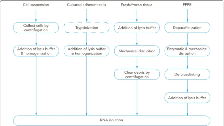
Fig. 7 Overview of commonly used RNA isolation protocols. Preparation of BBB‑derived samples according to the type of sample. Cells in suspension are first collected by centrifugation, while adherent cultured cells are commonly trypsinized; then lysis buffer is added and cells are homogenized before proceeding to isolation of the RNA. Fresh frozen tissue can be mechanically disrupted in lysis buffer; debris should be removed by centrifugation before RNA isolation. Formalin‑fixed paraffin‑embedded (FFPE) tissue is first deparaffinized, and tissue disruption can be achieved by enzymatic (proteinase K) and/or mechanical means; de‑crosslinking is followed by addition of lysis buffer, and then RNA is isolated