pathogens
Article
Beneficial Immunomodulatory Effects of Fluticasone Propionate in Chlamydia pneumoniae-Infected Mice
Dóra Paróczai1,2,* , Anita Sejben3, Dávid Kókai2, Dezs ˝o P. Virok2, Valéria Endrész2 and Katalin Burián2
Citation: Paróczai, D.; Sejben, A.;
Kókai, D.; Virok, D.P.; Endrész, V.;
Burián, K. Beneficial Immunomodulatory Effects of Fluticasone Propionate inChlamydia pneumoniae-Infected Mice.Pathogens 2021,10, 338. https://doi.org/
10.3390/pathogens10030338
Academic Editor: Lee Ann Campbell
Received: 17 February 2021 Accepted: 12 March 2021 Published: 14 March 2021
Publisher’s Note:MDPI stays neutral with regard to jurisdictional claims in published maps and institutional affil- iations.
Copyright: © 2021 by the authors.
Licensee MDPI, Basel, Switzerland.
This article is an open access article distributed under the terms and conditions of the Creative Commons Attribution (CC BY) license (https://
creativecommons.org/licenses/by/
4.0/).
1 Department of Pulmonology, University of Szeged, Alkotmány str. 36., 6772 Deszk, Hungary
2 Department of Medical Microbiology and Immunobiology, University of Szeged, Dóm sqr. 10.,
6720 Szeged, Hungary; kokai.david@med.u-szeged.hu (D.K.); virok.dezso.peter@med.u-szeged.hu (D.P.V.);
endresz.valeria@med.u-szeged.hu (V.E.); burian.katalin@med.u-szeged.hu (K.B.)
3 Department of Pathology, University of Szeged,Állomás str. 1., 6720 Szeged, Hungary;
sejben.anita@med.u-szeged.hu
* Correspondence: paroczai.dora@med.u-szeged.hu
Abstract: The associations between inhaled corticosteroid (ICS) use and pulmonary infections remains controversial. Chlamydia pneumoniae (C. pneumoniae)accounts for asthma exacerbations;
however, there are no data regarding ICS effects onC. pneumoniaeinfections. Thus, we investigated whether fluticasone propionate (FP) or budesonide (BUD) could affectC. pneumoniaeinfection in vitro and in vivo, focusing on the possible mechanisms that lead to potential anti-chlamydial outcomes.
We performed direct qPCR to detectC. pneumoniaegrowth in infected, FP-treated, and BUD-treated A549 cells. Furthermore, FP or BUD was administered by inhalation toC. pneumoniae-infected mice.
The recoverableC. pneumoniaewas determined by indirect immunofluorescence. Expression levels of interferon (IFN)-γand IFN-γinducible chemokines were assessed by qPCR. We measured the protein concentrations of IFN-γand of other cytokines that potentially participate in the anti-chlamydial response by ELISA. We found that FP treatment suppressedChlamydiagrowth in A549 cells and in mice. Higher levels of IFN-γgene expression were observed in FP-treated mice compared to the untreated and BUD-treated mice (p< 0.0001). IFN-γand anti-chlamydial protein MIG/CXCL9 values were significantly higher after FP inhalation. Collectively, FP, but not BUD, suppressedC. pneumoniae growthin vitroandin vivo, which was likely due to the enhanced IFN-γrelated responses.
Keywords: inhaled corticosteroid; fluticasone; Chlamydia pneumoniae; respiratory infection;
chemokine; interferon
1. Introduction
Inhaled corticosteroids (ICSs) are regarded as the most effective treatment for asthma and chronic obstructive pulmonary disease (COPD) to reduce the risk of exacerbation and improve lung function; however, ICSs have been associated with increased risk of pneumonia [1]. Earlier studies have provided controversial data about the potential risk of pneumonia in patients using ICSs and emphasise the differences in their mechanisms of action [2,3]. It is well established that budesonide (BUD) and fluticasone propionate (FP) show differences in their pharmacokinetic, physicochemical, and even in immuno- suppressive properties, which can explain their distinct effects on respiratory infections and exacerbations [4,5]. As ICSs may have anti-inflammatory effects on the outcomes of respiratory infections, BUD and FP were studied to determine whether they can affect common viral or bacterial infections associated with COPD and asthma exacerbations.
Previous studies have revealed that BUD can inhibit rhinovirus replication and beneficially modulate cytokine responses in vitro depending on the type of infected cell [6,7]. Al- though both BUD and FP can suppress pro-inflammatory cytokine expression, BUD had a greater impact on antimicrobial proteins [8]. Furthermore, BUD came into focus during the SARS-CoV-2 pandemic, and it was examined in coronavirus HCoV-229E infection, where
Pathogens2021,10, 338. https://doi.org/10.3390/pathogens10030338 https://www.mdpi.com/journal/pathogens
Pathogens2021,10, 338 2 of 13
it was found to decrease the expression of the viral entry receptor and infection-induced cytokines, especially interleukin (IL)-6, IL-8, and interferon (IFN)-γ, resulting in inhibited viral replication in vitro [9]. The transcription of genes involved in cytokine and chemokine responses, particularly CCL-5, also known as RANTES (regulated on activation, normal T cell expressed and secreted); and nuclear factorκB (NF-κB)-dependent gene expression, is altered more extensively by FP than by BUD [10].
However, it remains unclear why ICSs have different effects on the immune responses to respiratory infections. FP has been reported to cause a ten-fold more potent inhibitory effect on airway immune cells and pro-inflammatory cytokine production due to its pro- longed presence in the airway mucus, as compared to BUD [11,12]. Since BUD is in conjugated form intracellularly and creates a depot before it is activated [13], its effect is still unexplained in intracellular bacterial infections. In contrast to in vitro studies, BUD at- tenuated pulmonary antibacterial host defence and increased the number of viable bacteria in mouse lungs [14].
Chlamydia pneumoniae(C. pneumoniae) is an obligate intracellular bacterium that propa- gates in respiratory epithelial cells, is responsible for community-acquired atypical pneumo- nia, bronchitis, pharyngitis, sinusitis, and is implicated in the development of severe asthma and acute exacerbations [15,16]. We have previously revealed that a formerC. pneumoniae infection was associated with altered cytokine responses in patients with asthma [17].
However, to our knowledge, the consequences of ICS use onC. pneumoniaeinfection have not been investigated yet. To address the question of whether ICS use could directly or indirectly influenceC. pneumoniaeinfection, we investigated the effects of FP and BUD treatment in an infected mouse model. We hypothesised thatC. pneumoniaereplication and infection-induced immune responses, especially anti-chlamydial IFN-γ, IFN-related chemokine production, and IFN-γ triggered gene expressions, could be influenced by the administration of ICSs. Since BUD and FP have different immunomodulatory effects, they can cause distinct alterations in the immune response toC. pneumoniaeinfection. To investigate this, we assessed the effects of FP and BUD on the in vitro and in vivo growth ofC. pneumoniaein airway epithelial cells and in mice.
2. Results
2.1. FP Suppressed C. pneumoniae Replication in A549 Cells
We testedC. pneumoniae-infected A549 cells to investigate whether FP or BUD treat- ment could modulate bacterial growth. As shown earlier, assessment of chlamydial genome concentration by direct qPCR correlated with manual fluorescent microscopic quantitation, wherein qPCR was used to measure the concentration ofChlamydiain infected cells [18]. We assessedC. pneumoniaegrowth in FP- and BUD-treated epithelial cells based on the cycle threshold (Ct) values. FP treatment resulted in significantly higher Ct values, indicating suppressedC. pneumoniaegrowth, compared to that measured in BUD-treated (32.35±0.51 vs. 30.81±0.55,p< 0.01) and untreated control (32.35±0.51 vs. 31.41±0.39,p< 0.01) cells. BUD treatment did not affectC. pneumoniaegrowth significantly (Figure1). Given that ourin vitroobservations could have significant clinical relevance, we next addressed the elucidation of possible immunomodulating effects of FP and BUD in mice to explore the underlying mechanisms.
Pathogens2021,10, 338 3 of 13
Pathogens 2021, 10, x FOR PEER REVIEW 3 of 13
Figure 1. C. pneumoniae growth in A549 cells treated with budesonide (BUD) or fluticasone propi- onate (FP). A549 cells were treated with BUD or FP for 24 h before, and for 48 h after infection with C. pneumoniae (0.01 MOI). Growth of C. pneumoniae was estimated by direct qPCR, as described in the Materials and Methods section. The concentration of C. pneumoniae is shown in terms of Ct value. Error bars denote the mean ±SD of five parallel cultures. Asterisks indicate significant dif- ferences, ** p < 0.01; ns means not significant difference.
2.2. FP Inhibited C. pneumoniae Growth in the Lungs of Mice
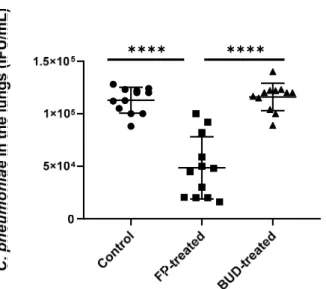
We found that the viable number of C. pneumoniae was significantly lower in FP-treated mice compared to the control group (5.33 × 10⁴ ± 3.42 × 10⁴ inclusion-forming unit (IFU)/mL vs. 1.13 × 10⁵ ± 1.28 × 10⁵ IFU/mL, p < 0.0001) (Figure 2). A similar trend was observed upon comparing FP-treated mice with BUD-treated mice (5.33 × 10⁴ ± 3.42 × 10⁴ IFU/mL vs. 1.15 × 10⁵ ± 1.42 × 10⁴ IFU/mL, p < 0.001). In contrast, no inhibition was de- tected in BUD-treated mice compared to the control (Figure 2).
Figure 2. Quantity of recoverable C. pneumoniae in mouse lungs. Supernatants of lung homogenates were cultured on McCoy cells, and the number of recoverable C. pneumoniae was counted by indi- rect immunofluorescence test after two days of incubation. Symbols show data from individual mice. Horizontal lines indicate mean ± SD. Significant differences are indicated by asterisks, **** p <
0.0001.
2.3. Effects of FP and BUD on Chlamydia-Infected Lung Tissue Histopathology
In the haematoxylin–eosin (HE)-stained mouse lung tissues, a distinctive difference in the general blueish appearance of the background in the control and BUD-treated specimen was observed, which was caused by extensive lymphoid infiltration. Even
✱✱ ✱✱
✱✱✱✱ ✱✱✱✱
Figure 1.C. pneumoniaegrowth in A549 cells treated with budesonide (BUD) or fluticasone propionate (FP). A549 cells were treated with BUD or FP for 24 h before, and for 48 h after infection with C. pneumoniae(0.01 MOI). Growth ofC. pneumoniaewas estimated by direct qPCR, as described in the Materials and Methods section. The concentration ofC. pneumoniaeis shown in terms of Ct value.
Error bars denote the mean±SD of five parallel cultures. Asterisks indicate significant differences,
**p< 0.01; ns means not significant difference.
2.2. FP Inhibited C. pneumoniae Growth in the Lungs of Mice
We found that the viable number ofC. pneumoniaewas significantly lower in FP-treated mice compared to the control group (5.33×10⁴±3.42×10⁴inclusion-forming unit (IFU)/mL vs. 1.13×10⁵±1.28×10⁵IFU/mL,p< 0.0001) (Figure2). A similar trend was observed upon comparing FP-treated mice with BUD-treated mice (5.33×10⁴±3.42×10⁴IFU/mL vs. 1.15×10⁵±1.42×10⁴IFU/mL,p< 0.001). In contrast, no inhibition was detected in BUD-treated mice compared to the control (Figure2).
Pathogens 2021, 10, x FOR PEER REVIEW 3 of 13
Figure 1. C. pneumoniae growth in A549 cells treated with budesonide (BUD) or fluticasone propi- onate (FP). A549 cells were treated with BUD or FP for 24 h before, and for 48 h after infection with C. pneumoniae (0.01 MOI). Growth of C. pneumoniae was estimated by direct qPCR, as described in the Materials and Methods section. The concentration of C. pneumoniae is shown in terms of Ct value. Error bars denote the mean ±SD of five parallel cultures. Asterisks indicate significant dif- ferences, ** p < 0.01; ns means not significant difference.
2.2. FP Inhibited C. pneumoniae Growth in the Lungs of Mice
We found that the viable number of C. pneumoniae was significantly lower in FP-treated mice compared to the control group (5.33 × 10⁴ ± 3.42 × 10⁴ inclusion-forming unit (IFU)/mL vs. 1.13 × 10⁵ ± 1.28 × 10⁵ IFU/mL, p < 0.0001) (Figure 2). A similar trend was observed upon comparing FP-treated mice with BUD-treated mice (5.33 × 10⁴ ± 3.42 × 10⁴ IFU/mL vs. 1.15 × 10⁵ ± 1.42 × 10⁴ IFU/mL, p < 0.001). In contrast, no inhibition was de- tected in BUD-treated mice compared to the control (Figure 2).
Figure 2. Quantity of recoverable C. pneumoniae in mouse lungs. Supernatants of lung homogenates were cultured on McCoy cells, and the number of recoverable C. pneumoniae was counted by indi- rect immunofluorescence test after two days of incubation. Symbols show data from individual mice. Horizontal lines indicate mean ± SD. Significant differences are indicated by asterisks, **** p <
0.0001.
2.3. Effects of FP and BUD on Chlamydia-Infected Lung Tissue Histopathology
In the haematoxylin–eosin (HE)-stained mouse lung tissues, a distinctive difference in the general blueish appearance of the background in the control and BUD-treated specimen was observed, which was caused by extensive lymphoid infiltration. Even
✱✱ ✱✱
✱✱✱✱ ✱✱✱✱
Figure 2.Quantity of recoverableC. pneumoniaein mouse lungs. Supernatants of lung homogenates were cultured on McCoy cells, and the number of recoverableC. pneumoniaewas counted by indirect immunofluorescence test after two days of incubation. Symbols show data from individual mice.
Horizontal lines indicate mean±SD. Significant differences are indicated by asterisks, ****p< 0.0001.
2.3. Effects of FP and BUD on Chlamydia-Infected Lung Tissue Histopathology
In the haematoxylin–eosin (HE)-stained mouse lung tissues, a distinctive difference in the general blueish appearance of the background in the control and BUD-treated specimen was observed, which was caused by extensive lymphoid infiltration. Even though the inflammation appeared diffusely, accentuated perivascular and peribronchial infiltration was also observed at higher magnification (Figure 3A,B). Similarly, in the FP-treated
Pathogens2021,10, 338 4 of 13
mouse lung tissues, lymphocytic and plasmacytic infiltration was observed; centriacinar emphysema was also visible, with thin alveolar septa (Figure3C).
Pathogens 2021, 10, x FOR PEER REVIEW 4 of 13
though the inflammation appeared diffusely, accentuated perivascular and peribronchial infiltration was also observed at higher magnification (Figure 3A,B). Similarly, in the FP-treated mouse lung tissues, lymphocytic and plasmacytic infiltration was observed;
centriacinar emphysema was also visible, with thin alveolar septa (Figure 3C).
Figure 3. The effects of inhaled corticosteroids (ICSs) on Chlamydia-induced histopathology in BALB/c mouse lung tissues. Representative HE-stained sections of C. pneumoniae-infected, un- treated (A), C. pneumoniae-infected, BUD-treated (B), and C. pneumoniae-infected, FP treated (C) lung tissues (magnification 200×).
2.4. Effects of FP and BUD Treatment on Gene Expressions Related to IFN- and Corticosteroid Responses in C. pneumoniae Infected Mice
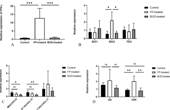
Next, we explored whether the expression of IFN-γ and IFN-γ induced genes was altered in FP- and BUD-treated lung tissues, including the typical, inducible defence genes against Chlamydia infection, such as indoleamine 2,3-dioxygenase 1 (IDO1), IDO2, MIG/CXCL9, IP-10/CXCL10 and I-TAC/CXCL11. qPCR using total RNA isolated from homogenised lung tissues revealed that the relative expression of IFN-γ was significantly enhanced in FP-treated mice (p < 0.001) compared to BUD-treated and control mice, the relative expression was 12.8 ± 5.8 vs. 0.9 ± 0.43 and 12.8 ± 5.8 vs. 0.75 ± 0.1, respectively (Figure 4A).
Our previous studies showed that the IFN-γ inducible IDO1 and IDO2 are highly expressed in C. pneumoniae-infected mouse lungs [19]. Thus, we next investigated the ef- fects of FP and BUD treatment on the expression of IFN-γ inducible IDO1, IDO2, and tryptophan 2,3-dioxygenase (TDO) involved in the metabolism of amino acid tryptophan that is essential for Chlamydia growth. Our results indicated a significantly increased IDO2 expression in the FP-treated mice compared to the control and BUD-treated group (p < 0.05). This phenomenon was not observed in IDO1 and TDO expression (Figure 4B).
To test the consequence of increased IFN- release, we determined the relative ex- pression levels of IFN- related chemokines. Unexpectedly, we found that BUD signif- icantly decreased the expression of MIG/CXCL9 (p < 0.05) and IP-10/CXCL10 (p < 0.01) compared to untreated C. pneumoniae infected control mice. However, the relative ex- pression of MIG/CXCL9 and IP-10/CXCL10 was not altered significantly in FP-treated mice. I-TAC/CXCL11 showed similar expression levels in all groups (Figure 4C).
Furthermore, we investigated the expression of glucocorticoid receptor (GR) and Vitamin D receptor (VDR) genes, as both receptors perform immunomodulatory func- tions [20,21]. Interestingly, our results demonstrated that FP treatment increased VDR expression significantly compared to control and BUD-treated mice (p < 0.01), whereas BUD treatment did not affect VDR expression. We found no statistically significant dif- ference in GR expressions in C. pneumoniae-infected BUD- and FP-treated mice (Figure 4D).
Figure 3.The effects of inhaled corticosteroids (ICSs) onChlamydia-induced histopathology in BALB/c mouse lung tissues.
Representative HE-stained sections ofC. pneumoniae-infected, untreated (A),C. pneumoniae-infected, BUD-treated (B), and C. pneumoniae-infected, FP treated (C) lung tissues (magnification 200×).
2.4. Effects of FP and BUD Treatment on Gene Expressions Related to IFN-γand Corticosteroid Responses in C. pneumoniae Infected Mice
Next, we explored whether the expression of IFN-γand IFN-γinduced genes was altered in FP- and BUD-treated lung tissues, including the typical, inducible defence genes againstChlamydiainfection, such as indoleamine 2,3-dioxygenase 1 (IDO1), IDO2, MIG/CXCL9, IP-10/CXCL10 and I-TAC/CXCL11. qPCR using total RNA isolated from homogenised lung tissues revealed that the relative expression of IFN-γwas significantly enhanced in FP-treated mice (p< 0.001) compared to BUD-treated and control mice, the relative expression was 12.8±5.8 vs. 0.9±0.43 and 12.8±5.8 vs. 0.75±0.1, respectively (Figure4A).
Pathogens 2021, 10, x FOR PEER REVIEW 5 of 13
Figure 4. Relative gene expressions in C. pneumoniae-infected and treated mouse lungs. Gene ex- pression of IFN-γ (A), IDO1, IDO2, TDO (B), IFN-γ-inducible chemokines (C), VDR, and GR (D) determined by qPCR. Relative expression was normalised to β-actin gene expression and calcu- lated by the 2−(ΔΔCt) method. Significant differences are indicated with asterisks, * p < 0.05, ** p < 0.01,
*** p < 0.001; ns means not significant difference.
2.5. Anti-chlamydial IFN- and MIG/CXCL9 Protein Production are Enhanced by FP Treat- ment
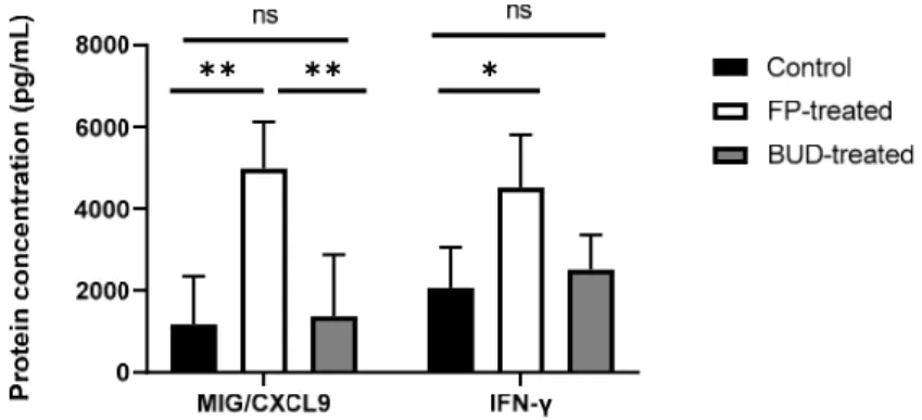
As previously described, IFN-γ exhibits crucial anti-chlamydial activity by inducing chemokine production and increasing anti-chlamydial gene expression [22]. To deter- mine whether ICSs altered gene expression, and influenced the levels of IFN-γ and re- lated chemokines, we estimated IFN-γ and MIG/CXCL-9 protein concentrations in the supernatants of lung homogenates for C. pneumoniae-infected mice (Figure 5). According to our results, IFN-γ production was significantly increased after FP treatment compared to untreated infected control (4519.77 ± 1289.08 pg/mL vs. 2060.07 ± 995.76 pg/mL, p <
0.05). BUD treatment did not affect IFN-γ production significantly compared to FP-treated or control mice.
We examined whether MIG/CXCL9 production at the protein level was changed in association with alterations in IFN-γ production in vivo. We found that FP-treated mice showed a higher protein level of MIG/CXCL9, compared to that in untreated controls (4984 ± 1137 pg/mL vs. 1169 ± 1178 pg/mL, p < 0.01). Furthermore, a significant increment in the level of MIG/CXCL9 was detected in FP-treated lung tissues compared to the BUD-treated mouse lungs (4984 ± 1137 pg/mL vs. 1370 ± 1509 pg/mL, p < 0.01) (Figure 5).
Figure 4. Relative gene expressions inC. pneumoniae-infected and treated mouse lungs. Gene expression of IFN-γ(A), IDO1, IDO2, TDO (B), IFN-γ-inducible chemokines (C), VDR, and GR (D) determined by qPCR. Relative expression was normalised toβ-actin gene expression and calculated by the 2−(∆∆Ct)method. Significant differences are indicated with asterisks, *p< 0.05, **p< 0.01, ***p< 0.001; ns means not significant difference.
Pathogens2021,10, 338 5 of 13
Our previous studies showed that the IFN-γinducible IDO1 and IDO2 are highly expressed inC. pneumoniae-infected mouse lungs [19]. Thus, we next investigated the effects of FP and BUD treatment on the expression of IFN-γinducible IDO1, IDO2, and tryptophan 2,3-dioxygenase (TDO) involved in the metabolism of amino acid tryptophan that is essential forChlamydiagrowth. Our results indicated a significantly increased IDO2 expression in the FP-treated mice compared to the control and BUD-treated group (p< 0.05).
This phenomenon was not observed in IDO1 and TDO expression (Figure4B).
To test the consequence of increased IFN-γrelease, we determined the relative expres- sion levels of IFN-γrelated chemokines. Unexpectedly, we found that BUD significantly decreased the expression of MIG/CXCL9 (p< 0.05) and IP-10/CXCL10 (p< 0.01) com- pared to untreatedC. pneumoniaeinfected control mice. However, the relative expression of MIG/CXCL9 and IP-10/CXCL10 was not altered significantly in FP-treated mice. I- TAC/CXCL11 showed similar expression levels in all groups (Figure4C).
Furthermore, we investigated the expression of glucocorticoid receptor (GR) and Vitamin D receptor (VDR) genes, as both receptors perform immunomodulatory func- tions [20,21]. Interestingly, our results demonstrated that FP treatment increased VDR expression significantly compared to control and BUD-treated mice (p< 0.01), whereas BUD treatment did not affect VDR expression. We found no statistically significant difference in GR expressions inC. pneumoniae-infected BUD- and FP-treated mice (Figure4D).
2.5. Anti-Chlamydial IFN-γand MIG/CXCL9 Protein Production Are Enhanced by FP Treatment As previously described, IFN-γexhibits crucial anti-chlamydial activity by inducing chemokine production and increasing anti-chlamydial gene expression [22]. To determine whether ICSs altered gene expression, and influenced the levels of IFN-γand related chemokines, we estimated IFN-γand MIG/CXCL-9 protein concentrations in the super- natants of lung homogenates forC. pneumoniae-infected mice (Figure5). According to our results, IFN-γproduction was significantly increased after FP treatment compared to untreated infected control (4519.77±1289.08 pg/mL vs. 2060.07±995.76 pg/mL,p< 0.05).
BUD treatment did not affect IFN-γproduction significantly compared to FP-treated or control mice.
Pathogens 2021, 10, x FOR PEER REVIEW 6 of 13
Figure 5. Effect of ICSs on IFN- and MIG/CXCL9 production in C. pneumoniae-infected mouse lungs. IFN- and MIG/CXCL9 protein concentrations were determined by ELISA in the superna- tants of homogenised lung tissues of infected FP-treated, BUD-treated, and untreated mice. Data are shown as pg/mL. Bars denote mean ±SD of each group (n = 12). Significant differences are in- dicated with asterisks, * p < 0.05, ** p < 0.01; ns means not significant difference.
2.6. Effects of BUD and FP Treatment on the Secretion of Th17 and Th2 cytokines in C. pneu- moniae-infected Lung Tissues
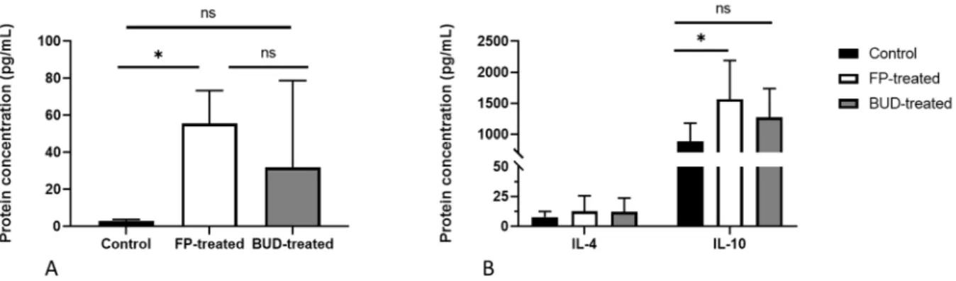
ELISA of supernatants from control and treated lung samples was performed to elucidate the influence of ICS treatment on cytokine production. As previously reported by us, IL-17A has an indirect anti-chlamydial activity in vivo [23]. Thus, we investigated the impact of ICSs on IL-17A production in infected mouse lungs (Figure 6A). We found a significantly elevated IL-17A level in FP-treated mouse lungs compared to the control group (55.59 ± 17.7 pg/mL vs. 3.06 ± 0.67 pg/mL, p < 0.01). Although increased IL-17A production was observed in BUD-treated mice, it did not differ significantly from the control and FP- treated mice.
As Th2 cytokines play a pivotal role in modulating lung inflammation, we evaluated whether FP and BUD could affect IL-4 and IL-10 secretion in C. pneumoniae-infected mice (Figure 6B). Our results indicated unaltered IL-4 production, whereas IL-10 levels changed in a different manner. We detected a significantly higher amount of IL-10 in FP-treated lung tissues, but not in the BUD-treated lungs, in comparison with the un- treated infected mice (1757.28 ± 602.93 pg/mL vs. 739.67 ± 19.70 pg/mL, p < 0.05).
Figure 6. Effects of BUD and FP on the secretion of interleukin (IL)-17A, IL-4 and IL-10 in control and treated mice. IL-17A (A), IL-4, and IL-10 (B) protein concentrations were determined by ELISA in the supernatants of homogenised lung tis- sues of infected FP-treated, BUD-treated, and untreated mice. Bars denote mean± SD of each group (n = 12). * p < 0.05, ns means not significant difference.
✱✱ ✱✱ ✱
Figure 5.Effect of ICSs on IFN-γand MIG/CXCL9 production inC. pneumoniae-infected mouse lungs.
IFN-γand MIG/CXCL9 protein concentrations were determined by ELISA in the supernatants of homogenised lung tissues of infected FP-treated, BUD-treated, and untreated mice. Data are shown as pg/mL. Bars denote mean±SD of each group (n = 12). Significant differences are indicated with asterisks, *p< 0.05, **p< 0.01; ns means not significant difference.
We examined whether MIG/CXCL9 production at the protein level was changed in association with alterations in IFN-γproduction in vivo. We found that FP-treated mice showed a higher protein level of MIG/CXCL9, compared to that in untreated controls (4984±1137 pg/mL vs. 1169±1178 pg/mL,p< 0.01). Furthermore, a significant incre- ment in the level of MIG/CXCL9 was detected in FP-treated lung tissues compared to the BUD-treated mouse lungs (4984±1137 pg/mL vs. 1370 ±1509 pg/mL,p< 0.01) (Figure5).
Pathogens2021,10, 338 6 of 13
2.6. Effects of BUD and FP Treatment on the Secretion of Th17 and Th2 Cytokines in C. pneumoniae-Infected Lung Tissues
ELISA of supernatants from control and treated lung samples was performed to elucidate the influence of ICS treatment on cytokine production. As previously reported by us, IL-17A has an indirect anti-chlamydial activity in vivo [23]. Thus, we investigated the impact of ICSs on IL-17A production in infected mouse lungs (Figure6A). We found a significantly elevated IL-17A level in FP-treated mouse lungs compared to the control group (55.59±17.7 pg/mL vs. 3.06±0.67 pg/mL,p< 0.01). Although increased IL-17A production was observed in BUD-treated mice, it did not differ significantly from the control and FP- treated mice.
Pathogens 2021, 10, x FOR PEER REVIEW 6 of 13
Figure 5. Effect of ICSs on IFN- and MIG/CXCL9 production in C. pneumoniae-infected mouse lungs. IFN- and MIG/CXCL9 protein concentrations were determined by ELISA in the superna- tants of homogenised lung tissues of infected FP-treated, BUD-treated, and untreated mice. Data are shown as pg/mL. Bars denote mean ±SD of each group (n = 12). Significant differences are in- dicated with asterisks, * p < 0.05, ** p < 0.01; ns means not significant difference.
2.6. Effects of BUD and FP Treatment on the Secretion of Th17 and Th2 cytokines in C. pneu- moniae-infected Lung Tissues
ELISA of supernatants from control and treated lung samples was performed to elucidate the influence of ICS treatment on cytokine production. As previously reported by us, IL-17A has an indirect anti-chlamydial activity in vivo [23]. Thus, we investigated the impact of ICSs on IL-17A production in infected mouse lungs (Figure 6A). We found a significantly elevated IL-17A level in FP-treated mouse lungs compared to the control group (55.59 ± 17.7 pg/mL vs. 3.06 ± 0.67 pg/mL, p < 0.01). Although increased IL-17A production was observed in BUD-treated mice, it did not differ significantly from the control and FP- treated mice.
As Th2 cytokines play a pivotal role in modulating lung inflammation, we evaluated whether FP and BUD could affect IL-4 and IL-10 secretion in C. pneumoniae-infected mice (Figure 6B). Our results indicated unaltered IL-4 production, whereas IL-10 levels changed in a different manner. We detected a significantly higher amount of IL-10 in FP-treated lung tissues, but not in the BUD-treated lungs, in comparison with the un- treated infected mice (1757.28 ± 602.93 pg/mL vs. 739.67 ± 19.70 pg/mL, p < 0.05).
Figure 6. Effects of BUD and FP on the secretion of interleukin (IL)-17A, IL-4 and IL-10 in control and treated mice. IL-17A (A), IL-4, and IL-10 (B) protein concentrations were determined by ELISA in the supernatants of homogenised lung tis- sues of infected FP-treated, BUD-treated, and untreated mice. Bars denote mean± SD of each group (n = 12). * p < 0.05, ns means not significant difference.
✱✱ ✱✱ ✱
Figure 6.Effects of BUD and FP on the secretion of interleukin (IL)-17A, IL-4 and IL-10 in control and treated mice. IL-17A (A), IL-4, and IL-10 (B) protein concentrations were determined by ELISA in the supernatants of homogenised lung tissues of infected FP-treated, BUD-treated, and untreated mice. Bars denote mean±SD of each group (n = 12). *p< 0.05, ns means not significant difference.
As Th2 cytokines play a pivotal role in modulating lung inflammation, we evaluated whether FP and BUD could affect IL-4 and IL-10 secretion inC. pneumoniae-infected mice (Figure6B). Our results indicated unaltered IL-4 production, whereas IL-10 levels changed in a different manner. We detected a significantly higher amount of IL-10 in FP-treated lung tissues, but not in the BUD-treated lungs, in comparison with the untreated infected mice (1757.28±602.93 pg/mL vs. 739.67±19.70 pg/mL,p< 0.05).
3. Discussion
ICSs are widely used to treat COPD and asthma, as they show a broad range of anti- inflammatory properties and improve lung function. However, the association between ICS use and the risk of pneumonia remains unclear, as different ICSs have distinct effects on infections. Several studies have been conducted to reveal their possible connection to respi- ratory infections; nevertheless, clinical data tend to be contradictory [2,24,25]. ICSs have different pharmacological and immunomodulatory effects considering responses to viral infections, immune cell functions, and cytokine production [4]. As exacerbations of obstruc- tive lung diseases are mainly caused by viral or bacterial infections, ICSs were investigated from a new perspective, and their effects on antimicrobial defence were examined.
FP and BUD affected pro-inflammatory epithelial responses, and they presented antiviral and anti-inflammatory activities differentially. In vitro studies have shown that BUD is effective in counteracting rhinovirus replication, inhibiting cytokine production, and enhancing antimicrobial protein secretion; however, in vivo, it suppressed pulmonary host defence genes that are essential for bacterial clearance [6,14,26,27]. Moreover, inhalation of BUD has been shown to lead to significant alterations in the regulation of anti- and pro-inflammatory genes involved in eliminating respiratory pathogens [28].
FP is regarded as a more potent inhibitor of pro-inflammatory genes and cytokines, although it can preserve the production of several cytokines, especially IFN-γ, to reverse its exaggerated effect on Th2 response in allergic inflammation. Inhibitory effects of this
Pathogens2021,10, 338 7 of 13
topically active ICS are not well-established in respiratory infections, despite the fact that FP is far more potent in cytokine production than other ICSs [29].
C. pneumoniaeis implicated in asthma exacerbations, and there is increasing evidence indicating that it plays a role in asthma pathogenesis, leading to altered cytokine responses, decreased lung function, and heightened disease severity [30–32]. Thus, we aimed to investigate whether ICSs could have beneficial modulatory effects onChlamydiainfection in vitro and in vivo, and thus, define the possible underlying mechanisms.
Collectively, our reported data inspire several exciting concepts that could have prac- tical outcomes for respiratory physicians. Our in vitro results suggest thatC. pneumoniae growth is suppressed in infected epithelial cells by FP but not by BUD. Thus, we tested our hypothesis whether FP could inhibit the growth ofC. pneumoniaein vivo. We found that the number of recoverableC. pneumoniaedecreased inC. pneumoniae-infected mouse lungs after FP treatment, as observed in A549 epithelial cells. It is well established that the IFN-γ is the main regulator ofChlamydiaelimination and can trigger various mechanism leading to Chlamydia inhibition. IFN-γexposure induced IDO activity, which is responsible for tryptophan degradation. AsChlamydiaeare tryptophan auxotrophs, increased IDO activ- ity results in a restrictedChlamydiagrowth in vitro and in vivo [19,33]. Moreover, IFN-γ enhances the expression of genes involved in innate immunity, thus contributing further to the mechanisms inhibitingChlamydiainfections [34]. Our findings revealed that FP can significantly induce IFN-γnot only at the transcription level but also at the protein level, in contrast to BUD, indicating the previously described phenomenon that FP preserves IFN-γresponses [29]. IFN-γassociated MIG/CXCL9 was shown to have an antimicrobial effect toC. pneumoniae[35], similar toC. trachomatisandC. muridarum[34]. Our results demonstrated that MIG/CXCL9 levels increased at the protein level in parallel with IFN-γ after FP administration, compared to that in untreated control and BUD-treated mouse lungs. Interestingly, FP did not alter the expression of other IFN-γ-inducible chemokines, suggesting that other mechanisms may be playing a role in chemokine expression. Notably, BUD treatment significantly reduced the gene expression levels of CXCL-10 and CXCL-11, which is in accordance with a previous report [36]. Consistently, ICS treatments have a unique impact on IFN-γresponse and chemokine production.
IDO activity is a hallmark of tryptophan depletion and suppression ofChlamydia growth in cell cultures and mice. Furthermore, IDO activity is associated with several pulmonary diseases, including lung cancer [37]. Sputum IDO activity was enhanced in an IL-10 dependent manner in asthmatic patients receiving ICSs; the ICSs increased IL-10 secretion from macrophages in parallel with IDO activity, indicating that ICS use could generate IDO activity through IL-10 production [38]. Our results also indicate a significant increase in IL-10 production after FP administration in infected mice; conversely, BUD did not have an enhancing effect. Since it is well-known that FP can increase local secretion of IL-10 in vivo [39], we concluded that the observed IDO activity could have been derived from the additive effects of IFN-γand IL-10.
To further analyse the production of anti-chlamydial cytokines, we measured IL-17A levels, as it has been reported to trigger neutrophil recruitment in the lung, whereas its neutralisation results in a higherC. pneumoniaeburden [23]. FP was not able to inhibit IL- 17A, in contrast to BUD, which downregulates IL-23, the potential inducer of IL-17A [40,41].
Our current findings revealed that FP-treated mice produced significantly higher amounts of IL-17A in response toC. pneumoniaeinfection, suggesting that this increase in IL-17A production might also result in the development ofC. pneumoniaeinhibition; as observed in a previous study, IL-17A can synergise with IFN-γ, playing a protective role inChlamydia infection [42].
Lastly, we analysed the relative expression of GR and VDR, and we found that FP treatment significantly increased VDR expression compared to control and BUD-treated mice. VDR is one of the highly downregulated transcription factors inChlamydia-infected murine cells [34], suggesting that the elevated VDR expression in our study was due to the influence of FP in mice. This result raises the likelihood of the beneficial impact of FP to
Pathogens2021,10, 338 8 of 13
stem from VDR activation; a former study proved that the diminished VDR activity was associated with higherChlamydiaload in the lungs [43]. Vitamin D favours the curtailment of bacterial infections, and it had several implications in modulating adaptive and innate immunity to eliminate bacterial infections [44]. Therefore, our results raise the possibility that FP supports VDR functions, which can further influence cytokine and chemokine production. Thus, we recommend further examination of the interaction between ICS use and VDR activity, as Vitamin D supplementation is widely recommended in asthma.
In addition, we investigated whether FP or BUD could amend the histopathology of C. pneumoniae-infected mouse lungs and found that all specimens contained lymphocyte infiltration as a sign ofC. pneumoniaeinfection. However, emphysema was also visible in FP-treated lung samples. As FP treatment showed an exaggerated IFN-γresponse to theChlamydiainfection, the elimination ofC. pneumoniaethrough enhanced cytokine and chemokine production could lead to minimal emphysema in infected lungs. According to previous studies [45,46], emphysema can be associated with the IFN-γproduction and Chlamydiainfections.
To our knowledge, this is the first study that investigates the effects of FP and BUD on C. pneumoniaeinfection in vivo and in vitro. The most notable results of our research are as follows: (1) FP treatment inhibitedC. pneumoniaegrowth in A549 cells; (2) FP also inhibited C. pneumoniaereplication in vivo; (3) FP induced IFN-γat the gene expression and protein levels, leading to enhanced IDO activity and MIG/CXCL9 production; (4) FP promoted the expression of VDR.
4. Materials and Methods 4.1. In Vitro Study Design
A549 human airway epithelial cells (ATCC, Manassas, VA, USA) were transferred to a 96-well plate at a density of 4×10⁴cells/well in 100µL of minimal essential medium (MEM) with Earle’s salts supplemented with 10% heat-inactivated foetal bovine serum (FBS), 2 mmol/L-glutamine, 1x non-essential amino acids, 4 mM HEPES and 25µg/mL gentamycin. The cells were pre-treated and incubated for 24 h at 37◦C, 5% CO2with FP (Sigma-Aldrich, Saint Louis, MO, USA) or BUD (Sigma-Aldrich, Saint Louis, MO, USA) or left untreated. The highest non-toxic drug concentrations (FP: 3.5×10⁻⁴mM, BUD:
7×10⁻⁴mM) determined by the 3-(4,5-dimethylthiazol -2yl)-2,5-diphenyl-2H-tetrazolium bromide (MTT) cytotoxicity test. After 24 h treatment, the wells were washed twice with phosphate-buffered saline (PBS) and the cells were infected withC. pneumoniaeat a multiplicity of infection (MOI) of 0.01. The cells were inoculated in 0.5% (w/v) glucose medium and centrifuged (800×g, 60 min), which was followed by the addition of FP or BUD to the wells. Control-infected cells were left untreated. After infection, the plates were incubated at 37◦C, under 5% CO2for 48 h. Subsequently, the wells were washed twice with PBS, and 100µL sucrose–phosphate–glutamic acid (SPG) solution was added to each well.
The plates were subjected to two freeze–thaw cycles with a quick freezing (−80◦C, 15 min) to obtain cell lysates, which were used directly as templates for quantitative polymerase chain reaction (qPCR). To evaluateC. pneumoniaepropagation, direct qPCR was performed as described previously [18,47].
4.2. Inoculum Preparation and Immunostaining
C. pneumoniaestrain CWL-029, kindly gifted by Agathe Subtil (Pasteur Institute, Paris, France), was propagated on HEp-2 cells as described previously [48]. The elementary bodies (EBs) were purified and concentrated, and subsequently aliquoted in SPG, which was followed by storage at−80◦C until further use. Indirect immunofluorescence was performed to determine the concentration of infectiousC. pneumoniaeEBs. Serial dilutions of purified EBs were inoculated onto McCoy cell monolayers (ECACC, London UK). After incubation for 48 h, the infected cells were fixed with acetone at−20◦C and stained with monoclonal anti-ChlamydiaLPS antibody (AbD Serotec, Oxford, UK) and fluorescein isoth- iocyanate (FITC)-labelled anti-mouse immunoglobulin (Ig) G (Sigma-Aldrich, St. Louis,
Pathogens2021,10, 338 9 of 13
MO, USA). The number ofC. pneumoniaeinclusions was counted under a UV microscope, and the titre was expressed as inclusion-forming units (IFUs)/mL.
4.3. Corticosteroid Treatment in Mice
FP and BUD powder were obtained, and dimethyl sulfoxide (DMSO) was used as a vehicle for the drugs. Mice were exposed to nebulised BUD (40µg, 1000µg/kg) and FP (25µg, 625µg/kg) in an inhalation chamber for 15 min once a day, as described previ- ously [49]. We used BUD and FP at equivalent concentrations with the ratio FP:BUD = 1:1.6, based on former clinical studies [50] and the higher potency of FP [10].
4.4. Animals and Experimental Design
Female BALB/c mice (6–8-weeks-old) were obtained from Charles River Laboratories (Hungary). The mice were kept under standard husbandry conditions at the animal facility of the Department of Medical Microbiology and Immunobiology, University of Szeged.
Animals were fed regular mouse chow and provided with waterad libitum. The mice were randomly divided into three groups: the control, the BUD-treated, and FP-treated (n = 16 in each group). Mice received either BUD, FP, or vehicle alone, for three days prior to infection, and then for seven days after infection. On day 3, the mice were sedated with intraperitoneal injection of sodium pentobarbital (200µL, 7.5 mg/mL) and were infected with 5×10⁵IFUC. pneumoniaein 20µL SPG. On day 10, i.e., seven days after infection, the mice were anaesthetised and sacrificed. The lungs were removed and homogenised with acid-purified sea sand (Fluka Chemie AG, Buchs, Switzerland) using a mortar with a pestle. One half of the homogenised lungs was prepared for total RNA extraction, and the other half was suspended in 1 mL SPG for the detection of recoverableC. pneumoniaeand for cytokine measurements. The experiments were implemented with the approval of the Animal Welfare Committee of the University of Szeged, Hungary and conformed to the Directive 2010/63/EU.
4.5. Culturing of C. pneumoniae from the Lungs
One half of the homogenised lungs was centrifuged (10 min, 400×g), and serial dilutions of the supernatants were inoculated onto McCoy cell monolayers and centrifuged (60 min, 800×g). The number of recoverableC. pneumoniaeinclusions was determined by indirect immunofluorescence as described earlier, and expressed in terms of IFU/mL.
4.6. mRNA Extraction and cDNA Synthesis
Total RNA was extracted from the other half of the homogenised lung tissues of the control (n = 12), as well as BUD- and FP-treated mice (n = 12 for each group) using TRI reagent (Sigma-Aldrich, Saint Louis, MO, USA) according to the manufacturer’s protocol.
Total RNA concentrations and purity were measured using a NanoDrop spectrophotometer (Thermo Scientific, Waltham, MA, USA). First-strand cDNA was synthesised from 2µg of total RNA using Maxima First Strand cDNA Synthesis Kit, and 20 pM random hexamer primer in 20µL reaction buffer according to the manufacturer’s protocol (Thermo Fisher Scientific Inc. Waltham, MA, USA).
4.7. qPCR Validation
qPCR was performed in a Bio-Rad CFX96 real-time system with SsoFast™ Eva- Green®qPCR Supermix (Bio-Rad, Hercules, CA, USA) master mix and murine specific primer pairs:β-actin sense, 50-TGGAATCCTGTGGCATCCATGAAAC-30;β-actin antisense, 50-TAAAACGCAGCTCAGTAACAGTCCG-30; IDO1 sense, 50-GCTTCTTCCTCGTCTCTCT ATTG-30; IDO1 antisense, 50-TCTCCAGACTGGTAGCTATGT-30; IDO2 sense, 50-CCTGGAC TGCAGATTCCTAAAG-30; IDO2 antisense, 50-CCAAGTTCCTGGATACCTCAAC-30; TDO sense, 50-GGCATGGCTGGAAAGAACAC-30; TDO antisense, 50-CTCCCTGGAGTGCACG GTAT-30; IFN-γsense, 50-CAAGTGGCATAGATGTGGAAGA-30; IFN-γantisense, 50-GCTG TTGCTGAAGAAGGTAGTA-30; MIG/CXCL9 sense, 50-ACGTAGGTTTCGAGACCAGGGA
Pathogens2021,10, 338 10 of 13
TT-30; MIG/CXCL9 antisense, 50-CAACACCAAGTGTTCTGCCACCAA-30; IP10/CXCL10 sense, 50-TGGCTAGTCCTAATTGCCCTTGGT-30; IP10/CXCL10 antisense, 50-TCAGGACC ATGGCTTGACCATCAT-30; ITAC/CXCL11 sense, 50-TACCCGAGTAACAGCTGCGACAA A-30; ITAC/CXCL11 antisense, 50-TATGAGGCGAGCTTGCTTGGATCT-30; VDR sense, 50- TACACCCCCTCACTGGACATGAT-30; VDR antisense, 50-CGATGACCTTTTGGATGCTGT AA-30; GR sense, 50-GTTCCTAAGGAAGGTCTGAAGAG-30; and GR antisense, 50-CAATTC TGACTGGAGTTTCC-30.
Cycle threshold (Ct) values were calculated forβ-actin, IDO1, IDO2, TDO, IFN-γ MIG/CXCL9, IP10/CXCL10, ITAC/CXCL11, VDR, and GR, and the relative gene ex- pression levels were determined by the 2−(∆∆Ct)method. The relative expression level was indicated as 2−(∆∆Ct), where∆∆Ct =∆Ct for the experimental sample−∆Ct for the control sample.
4.8. Lung Histology
Microscopic examination of the lungs of infected control as well as BUD-treated and FP-treated mice (n = 4 from each group) was performed. After the removal of the lungs, tissues of individual mice were immediately placed into plastic tubes, pre-filled with 10%
formalin, resulting in 1:10 of tissue/formalin ratio. During dissection, tissue samples in the tube were cut into 1 mm slices and embedded into paraffin blocks. Four-micrometre sections were cut, and regular haematoxylin–eosin (HE) staining was performed. All tissue fragments were examined by light microscopy.
4.9. Cytokine and Chemokine Measurements from the Lungs
The supernatants of homogenised lung tissues were centrifuged (12,000×g, 5 min) and enzyme-linked immunosorbent assay (ELISA) for IFN-γ, IL-4, IL-10, IL17-A, MIG/CXCL-9 was performed according to the manufacturers’ instructions. MIG/CXCL-9 concentration was determined using a mouse MIG/CXCL-9 ELISA Kit (Sigma-Aldrich, Saint Louis, MO, USA) and IL-17A was measured using Quantikine mouse IL-17 immunoassay (R&D Systems, Minneapolis, MN, USA). IFN-γ, IL-4, and IL-10 concentrations were detected with Invitrogen mouse ELISA kits (Thermo Fisher Scientific Inc., Waltham, MA, USA).
Sensitivity for IFN-γ, IL-4, IL-10, IL-17A, and MIG/CXCL-9 measurement ranges were 15–2000, 4–500, 32–4000, 10.9–700, and 2.741–2000 pg/mL, respectively.
4.10. Statistical Analysis
Statistical analysis of data was performed with GraphPad Prism 8.0.1. software, using one-way and two-way ANOVA, and Kruskal–Wallis test. All post hoc comparisons were performed using Tukey’s method. Data are expressed as mean±standard deviation (SD).
Differences atp< 0.05 were considered statistically significant.
5. Conclusions
Our results demonstrate that FP treatment can lead to favourable outcomes in C. pneumoniae infection by enhancing IFN-γ responses, and inducing potential anti- chlamydial cytokine and chemokine production. Our observations revealed that the effects of FP were different from other ICSs; therefore, we hypothesised that FP is beneficial inC. pneumoniaeinfections. Recently, patients with asthma are controlled in new ways via telemedicine and experienced fewer outpatient visits due to COVID- 19 pandemic. Therefore, investigating the associations of ICSs use and respiratory infections is especially important to avoid further asthma exacerbations. Our study can contribute to a better management of asthma andC. pneumoniaeinfections, and it could assist therapeutic choices.
Author Contributions: D.P. conceived, designed, and performed the experiments, analysed the data, and wrote the paper. A.S. prepared lung tissues for histopathological analysis, performed HE staining, interpreted histological findings, and was involved in manuscript writing. D.K. was involved in performing qPCR, ELISA, and statistical analysis. V.E. contributed to animal testing
Pathogens2021,10, 338 11 of 13
and manuscript writing. D.P.V. was involved in interpreting lab data and manuscript writing. K.B.
conceived the study design, reviewed the manuscript, and supervised the experiment. All authors have read and agreed to the published version of the manuscript.
Funding:The study was supported by GINOP-2.3.2-15-2016-00012.
Institutional Review Board Statement: The experiments were approved by the Animal Welfare Committee of the University of Szeged, Hungary, and conformed to the Directive 2010/63/EU.
Informed Consent Statement:Informed consent was obtained from all subjects involved in the study.
Data Availability Statement: The data presented in this study are available on request from the corresponding author. The data are not publicly available due to privacy.
Acknowledgments: We thank MüllernéDeák Györgyi and VigyikánnéVáradi Anikó for their excellent technical support, and Dezs˝o Mihály for his assistance in the microscopic pictures. D.P. was supported by the Hungarian Respiratory Foundation.
Conflicts of Interest:The authors declare no conflict of interest.
References
1. Morjaria, J.B.; Rigby, A.; Morice, A.H. Inhaled Corticosteroid Use and the Risk of Pneumonia and COPD Exacerbations in the UPLIFT Study.Lung2017,195, 281–288. [CrossRef]
2. Sin, D.D.; Tashkin, D.; Zhang, X.; Radner, F.; Sjöbring, U.; Thorén, A.; Calverley, P.M.A.; Rennard, S.I. Budesonide and the Risk of Pneumonia: A Meta-Analysis of Individual Patient Data.Lancet2009,374, 712–719. [CrossRef]
3. McKeever, T.; Harrison, T.W.; Hubbard, R.; Shaw, D. Inhaled Corticosteroids and the Risk of Pneumonia in People with Asthma:
A Case-Control Study.Chest2013,144, 1788–1794. [CrossRef] [PubMed]
4. Janson, C.; Stratelis, G.; Miller-Larsson, A.; Harrison, T.W.; Larsson, K. Scientific Rationale for the Possible Inhaled Corticosteroid Intraclass Difference in the Risk of Pneumonia in COPD.Int. J. Chronic Obstr. Pulm. Dis.2017,12, 3055–3064. [CrossRef]
5. Belchamber, K.B.; Thomas, C.M.; Dunne, A.E.; Barnes, P.J.; Donnelly, L.E. Comparison of Fluticasone Propionate and Budesonide on COPD Macrophage and Neutrophil Function.Int. J. Chronic Obstr. Pulm. Dis.2018,13, 2883–2897. [CrossRef]
6. Yamaya, M.; Nishimura, H.; Nadine, L.; Kubo, H.; Nagatomi, R. Formoterol and Budesonide Inhibit Rhinovirus Infection and Cytokine Production in Primary Cultures of Human Tracheal Epithelial Cells. Respir. Investig. 2014,52, 251–260. [CrossRef]
[PubMed]
7. Bochkov, Y.A.; Busse, W.W.; Brockman-Schneider, R.A.; Evans, M.D.; Jarjour, N.N.; McCrae, C.; Miller-Larsson, A.; Gern, J.E.
Budesonide and Formoterol Effects on Rhinovirus Replication and Epithelial Cell Cytokine Responses.Respir. Res.2013,14, 98.
[CrossRef]
8. Van den Berge, M.; Jonker, M.R.; Miller-Larsson, A.; Postma, D.S.; Heijink, I.H. Effects of Fluticasone Propionate and Budesonide on the Expression of Immune Defense Genes in Bronchial Epithelial Cells.Pulm. Pharmacol. Ther.2018,50, 47–56. [CrossRef]
9. Yamaya, M.; Nishimura, H.; Deng, X.; Sugawara, M.; Watanabe, O.; Nomura, K.; Shimotai, Y.; Momma, H.; Ichinose, M.;
Kawase, T. Inhibitory Effects of Glycopyrronium, Formoterol, and Budesonide on Coronavirus HCoV-229E Replication and Cytokine Production by Primary Cultures of Human Nasal and Tracheal Epithelial Cells. Respir. Investig.2020,58, 155–168.
[CrossRef]
10. Jaffuel, D.; Demoly, P.; Gougat, C.; Balaguer, P.; Mautino, G.; Godard, P.; Bousquet, J.; Mathieu, M. Transcriptional Potencies of Inhaled Glucocorticoids.Am. J. Respir. Crit. Care Med.2000,162, 57–63. [CrossRef]
11. Ek, A.; Larsson, K.; Siljerud, S.; Palmberg, L. Fluticasone and Budesonide Inhibit Cytokine Release in Human Lung Epithelial Cells and Alveolar Macrophages.Allergy1999,54, 691–699. [CrossRef]
12. Edsbäcker, S.; Wollmer, P.; Selroos, O.; Borgström, L.; Olsson, B.; Ingelf, J. Do Airway Clearance Mechanisms Influence the Local and Systemic Effects of Inhaled Corticosteroids?Pulm. Pharmacol. Ther.2008,21, 247–258. [CrossRef] [PubMed]
13. Miller-Larsson, A.; Mattsson, H.; Hjertberg, E.; Dahlbäck, M.; Tunek, A.; Brattsand, R. Reversible Fatty Acid Conjugation of Budesonide. Novel Mechanism for Prolonged Retention of Topically Applied Steroid in Airway Tissue.Drug Metab. Dispos.1998, 26, 623–630. [PubMed]
14. Wang, P.; Wang, X.; Yang, X.; Liu, Z.; Wu, M.; Li, G. Budesonide Suppresses Pulmonary Antibacterial Host Defense by Down- Regulating Cathelicidin-Related Antimicrobial Peptide in Allergic Inflammation Mice and in Lung Epithelial Cells.BMC Immunol.
2013,14, 7. [CrossRef]
15. Kuo, C.C.; Jackson, L.A.; Campbell, L.A.; Grayston, J.T. Chlamydia Pneumoniae (TWAR).Clin. Microbiol. Rev.1995,8, 451–461.
[CrossRef] [PubMed]
16. Carr, T.F.; Kraft, M. Chronic Infection and Severe Asthma.Immunol. Allergy Clin. N. Am.2016,36, 483–502. [CrossRef] [PubMed]
17. Paróczai, D.; Mosolygó, T.; Kókai, D.; Endrész, V.; Virok, D.P.; Somfay, A.; Burián, K. Chlamydia Pneumoniae Influence on Cytokine Production in Steroid-Resistant and Steroid-Sensitive Asthmatics.Pathogens2020,9. [CrossRef] [PubMed]
18. Eszik, I.; Lantos, I.; Önder, K.; Somogyvári, F.; Burián, K.; Endrész, V.; Virok, D.P. High Dynamic Range Detection of Chlamydia Trachomatis Growth by Direct Quantitative PCR of the Infected Cells.J. Microbiol. Methods2016,120, 15–22. [CrossRef] [PubMed]
Pathogens2021,10, 338 12 of 13
19. Virok, D.P.; Raffai, T.; Kókai, D.; Paróczai, D.; Bogdanov, A.; Veres, G.; Vécsei, L.; Poliska, S.; Tiszlavicz, L.; Somogyvári, F.; et al.
Indoleamine 2,3-Dioxygenase Activity in Chlamydia Muridarum and Chlamydia Pneumoniae Infected Mouse Lung Tissues.
Front. Cell Infect. Microbiol.2019,9, 192. [CrossRef]
20. Marcellini, A.; Swieboda, D.; Guedán, A.; Farrow, S.N.; Casolari, P.; Contoli, M.; Johnston, S.L.; Papi, A.; Solari, R. Glucocorticoids Impair Type I IFN Signalling and Enhance Rhinovirus Replication.Eur. J. Pharmacol.2020,893, 173839. [CrossRef]
21. Baeke, F.; Takiishi, T.; Korf, H.; Gysemans, C.; Mathieu, C. Vitamin D: Modulator of the Immune System.Curr. Opin. Pharmacol.
2010,10, 482–496. [CrossRef]
22. Nelson, D.E.; Virok, D.P.; Wood, H.; Roshick, C.; Johnson, R.M.; Whitmire, W.M.; Crane, D.D.; Steele-Mortimer, O.; Kari, L.;
McClarty, G.; et al. Chlamydial IFN-Gamma Immune Evasion Is Linked to Host Infection Tropism.Proc. Natl. Acad. Sci. USA 2005,102, 10658–10663. [CrossRef] [PubMed]
23. Mosolygó, T.; Korcsik, J.; Balogh, E.P.; Faludi, I.; Virók, D.P.; Endrész, V.; Burián, K. Chlamydophila Pneumoniae Re-Infection Triggers the Production of IL-17A and IL-17E, Important Regulators of Airway Inflammation.Inflamm. Res.2013,62, 451–460.
[CrossRef]
24. Yeh, J.-J.; Lin, C.-L.; Kao, C.-H. Associations among Chronic Obstructive Pulmonary Disease with Asthma, Pneumonia, and Corticosteroid Use in the General Population.PLoS ONE2020,15, e0229484. [CrossRef] [PubMed]
25. Kim, M.H.; Rhee, C.K.; Shim, J.S.; Park, S.Y.; Yoo, K.H.; Kim, B.Y.; Bae, H.W.; Sim, Y.S.; Chang, J.H.; Cho, Y.J.; et al. Inhaled Corticosteroids in Asthma and the Risk of Pneumonia.Allergy Asthma Immunol. Res.2019,11, 795–805. [CrossRef] [PubMed]
26. Heijink, I.H.; Jonker, M.R.; de Vries, M.; van Oosterhout, A.J.M.; Telenga, E.; Ten Hacken, N.H.T.; Postma, D.S.; van den Berge, M.
Budesonide and Fluticasone Propionate Differentially Affect the Airway Epithelial Barrier.Respir. Res.2016,17, 2. [CrossRef]
[PubMed]
27. Kim, S.-R.; Song, J.-H.; Ahn, J.-H.; Lee, G.-S.; Ahn, H.; Yoon, S.-I.; Kang, S.G.; Kim, P.-H.; Jeon, S.-M.; Choi, E.-J.; et al. Antiviral and Anti-Inflammatory Activity of Budesonide against Human Rhinovirus Infection Mediated via Autophagy Activation.Antivir. Res.
2018,151, 87–96. [CrossRef]
28. Leigh, R.; Mostafa, M.M.; King, E.M.; Rider, C.F.; Shah, S.; Dumonceaux, C.; Traves, S.L.; McWhae, A.; Kolisnik, T.; Kooi, C.; et al.
An Inhaled Dose of Budesonide Induces Genes Involved in Transcription and Signaling in the Human Airways: Enhancement of Anti- and Proinflammatory Effector Genes.Pharmacol. Res. Perspect.2016,4. [CrossRef] [PubMed]
29. Umland, S.P.; Nahrebne, D.K.; Razac, S.; Beavis, A.; Pennline, K.J.; Egan, R.W.; Billah, M.M. The Inhibitory Effects of Topically Ac- tive Glucocorticoids on IL-4, IL-5, and Interferon-Gamma Production by Cultured Primary CD4+ T Cells.J. Allergy Clin. Immunol.
1997,100, 511–519. [CrossRef]
30. Smith-Norowitz, T.A.; Loeffler, J.; Huang, Y.; Klein, E.; Norowitz, Y.M.; Hammerschlag, M.R.; Joks, R.; Kohlhoff, S. Chlamydia Pneumoniae Immunoglobulin E Antibody Levels in Patients with Asthma Compared with Non-Asthma. Heliyon2020, 6.
[CrossRef] [PubMed]
31. Smith-Norowitz, T.A.; Chotikanatis, K.; Weaver, D.; Ditkowsky, J.; Norowitz, Y.M.; Hammerschlag, M.R.; Joks, R.; Kohlhoff, S.
Chlamydia Pneumoniae-Induced Tumour Necrosis Factor Alpha Responses Are Lower in Children with Asthma Compared with Non-Asthma.BMJ Open Respir. Res.2018,5, e000239. [CrossRef] [PubMed]
32. Black, P.N.; Scicchitano, R.; Jenkins, C.R.; Blasi, F.; Allegra, L.; Wlodarczyk, J.; Cooper, B.C. Serological Evidence of Infection with Chlamydia Pneumoniae Is Related to the Severity of Asthma.Eur. Respir. J.2000,15, 254–259. [CrossRef] [PubMed]
33. Byrne, G.I.; Lehmann, L.K.; Landry, G.J. Induction of Tryptophan Catabolism Is the Mechanism for Gamma-Interferon-Mediated Inhibition of Intracellular Chlamydia Psittaci Replication in T24 Cells.Infect. Immun.1986,53, 347–351. [CrossRef] [PubMed]
34. Burian, K.; Endresz, V.; Deak, J.; Kormanyos, Z.; Pal, A.; Nelson, D.; Virok, D.P. Transcriptome Analysis Indicates an Enhanced Ac- tivation of Adaptive and Innate Immunity by Chlamydia-Infected Murine Epithelial Cells Treated with Interferonγ.J. Infect. Dis.
2010,202, 1405–1414. [CrossRef]
35. Balogh, E.P.; Faludi, I.; Virók, D.P.; Endrész, V.; Burián, K. Chlamydophila Pneumoniae Induces Production of the Defensin-like MIG/CXCL9, Which Has in Vitro Antichlamydial Activity.Int. J. Med. Microbiol.2011,301, 252–259. [CrossRef] [PubMed]
36. Davies, J.M.; Carroll, M.L.; Li, H.; Poh, A.M.; Kirkegard, D.; Towers, M.; Upham, J.W. Budesonide and Formoterol Reduce Early Innate Anti-Viral Immune Responses in Vitro.PLoS ONE2011,6, e27898. [CrossRef] [PubMed]
37. Dora, D.; Rivard, C.; Yu, H.; Bunn, P.; Suda, K.; Ren, S.; Lueke Pickard, S.; Laszlo, V.; Harko, T.; Megyesfalvi, Z.; et al.
Neuroendocrine Subtypes of Small Cell Lung Cancer Differ in Terms of Immune Microenvironment and Checkpoint Molecule Distribution.Mol. Oncol.2020,14, 1947–1965. [CrossRef]
38. Maneechotesuwan, K.; Supawita, S.; Kasetsinsombat, K.; Wongkajornsilp, A.; Barnes, P.J. Sputum Indoleamine-2, 3-Dioxygenase Activity Is Increased in Asthmatic Airways by Using Inhaled Corticosteroids.J. Allergy Clin. Immunol.2008,121, 43–50. [CrossRef]
39. Feng, E.; Wan, R.; Yang, S.; Yan, Z.; Wang, S.; He, W.; Zhang, Y.; Yin, H.; Chen, Z.; Liu, R. Expression Levels of Induced Sputum IL-8 and IL-10 and Drug Intervention Effects in Patients with Acute Exacerbated COPD Complicated with Chronic Cor Pulmonale at High Altitude.Exp. Ther. Med.2013,6, 747–752. [CrossRef]
40. Li, H.-T.; Lin, Y.-S.; Ye, Q.-M.; Yang, X.-N.; Zou, X.-L.; Yang, H.-L.; Zhang, T.-T. Airway Inflammation and Remodeling of Cigarette Smoking Exposure Ovalbumin-Induced Asthma Is Alleviated by CpG Oligodeoxynucleotides via Affecting Dendritic Cell-Mediated Th17 Polarization.Int. Immunopharmacol.2020,82, 106361. [CrossRef]
Pathogens2021,10, 338 13 of 13
41. Honda, K.; Wada, H.; Nakamura, M.; Nakamoto, K.; Inui, T.; Sada, M.; Koide, T.; Takata, S.; Yokoyama, T.; Saraya, T.; et al. IL-17A Synergistically Stimulates TNF-α-Induced IL-8 Production in Human Airway Epithelial Cells: A Potential Role in Amplifying Airway Inflammation.Exp. Lung Res.2016,42, 205–216. [CrossRef] [PubMed]
42. Zhang, Y.; Wang, H.; Ren, J.; Tang, X.; Jing, Y.; Xing, D.; Zhao, G.; Yao, Z.; Yang, X.; Bai, H. IL-17A Synergizes with IFN-γto Upregulate INOS and NO Production and Inhibit Chlamydial Growth.PLoS ONE2012,7, e39214. [CrossRef] [PubMed]
43. He, Q.; Ananaba, G.A.; Patrickson, J.; Pitts, S.; Yi, Y.; Yan, F.; Eko, F.O.; Lyn, D.; Black, C.M.; Igietseme, J.U.; et al. Chlamydial Infection in Vitamin D Receptor Knockout Mice Is More Intense and Prolonged than in Wild-Type Mice. J. Steroid Biochem.
Mol. Biol.2013,135, 7–14. [CrossRef] [PubMed]
44. Youssef, D.A.; Miller, C.W.; El-Abbassi, A.M.; Cutchins, D.C.; Cutchins, C.; Grant, W.B.; Peiris, A.N. Antimicrobial Implications of Vitamin D.Dermatoendocrinology2011,3, 220–229. [CrossRef]
45. Qiu, S.-L.; Kuang, L.-J.; Tang, Q.-Y.; Duan, M.-C.; Bai, J.; He, Z.-Y.; Zhang, J.-Q.; Li, M.-H.; Deng, J.-M.; Liu, G.-N.; et al. Enhanced Activation of Circulating Plasmacytoid Dendritic Cells in Patients with Chronic Obstructive Pulmonary Disease and Experimental Smoking-Induced Emphysema.Clin. Immunol.2018,195, 107–118. [CrossRef]
46. Theegarten, D.; Anhenn, O.; Hotzel, H.; Wagner, M.; Marra, A.; Stamatis, G.; Mogilevski, G.; Sachse, K. A Comparative Ultrastructural and Molecular Biological Study on Chlamydia Psittaci Infection in Alpha-1 Antitrypsin Deficiency and Non- Alpha-1 Antitrypsin Deficiency Emphysema versus Lung Tissue of Patients with Hamartochondroma.BMC Infect. Dis2004,4, 38.
[CrossRef]
47. Virók, D.P.; Eszik, I.; Mosolygó, T.; Önder, K.; Endrész, V.; Burián, K. A Direct Quantitative PCR-Based Measurement of Herpes Simplex Virus Susceptibility to Antiviral Drugs and Neutralizing Antibodies.J. Virol. Methods2017,242, 46–52. [CrossRef]
48. Burián, K.; Hegyesi, H.; Buzás, E.; Endrész, V.; Kis, Z.; Falus, A.; Gönczöl, E. Chlamydophila (Chlamydia) Pneumoniae Induces Histidine Decarboxylase Production in the Mouse Lung.Immunol. Lett.2003,89, 229–236. [CrossRef]
49. Zu´ska-Prot, M.; Ma´slanka, T. Effect of Inhaled and Systemic Glucocorticoid Treatment on CD4+ Regulatory and Effector T Cells in a Mouse Model of Allergic Asthma.Int. Immunopharmacol.2017,45, 98–109. [CrossRef]
50. Becker, A.B.; Abrams, E.M. Asthma Guidelines: The Global Initiative for Asthma in Relation to National Guidelines.Curr. Opin.
Allergy Clin. Immunol.2017,17, 99–103. [CrossRef]