THESIS OF THE Ph.D. DISSERTATION
University of West Hungary,
Faculty of Agricultural and Food Sciences Institute of Animal Science
Ph.D School for the Biological, Technological, Ecological, Feeding and Economical Questions of Animal Production
Chairman of the Ph.D School Dr. Pál Benedek
Improvement and Breeding Technology Considerations of Animal Production Program
Program leader Dr. Katalin Kovácsné Gaál
Supervisor Dr. Ágnes Bali Papp
ANALYSES OF PORCINE IN VITRO EMBRYO PRODUCTION SYSTEM
Author
ERIKA VARGA
candidate
Mosonmagyaróvár
2007
Analyses of porcine in vitro production system
LIST OF ABBREVIATIONS
LIST OF ABBREVIATIONS
COC Cumulus−oocyte complex CPA Cryoprotectant Agent CSF Cytostatic Factor
CX Cycloheximide
6-DMAP 6-Dymethyl-aminopurine GV Germinal Vesicle
hCG Human Chorion Gonadotrophin IVC In Vitro Cultivation
IVF In Vitro Fertilization IVM In Vitro Maturation
IVP In Vitro embryo Production
NCSU-23 North Carolina State University medium 23 NCSU-37 North Carolina State University medium 37 OPS Open Pulled Straw vitrification method PFF Pig Follicular Fluid
PMSG Pregnant Mare Serum Gonadotrophine
PN Pronucleus
SrCl2 Strontium-chloride
TCM-199 Tissue Culture Medium 199 ZP Zona Pellucida
Analyses of porcine in vitro production system
OBJECTIVES
1. OBJECTIVES
The applied methods of biotechnology based on in vitro produced embryos, which suppose an existence of successful in vitro embryo production (IVP) system.
IVP has several advances for animal breeding: thus IVP enables us to produce a larger number of embryos with less cost and less time. These embryos serve as recipient embryos for other assisted reproductive techniques (Braga et al., 2007) such as cloning (Betthauser et al., 2000), transgenic pig production (Brem et al., 1985) and xenotransplantation (Cascalho et al., 2006).
In vitro embryo production could be instrumental in gene banking of rare breeds, endangered species and precious individuals as well and reservation and expansion of bio-diversity.
The cryopreservation of porcine oocytes is still an open problem because of their structural sensitivity to the cooling and freezing process and to the exposure to cryoprotectants.
In the past decades several new techniques were performed to improve IVP system and the cryopreservation methods but more research is needed in this area in the future.
The main objectives of this study were:
1. In vitro production of porcine embryos from oocytes activated by chemical agents.
2. Vitrification of porcine oocytes with Open Pulled Straw (OPS) method
Evaluate the effect of cumulus cells on viability and fertilizability of in vitro matured, vitrified porcine oocytes.
Examination of role of different meiotic stages (GV and M ll) on sensibility of oocytes to Open Pulled Straw vitrification.
3. In vitro maturation, cryoprezervation and in vitro fertilization of Mangalica (Hungarian native pig breed) oocytes and in vitro embryo cultivation.
Analyses of porcine in vitro production system
MATERIALS AND METHODS
2. MATERIALS AND METHODS
Experiments were made in the laboratory of the Institute of Animal Science at the University of West Hungary, Faculty of Agricultural and Food Sciences and in the laboratory of University of Murcia, Faculty of Veterinary Sciences between 2004 and 2007.
All chemical reagents used for the experiments were purchased from Sigma−Aldrich Chemical Co. (Budapest) and Werft−Chemie GmbH (Wien).
Three replicates of each experiment were performed.
All data were analyzed by ANOVA system of STATISTICA program, followed Duncan’s multiple range test. Differences with P<0.05 was considered significant.
2.1. Activation of porcine oocytes
Cumulus−oocyte complexes (COCs) from slaughterhouse ovaries of Hungarian Large White gilts were used in the experiments.
COCs were matured in TCM-199 medium supplemented with 10 % pig follicular fluid (PFF), 0.9 mM Na-pyruvate, 100 µM cysteamine, 0.25 mM L−glutamine, 0.1 mg/ml streptomycin sulphate for 42 hours, and in 10 IU/ml PMSG and 10 IU/ml hCG in the first 20 hours of maturation.
All the 2401 oocytes were examined in three replicates.
Experiment 1.
Cumulus free IVM oocytes were exposed to 10 mM strontium-chloride in S group (number of oocytes (n) were 145), treated with 2 mM 6-dimethyl- aminopurine in D group (n=144) and activate with 0.04 mM cycloheximide in CX group (n=143). Oocytes were treated with strontium-chloride (15.85 mg/ml) combined with cycloheximide (1 mg/ml) in SCX group (n=142) and strontium-chloride (15.85 mg/ml) combined with 6-DMAP (32.36 mg/ml) in SD group (n=144).
Seven hours after incubation the rate of activation was judged by morphological appearance of oocytes. Those oocytes that formed visible pronucleus (PN) were recorded as activated; the rate of oocyte degeneration was also determined.
Analyses of porcine in vitro production system
MATERIALS AND METHODS
Experiment 2.
Oocytes were treated with the same chemicals as in Experiment 1. [S group (n=188), D group (n=169), CX group (n=159), SD group (n=191), SCX group (n=158)].
After treatments oocytes were incubated in NCSU-37 for 48 hours.
Oocytes in control group (n=90) were not treated, only matured for 42 hours and then cultured in NCSU-37 medium for 48 hours.
To evaulate the cleavage rate, 48 hours after activation oocytes/zygotes were mounted on glass slides with coverslips and fixed in acetic alcohol (1/3 w/v) for at least three days. Fixed oocytes were than stained with 0.1 % (w/v) orcein in 45 % (v/v) acetic acid and evaluated under phase contrast microscopy.
Experiment 3.
Oocytes were treated in the same way as in Experiment 1. [S group (n=68), D group (n=97), CX group (n=95), SD group (n=80), SCX group (n=81)].
After treatments oocytes/zygotes were cultured in 500 µl NCSU-37 medium for 6 days.
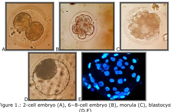
Oocytes in control group (n=90) were matured for 42 hours and cultured in NCSU-37 medium for 6 days. Thereafter, the developmental stages of embryos were determined after orcein staining.
A B C
D E
Figure 1.: 2-cell embryo (A), 6−8-cell embryo (B), morula (C), blastocyst (D,E)
Analyses of porcine in vitro production system
MATERIALS AND METHODS
2.2. Vitrification of porcine oocytes
Open Pulled Straw (OPS) vitrification of oocytes from slaughterhouse ovaries was performed in the experiments as described by Vajta (1997).
A total of 2237 oocytes were examined in three replicates.
Experiment 1.
In vitro matured (42 h), cumulus−surrounded oocytes (COC group;
n=255), and in vitro matured oocytes (completely) denuded of cumulus cells after maturation (D group; n=215) were vitrified with OPS method.
Oocytes in control group (n=217) were matured in vitro for 42 hours, thereafter, they were fertilized and cultivated in NCSU-23 medium for 24 hours to evaluate the fertilization rate.
The morphology of frozen/thawed oocytes and the rate of oocyte degeneration were determined after vitrification.
Some oocytes from each group were treated with 0.1 % pronase to evaulate the structure of zona pellucida (ZP) and the integrity of plasma membrane of oocyte.
Fertilization rate was determined after 24 hours of in vitro fertilization.
Experiment 2.
Immature [cumulus−covered = GCOC group (n=510); cumulus−denuded=
GD group (n=560)] and in vitro matured, cumulus−surrounded [MCOC group (n=350)] oocytes were vitrified with OPS method. Survival and developmental potential of each group were determined after vitrification.
After thawing, oocytes in MCOC group were fertilized; oocytes in GCOC group and GD group were first in vitro matured and then fertilized.
Oocytes in control group (n=103) were matured in vitro then fertilized, and cultivated in NCSU-23 medium for 24 hours.
Changes in oocyte morphology after thawing were examined in different groups and fertilization rate were also determined in each group.
Analyses of porcine in vitro production system
MATERIALS AND METHODS
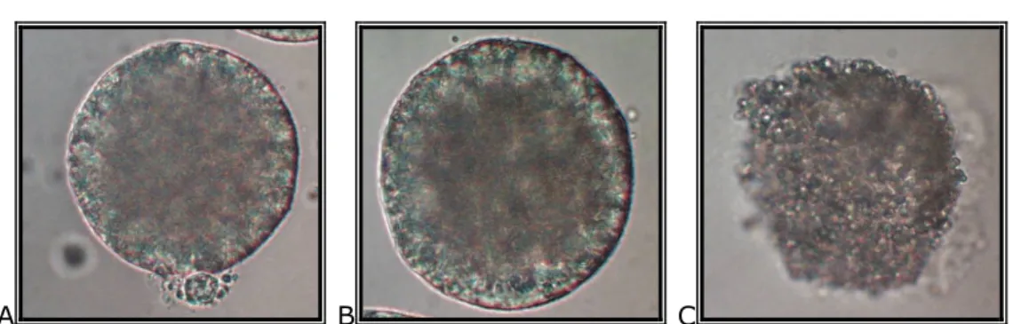
A B C
2. Figure: Frozen/thawed porcine oocytes after removing ZP: oocyte with intact plasma membrane, and visible polar body (A), oocytes with intact plasma membrane (B), degenerated oocyte without plasma membrane (C)
2.3. In vitro maturation and cryopreservation of Mangalica (Hungarian native pig breed) oocytes
No studies were found about in vitro maturation and Open Pulled Straw (OPS) vitrification of Mangalica oocytes in the scientific literature.
A total of 658 Mangalica and 676 Hungarian Large White pig oocytes were vitrified in the experiments.
In vitro matured, cumulus−surrounded Mangalica (M group) and Hungarian Large White (LW group) oocytes from slaughterhouse ovaries were used for OPS vitrification.
Oocytes in control Mangalica (CM group) and control Hungarian Large White groups (CLW group) were in vitro matured and fertilized without vitrification. Experiments were repeated three times.
Experiment 1.
The effectiveness of in vitro maturation system on Mangalica [M group (n=310)] and Large White [LW group (n=330)] oocytes were compared.
The meiotic maturation and cumulus−expansion rate were determined.
Experiment 2.
The sensitivity of Mangalica
[
M group (n=171)] and Large White [LW group (n=183)] oocytes on vitrification method was compared.Experiment 3.
The embryonic development of frozen/thawed fertilized Mangalica
[
M group (n=28); CM group (n=130)] and Large White [LW group (n=53); CLW group (n=136)]oocytes were examined.Analyses of porcine in vitro production system
RESULTS
3. RESULTS
3.1. Activation of porcine oocytes
After 42 h of the initiation of oocyte maturation, 93.33±1.92 % cumulus−expansion rate and 77.78±1.92 % meiotic maturation rate was observed (n=90).
Results of experiment 1.
The amount of oocytes formed pronuclei was significantly higher (P<0.05) in CX group (57.3±1.52 %) than in S group (50.96±3.72
%), D group (45.91±3.39 %) and SD group (47.25±1.08 %).
The activation rate was also significantly higher (P<0.05) in SCX group (57.19±4.5 %) than in S group (50.96±3.72 %), D group (45.91±3.39 %) and SD group (47.25±1.08 %).
There was a significant (P<0.05) effect of the different treatments on oocyte activation: oocytes in the treated groups showed significantly higher (P<0.05) activation rate than oocytes in the control (non−treated) group. In the control group 0.78±0.45 % of oocytes showed spontaneous pronucleus formation.
Degeneration rate after treatments were 5 to 11 %: S group:
6.89±3.18 %; D group: 8.4±2.44 %; CX group: 5.71±3.48 %; SD group: 6.95±1.22 %; SCX group: 10.63±2.42 %.
Results of experiment 2.
Cleavage rate was significantly higher (P<0.05) in the treated groups compared to control group. Spontaneous embryonic development was observed (1.11±1.92 %) in the control group. One of the 90 oocytes showed spontneous PN formation and started embryonic development but the cleavage stopped in 2−cell stage.
More than 46 % of oocytes started embryonic development in each group, but there were no significant differences (P<0.05) between the treated groups.
Analyses of porcine in vitro production system
RESULTS
Results of experiment 3.
About 10 to 19 % of the embryos stopped their development at 2−cell stage on the sixth day of IVC in the treated groups. Significant differences (P<0.05) were only found between SD group (18.75±4.73 %) and SCX group (9.88±2.42 %).
Observing the 4−cell stage embryos, there were no significant (P<0.05 and P<0.001) differences between the treated groups.
In S group (36.76±7.41 %) and SD group (25.0±6.44 %) significantly higher (P<0.05) number of embryos stopped their development at 8−cell stage respectively, in SCX group (14.81±7.55
%) significantly less embryo showed 8−cell stage than for CX group (18.95±2.47 %).
Examining morula stage embryos, no significant difference was observed between the different groups (P<0.05 and P<0.001).
The different treatments have significant effect (P<0.05) on blastocyst formation as blastocyst stage embryos could only observed in S group, CX group, and SCX group. A significantly higher (P<0.05) portion of the embryos reach the blastocyst stage in SCX group (25.93±4.26 %) and CX group (18.95±2.47 %) compared to S group (13.24±5.12 %). However, significant difference (P<0.05) was also observed between SCX group (25.93±4.26 %) and CX group (18.95±2.47 %) .
3.2. Vitrification of porcine oocytes
Results of experiment 1.
After 42 hours of maturation the cumulus−expansion rate was 89.33±6.11 % and 82.67±6.11 % of the oocytes (n=75) showed M ll stage.
Degeneration rate of frozen/thawed oocytes in D group (78.08±1.88
%) was significantly higher (P<0.05) compared to COC group (57.04±1.55 %).
Zona pellucida digestion needed significantly less (P<0.05) time in COC group (218.73 sec) and D group (83.07 sec) than for the control (non−vitrified) group (255.24 sec).
According to the zona pellucida digestion significant difference (P<0.05) was observed between COC group (218.73 sec) and D group (83.07 sec) too.
According to the integrity of the plasma membrane after warming, a significantly higher (P<0.05) rate of intact plasma membrane was
Analyses of porcine in vitro production system
RESULTS
observed in control group (93.33±6.67 %) compared to COC group (68.89±10.18 %) and D group (60±6.67 %) respectively.
Fertilization rate was significantly less (P<0.05) in the frozen/thawed oocytes (COC group: 36.63±4.64 %; D group: 19.66±4.78 %) than for control group (57.89±3.13 %). Furthermore, significant difference (P<0.05) was found between COC group (36.63±4.64 %) and D group (19.66±4.78 %) too.
Results of experiment 2.
In vitro maturation before vitrification (control group) was significantly (P<0.05) more effective than maturation of frozen/thawed oocytes.
Significantly higher (P<0.05) portion of the oocytes were degenerated in GCOC group (78.57±2.04 %) and GD group (85.71±1.31 %) as it was showed in MCOC group (62.5±3.63 %). In addition, significant difference (P<0.05) was also observed between GCOC group and GD groups.
The obtained fertilization rate was significantly higher (P<0.05) in control group (51.83±3.55 %) as in the other groups. Comparing the results of the treated groups it was exhibited that MCOC group (35.73±2.15%) showed significantly higher (P<0.05) fertilization rate than for GCOC (13.33±2.89 %) and GD (9.4±0.96 %) groups.
3.3. In vitro maturation and cryopreservation of Mangalica (Hungarian native pig breed) oocytes
Results of experiment 1.
The applied maturation system of our experiments seemed to be suitable for the maturation of immature oocytes from slaughterhouse Mangalica ovaries.
Comparing cumulus−expansion rate, results were significantly higher (P<0.05) in LW group (87.63±2.13 %), than for M group (82.99±2.32 %) after 42 hours maturation, whereas the examined meiotic maturation rate showed no significant differences (P<0.05) between Mangalica (71.11±5.09 %) and Large White (74.44±1.92
Analyses of porcine in vitro production system
RESULTS
Results of experiment 2.
The obtained results show that the sensitivity of the oocytes to vitrification method is different between species. Oocytes of Mangalica had less tolerance to cryoinjuries; oocytes of Mangalica showed significantly higher (P<0.05) abnormal morphology after thawing than for Large White oocytes. Microscopic oocyte evaluation after warming exhibited a significantly higher (P<0.05) degeneration rate in Mangalica group (69.95±3.91 %) as in Large White group (56.31±4.89 %).
Results of experiment 3.
More than 50 % of cleavage rate was noticed in all groups (M group 50.62±5.97 %; LW group: 63.77±8.29 %).
More than 60 % of fertilized oocytes started embryonic development in the control gruops (CM group: 64.52±4.18 %; CLW group:
71.22±5.82 %). Cleavage rate was significantly lower (P<0.05) in LW group comparing to LW control group.
According to the embryonic developmental stages, there were no significant differences (P<0.05 and P<0.001) observed between the different groups.
Morula embryos at the end of 4−day IVC could only be observed in control M group (16.72±4.8 %), control LW group (14.09±4.32 %) and LW group (17.38±2.55 %) .
Analyses of porcine in vitro production system
NEW SCIENTIFIC RESULTS
4. NEW SCIENTIFIC RESULTS
4.1. Activation of porcine oocytes
The applied chemical agents had significant effect on the activation of in vitro matured porcine oocytes: a significantly higher percentage of oocytes activated in the treated groups than for the control (non−treated) group; strontium-chloride, cycloheximide and cycloheximide strontium-chloride combination treatment resulted 13.24 %, 18.95 % and 25.93 % blastocysts respectively.
4.2. Vitrification of porcine oocytes
In vitro matured, cumulus−denuded oocytes had less cryotolerance, and their zona pellucida could be eliminate within shorter time with 0.1% pronase treatment. Additionally, their fertilization rate was lower compared to in vitro matured, frozen/thawed cumulus−oocyte complexes.
Furthermore, in vitro maturation of oocytes after vitrification had a lower efficiency than in vitro maturation before vitrification.
Efficiency of the Open Pulled Straw (OPS) vitrification method showed the worst results for cumulus−freed oocytes in germinal vesicle, as it was the most effective in oocytes surrounded with cumulus cells at metaphase ll stage.
4.3. In vitro maturation and cryopreservation of Mangalica (Hungarian native pig breed) oocytes
No data were found about in vitro maturation and OPS vitrification of Mangalica oocytes in the scientific literature before these experiments.
Mangalica oocytes from sloughterhouse ovaries could be successfully in vitro matured for 42 hours.
Mangalica ooytes had less cryotolerance than Large White oocytes,
Analyses of porcine in vitro production system
RECOMMENDATIONS
5. RECOMMENDATIONS
5.1. Activation of porcine oocytes
Activation of the recipient oocyte after nuclear transfer is an extremely considerable step towards the cloning procedure.
Oocyte activation can be used for cytogenetic studies of embryos because maternal chromosomes resulted by parthenotes can be analyzed independently from paternal chromosomes. Furthermore, the method has advances – like IVP techniques – to examine the events of fertilization and early embryonic development (Lee et al., 2004).
Mammalian oocyte activation, naturally triggered by fertilization, allows the oocyte to resume meiosis and proceed to embryonic development. Meiosis is completed with the extrusion of the second polar body and the resulting of haploid chromosomal state of the oocyte.
Oocytes can be activated by electrical stimuli (Ozil et al., 2001) and a variety of chemical agents such as ethanol (Meo et al., 2004), ionophore A23187 (Wang et al., 1998), cycloheximide (Nussbaum and Prather, 1999), strontium (Fraser, 1987), 6-dimethyl-aminopurine (Grupen et al., 2002) and calcium-chloride (Macháty et al., 1996) or a combined electrical and chemical stimulation.
Electrical oocyte activation need special and expensive equipments, that is why the aim of this experiment was to develop an effective and cheap activation system.
The obtained results showed that the applied chemicals (strontium-chloride, cycloheximide, 6-DMAP and the combinations of theese agents) could induce the parthenogenetic activation of in vitro matured porcine oocytes.
Activation by strontium-chloride combined with cycloheximide resulted the highest blastocyst rate. It is presumable that SrCl2−induced Ca2+−elevation inactivated the existing cytostatic factor (CSF), and the subsequent cycloheximide exposure prevented the renewal of CSF synthesis in the oocyte.
In spite of the higher activation rate of the electrical activation method, the use of chemical agents for oocyte activation is recommended on the basis of our results, because this method is simple, cheap and need less equipments.
Analyses of porcine in vitro production system
RECOMMENDATIONS
5.2. Vitrification of porcine oocytes
Nowadays, there are different opinions about the necessity of the presence of cumulus cells surrounding the oocyte during vitrification (Modina et al., 2004).
Our results showed that oocytes surrounded with expanded cumulus cells had better tolerance to cryoinjuries, and indicated a higher fertilization rate after warming than oocytes freed of cumulus cells before freezing.
Based on these results, the use of cumulus−oocyte complexes for porcine oocyte vitrification with „Open Pulled Straw” (OPS) method is recommended.
It has also been suggested that the presence of cumulus cells surrounding the oocyte is beneficial for subsequent development of matured oocytes after vitrification.
A great deal of attention has been focused recently on the cryopreservation of matured (M ll) oocytes. However, it is known that the meiotic spindle is extremely sensitive to low temperature on this stage of meiosis in most domestic species (Aman and Park, 1994; Rojas et al., 2004).
The effect of vitrification of oocytes on viability and developmental potential after warming at different meiotic stages (GV and M ll) was examined in this study. It is probable that GV stage oocytes are less permeable to ethylene- glicol. This feature could explain the very low survival rate of oocytes after cryopreservation at the GV stage (Pedro et al., 1996).
In spite of the sensitivity of meiotic spindle, however, the use of matured oocytes for oocyte cryopreservation has been suggested. Oocytes at germinal vesicle (GV) stage showed higher degeneration rate and lower fertilization rate than oocytes vitrified at M ll stage after vitrification.
5.3. In vitro maturation and cryopreservation of Mangalica (Hungarian native pig breed) oocytes
In the third part of this study, in vitro maturation and cryopreservation of Mangalica oocytes were investigated. Our results show that the protocol used for Large White oocytes is suitable for maturation of Mangalica oocytes too.
Analyses of porcine in vitro production system
RECOMMENDATIONS
cytosceletal stabilizer. It makes the cytosceleton more elastic, therefore saves the oocyte during vitrification and prevent plasma membrane disruption.
It is planned to try the cytochalasin B pre−treatment to increase the cryotolerance of oocytes during vitrification in Mangalica oocytes.
It seems that the sensitivity of oocytes to cooling not only differ in species but differ in types, too. However, to confirm this statement more investigations (analyses of microfilamentes and other cell−organelles, chormosome analyses) will be needed in the future.
3. Figure: Mangalica pig with its piglets
Analyses of porcine in vitro production system
REFERENCES
6. REFERENCES
Aman RR, Parks JF (1994): Effects of cooling and rewarming on the meiotic spindle and chromosomes of in vitro matured bovine oocytes. Biol. Reprod.
50: 103−110.
Betthauser J, Forsberg E, Augenstein M, Childs L, Eilertsen K, Enos J, Forsythe T, Gloueke P, Jurgella G, Koppang R, Lesmeister T, Mallon K, Mell G, Misica P, Pace M, Pfister-Genskow M, Strelchenko N, Voelker G, Watt S, Thompson S, Bishop M (2000): Production of cloned pigs from in vitro systems. Nat. Biotech. 18: 1055−1059.
Braga DPAF, Pasqualotto FF, Madaschi C, Carvalho T, Bonetti S, Rodriguez D, Iaconelli A, Borges E (2007): Use of pig oocytes for training new professionals in human assisted reproduction. Fert. Ster. In press.
Brem G, Brening B, Goodman HM, Selden RC, Graf F, Kruff B, Springmann K, Hondele J, Meyer J, Winnaker EL, Krausslich H (1985): Production of transgenic mice, rabbits and pigs by microinjection into pronuclei. Zuchthyg.
20: 251−252.
Cascalho M, Ogle BM, Platt JL (2006): The future of organ transplantation.
Ann. Transplant. 11: 44−47.
Fraser LR (1987): Strontium supports capacitation and the acrosome reaction in mouse sperm and rapidly activates mouse eggs. Gamete Res.
18: 363−374.
Grupen CG, Mau JC, McLlfitrick SM, Maddocks S, Nottle MB (2002): Effect of 6-dimethylaminopurine on electrically activated in vitro matured porcine oocytes. Mol. Reprod. Dev. 62: 387−396.
Lee WJ, Tian CX, Yang X (2004): Optimization of parthenogenetic activation protocol in porcine. Mol. Reprod. Dev. 68: 51−57.
Macháty Z, Funahasi H, Mayes MA, Day BN (1996): Effects of injecting calcium chloride into in vitro matured porcine oocytes. Biol. Reprod. 54:
316−322.
Meo SC, Leal CL, Garcia JM (2004): Activation and early parthenogenesis of bovine oocytes treated with ethanol and strontium. Anim. Reprod. Sci. 81:
35−46.
Modina S, Bretta M, Lodde V, Laurina A, Luciano AM (2004): Cytoplasmic changes and developmental competence of bovine oocytes cryopreserved
Analyses of porcine in vitro production system
REFERENCES
Pedro PB, Kasai M, Mammaru Y, Yokoyama E, Edashige K (1996): Changes in the permeability to different cryoprotectants of bovine oocytes and embryos during maturation and development. In: 31th Int. Congr. Anim.
Reprod. 3: 15−19.
Rojas C, Palomo MJ, Albarracín JL, Mogas T (2004): Vitrification of immature and in vitro matured pig oocytes: study of distribution of chromosomes, microtubules, and actin microfilaments. Cryobiol. 49:
211−220.
Vajta G, Holm P, Greve T, Callesen H (1997): Vitrification of porcine embryos using the Open Pulled Straw (OPS) method. Acta. Vet. Scand. 38:
349−352.
Wang WH, Macháty Z, Abeydera LR, Prather RS, Day BN (1998):
Parthenogenetic activation of pig oocytes with Ca−ionophore and the block to sperm penetration after activation. Biol. Reprod. 58: 1357−1366.
Analyses of porcine in vitro production system
LIST OF PUBLICATIONS MADE IN THE TEAM OF THE DISSERTATION
7. LIST OF PUBLICATIONS MADE IN THE TEAM OF THE DISSERTATION
7.1. Publications in supervised scientific journals
Varga E, Makkosné Petz B, Gajdócsi E, Salamon I, Bali Papp Á (2007): Vitrification of in vitro matured oocytes of Mangalica (Hungarian native pig breed) and Large White pig. Acta Veterinaria Hungarica. (To review)
Varga E, Lırincz Zs, Koltai J, Bali Papp Á (2007):
Parthenogenetic development of in vitro matured porcine oocytes treated with chemical agents. Animal Reproduction Science. (In press)[IF: 2.18]
Varga E, Gardón JC, Bali Papp Á (2006): Effect of Open Pulled Straw (OPS) vitrification on the fertilization ability and developmental competence of porcine oocytes. Acta Veterinaria Hungarica. 54:
107−116. [IF: 0.535]
Varga E, Matas C, Ruíz S, Bali Papp Á (2006): Sertés petesejtek vitrifikálása nyitott végő mőszalma eljárással. Állattenyésztés és Takarmányozás. 55: 475−481.
Bali Papp Á, Somfai T, Tartaglione M, Varga E, Gardón JC (2005): The effect of nerve growth factor on nuclear progression of porcine oocytes during in vitro maturation and embryo development.
Acta Veterinaria Hungarica. 53: 91−101. [IF: 0.535]
Bali Papp Á, Varga E, Kiss V (2004): Sertés embriók mélyhőtésének lehetıségei. Állattenyésztés és Takarmányozás. 53:
167−168.
7.2. Abstracts in supervised scientific journals
Varga E, Romar R, Garcia-Vazquez FA, Coy P, Bali Papp A, Grullón L, Ruiz S, Matas C (2007): Influence of the vitrification procedure on zona pellucida solubility in pig oocytes. Reproduction in Domestic Animals. 42: 77.[IF: 1.503]
Matás C, Garcia-Vázquez F, Varga E, Gadea J, Coy P, Ruiz S (2006): Sperm source and sperm treatment affect the IVF yield in
Analyses of porcine in vitro production system
LIST OF PUBLICATIONS MADE IN THE TEAM OF THE DISSERTATION
bovine oocytes. Reproduction in Domestic Animals. 40: 373. [IF:
1.503]
Bali Papp Á, Somfai T, Varga E, Marosán M (2005): Survival of porcine oocytes at germinal vesicle stage after vitrification with Open Pulled Straw method. Reproduction Fertility and Development. 17:
189. [IF: 1.515]
7.3. Performances
Varga E (2007): Master Program. „Biology and technology of reproduction in domestic animals”. Cryopreservation of gametes and embryos in domestic animals. Murcia, Spain, 22 Jan–2 Febr.
Varga E (2006): Master Program. „Biology and technology of reproduction”: Preservation of gametes and embryos in domestic animals. Murcia, Spain. 13−17 March.
Varga E, Bali Papp Á (2006): Néhány biotechnológiai módszer a sertésnemesítésben. Xlll. Szaporodásbiológia Találkozó: „Az állattenyésztés szaporodásbiológiai vonatkozásai”. Budapest, június 26−27.
Varga E, Gajdócsi E, Bali Papp Á (2006): Különbözı fajtájú sertés petesejtek vitrifikációs hőtése, visszaolvasztás utáni fertilizációja és az embriók fejlıdése. Állatbiotechnológiai kutatások Magyarországon.
Budapest, MTA Székház, szeptember 29.
Bali Papp Á, Varga E (2005): Sertés petesejtek vitrifikálása nyitott végő mőszalma (OPS) eljárással. Xll. Szaporodásbiológia Találkozó:
„Szaporodásbiológiai gondozás a fenntartható állattenyésztésben”.
Hajdúszoboszló, november 4−5.
7.4. Proceedings
Varga E (2006): Mangalica sertés génmegırzésének egyik lehetséges útja: a vitrifikációs hőtés. Nemzetközi Diákkonferencia, Mezıtúr.
Varga E, Gajdócsi E, Bali Papp Á (2006): In vitro maturált sertés petesejtek aktiválása. XXXl. Óvári Tudományos Nap: „Élelmiszer- alapanyag elıállítás - Quo vadis?”. Mosonmagyaróvár, október 6.
Varga E, Gardón JC, Bali Papp Á (2005): Effect of Open Pulled Straw (OPS) vitrification on fertilization ability and developmental competence of in vitro matured and immature denuded or cumulus covered porcine oocytes. 21th Scientific meeting of European Embryo Transfer Association. Keszthely, Hungary. 9−10 Sept.
Analyses of porcine in vitro production system
LIST OF PUBLICATIONS MADE IN THE TEAM OF THE DISSERTATION
Bali Papp Á, Varga E, Kiss V (2004): Sertésembriók vitrifikációs hőtése XXX. Óvári Tudományos Napok: „Agrártermelés − harmóniában a természettel”. Mosonmagyaróvár, október 7.
Varga E, Somfai T, Tartaglione M, Gardón JC, Bali Papp Á (2004): Az idegnövekedési faktor hatása sertés petesejtek meiotikus osztódására az in vitro érlelés során. XXX. Óvári Tudományos Napok: „Agrártermelés − harmóniában a természettel”.
Mosonmagyaróvár, október 7.
Bali Papp Á, Somfai T, Varga E, Tartaglione M, Gardón JC (2004): How can influence nerve growth factor the in vitro maturation of pig oocytes 15th International Congress on Animal Reproduction. Porto Seguro, Brazil. 8−12 August.
Bali Papp Á, Makkosné Petz B, Varga E (2003): Evaluation of boar semen quality by different staining methods. 15th European A.I.
Vets Meeting. Budapest, október 8−11.