buffalo ( Bubalus bubalis ) hindlimb digit using anatomical and contrast arthrography-guided landmarks
MOHAMED HAMED
1†, EL-SAYED EL-SHAFAEY
2,3†p, EMAN ABO ELFADL
4and AHMED ABDELLATIF
51Department of Surgery, Anaesthesiology and Radiology, Faculty of Veterinary Medicine, Aswan University, Aswan City, Egypt
2Department of Surgery, Anaesthesiology and Radiology, Faculty of Veterinary Medicine, Mansoura University, Mansoura City, Egypt
3Department of Veterinary Medicine, College of Agriculture and Veterinary Medicine, Qassim University, Buraydah City, Qassim, 51452, Saudi Arabia
4Department of Animal Husbandry and Development of Animal Wealth, Faculty of Veterinary Medicine, Mansoura University, Mansoura-City, Egypt
5Department of Anatomy and Embryology, Faculty of Veterinary Medicine, Mansoura University, Mansoura-City, Egypt
Received: January 31, 2020 • Accepted: April 21, 2020 Published online: October 30, 2020
ABSTRACT
This study was designed to evaluate and compare the optimal sites for intra-articular (IA) injection into the digits of buffalo by discrimination of the injection criteria. Forty-eight cadaveric hind digits of adult buffalos and nine live ones were assigned for three trial investigations. In the first division, eighteen sound cadaveric limbs were used to describe the anatomical features of the hind digit. In the second division, thirty cadaveric limbs (ten for each approach) were injected with an equal volume of iopamidol through relevant joint pouches to compare the dorsal, lateral and plantar IA approaches for each joint.
The former technique was applied to nine live, healthy adult buffaloes to evaluate the accuracy of IA injection of the hind digitin vivo. Injection criteria were assessed, scored and statistically compared among the three approaches. The summation of injection criteria scores showed a significant increase (P
< 0.05) in the dorsal and lateral approaches for IA injection of the fetlock, pastern and coffin joints in the buffalo digit compared to the plantar one. However, median and range of injection criteria scores between the dorsal and lateral approaches were slightly less significant. In conclusion, the present study established a reference for IA injection of the buffalo digit that could aid the diagnosis and treatment of digit-related lameness.
KEYWORDS
anatomy, arthrography, buffalo, digit, intra-articular, injection
INTRODUCTION
Egypt has approximately 2% of the total world population of water buffaloes, and they constitute the main source of milk, meat, and leather production, among other agriculture purposes (Soliman and Sadek, 2004). Lameness is a major problem in buffaloes, causing economic losses to farmers due to the high costs of treatment, reduced milk production, and impaired fertility. Digit lameness, especially that of the hindlimbs, represents approximately 80% of all lameness cases in buffaloes (Enting et al., 1997). Reducing the incidence of digit
Acta Veterinaria Hungarica
68 (2020) 3, 310–317 DOI:
10.1556/004.2020.00046
© 2020 Akademiai Kiado, Budapest
RESEARCH ARTICLE
yThese authors contributed equally to this manuscript.
*Corresponding author.
Tel.:þ966 1568921799.
E-mail:sayedelshafaey@yahoo.com
lameness improves the welfare and productivity of the ani- mals and could bring substantial benefits to the farmers and the national economies (Ettema and Ostergaard, 2006).
Several techniques have been used for the diagnosis and treatment of digit afflictions in animals (Alsobayil et al., 2015; Abdellatif et al., 2018). Among these techniques, intra- articular (IA) injection is a method frequently used in vet- erinary practice for diagnostic and therapeutic purposes, and it is gaining considerable importance for the early diagnosis of joint injury (Mostafa et al., 1993; Smith et al., 1998; Baxter and Stashak, 2011). This technique is easy and cost effective to perform and can be carried out under field conditions without special equipment (Courtney and Doherty, 2009;
Al-Akraa et al., 2014).
Many reports have described IA injection of the digit in horses (Just et al., 2007; Poore et al., 2011), cattle (Nuss et al., 2002; Blaser et al., 2012), camels (Alsobayil et al., 2015) and small animals (Smith et al., 1998). However, to the best of our knowledge there are no comprehensive data in the available literature covering all aspects of IA injection of the buffalo digit; instead, a horse or cattle model has been widely applied without considering species-specific variations in digit anatomy. Therefore, the present investigation was designed to study and evaluate the optimal sites for IA in- jection of the buffalo digit based on constant anatomical and arthrography-guided landmarks.
MATERIALS AND METHODS
Buffaloes
A total of 48 hindlimb digits collected from apparently healthy adult Egyptian buffalos (48 ± 12 months old and weighing 425 ± 75 kg) immediately following slaughter at the local abattoir of Dakahlia governorate (Egypt). In addi- tion, nine clinically and radiographically sound live buffaloes of the same age were randomly selected for thein vivostudy.
For all experiments, the digits were clipped and thoroughly cleaned with warm water and soap. The study protocol was approved by the Committee of Animal Welfare and Ethics, Faculty of Veterinary Medicine, Mansoura University, Egypt.
Anatomical study ( n 5 18)
Three cadaveric buffalo digits were randomly selected for the skeletal preparation of the hindlimb digit, as described by Onwuama et al. (2012). In addition, three specimens were dissected to study the regional anatomy of the fetlock, pastern and coffin joints and to set anatomical landmarks to be used for IA injection. Additionally, 12 cadaveric digits were subjected to IA injection of the phalangeal joints with 1% methylene blue solution. Following their injection, the specimens were labelled with numbers, sealed in plastic bags and stored at–208C for one week to allow complete staining of the joint capsule and its pouches. The injected specimens were then dissected to reveal the anatomy of joints and their related structures.
Cadaveric study ( n 5 30)
In this division, the sites for IA injection of each joint of the 30 cadaveric digits (10 for each approach) were determined and thoroughly cleaned. The sole of the digit specimen was parallel to the ground surface to mimic the weight-bearing position in standing sedated buffaloes.
Injection technique
For all specimens, the injections were performed by a well-trained veterinary surgeon in a prospective, controlled trial. During all attempts to inject the fetlock, pastern and coffin joints, the limbs were held by the same assistant. Based on proposed anatomical landmarks, the sites selected for joint injections were devoid of large nerves and blood vessels. Three approaches (dorsal, lateral and plantar) were applied for the IA injection of each joint (Table 1). By palpating the joint space related to the target joint, a 20-gauge needle (Med, Eldawlia Ico, Egypt) was inserted in the joint in a correct manner until successful injection, except for the plantar approach of the coffin joint that necessitates a 22-gauge spinal needle to reach the joint cavity. Each joint was injected with an equal volume of radiopaque iopamidol contrast agent (5–20 mL) according to joint capacity. Immediately after injec- tion, the injected joint was fullyflexed and extended three times. Subsequently, lateromedial (LM), dorsoplantar (DP) and plantodorsal (PD) radiographic projections of each joint were obtained using a radiography unit (Sam- sung-dong, SY-31-100-P, Seoul, Korea) with 70 kVp, 2.0 mAs and a 70-cm focalfilm distance. Successful insertion of the needle into the joint space revealed little resistance with the injection of the contrast agent, and visible distension of a joint pouch or fluid could be identified following injection. The presence of contrast agent in the joint on contrast arthrography was regarded as confir- mation of a successful injection.
In vivo study ( n 5 9)
To assess the reliability and accuracy of the blind IA injec- tion of the buffalo hindlimb digit, nine live healthy adult buffaloes were used. The above-mentioned procedure was performed with the animals well restrained and sedated with xylazine hydrochloride (Xylaject, Adwia, Egypt) at a dose of 0.05 mg/kg IV. Each joint region was aseptically prepared and up to 2 mL of the synovial fluid was aspirated before IA injection.
Evaluation parameters
Evaluation of the IA injection criteria was carried out by an individual expert. The perceived confidence of the expert at injection was assessed and scored on a subjective scoring system for ease of correct needle penetration, difficulty of injection, number of trials and performance time on a scale from 0 to 2 (Tables 1–4), according to El-Shafaey et al.
(2017).
Statistical analysis
Statistical analysis was performed using the GraphPad Prism statistical software program (GraphPad Prism win. version 5.0, GraphPad software Inc., USA). The injection criteria scores for IA injection of the buffaloes’digit were compared among the three injection techniques by the Kruskal–Wallis nonparametric ANOVA test. Data were presented as median and range, and differences between median and range at P< 0.05 were considered significant.
RESULTS
Anatomical findings
Gross anatomical dissection of the buffaloes’hindlimb digits showed several tendinous, ligamentous, vascular and neural structures that were vulnerable to injury during accession of the joints of the digit (Fig. 1). Therefore, the sites for needle insertion into the joint pouches were carefully selected to avoid damage of the sensitive structures of the digit (Table 1 Table 2.Effect of performance time (min) on the injection scores for intra-articular injection of the buffalo digit
Approach Fetlock Pastern Coffin Kruskal–Wallis test Pvalue
Dorsal 2 (1–2)aA 1 (0–2)aA 2 (1–2)aA 3.103NS 0.2119
Lateral 1 (0–2)aAB 1 (0–1)aAB 1 (0–1)aAB 0.2268NS 0.8920
Plantar 0 (0–0)aB 0 (0–0)aB 0 (0–0)aB 0.3295NS 0.8481
Kruskal–Wallis test 18.29 14.77 17.81
Pvalue 0.0001✻✻✻ 0.0006✻✻ 0.0001✻✻✻
Performance time (min): 0515, 1510, 255.
Different small superscript letters show medians and ranges with significant difference in the same row atP< 0.05, while different capital superscript letters show medians and ranges with significant difference in the same column atP< 0.05.
Table 3.Effect of correct needle penetration on the injection scores for intra-articular injection of the buffalo digit
Approach Fetlock Pastern Coffin Kruskal–Wallis test Pvalue
Dorsal 2 (1–2)aA 2 (1–2)aA 2 (1–2)aA 0.2900NS 0.8650
Lateral 1.5 (1–2)aAB 1 (1–1)aAB 1 (1–2)aAB 6.84* 0.0326
Plantar 0 (0–0)aB 0 (0–0)aB 0 (0–0)aB 1.79NS 0.4075
Kruskal–Wallis test 7.81 13.51 14.47
Pvalue 0.0201✻ 0.0012✻✻ 0.0007✻✻✻
Correct penetration: 05Poor, out of the target joint capsule; 15Good, in the way but did not enter the target joint capsule; 25Excellent, in the target joint capsule.
Different small superscript letters in the same row show medians and ranges with significant difference atP< 0.05, while different capital superscript letters in the same column show medians and ranges with significant difference atP< 0.05.
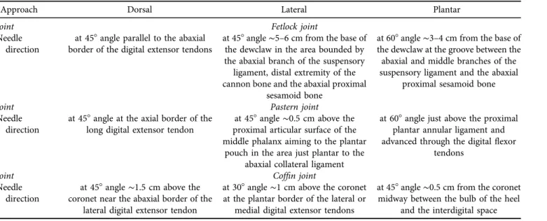
Table 1.Approximate sites selected for localising the joint pouches during intra-articular injection of buffalo digit using dorsal, lateral and plantar approaches
Approach Dorsal Lateral Plantar
Joint Fetlock joint
Needle direction
at 458angle parallel to the abaxial border of the digital extensor tendons
at 458angle∼5–6 cm from the base of the dewclaw in the area bounded by the abaxial branch of the suspensory ligament, distal extremity of the cannon bone and the abaxial proximal
sesamoid bone
at 608angle∼3–4 cm from the base of the dewclaw at the groove between the abaxial and middle branches of the suspensory ligament and the abaxial
proximal sesamoid bone
Joint Pastern joint
Needle direction
at 458angle at the axial border of the long digital extensor tendon
at 458angle∼0.5 cm above the proximal articular surface of the middle phalanx aiming to the plantar
pouch in the area just plantar to the abaxial collateral ligament
at 608angle just above the proximal plantar annular ligament and advanced through the digitalflexor
tendons
Joint Coffin joint
Needle direction
at 458angle∼1.5 cm above the coronet near the abaxial border of the
lateral digital extensor tendon
at 308angle∼1 cm above the coronet at the plantar border of the lateral or
medial digital extensor tendons
at 458angle∼0.5 cm from the coronet midway between the bulb of the heel
and the interdigital space
andFig. 2). The mean distance from the skin surface to the target joint was approximately 4.0±0.65 cm, which differed according to the site of the joint and its surrounding structures. Each joint revealed dorsal and plantar pouches.
The latter pouches were also accessible from the lateral aspect of the digit. An anatomic communication was evident in all injected specimens between the two fetlock joints of
each limb, as the methylene blue solution injected through the dorsal pouch of one fetlock joint reached the cavity and pouches of the other joint of the same limb immediately following IA injection (Fig. 1A and G). The communication site was found to be between the two plantar pouches at the area between the interdigital band of the interosseous muscle and the caudal surface of the cannon bone (Fig. 1D).
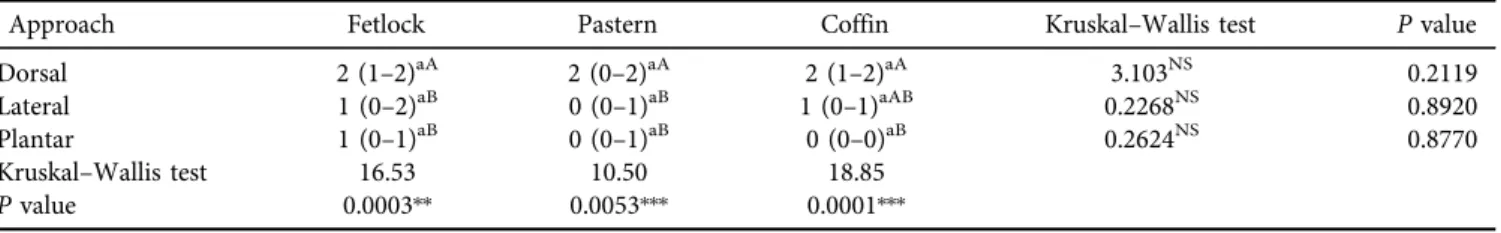
Table 4.Effect of the number of trial on the injection scores for intra-articular injection of buffalo digit
Approach Fetlock Pastern Coffin Kruskal–Wallis test Pvalue
Dorsal 2 (1–2)aA 2 (0–2)aA 2 (1–2)aA 3.103NS 0.2119
Lateral 1 (0–2)aB 0 (0–1)aB 1 (0–1)aAB 0.2268NS 0.8920
Plantar 1 (0–1)aB 0 (0–1)aB 0 (0–0)aB 0.2624NS 0.8770
Kruskal–Wallis test 16.53 10.50 18.85
Pvalue 0.0003✻✻ 0.0053✻✻✻ 0.0001✻✻✻
Number of trials: 05five, 15three, 25one.
Different small superscript letters in the same row show medians and ranges with significant difference atP< 0.05, while different capital superscript letters in the same column show medians and ranges with significant difference atP< 0.05.
Fig. 1.Gross anatomical dissections of the pouches of buffalo fetlock, pastern and coffin joints and their related structures. The joints are injected with appropriate amounts of 1% methylene blue solution. The site of communication between two fetlock joints is shown inD (arrowheads).A–C:dorsal views;D:proximal view of a cross section in the fetlock joints at the level of their plantar pouches;E:lateral view;
F–G:plantar views
Results of the cadaveric study
The anatomical landmarks determining the point of needle insertion for each joint were successfully identified in all cadavers and properly injected in all instances, as confirmed by arthrography (Fig. 3). Anatomical and arthrography- guided techniques for IA injection of the hindlimbs in buffalos could precisely discriminate each target joint.
Collectively, the summation of injection criteria scores showed a significant increase (P < 0.05) in the dorsal and lateral approaches for IA injection of the fetlock, pastern and coffin joints in the buffalo digit compared with the plantar approach. However, the median and range of the injection criteria scores presented between the dorsal and lateral ap- proaches were slightly less significant. The median and range for the injection criteria scores in the three injection tech- niques are presented in Tables 2–5.
Comparison of the injection criteria between the dorsal, lateral and plantar approaches for IA injection of the fetlock, pastern and coffin joints in buffalo limbs showed that the performance time required for accurate needle placement was significantly (P < 0.05) lower when using the dorsal approach than by the lateral and plantar approaches (5 min vs. 10 min and 15 min, respectively; Table 2). The correct needle penetration was significantly higher (P< 0.05) in the dorsal and lateral approaches than in the plantar one (Ta- ble 3). The number of trials and the difficulty of injection were significantly (P< 0.05) higher in the plantar approach than in the dorsal or lateral approach (Tables 4 and 5). The average number of trials required to inject the joint suc- cessfully were 1, 3 and 5 for the dorsal, lateral and plantar approach, respectively. The amount of contrast material required for the joint capsule in the three approaches was higher for the fetlock joint than in either of the other joints (20 mL vs. 5 mL, respectively). Additionally, plantar injec- tion of the coffin joint required a larger needle size than did
the other procedures (22-gauge vs. 20-gauge). For accurate IA injection of the buffalo digit, a flexed digit position is preferred in all approaches to the fetlock, pastern and coffin joints.
Results of the in vivo study
The IA injection technique for the hindlimb digits was well tolerated in live buffaloes. In overall outcomes, no significant differences were noted between cadaveric and live animals in the IA injection criteria. Identification of the landmarks for IA injection of the digit was possible in all cases. Moreover, in the contrast arthrography-guided method, it was possible to visualise the same structures as observed in the cadavers.
Arthrocentesis was attempted prior to each injection to convey appropriate needle placement for joint injections.
Successful IA injection was achieved in all cases without any gross orthopaedic abnormalities during or following this procedure.
DISCUSSION
A sound IA injection technique with a higher success rate is essential for the proper treatment of joint affections in farm animals (Moyer et al., 2011; Blaser et al., 2012;
Alsobayil et al., 2015). Although the equine and bovine models of IA injection are commonly employed for other animal species including the buffalo, the success of the technique is usually challenged by anatomical and genetic variations which in turn can influence limb conformation and joint orientation (Desrochers et al., 1997; Al-Akraa et al., 2014). Thus, the aim of the present work was to establish an efficient and specific model for IA injection of buffalo digits based on fixed anatomical and arthrog- raphy-guided landmarks.
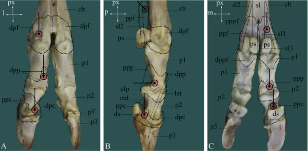
Fig. 2.A skeletal preparation of buffalo digit illustrating the approximate sites selected for injection into phalangeal joints. The dotted areas schematically mark the delineations of the joint capsules and their pouches. The red circles are the sites at which the needles are inserted,
and the black arrows refer to the needle direction during injection. The views are dorsal (A), lateral (B) and plantar (C)
To our knowledge, this is the first study comparing different approaches for IA injection of the buffalo digit. The accuracy rates for injection of the hindlimb digit joints in buffalo cadavers were as high as desired, which agreed with
those reported by Nuss et al. (2002), Francoz et al. (2007) and Courtney and Doherty (2009) in cattle. The in vivo study was performed to exclude postmortem changes to local tissues in cadaveric samples and demonstrate vital Table 5.Effect of the difficulty of injection on the injection scores for intra-articular injection of buffalo digit
Approach Fetlock Pastern Coffin Kruskal–Wallis test Pvalue
Dorsal 2 (1–2)aA 2 (0–2)aA 2 (0–2)aA 0.2900NS 0.8650
Lateral 2 (1–2)aA 1 (0–2)aAB 1 (1–2)aAB 1.521NS 0.4674
Plantar 0 (0–1)aB 0 (0–1)aB 0 (0–1)aB 3.366NS 0.1858
Kruskal–Wallis test 12.69 15.59 16.24
Pvalue 0.0018✻✻✻ 0.0004✻✻ 0.0003✻✻✻
Difficulty of injection: 05difficult, several attempts with low confidence; 15moderate, several attempts until successful injection; 25 Easy, immediate and confident injection.
Different small superscript letters in the same row show medians and ranges with significant difference atP< 0.05, while different capital superscript letters in the same column show medians and ranges with significant difference atP<0.05.
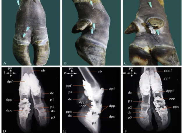
Fig. 3.Dorsal (A), lateral (B) and plantar (C) sites of articular puncture of the fetlock, pastern and coffin joints in the hindlimb of an adult buffalo. Dorsoplantar (D), lateral (E) and plantodorsal (F) radiographic images of buffalo fetlock, pastern and coffin joints immediately after
their injection with contrast medium.
Abbreviations used inFigs. 1–3. alf:annular ligament of fetlock joint;cb:cannon bone;ccl:common axial collateral ligament;clf:collateral ligaments of fetlock joint;clp:collateral ligaments of pastern joint;cnl:collateral navicular ligament;co:coronary border of hoof;d:dorsal; dal:
digital annular ligament;dc:dewclaw;dcn:dorsal common digital nerve III;dcv:dorsal common digital vein III;ddft:deep digitalflexor tendon;
dil:distal interdigital ligament;dmv:dorsal metatarsal vein III;dpc:dorsal pouch of coffin joint;dpf:dorsal pouch of fetlock joint;dpp:dorsal pouch of pastern joint;dppf:distal plantar pouch of fetlock joint;ds:distal sesamoid bone;dsh:digital sheath;i:injected fetlock joint;itn:
intertrochlear notch;l:lateral;ldt:lateral digital extensor tendon;llt:lateral long digital extensor tendon;lpn:lateral plantar nerve;lpv:lateral plantar vein;m:medial;mlt:medial long digital extensor tendon;n:non-injected fetlock joint;p:plantar;p1:proximal phalanx;p1’:prox- imoplantar tubercle of p1;p2:middle phalanx;p2’:proximoplantar tubercle of p2;p3:distal phalanx;p3’:extensor process of p3;pil:proximal interdigital ligament;ppc:plantar pouch of coffin joint;ppf:plantar pouch of fetlock joints;ppp:plantar pouch of pastern joint;ppf:proximal plantar pouch of fetlock joint;ps:proximal sesamoid bones;px:proximal;sdft:superficial digitalflexor tendon;sl:suspensory ligament;sl1:axial
branches of sl;sl2:abaxial branches of sl;sl2’:extensor branch of sl2;sl3:interdigital band of sl
parameters such as the temperament, pain and movement of the live buffalo during injection. There were no significant differences between the IA injection techniques in fresh cadavers and live buffaloes, except for easier aspiration of synovialfluid prior to IA injection in live buffalo that might be due to the absence of weight-bearing effects on cadaveric limbs. These findings were in accordance with those of Piccot-Crezollet et al. (2005) in the horse and El-Shafaey et al. (2017)in camel, and confirmed the consistency of the techniques proposed by us.
Contrast injection followed by radiography is a superior method of detecting extra-articular injection, as dissection depends on every plane of tissue being exactly exposed whereas any radiographic contrast is easily detected on a single radiograph (Poore et al., 2011; Alsobayil et al., 2015).
In this study, contrast arthrography of the buffalo digit provided high-quality images allowing good differentiation of the radiographic features of the hindlimb synovial cav- ities, as well as a marker for successful injection through IA localisation of the needle or contrast agent.
Full flexion and extension of the injected joint several times is an essential step for increasing the space within the joint, which subsequently facilitates IA injection of the limb.
In this study, all IA injection approaches of the hindlimb digit joints were successful and easily performed by flexion with no post-procedural clinical abnormalities. However, the dorsal and lateral approaches revealed significant improve- ment of the injection criteria in comparison to the plantar approach. These findings were in accordance with the results reported byLewis (1996), Nuss et al. (2002) and Poore et al.
(2011).
In this study, IA injection of the fetlock joint was easily performed by the dorsal approach, and one injection was sufficient for the whole two joints. This is due to the anatomical communication between the lateral and medial synovial pouches in the distal plantar area of the fetlock joint in buffalo. This finding is in line with that reported by Desrochers et al. (1997)in the bovine species and comple- ments our previous report in buffalo (Abdellatif et al., 2018).
The needle was inserted distally in the dorsal pouch of the fetlock joint at a 458angle parallel to the abaxial border of the lateral or medial digital extensor tendons to avoid pricking the common digital extensor tendon. Similar findings were reported in cattle (Al-Akraa et al., 2014) and camels (Alsobayil et al., 2015). However, the fetlock joint in horses and cattle can be punctured using the plantar and dorsal approaches just above the lateral proximal sesamoid bone, at the area between the third metatarsal bone and the suspensory ligament (Baxter and Stashak, 2011; Blaser et al., 2012; Alrtib et al., 2013).
In horses, IA injection of the pastern joint can be per- formed using a dorsolateral approach by inserting the needle in the vertical midline above the proximal epicondyle of the second phalanx (Just et al., 2007; Baxter and Stashak, 2011;
Poore et al., 2011). Additionally, in camels the optimal site for insertion of the needle is in the dorsal midline between the medial and lateral eminences on the distal end of the proximal phalanx (Alsobayil et al., 2015). However, in the
buffalo this appears to be different, as the pastern joint was easily punctured in the dorsal aspect of the digit by inserting the needle away from the lateral branch of the long digital extensor tendon.
The coffin joint of buffaloes can be punctured easily via the dorsal and/or lateral approach. In the dorsal approach, the needle was inserted at a 458angle∼1.5 cm above the coronet near the abaxial border of the terminal parts of the lateral digital extensor tendon. In the lateral approach, the needle was inserted distally at a 308angle approximately 1 cm above the coronet in the plantar pouch at the plantar border of the terminal parts of the lateral or medial digital extensor tendon.
These findings were in accordance with those reported in cattle (Van Amstel and Shearer, 2006) and horses (Poore et al., 2011; Alrtib et al., 2013). However, in camels, IA in- jection of theflexed coffin joint was easily performed via the dorsomedial or dorsolateral approaches with the needle inserted distally in the ventrolateral or ventromedial direction perpendicular to the weight-bearing surface of the foot (Mostafa et al., 1993; Alsobayil et al., 2015).
The limitations of the present study were as follow: first, the number of animals used was low; second, different ap- proaches were used for all joints. The shortcomings of the present study should be considered in further investigations to get a concrete conclusion and validate the effectiveness of the method on a larger sample size in clinical cases.
In conclusion, anatomical and arthrography-guided techniques offer considerable advantages for the character- isation and selection of appropriate sites to be used for IA injection of the buffalo digit. The present study establishes a reference approach that can be easily incorporated into diagnostic and therapeutic procedures related to digit lameness in buffaloes.
REFERENCES
Abdellatif, A. M., Hamed, M. A., El-Shafaey, E. A. and Eldoumani, A. H. (2018): Normal magnetic resonance anatomy of the hind foot of Egyptian buffalo (Bubalus bubalis): a correlative low- field T1- and T2-weighted MRI and sectional anatomy atlas.
Anat. Histol. Embryol.47, 599–608.
Al-Akraa, A. M., El-Kasapy, A. H. and El-Shafey, A. A. (2014):
Intra-articular injection, computed tomography and cross sectional anatomy of the metacarpus and digits of cattle (Bos taurus)and buffalo(Bos bubalis). Glob. Vet.13, 1122–1128.
Alrtib, A. M., Philip, C. J., Abdunnabi, A. H. and Davies, H. S.
(2013): Morphometrical study of bony elements of the forelimb fetlock joints in horses. Anat. Histol. Embryol.42, 9–20.
Alsobayil, F. A., Allouch, J. A. and Ahmed, F. A. (2015): Articular puncture techniques and contrast arthrography of the forelimb in dromedary camels(Camelus dromedarius).Pak. Vet. J.35, 28–32.
Baxter, G. M. and Stashak, T. S. (2011): Perineural and intra- synovial anesthesia. In: Baxter, G. M. (ed.) Adams and Stashak’s Lameness in Horses. 6th edition. John Wiley and Sons, Hoboken, New Jersey, USA. pp. 173–202.
Blaser, M., Bertagnoli, A., R€aber, M., Nuss, K., Rasekh, M. and Steiner, A. (2012): Arthroscopic approaches to the fetlock joint of adult cattle: a cadaver study. Vet. J.193, 701–706.
Courtney, P. and Doherty, M. (2009): Joint aspiration and injection and synovialfluid analysis. Best Pract. Res. Clin. Rheumatol.23, 161–192.
Desrochers, A., St-Jean, G., Cash, W. C., Hoskinson, J. J. and DeBowes, R. M. (1997): Characterization of anatomic com- munications of the fetlock in cattle using intra-articular latex injection and positive contrast arthrography. Am. J. Vet. Res.
58, 710–712.
El-Shafaey, E., Hamed, M., Abdellatif, A. and Abo Elfadl, E. (2017):
Comparison of blind, ultrasound and computed tomographic- guided injection techniques for nerve block of the head in one- humped camel(Camelus dromedarius)cadavers. Pak. Vet. J.37, 180–184.
Enting, H., Kooij, D., Dijkhuizen, A. A., Huirne, R. M. and Noordhuizen-Stassen, E. N, (1997): Economic losses due to clinical lameness in dairy cattle. Livest. Prod. Sci.49, 259–267.
Ettema, J. F. and Ostergaard, S. (2006): Economic decision making on prevention and control of clinical lameness in Danish dairy herds. Livest. Prod. Sci.102, 92–106.
Francoz, D., Desrochers, A. and Latouch, J. (2007): Effect of repeated arthrocentesis and single joint lavage on cytologic evaluation of synovialfluid in 5 young calves. Can. J. Vet. Res.
71, 129–134.
Just, E. M., Patan, B. and Licka, T. F. (2007): Dorsolateral approach for arthrocentesis of the centrodistal joint in horses. Am. J. Vet.
Res.68, 946–952.
Lewis, R. D. (1996): Techniques for arthrocentesis of equine shoulder, elbow, stifle and hip joints. Proc. Am. Assoc. Equine Pract.42, 55–63.
Mostafa, M. B., Farag, K. A. and Rajab, G. A. (1993): Arthrography of the interphalangeal joints in the camel. Camel Newsletter10, 20–27.
Moyer, W., Schumacher, J. and Schumacher, J. (2011): Equine Joint Injection and Regional Anesthesia. 5th edition.
Academic Veterinary Solutions LLC, Chadds Ford, PA, USA.
pp. 70–73.
Nuss, K., Hecht, S., Maierl, J. and Matis, U. (2002): Arthrocentesis in cattle. Part 2: Pelvic limb. Tierarztl. Prax.30, 301–307.
Onwuama, K. T., Salami, S. O., Ali, M. and Nzalak, J. O. (2012):
Effect of different methods of bone preparation on the skeleton of the African giant pouched rat(Cricetomys gambianus).Inter.
J. Morphol.30, 425–427.
Piccot-Crezollet, C., Cauvin, E. R. and Lepage, O. M. (2005):
Comparison of two techniques for injection of the podotrochlear bursa in horses. J. Am. Vet. Med. Assoc.226, 1524–1528.
Poore, L. A., Lambert, K. L., Shaw, D. J. and Weaver, M. P.
(2011): Comparison of three methods of injecting the proximal interphalangeal joint in horses. Vet. Rec. 168, 302–305.
Smith, G. N., Myers, S. L., Brandt, K. D. and Mickler, E. A. (1998):
Effect of intraarticular hyaluronan injection in experimental canine osteoarthritis. Arthritis Rheum.41, 976–985.
Soliman, I. and Sadek, H. (2004): Impacts of productive and reproductive performances on investment efficiency of buffalo enterprises in Egypt. Proceedings of the 7th World Buffalo Congress, Manila, Philippines1, pp. 212–217.
Van Amstel, S. R. and Shearer, J. (2006): Subsolar ulcer. In: Van Amstel, S. R. and Shearer, J. (eds) Manual for Treatment and Control of Lameness in Cattle. First edition. Blackwell Pub- lishing, Ames, Iowa, USA. pp. 81–82.