https://doi.org/10.1007/s11064-019-02871-9 ORIGINAL PAPER
The Mitochondrial Targets of Neuroprotective Drug Vinpocetine on Primary Neuron Cultures, Brain Capillary Endothelial Cells, Synaptosomes, and Brain Mitochondria
Gergely Svab1 · Judit Doczi1 · Akos A. Gerencser1,2 · Attila Ambrus1 · Ferenc Gallyas3,4,5 · Balazs Sümegi3,4,5 · László Tretter1
Received: 29 August 2019 / Revised: 29 August 2019 / Accepted: 5 September 2019 / Published online: 18 September 2019
© The Author(s) 2019
Abstract
Vinpocetine is considered as neuroprotectant drug and used for treatment of brain ischemia and cognitive deficiencies for decades. A number of enzymes, channels and receptors can bind vinpocetine, however the mechanisms of many effects’
are still not clear. The present study investigated the effects of vinpocetine from the mitochondrial bioenergetic aspects. In primary brain capillary endothelial cells the purinergic receptor-stimulated mitochondrial Ca2+ uptake and efflux were stud- ied. Vinpocetine exerted a partial inhibition on the mitochondrial calcium efflux. In rodent brain synaptosomes vinpocetine (30 μM) inhibited respiration in uncoupler stimulated synaptosomes and decreased H2O2 release from the nerve terminals in resting and in complex I inhibited conditions, respectively. In isolated rat brain mitochondria using either complex I or complex II substrates leak respiration was stimulated, but ADP-induced respiration was inhibited by vinpocetine. The stimu- lation of oxidation was associated with a small extent of membrane depolarization. Mitochondrial H2O2 production was inhibited by vinpocetine under all conditions investigated. The most pronounced effects were detected with the complex II substrate succinate. Vinpocetine also mitigated both Ca2+-induced mitochondrial Ca2+-release and Ca2+-induced mitochon- drial swelling. It lowered the rate of mitochondrial ATP synthesis, while increasing ATPase activity. These results indicate more than a single mitochondrial target of this vinca alkaloid. The relevance of the affected mitochondrial mechanisms in the anti ischemic effect of vinpocetine is discussed.
Keywords Vinpocetine · Neuroprotection · Mitochondria · Reactive oxygen species · Calcium induced calcium release · Oxygen consumption · ATP synthesis · Uncoupling
Introduction
Vinpocetine has been marketed for more than 30 years.
Among its indication are ischemic neuronal damage [1], stroke [2, 3], cerebrovascular diseases [4], neurodegenerative diseases [5, 6], dementia and cognitive deficits [1, 7]. During this period many targets and mechanisms of actions has been proposed, including the inhibition of the cyclic nucleotide phosphodiesterase 1 (PDE1) [8, 9], voltage-dependent Na+
Balazs Sümegi: Deceased on Aug 1, 2019.
Special issue In honour of Professor Vera Adam-Vizi.
The authors dedicate this article to Prof. Vera Adam-Vizi as an appreciation for her all time support, encouragement, positive criticism, and high expectations.
* László Tretter
tretter.laszlo@med.semmelweis-univ.hu
1 Department of Medical Biochemistry, MTA-SE Laboratory for Neurobiochemistry, Semmelweis University, 37-47 Tuzolto Street, Budapest 1094, Hungary
2 Buck Institute for Research on Aging, Novato, CA, USA
3 Department of Biochemistry and Medical Chemistry, University of Pecs Medical School, Pecs, Hungary
4 Szentagothai Research Centre, University of Pecs, Pecs, Hungary
5 Nuclear-Mitochondrial Interactions Research Group, Hungarian Academy of Sciences, Budapest, Hungary
channels [10, 11], the IκB kinase, the NF-κB [12] and bind- ing to peripheral benzodiazepine receptors [13]. For a recent review see [14]. As a consequence of having numerous tar- gets, the drug can influence diverse functions. The present study focuses on the effects of vinpocetine on mitochondrial function. The brain is extremely sensitive to proper provision of energy, in which mitochodria play a central role. Most of the inborn errors of metabolism are associated with struc- tural and/or functional brain damage and mental retarda- tion. Mitochondria are not only cellular power stations, but also pollute their environment with reactive oxygen species (ROS). Although as a consequence of powerful antioxidant systems most of the mitochondrially produced ROS will not leave the mitochondria in physiological conditions [15], excessive mitochondrial ROS generation is considered as an important factor in cerebral ischemia/reperfusion injury [16–19], neurodegeneration [20] and glutamate toxicity [21, 22]. Early studies already addressed mitochondria as poten- tial vinpocetine targets, but only in a very few studies were mitochondria the focus of the investigation. Vinpocetine’s effect has been explained by binding to the peripheral ben- zodiazepine receptor [22], a mitochondrial outer membrane protein and putative component of the mitochondrial perme- ability transition pore (PTP). In the present study the effects of vinpocetine are investigated at three level of complexity, in cellular systems (primary neuronal and endothelial cell cultures), in isolated nerve terminals (synaptosomes) and in isolated guinea pig brain mitochondria. Complex biological phenomena like delayed calcium deregulation, intracellular calcium transients and Ca2+ release from in situ mitochon- dria have been investigated in cellular systems, while mito- chondrial oxygen consumption, ROS production, ATPase activity in isolated mitochondria. We conclude that the delayed PTP opening, mild mitochondrial depolarization and decreased H2O2 release may all play an important role in the beneficial effects of vinpocetine in pathological conditions associated with neuronal damage.
Materials and Methods
AnimalsFor cell culture and synaptosomal experiments Wistar rats were used. Mitochondria were isolated from guinea pig brain. Animal experiments were performed in accordance with the Guidelines for Animal Experiments at Semmelweis University.
Cell Cultures
Brain capillary endothelial cells from 3 to 5 month-old Wistar rats were prepared and seeded on extracellular
matrix coated glass coverslips as described earlier [23]. Cul- tures were kept in DMEM containing 17% plasma-derived bovine serum (First Link, UK), supplemented with 2 mM glutamine, 80 µg/ml heparin, 150 µg/ml endothelial cell growth supplement (Sigma), antibiotics, and trace factors (vitamin C, selenium, insulin, transferrin and glutathione).
After reaching confluence, experiments were performed on 6–10 days old primary cultures.
Primary neuron-enriched cultures were prepared from E17 Wistar rat embryos, plated on 6 mm glass coverslips coated with poly-l-ornithine plus laminin in 12-well plates or on Petri dishes coated with poly-d-lysine, and maintained in Neurobasal medium (Invitrogen, Carlsbad, CA) with 2% B27 supplement (Invitrogen) and 2 mM glutamine for 8–12 days at 37 °C, 5% CO2 without feeding [24].
Measurements of Mitochondrial Calcium Transients In situ mitochondrial and cytoplasmic calcium measure- ments were performed in brain capillary endothelial cell cultures (at 7–8 days in vitro) by X-Rhod-1-AM (Molecular Probes, Eugene, OR, USA), localized mainly in mitochon- dria, a calcium sensitive dye with fluorescence microscopy as described earlier [25]. Briefly, mitochondrial calcium transients ([Ca2+]m) were analyzed using highpass spatial filtering of fluorescence images using the “the original X-rhod-1 mito filter.flt” in Image Analyst MKII (Image Analyst Software, Novato, CA, USA). Cytosolic calcium transients ([Ca2+]c) were analyzed on the same images left unfiltered by the measurement of X-Rhod-1 fluorescence of the nuclear region in the cell [25]. Half decay time (τ1/2) was defined as the time between peak of the transient and the intensity decaying to half of the peak amplitude.
Fluorimetric measurements were performed to examine potential optical or chemical interaction of vinpocetine and calcium sensitive fluorescent dyes (X-Rhod-1 and Fura-FF), to exclude the possibility of recording artefacts due to the change in fluorescence. Fluorescence of vinpocetine in aque- ous solution was negligible both in the range of Fura-FF emission 510 nm (excitation: 340/380 nm) and in the range of X-Rhod-1 emission 570 nm (excitation: 535 nm). 30 μM vinpocetine had no effect on the fluorescence of hydrolyzed X-Rhod-1 or Fura-FF dyes either in high calcium (40 μM) containing or in calcium free intracellular solution (pH 7.4, 37 °C).
Measurement of Delayed Ca2+ Deregulation in Isolated Primary Rat Brain Neurons
Time-lapse fluorescence microscopy of rat primary corti- cal cultures was carried out using the above fluorescence
microscope setup using a UAPO 20 × dry 0.75 NA lens. Cul- tures were incubated with Fura-FF-AM (3 µM) for 15 min at 37 °C and subsequently rinsed with superfusion medium.
Fura-FF fluorescence was ratio imaged using 340/10 (center/
bandwidth in nm) and 380/10 exciters (Chroma, Rocking- ham, VT, USA), a 400DCLP dichroic mirror and a 470LP long pass emitter (Omega Optical, Brattleboro, VT, USA) [24].
Isolation of Synaptosomes from Rat Brain
Synaptosomes were isolated from rat brain cortex as described earlier [26]. The synaptosomal fraction was obtained after sucrose (0.8 M) gradient centrifugation.
Sucrose was diluted by ice cold distilled water to 0.32 M and synaptosomes were sedimented with 20,000×g for 20 min.
The pellet’s protein concentration was adjusted to about 20 mg/ml, and synaptosomes were kept on ice until use.
Most of the experiments were performed in standard extracellular medium composed of (mM): 140 NaCl, 3 KCl, 2 MgCl2, 2 CaCl2, 10 PIPES pH 7.38 and 10 glucose.
Incubations and measurements were done at 37 °C. Synap- tosomes retained their basic bioenergetic parameters for at least 6 h.
Isolation of Rodent Brain Mitochondria
Guinea pig mitochondria were isolated using discontinu- ous percoll gradient as described [27]. Mitochondria were incubated in the following incubation buffer (mM): 125 KCl, 20 HEPES, 2 K2HPO4, 1 MgCl2, 0.1 EGTA, at pH 7.0 adjusted by KOH. Incubation buffer was supplemented with 0.025 w/v % fatty acid free BSA. To use BSA was necessary, because mitochondria may lose their functional parameters very quickly as a consequence of liberated fatty acids [28].
Oxygen Consumption
of Synaptosomes and Mitochondria
Oxygen consumption of synaptosomes were measured by high resolution respirometry Oxygraph-2K (Oroboros Instruments, Innsbruck, Austria) [29] at 37 °C in 2-ml chambers. Data were digitally recorded and analyzed; oxy- gen flux was calculated as the negative temporal derivative of the oxygen concentration, cO2(t). Oxygen sensors were calibrated routinely in air saturated and oxygen depleted media. Synaptosomal protein concentration was 2 mg/ml, mitochondrial protein concentration was 0.1 mg/ml.
Measurement of Mitochondrial Membrane Potential (Δψm)
Mitochondrial membrane potential was measured by a cus- tom-made tetraphenylphosphorium (TPP+) electrode [30].
TPP+ is a lipophilic membrane permeable cation. With the TPP+ electrode the extramitochondrial concentration of TPP+ was measured as described earlier. Knowing the total concentration of the TPP+ Δψm was calculated using the Nernst equation.
Measurement of H2O2 Production in Mitochondria and in Synaptosomes
H2O2 released from mitochondria was detected with horse- radish peroxidase (5U/2 ml) and Amplex Ultrared (3 μM) [31]. As a result of peroxidase action Amplex Ultrared was converted to fluorescent resorufin in the presence of mito- chondria (0.1 mg/ml), and measured using PTI Deltascan fluorescence spectrophotometer (550 nm excitation, 585 nm emission wavelengths; Photon Technology International, Lawrenceville, NJ, USA). Each measurement was calibrated with known amounts of H2O2 at the end of the experiment.
Measurement of Mitochondrial Ca2+ Uptake
Mitochondria (0.05 mg/ml) were incubated in the follow- ing reaction medium (mM): 8 KCl, 110 K-gluconate, 10 NaCl, 10 HEPES, 2 KH2PO4, 4 MgCl2, 10 mannitol, 5 glu- tamate, 5 malate, 3 ATP, 0.25 ADP pH 7.25 (KOH) sup- plemented with 0.025% BSA. The free Ca2+ concentration was calculated with the WinMAXC software ([32, 33]. Ca2+
(CaCl2) was given to mitochondria in the form of 12.5 μM Ca2+ pulses. The Ca2+ level of the medium was followed by Calcium-Green 5 N (KD = 4.29 μM). Wavelengths for Calcium-green fluorescence were 505 nm excitation and 535 nm emission, respectively.
Measurement of Mitochondrial Swelling
Swelling of isolated mitochondria can reflect permeability pore opening (see [33, 34]) and was followed by light scat- tering at 590 nm in parallel with mitochondrial Ca2+ uptake using double excitation, double emission mode of PTI Del- tascan spectrofluorimeter. At the end of each measurement alamethicin (a pore forming peptide 80 μg/2 ml) was added to obtain maximal swelling.
Kinetic measurement of Mitochondrial ATP Synthesis
Mitochondrial ATP formation was monitored by a com- bined enzymatic system comprising of hexokinase and
glucose-6-phosphate dehydrogenase, as described ear- lier [35, 36]. Mitochondria (0.05 mg/ml) were incubated in the medium described in the isolation procedure.
This medium was supplemented with 3 mM NADP+, 1.5 U hexokinase, 0.5 U glucose-6-phosphate dehydro- genase, 5 mM glucose, 2 mM ADP and 200 µM AP5 (P1,P5-Di(adenosine-5′) pentaphosphate), an inhibitor of adenylate kinase) in 2 ml total volume (Melnick et al.
1979). In the presence of mitochondria, ADP and respira- tory substrates glutamate plus malate ATP was formed, which mostly left the mitochondria via the adenine nucleotide translocase. ATP phosphorylated glucose (to glucose-6-phosphate) by hexokinase. The resulted prod- uct glucose-6-phosphate was oxidized to 6-phosphogluco- nate by glucose-6-phospate dehydrogenase with the con- comitant reduction of NADP+ to NADPH. Thus, NADPH formation was stoichiometrically equal to ATP released from the mitochondria. The absorbance of NADPH (ɛ = 6220 M−1 cm−1) was recorded at 340 nm, 37 °C using a JASCO V-650 spectrophotometer (ABL&E-JASCO, Tokyo, Japan). ATP standards were used for calibration.
Measurement of ATPase Activity
ATP hydrolyzed by mitochondria in the absence of res- piratory substrates was measured by a combined enzyme assay [37]. The standard assay medium was supplemented with NADH (300 μM), lactate dehydrogenase (2 U/ml),
pyruvate kinase (PK; 2 U/ml), phosphoenolpyruvate (PEP; 2 mM), ATP (0.5 mM), and rotenone (1 μM). In the medium, ADP was phosphorylated to ATP in the presence of PK and PEP, then pyruvate was reduced to lactate with the concomitant oxidation of NADH to NAD+. Absorb- ance of NADH was monitored at 340 nm using a JASCO spectrophotometer. Measurements were calibrated with known amounts of ADP.
Statistics
Statistical differences were evaluated with ANOVA in Sig- mastat wherever multiple comparisons were made. Values are indicated as mean ± standard error, p < 0.05 were con- sidered statistically significant.
Results
In Situ Mitochondrial Ca2+ Measurements in Brain Capillary Endothelial Cells
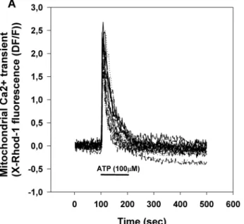
Brain capillary endothelial cells in primary culture were stimulated by the application of 100 μM ATP (Fig. 1).
ATP triggers a transient rise of cytoplasmic [Ca2+] ([Ca2+]c, Fig. 1b) and mitochondrial [Ca2+] ([Ca2+]m, Fig. 1a) by activating purinergic receptors and IP3-mediated Ca2+ release from the endoplasmic reticulum [23]. Mito- chondria take up Ca2+ from the cytoplasm by the calcium
Fig. 1 Mitochondrial (a) and cytosolic (b) calcium transients of pri- mary brain capillary endothelial cells stimulated by the addition of 100 μM ATP. ATP was provided by superfusion at the indicated time.
Mitochondrial and cytosolic Ca2+ transients were extracted from the same recordings using image processing. Pooled from 3 measure- ments, total cell number: 17
uniporter any Ca2+ is predominantly released from mito- chondria by the Na+/Ca2+ exchanger.
In the stimulation paradigm depicted in Fig. 1, any change to Ca2+ ‘handling’ in mitochondria is indicated by the altera- tion of the peak amplitude or the half decay time of the mitochondrial Ca2+ transient. Alteration of the peak ampli- tude indicates changes both in the calcium influx and efflux while alteration of half decay time indicates change in the calcium efflux.
Effect of Vinpocetine on ATP Induced [Ca2+]m and [Ca2+]c Transients
Vinpocetine up to 10 μM had no significant effects on the parameters of [Ca2+]m, or [Ca2+]c transients (not shown). However, 30 μM vinpocetine pretreatment sig- nificantly increased half decay time of the mitochondrial calcium transient (from τ1/2 = 23.5 ± 6.8 s to 42 ± 6.2 s, p < 0.05), but peak amplitude was not changed (from ampitude 2.24 ± 0.48 to 2.22 ± 0.77; given as relative fluo- rescence change from baseline, ΔF/F0) (Fig. 2).
Parameters of the cytosolic calcium transients were not significantly different (from τ1/2 = 33.3 ± 5.2 s to τ1/2 = 29
± 8.69 s and from ampitude 1.33 ± 0.13 to 1.53 ± 0.15).
Figure not shown.
The Effect of Vinpocetine on Delayed Ca2+
Deregulation (DCD)
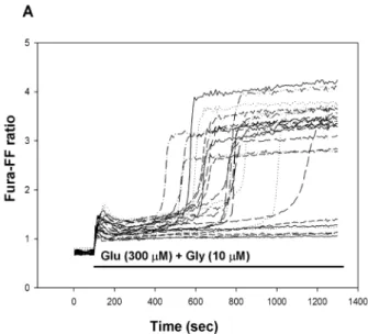
DCD is a model of glutamate toxicity, where excessive calcium load through Ca2+ permeable glutamate receptors could result in deterioration of calcium and energy homeo- stasis and eventually lead to cell death [38]. Primary cor- tical neurons were stimulated with glutamate (300 μM) plus glycine (10 μM) for 20 min in Mg2+-free solution and cytoplasmic [Ca2+] was measured using a low affinity calcium-sensitive fluorescent dye Fura-FF in single cells.
The glutamate-induced initial Ca2+ peak was followed by a transient plateau where the cytosolic [Ca2+] remained lower than the peak, but higher than before the stimulation.
The length of this period had large cell to cell variations
Fig. 2 Effect of Vinpocetine (30 μM) pretreatment on the mitochon- drial calcium transient. Data are expressed as mean ± SE of n = 3 experiments; ctrl, untreated control in matched culture preparations
Fig. 3 Delayed Ca2+ deregulation in primary cortical neurons in the absence (a) or presence (b) of vinpocetine (30 μM). Glutamate plus glycine (Glu + Gly) (a, b) and vinpocetine (vinpo) (b) were applied as
indicated. The Fura-FF fluorescence excitation ratio reflects the cyto- solic Ca2+ levels. Representative traces are shown
and was followed by a sharp, irreversible elevation of [Ca2+]c termed as DCD (Fig. 3a). Vinpocetine pretreat- ment (Fig. 3b) was unable to protect cells against DCD.
There was neither a change in the mean time of DCD, nor in the fraction of cells exhibiting DCD within the time- frame of the experiment (20 min).
The Effects of Vinpocetine on Synaptosomal Oxygen Consumption
Oxygen consumption of synaptosomes reflects substrate oxi- dation by in situ synaptosomal mitochondria, whereas con- taminating, free mitochondria are assumed to be damaged and metabolically silent in the presence of mM extracellular Ca2+. Vinpocetine inhibited both resting and uncoupler-stim- ulated respiration of nerve endings. energized by glucose (Fig. 4). Replacing glucose by lactate resulted in similar results (data not shown).
Effects of Vinpocetine on Synaptosomal H2O2 Production
In synaptosomes, ROS production can primarily be attrib- uted to mitochondria. To address the effects of vinpocetine on ROS generation, cortical synaptosomes supplied with glucose were preincubated with vinpocetine (10 or 30 μM) for 5 min. Vinpocetine lowered H2O2 release by 22%. In the presence of the complex I inhibitor rotenone-vinpocetine also significantly decreased H2O2 production by 19.7% (Fig. 5).
Effects of Vinpocetine on Mitochondrial Membrane Potential
In isolated guinea pig brain mitochondria Δψm was meas- ured with TPP+-electrode. Mitochondria were energized either by glutamate plus malate or succinate. After the development of Δψm vinpocetine (30 μM) was given. Vin- pocetine decreased Δψm in glutamate plus malate sup- ported mitochondria by 4.0 ± 0.6 mV. In succinate sup- ported mitochondria similar depolarization was detected (data not shown).
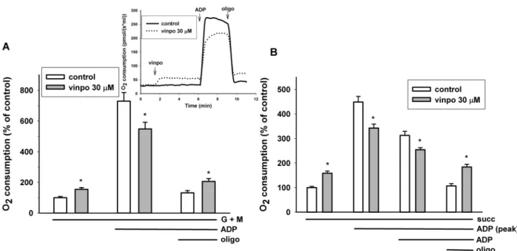
Effects of Vinpocetine on the Respiration of Isolated Guinea Pig Brain Mitochondria
Isolated mitochondria are intact cellular organelles with complex bioenergetic functions, e.g. membrane potential, ATP synthesis and Ca2+ transport. Brain mitochondria were supported with complex I (glutamate plus malate) or complex II (succinate) substrates. Adding vinpocetine during resting respiration (state 2, substrate only) stimu- lated oxygen consumption. In contrast in the presence of ADP (state 3 respiration) the rate of oxygen consumption was decreased by vinpocetine irrespective of the substrates applied (Fig. 6). Similarly, if vinpocetine was added after ADP, there was an immediate decrease in the respiration rate
Fig. 4 The effect of vinpocetine on the O2 consumption of syn- aptosomes. Synaptosomes were preincubated in glucose contain- ing standard medium for 5 min under control conditions (C, white bars), or in the presence of vinpocetine (V, grey bars, 10 or 30 μM).
First basal respiration (C baseline, V10 baseline, V30 baseline) was measured and then respiration was stimulated by the uncoupler FCCP (500 nM, C FCCP, V10 FCCP, V30 FCCP). * indicate significant dif- ference (p < 0.05, n > 4) from the corresponding control measured in the absence of vinpocetine
Fig. 5 Effect of vinpocetine on H2O2 production of synaptosomes.
Synaptosomes were preincubated in glucose-containing standard medium for 5 min under control conditions (C) or with vinpocetine (10 or 30 μM, hatched and cross hatched bars). Recording of baseline H2O2 formation was started after addition of Amplex Ultrared and horseradish peroxidase. After 200 s rotenone (1 μM, gray bars) was given. * indicate significant difference (p < 0.05, n > 4) compared to the corresponding control measured in the absence of vinpocetine
(not shown). Addition of oligomycin decreased the rate of respiration, but in the presence of vinpocetine the remaining respiration rate (leak respiration) was always higher than under control conditions, indicating the uncoupling effect of this vinca derivative.
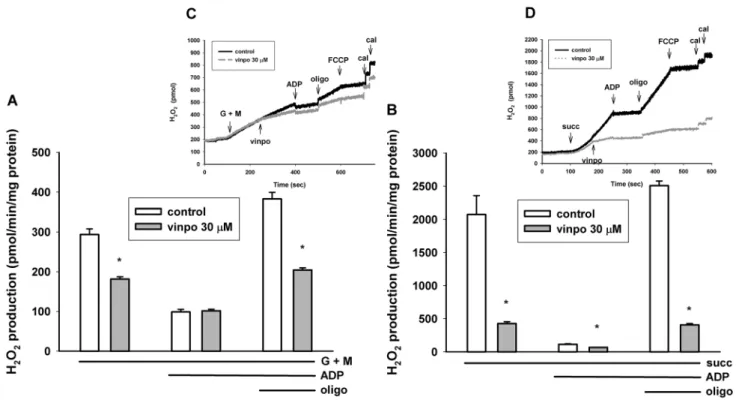
Effects of Vinpocetine on H2O2 Formation in Isolated Mitochondria
Usually there is a close correlation between the rate of oxi- dation and H2O2 production. The mitochondrial H2O2 for- mation has been investigated with glutamate plus malate and succinate substrates.
Using the complex I substrates glutamate plus malate, basal ROS production was only a fraction of that detected in succinate-supported mitochondria. This observation agrees with our and others previous studies [39, 40].
Vinpocetine also decreased the rate of H2O2 formation in the presence of glutamate plus malate (Fig. 7). Stimu- lation of respiration with ADP resulted in a decrease of ROS production irrespective of the presence or absence of vinpocetine. Inhibition of ATP synthesis with oligo- mycin stimulated H2O2 formation, and this increase was more modest (201% vs. 387%) in vinpocetine-treated mitochondria (Fig. 7a).
In succinate supported mitochondria ROS production was 2074 ± 282 pmol/min/mg protein. Under control con- ditions ADP dramatically decreased ROS production by 94.6% Vinpocetine added after succinate decreased H2O2 formation by 79.4%. Addition of the ATP synthesis inhibi- tor oligomycin stimulated H2O2 release by a factor of 22.2, while in the presence of vinpocetine only by a factor of only 6.1 (Fig. 3b).
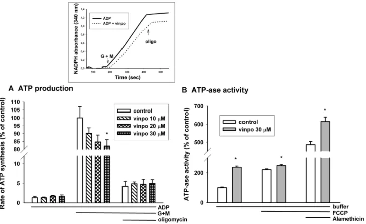
ATP Synthesis in Mitochondria
The most important bioenergetic function of mitochon- dria is ATP production. Alterations in state 3 respiration and membrane potential suggested a decreased capacity for ATP production. Measurement of ATP release from mitochondria in the presence of respiratory substrates and ADP provides information about this complex function.
Mitochondrial ATP synthesis was measured in gluta- mate plus malate supported mitochondria. In the pres- ence of glutamate plus malate vinpocetine evoked a dose-dependent inhibition of ATP synthesis (Fig. 8a).
Applying 30 μM vinpocetine decreased ATP production by 17.9 ± 4.6%. Addition of oligomycin inhibited ATP pro- duction by about 95% indicating that most of the ATP pro- duction could be attributed to oxidative phosphorylation.
Fig. 6 Effects of vinpocetine on mitochondrial respiration. Mitochon- dria supported by glutamate plus malate respiratory substrates (a) or by succinate (b) were incubated in standard mitochondrial medium.
Vinpocetine (vinpo; 30 μM), ADP (2 mM) and oligomycin (oligo;
5 μM) were added as indicated in the original traces of the inset. The
effects of vinpocetine (gray bars) were compared to the control (white bars, vehicle treated). Data are % of mean control oxygen consump- tion. Mean ± SE; n > 5; * significant difference from the correspond- ing solvent controls
ATPase Activity of Mitochondria
The formation of ATP by oxidative phosphorylation is influ- enced by many factors, e.g. transport of respiratory sub- strates, ADP, activity of respiratory complexes, activity of citric acid cycle enzymes. Therefore, for specific assessment of ATP synthase activity mitochondria were supported by ATP only and the rate of mitochondrial ADP release was measured. Mitochondria were incubated in the presence of vinpocetine (30 μM) or vehicle. In the presence of vinpoce- tine the rate of ADP hydrolysis more than doubled (increased by 137%). The uncoupler FCCP stimulated ADP hydrolysis and mitigated the difference between the effect of vinpoce- tine. In order to detect the maximal reverse activity of ATP synthase, mitochondria were permeabilized by the pore form- ing alamethicin. Vinpocetine also stimulated ATP hydrolysis in fully permeabilized mitochondria (Fig. 8b).
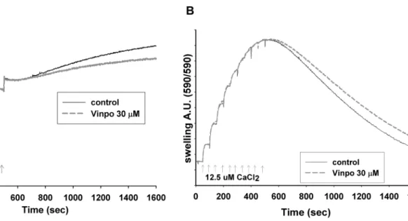
Mitochondrial Ca2+ Uptake and Ca2+‑Induced Ca2+
Release
Another function of mitochondria is Ca2+-handling. To assess the Ca2+ uptake capacity, Ca2+ pulses (12.5 μM) were added and taken up by the mitochondria in 50 s intervals (Fig. 9a). The uptake was fast (Vmax for the Ca2+ uniporter was 1200 nmol Ca2+/min/mg protein. After adding multiple
Ca2+ pulses, the Ca2+ uptake capacity of mitochondria was gradually lost. When Ca2+ uptake stopped, mitochondria started to release the previously accumulated Ca2+ due to the mPTP opening (Ca2+-induced Ca2+ release (mCICR)).
In the presence of vinpocetine (30 μM), the maximal Ca2+
uptake capacity of mitochondria remained unaltered, how- ever the mCICR decreased.
Effect of Vinpocetine on Ca2+‑Induced Swelling of Mitochondria
In the above mCICR assay, in parallel with Ca2+ record- ing, swelling of mitochondria was measured from the same samples (Fig. 9b). In the presence of vinpocetine (30 μM) mitochondrial swelling (an indicator of mPTP opening) was decreased, indicated by the smaller decrease in light scatter after the onset of mCICR.
Discussion
Vinpocetine was introduced to clinical therapy decades ago, and many possible molecules and structures were associated as targets for its beneficial effects. It is obvious that energy homeostasis is a crucial factor in the cell survival and can
Fig. 7 Effects of vinpocetine on mitochondrial H2O2 formation in glutamate plus malate (a) and succinate-supported (b) mitochondria.
Mitochondria were incubated in the standard mitochondrial medium.
Vinpocetine (vinpo; 30 μM), ADP (2 mM), oligomycin (oligo;
5 μM), and FCCP (250 nM) were given as indicated in the insets. At the end of each measurement 100 pmol H2O2 was added for calibra- tion (cal). * indicates significant difference (p < 0.05, n > 4) from the corresponding vinpocetine-free controls
also determine the type of cell death. Although the binding of vinpocetine to the mitochondrial peripheral benzodiaz- epine receptor (PBR) has already been demonstrated [41, 42], systematic mitochondrial studies have not followed this early observation.
In the present study, the bioenergetic aspects of the effects of vinpocetine were investigated on primary brain capillary endothelial cells, neurons, isolated nerve terminals (synap- tosomes) and in isolated mitochondria.
Measurements in Cellular Systems
Ca2+ Transients in Primary Brain Capillary Endothelial Cells Integrity of the blood brain barrier is an important factor in the maintenance of CNS homeostasis [43]. In brain capillary endothelial cells, purinergic signaling evoked cytoplasmic
and mitochondrial Ca2+ transients (Fig. 1). In the presence of vinpocetine the peak height of the Ca2+ transient remained unaltered, however, the half decay time of the mitochon- drial Ca2+ transient was increased, thus Ca2+ release was slower in the presence of vinpocetine (Fig. 2). The phenom- enon may be attributed to inhibition of the mitochondrial Na+/Ca2+ exchange, the major route of the mitochondrial Ca2+ release [44]. This effect of vinpocetine is similar to that found with CGP-37157, a dedicated inhibitor of the mito- chondrial Na+-dependent Ca2+ release [25]. CGP-37157 has neuroprotective effects [45]. Inhibition of ion channels is also in the spectrum of vinpocetine, e.g. it is an efficient blocker of the tetrodotoxin-sensitive voltage dependent Na+ channel. Nevertheless, there are further alternative explana- tions. Depolarization of mitochondria decreases the driving force of transport processes including the energy required for the forward operation of the Na+/Ca2+ exchanger [46].
Fig. 8 Effects of vinpocetine on mitochondrial ATP synthesis (a) and ATPase activity (b). The rate of ATP synthesis in glutamate plus malate (G + M) supported mitochondria (0.5 mg/ml mitochondrial protein was measured in standard medium supplemented with the inhibitor of adenylate kinase, ADP (2 mM) in the presence (hatched bars on (a) and dotted line on the inset) or absence (white bars on (a), solid line on the inset) of vinpocetine. At 200 s G + M initiated oxi- dative substrate-dependent ATP synthesis. Oligomycin (oligo; 5 μM)
inhibited oxidative phosphorylation. ATPase activity (b) was meas- ured on the basis of hydrolysis of exogenous ATP given to mitochon- dria. ATP hydrolysis was measured under resting conditions (buffer), in the presence of uncoupler FCCP (250 nM) and in the presence of alamethicin, a pore-forming antibiotics. Vinpocetine (vinpo; 30 μM;
gray bars). * indicates significant difference (p < 0.05, n > 4) from the corresponding vinpocetine-free controls (white bars)
Delayed Ca2+ Deregulation (DCD) in Cortical Neurons
DCD is a model of glutamate toxicity, where excessive Ca2+ loads through Ca2+ permeable glutamate channels could result in deterioration of calcium and energy homeo- stasis. Vinpocetine did not affect DCD in primary cortical neurons. Our results somewhat differ from those published by others [12]. The reason of discrepancy is possibly attrib- uted to the higher glutamate concentration applied in the present study (300 μM for 20 min vs. 25 μM for 30 min).
Measurements on Isolated Nerve Terminals (Synaptosomes)
Oxygen Consumption Measurements
Oxygen consumption of synaptosomes is attributed to the substrate oxidation of in situ mitochondria [47]. Thus, glu- cose used as an energy supply for synaptosomes is metabo- lised in glycolysis and subsequently in the mitochondria.
Compromised mitochondrial O2 consumption in synap- tosomes energized by external glucose (Fig. 4) could be also explained by inhibition of either the glucose transport or glycolysis, besides mitochondrial effects. In order to rule out these possibilities, lactate was also used as respiratory substrate. Vinpocetine inhibited synaptosomal O2 consump- tion similarly in the presence of glucose or lactate, ruling out
that this inhibition was be specific to the glucose metabo- lism. These observations directed our attention towards iso- lated mitochondria.
Release of H2O2 from Synaptosomes
Vinpocetine inhibited H2O2 formation in synaptosomes.
Measurement of synaptosomal H2O2 production using Amplex Ultrared dye and horseradish peroxidase gives indirect information about the ROS homeostasis. The infor- mation are indirect, because (i) ROS sources other than mitochondria e.g. NADPH oxidases [47, 48], and nitrogen monoxide synthase (NOS) [49] can also be found in synap- tosomes (ii) the cytoplasm possesses many enzymatic and nonenzymatic ROS scavenging systems [50, 51] and our detection system for H2O2 is extra-synaptosomal; in syn- aptosomes between the mitochondrial membranes and the plasma membrane of the synaptosomes both ROS generating and scavenging mechanisms can be found, therefore more direct information was acquired using isolated mitochondria.
Measurements in Isolated Guinea Pig Brain Mitochondria
Effect of Vinpocetine on Mitochondrial Membrane Potential, Oxygen Consumption and H2O2 Formation In isolated mitochondria membrane potential was slightly depolarized (4.0 ± 0.6 mV) by vinpocetine. This together
Fig. 9 Effect of vinpocetine on mitochondrial Ca2+ handling. Effect of vinpocetine on the Ca2+ uptake capacity and mCICR. a Rat brain mitochondria (0.05 mg/ml) were incubated in reaction medium.
CaCl2 pulses were added as indicated. The free [Ca2+] of the medium was detected by Calcium-Green 5 N fluorescence in the absence (con- trol) or in the presence of vinpocetine (vinpo, 30 μM) as indicated.
The loss of Ca2+ buffering capacity is followed by mCICR. Repre- sentative traces n > 3. Calcium induced swelling of mitochondria (b).
Swelling of mitochondrial matrix was measured simultaneously with Ca2+ uptake by the light scattering method. More that swelling and the loss of Ca2+ buffering capacity were developed at the same time.
Representative traces n > 3 from independent experiments
with an increased respiration in the absence of ADP (Fig. 6) suggests an uncoupler-like effect that could be detected with both glutamate plus malate and succinate as respir- atory substrates. In the absence of ADP (substrate only conditions) or in the presence of oligomycin the mitochon- drial membrane potential is high and redox centers are very reduced. In this condition a high rate of H2O2 production was detected both with complex I and complex II substrates (Fig. 7). Even slight depolarization could exponentially decrease the ROS formation under these conditions [40, 52, 53]. Therefore, the ROS lowering effect of vinpocetine may be explained by mild uncoupling. The ADP stimulated respiration was strongly inhibited in the presence of vin- pocetine. Like the depolarization in ADP-free conditions, the respiratory inhibition was also independent from what type of substrates (complex I or complex II) supported the mitochondria. This finding raises the possibility that either both complex I and II, or complex III, or complex IV or their combination were inhibited by vinpocetine.
ATP Synthetic and ATP Hydrolytic Rates in the Presence of Vinpocetine
Vinpocetine decreased the rate of ATP synthesis with glu- tamate plus malate as respiratory substrates, but increased the rate of ATP hydrolysis in the absence of respiration- supporting substrates. The concrete reasons behind a com- promised mitochondrial ATP synthesis is generally rather obscure. In order to produce ATP efficiently, tens of mech- anisms behind the four postulates of Mitchell should work perfectly and in synchrony. It is rather difficult to evalu- ate our findings from the aspect of neuroprotection. The slight decrease in the rate of ATP synthesis in the pres- ence of vinpocetine might be attributed to the uncoupling detected in O2 consumption measurements. A less efficient ATP synthesis is not beneficial from the point of cell sur- vival. On the other hand, an uncoupling-evoked decrease in membrane potential would decrease the rate of ROS production and could also accelerate ROS elimination by stimulating NADPH formation via the NADP+-dependent isocitrate dehydrogenase or glutamate dehydrogenase.
Therefore, this phenomenon might improve the redox homeostasis in mitochondria and the cell. The increased ATP hydrolysis rate suggests that vinpocetine does not inhibit the reverse mode of ATP synthase (and perhaps does not inhibit its forward mode, either).
Seeking the Mechanism and Conclusions
In the present paper, we attempted to describe the effects of vinpocetine on selected mitochondrial functions in light of
the well documented beneficial effects of the drug on brain functions. Various findings presented above are somewhat controversial. Some of them may first seem to be nega- tive, e.g. inhibition of mitochondrial respiration and ATP synthesis. Some of the effects are subjects of various inter- pretations, e.g. mild uncoupling (its benefits are debated), the increased half time of Ca2+ release from endothelial cells, the possible inhibition of the mitochondrial Na+/Ca2+
exchanger, all may or may not have beneficial physiological effects. Finally, inhibition of the Ca2+-induced mitochondrial swelling, delayed CICR, and the very strong inhibition of the mitochondrial H2O2 release are all unequivocally positive.
Considering that vinpocetine does not display any severe side effects, we may conclude that the positive effects dom- inate under in vivo conditions. We also conclude that on the basis of the results presented above it is very likely that vinpocetine possesses more than one molecular target in mitochondria.
Acknowledgement Open access funding provided by Semmelweis University (SE). Authors are indebted to Christos Chinopoulos (Sem- melweis University) for helping us in Ca2+ handling experiments on isolated mitochondria. Authors are also grateful to Katalin Takacs and Andrea Varnagy for the excellent technical assistance. This work was supported by the Hungarian Brain Research Program (KTIA_13_NAP- A-III/6 and 2017-1.2.1-NKP-2017-00002), OTKA (K 112230), Hun- garian Higher Education Institution Excellence Program [FIKP 61822 64860 EATV and FIKP 61826 690289 EATV] and the Hungarian Academy of Sciences (MTA TKI 02001), all to Vera Adam-Vizi.
Compliance with Ethical Standards
Conflict of interest Part of this work was supported by the Gedeon Richter Plc. AAG has financial interest in Image Analyst Software.
Open Access This article is distributed under the terms of the Crea- tive Commons Attribution 4.0 International License (http://creat iveco mmons .org/licen ses/by/4.0/), which permits unrestricted use, distribu- tion, and reproduction in any medium, provided you give appropriate credit to the original author(s) and the source, provide a link to the Creative Commons license, and indicate if changes were made.
References
1. Nivison-Smith L, O’Brien BJ, Truong M, Guo CX, Kalloniatis M, Acosta ML (2015) Vinpocetine modulates metabolic activity and function during retinal ischemia. Am J Physiol Cell Physiol 308:C737–C749
2. Bonoczk P, Panczel G, Nagy Z (2002) Vinpocetine increases cer- ebral blood flow and oxygenation in stroke patients: a near infra- red spectroscopy and transcranial Doppler study. Eur J Ultrasound 15:85–91
3. Szobor A, Klein M (1992) Examinations of the relative fluidity in cerebrovascular disease patients. Ther Hung 40:8–11 4. Rischke R, Krieglstein J (1991) Protective effect of vinpoce-
tine against brain damage caused by ischemia. Jpn J Pharmacol 56:349–356
5. Medina AE (2011) Therapeutic utility of phosphodiesterase type I inhibitors in neurological conditions. Front Neurosci 5:21 6. Heckman PR, Wouters C, Prickaerts J (2015) Phosphodiesterase
inhibitors as a target for cognition enhancement in aging and Alzheimer’s disease: a translational overview. Curr Pharm Des 21:317–331
7. Paroczai M, Kiss B, Karpati E (1998) Effect of RGH-2716 on learning and memory deficits of young and aged rats in water- labyrinth. Brain Res Bull 45:475–488
8. Chiu PJ, Tetzloff G, Ahn HS, Sybertz EJ (1988) Comparative effects of vinpocetine and 8-Br-cyclic GMP on the contraction and 45Ca-fluxes in the rabbit aorta. Am J Hypertens 1:262–268 9. Hagiwara M, Endo T, Hidaka H (1984) Effects of vinpocetine
on cyclic nucleotide metabolism in vascular smooth muscle.
Biochem Pharmacol 33:453–457
10. Erdo SA, Molnar P, Lakics V, Bence JZ, Tomoskozi Z (1996) Vincamine and vincanol are potent blockers of voltage-gated Na + channels. Eur J Pharmacol 314:69–73
11. Tretter L, Adam-Vizi V (1998) The neuroprotective drug vin- pocetine prevents veratridine-induced [Na+]i and [Ca2+]i rise in synaptosomes. NeuroReport 9:1849–1853
12. Jeon KI, Xu X, Aizawa T, Lim JH, Jono H, Kwon DS, Abe J, Berk BC, Li JD, Yan C (2010) Vinpocetine inhibits NF-kappaB- dependent inflammation via an IKK-dependent but PDE-inde- pendent mechanism. Proc Natl Acad Sci USA 107:9795–9800 13. Tarnok K, Kiss E, Luiten PG, Nyakas C, Tihanyi K, Schlett
K, Eisel UL (2008) Effects of Vinpocetine on mitochondrial function and neuroprotection in primary cortical neurons. Neu- rochem Int 53:289–295
14. Zhang YS, Li JD, Yan C (2018) An update on vinpocetine:
new discoveries and clinical implications. Eur J Pharmacol 819:30–34
15. Andreyev AY, Kushnareva YE, Starkov AA (2005) Mitochondrial metabolism of reactive oxygen species. Biochemistry (Moscow) 70:200–214
16. Torres-Cuevas I, Corral-Debrinski M, Gressens P (2019) Brain oxidative damage in murine models of neonatal hypoxia/ischemia and reoxygenation. Free Radic Biol, Med
17. Starkov AA, Chinopoulos C, Fiskum G (2004) Mitochondrial cal- cium and oxidative stress as mediators of ischemic brain injury.
Cell Calcium 36:257–264
18. Love S (1999) Oxidative stress in brain ischemia. Brain Pathol 9:119–131
19. Halliwell B (1992) Reactive oxygen species and the central nerv- ous system. J Neurochem 59:1609–1623
20. Dumont M, Beal MF (2011) Neuroprotective strategies involving ROS in Alzheimer disease. Free Radic Biol Med 51:1014–1026 21. Nicholls DG (2009) Mitochondrial calcium function and dys-
function in the central nervous system. Biochim Biophys Acta 1787:1416–1424
22. Gulyas B, Toth M, Vas A, Shchukin E, Kostulas K, Hillert J, Halldin C (2012) Visualising neuroinflammation in post-stroke patients: a comparative PET study with the TSPO molecular imaging biomarkers [11C]PK11195 and [11C]vinpocetine. Curr Radiopharm 5:19–28
23. Domotor E, Abbott NJ, Adam-Vizi V (1999) Na+-Ca2+ exchange and its implications for calcium homeostasis in primary cul- tured rat brain microvascular endothelial cells. J Physiol 515(Pt 1):147–155
24. Chinopoulos C, Gerencser AA, Doczi J, Fiskum G, Adam-Vizi V (2004) Inhibition of glutamate-induced delayed calcium deregula- tion by 2-APB and La3+ in cultured cortical neurones. J Neuro- chem 91:471–483
25. Gerencser AA, Adam-Vizi V (2001) Selective, high-resolution fluorescence imaging of mitochondrial Ca2+ concentration. Cell Calcium 30:311–321
26. Hajos F (1975) An improved method for the preparation of syn- aptosomal fractions in high purity. Brain Res 93:485–489 27. Sims NR (1990) Rapid isolation of metabolically active mitochon-
dria from rat brain and subregions using Percoll density gradient centrifugation. J Neurochem 55:698–707
28. Tretter L, Adam-Vizi V (2007) Moderate dependence of ROS formation on DeltaPsim in isolated brain mitochondria supported by NADH-linked substrates. Neurochem Res 32:569–575 29. Gnaiger E (2001) Bioenergetics at low oxygen: dependence of
respiration and phosphorylation on oxygen and adenosine diphos- phate supply. Respir Physiol 128:277–297
30. Kamo N, Muratsugu M, Hongoh R, Kobatake Y (1979) Membrane potential of mitochondria measured with an electrode sensitive to tetraphenyl phosphonium and relationship between proton electro- chemical potential and phosphorylation potential in steady state.
J Membr Biol 49:105–121
31. Tretter L, Adam-Vizi V (2007) Moderate dependence of ROS formation on DeltaPsim in isolated brain mitochondria supported by NADH-linked substrates. Neurochem Res 32:569–575 32. Schoenmakers TJ, Visser GJ, Flik G, Theuvenet AP (1992) CHE-
LATOR: an improved method for computing metal ion concentra- tions in physiological solutions. Biotechniques 12:870–879 33. Gunter TE, Pfeiffer DR (1990) Mechanisms by which mitochon-
dria transport calcium. Am J Physiol 258:C755–C786
34. Zoratti M, Szabo I (1995) The mitochondrial permeability transi- tion. Biochim Biophys Acta 1241:139–176
35. Komlodi T, Tretter L (2017) Methylene blue stimulates substrate- level phosphorylation catalysed by succinyl-CoA ligase in the cit- ric acid cycle. Neuropharmacology 123:287–298
36. Williamson JR, Corkey BE (1979) Assay of citric acid cycle intermediates and related compounds–update with tissue metab- olite levels and intracellular distribution. Methods Enzymol 55:200–222
37. Teruel JA, Tudela J, Fernandez-Belda F, Garcia-Carmona F, Gar- cia-Canovas F, Gomez-Fernandez JC (1986) A kinetic study of the irreversible inhibition of an enzyme measured in the presence of coupled enzymes. Fluorescein isothiocyanate as inhibitor of the adenosinetriphosphatase activity from sarcoplasmic reticulum.
Biochim Biophys Acta 869:8–15
38. Nicholls DG, Johnson-Cadwell L, Vesce S, Jekabsons M, Yadava N (2007) Bioenergetics of mitochondria in cultured neu- rons and their role in glutamate excitotoxicity. J Neurosci Res 85:3206–3212
39. Komary Z, Tretter L, Adam-Vizi V (2008) H2O2 generation is decreased by calcium in isolated brain mitochondria. Biochim Biophys Acta 1777:800–807
40. Starkov AA, Polster BM, Fiskum G (2002) Regulation of hydro- gen peroxide production by brain mitochondria by calcium and Bax. J Neurochem 83:220–228
41. Gulyas B, Halldin C, Vas A, Banati RB, Shchukin E, Finnema S, Tarkainen J, Tihanyi K, Szilagyi G, Farde L (2005) [11C]vinpoce- tine: a prospective peripheral benzodiazepine receptor ligand for primate PET studies. J Neurol Sci 229–230:219–223
42. Vas A, Shchukin Y, Karrenbauer VD, Cselenyi Z, Kostulas K, Hillert J, Savic I, Takano A, Halldin C, Gulyas B (2008) Func- tional neuroimaging in multiple sclerosis with radiolabelled glia markers: preliminary comparative PET studies with [11C]vin- pocetine and [11C]PK11195 in patients. J Neurol Sci 264:9–17 43. Abbott NJ, Patabendige AA, Dolman DE, Yusof SR, Begley DJ
(2010) Structure and function of the blood-brain barrier. Neuro- biol Dis 37:13–25
44. Hoppe UC (2010) Mitochondrial calcium channels. FEBS Lett 584:1975–1981
45. Garcia-Casas P, Arias-Del-Val J, Alvarez-Illera P, Wojnicz A, de Los RC, Fonteriz RI, Montero M, Alvarez J (2018) The
neuroprotector benzothiazepine CGP37157 extends lifespan in C.
elegans worms. Front Aging Neurosci 10:440
46. Kim B, Matsuoka S (2008) Cytoplasmic Na+-dependent modula- tion of mitochondrial Ca2+ via electrogenic mitochondrial Na+- Ca2+ exchange. J Physiol 586:1683–1697
47. Abdel-Rahman EA, Mahmoud AM, Aaliya A, Radwan Y, Yasseen B, Al-Okda A, Atwa A, Elhanafy E, Habashy M, Ali SS (2016) Resolving contributions of oxygen-consuming and ROS-generat- ing enzymes at the synapse. Oxid Med Cell Longev 2016:1089364 48. Valencia A, Sapp E, Kimm JS, McClory H, Reeves PB, Alexander
J, Ansong KA, Masso N, Frosch MP, Kegel KB, Li X, DiFiglia M (2013) Elevated NADPH oxidase activity contributes to oxidative stress and cell death in Huntington’s disease. Hum Mol Genet 22:1112–1131
49. Alekseenko AV, Lemeshchenko VV, Pekun TG, Waseem TV, Fedorovich SV (2012) Glutamate-induced free radical formation in rat brain synaptosomes is not dependent on intrasynaptosomal mitochondria membrane potential. Neurosci Lett 513:238–242 50. Bizzozero OA, Ziegler JL, De JG, Bolognani F (2006) Acute
depletion of reduced glutathione causes extensive carbonylation of rat brain proteins. J Neurosci Res 83:656–667
51. Cardoso SM, Pereira C, Oliveira CR (1998) The protective effect of vitamin E, idebenone and reduced glutathione on free radical mediated injury in rat brain synaptosomes. Biochem Biophys Res Commun 246:703–710
52. Korshunov SS, Skulachev VP, Starkov AA (1997) High protonic potential actuates a mechanism of production of reactive oxygen species in mitochondria. FEBS Lett 416:15–18
53. Miwa S, St-Pierre J, Partridge L, Brand MD (2003) Superoxide and hydrogen peroxide production by Drosophila mitochondria.
Free Radic Biol Med 35:938–948
Publisher’s Note Springer Nature remains neutral with regard to jurisdictional claims in published maps and institutional affiliations.