Co-Activation of Nuclear Factor- B and Myocardin/Serum Response Factor Conveys the Hypertrophy Signal of High Insulin Levels in Cardiac Myoblasts *
Received for publication, December 7, 2013, and in revised form, May 11, 2014Published, JBC Papers in Press, May 22, 2014, DOI 10.1074/jbc.M113.540559
Rosalinda Madonna‡§, Yong-Jian Geng‡, Roberto Bolli¶, Gregg Rokosh¶, Peter Ferdinandy储, Cam Patterson**, and Raffaele De Caterina§1
From the‡Texas Heart Institute and University of Texas Medical School in Houston, Houston, Texas 77030, the§Institute of Cardiology, and Center of Excellence on Aging, “G. d’Annunzio” University, 66100 Chieti, Italy, the¶Institute of Molecular Cardiology, University of Louisville, Louisville, Kentucky 40202, the储Department of Pharmacology and Pharmacotherapy, Semmelweis University, H-1085 Budapest, Hungary, and the**Center for Molecular Cardiology, The University of Texas Medical Branch at Galveston, Galveston, Texas 77555
Background:Mechanisms of cardiac hypertrophy in states of insulin resistance/high insulin remain poorly understood.
Results:In response to high insulin, myocardin/SRF expression in cardiac myoblasts is related to the development of cardiac myoblast hypertrophy.
Conclusion:Myocardin acts, with Nuclear Factor-B, as a nuclear effector of insulin, promoting cardiac hypertrophy.
Significance:Understanding these mechanisms may help designing strategies to prevent diabetic cardiomyopathy.
Hyperinsulinemia contributes to cardiac hypertrophy and heart failure in patients with the metabolic syndrome and type 2 diabetes. Here, high circulating levels of tumor necrosis factor (TNF)-␣may synergize with insulin in signaling inflammation and cardiac hypertrophy. We tested whether high insulin affects activation of TNF-␣-induced NF-B and myocardin/serum response factor (SRF) to convey hypertrophy signaling in car- diac myoblasts. In canine cardiac myoblasts, treatment with high insulin (10ⴚ8to 10ⴚ7M) for 0 –24 h increased insulin recep- tor substrate (IRS)-1 phosphorylation at Ser-307, decreased protein levels of chaperone-associated ubiquitin (Ub) E3 ligase C terminus of heat shock protein 70-interacting protein (CHIP), increased SRF activity, as well as-myosin heavy chain (MHC) and myocardin expressions. Here siRNAs to myocardin or NF-B, as well as CHIP overexpression prevented (while siRNA-mediated CHIP disruption potentiated) high insulin-in- duced SR element (SRE) activation and -MHC expression.
Insulin markedly potentiated TNF-␣-induced NF-B activa- tion. Compared with insulin alone, insulinⴙTNF-␣increased SRF/SRE binding and-MHC expression, which was reversed by the NF-B inhibitor pyrrolidine dithiocarbamate (PDTC) and by NF-B silencing. In the hearts of db/db diabetic mice, in which Akt phosphorylation was decreased, p38MAPK, Akt1, and IRS-1 phosphorylation at Ser-307 were increased, together with myocardin expression as well as SRE and NF-B activities.
In response to high insulin, cardiac myoblasts increase the expression or the promyogenic transcription factors myocar- din/SRF in a CHIP-dependent manner. Insulin potentiates TNF-␣in inducing NF-B and SRF/SRE activities. In hyperin-
sulinemic states, myocardin may act as a nuclear effector of insulin, promoting cardiac hypertrophy.
Myocardial hypertrophy, resulting from increased hemody- namic load or hormonal stimuli, tends to progress to cardiac dysfunction and heart failure (1).In vitro(2) andin vivoevi- dence from animal (3–5) and clinical studies (6 – 8), as well asex vivo evidence (from autopsy and biopsy samples of cardiac muscle from diabetic patients with congestive heart failure) (9), all indicate a link between insulin resistance (and the associated hyperinsulinemia), and cardiac hypertrophy/heart failure.
However the targets of insulin in hypertrophy signaling remain poorly defined.
Recent studies have suggested that the transcription factors myocardin and serum response factor (SRF)2may be potential regulators of cardiac gene expression in response to hypertro- phy signals (10). SRF, an MCM1, Agamous Deficiens, SRF (MADS)-box transcription factor related to myocyte enhancer factor-2c (MEFc2), interacts with myocardin to bind the DNA consensus sequence CCA/T6GG (CArG box), and is involved in the activation of several cardiac genes, including␣-actinin and myosin heavy chain (MHC) (11, 12). Myocardin physically interacts with, and is a target for, ubiquitin-mediated proteoly- sis via the chaperone-associated ubiquitin (Ub) E3 ligase C ter- minus of Heat shock Protein 70-interacting protein (CHIP), which functions in protein quality control and plays important roles in cell proliferation and apoptosis (13, 14).
There is a growing consensus that diabetes-induced activa- tion of nuclear factor-B (NF-B) contributes to the pro-in-
*This study was supported by Grants R01HL59249 and R01HL69509 from the National Institutes of Health (to Y.-J. G.) and the Italian Istituto Nazionale Ricerche Cardiovascolari (I.N.R.C.) and CARIPLO (to R. M. and R. D. C.).
1To whom correspondence should be addressed:Institute of Cardiology, “G.
d’Annunzio” University, Chieti, C/o Ospedale SS. Annunziata, Via dei Ves- tini, 66013 Chieti, Italy. Tel.: 39-0871-41512; Fax: 39-0871-553-461; E-mail:
rdecater@unich.it.
2The abbreviations used are: SRF, serum response factor; CHIP, C terminus of heat shock protein 70-interacting protein; MHC, myosin heavy chain;
PDTC, pyrrolidine dithiocarbamate; CM, cardiac myoblasts; siRNA, small interfering RNA; IR, insulin receptor; SRE, serum response element; MEF, myocyte enhancer factor; MYOCD, myocardin.
at SEMMELWEIS UNIV OF MEDICINE on August 27, 2018http://www.jbc.org/Downloaded from
flammatory diabetic milieu, including myocardial inflamma- tion, occurring in this condition (15–17). Tumor necrosis factor (TNF)-␣, which is found at high levels in patients with insulin resistance, activates NF-B (18). In turn, NF-B inter- acts with other transcription factors, including SRF (19), in inducing inflammation, immune responses, and cellular prolif- eration (20, 21). In patients with insulin resistance, insulin and TNF-␣may synergize in both pro-inflammatory and cardiac hypertrophy signaling. A number of clinical studies have indeed indicated a link between NF-B activation, cardiac hypertro- phy, and cardiovascular disease (22, 23), including diabetic car- diomyopathy (see (24) for a comprehensive review). Despite this body of evidence, the mechanisms and targets by which insulin signals inflammation and hypertrophy remain poorly understood.
Aims of this study were therefore to elucidate the mecha- nisms of cardiac hypertrophy in insulin resistance, to test whether high insulin levels affect TNF-␣-induced activation of NF-B, and to examine the relationship between myocardin, SRF, and NF-B in the regulation of the hypertrophy signaling in cardiac myoblasts.
EXPERIMENTAL PROCEDURES
Human Recombinant Insulin—TNF-␣, pyrrolidine dithio- carbamate (PDTC), and MTG132 were purchased from Sigma- Aldrich. The Akt/phosphatidyl inositol (PI3)-kinase inhibitor LY294002 was from Calbiochem, La Jolla, CA.
Animal Care—The study population comprised wild-type C57BL/6 (body weight: 22⫾4 g,n⫽3 male andn⫽3 female), and Leprdb(db/db) mice on a C57BL/6 background (homozy- gotes for a mutation in the leptin receptor gene leading to the loss of a functional leptin receptor) (25) (body weight: 76⫾5 g, n⫽3 male andn⫽3 female), of comparable age (12 months), purchased from The Jackson Laboratories, Bar Harbor, ME.
Physiological and biochemical parameters in db/db and C57BL/6 mice have been previously reported by us (26). Db/db mice on a C57BL/6J background develop hyperphagia, obesity, and insulin resistance, with severe depletion of the insulin-pro- ducing beta-cells of pancreatic islets (25). Their diabetic phe- notype, however, is less severe compared with db/db mice on a KsJ background (27). The latter, despite being more severely insulin resistant are less hyperglycemic compared with db/db mice on a C57BL/6J background (27). Db/db mice on C57BL/6J background develop higher plasma fatty acids levels compared with db/db mice on a KsJ background. All animals were specific pathogen-free and kept in a temperature-controlled environ- ment in a ventilated rack with a 12 h:12 h light:dark cycle. Food and water were givenad libitum. All procedures were approved by the Institutional Ethics Committee for Animal Research of the University of Texas Health Science Center at Houston). The investigations conformed to the Principles of Laboratory Ani- mal Care formulated by the National Society for Medical Research and the Guide for the Care and Use of Laboratory Animals published by the United States National Institutes of Health.
Cell Culture and Insulin Treatments—Cardiac myoblasts (CMs) were isolated from canine embryonic hearts by collagen- ase digestion, as previously reported (28). Cells were cultured in
Iscove’s medium supplemented with 15% embryonic stem cell- qualified fetal bovine serum (FBS) (Stem Cell Technologies, Vancouver, British Columbia, Canada), 2 mMglutamine, 100 IU/ml penicillin, and 0.1 mg/ml streptomycin. At subconflu- ence, passage 3 CMs were serum-starved for 12 h before being further incubated with insulin (10⫺8to 10⫺7mol/liter) for up to 24 h with or without TNF-␣(10 ng/ml) or LY294002 (50mol/
liter). Cell viability after treatments was assessed by evaluating several parameters, including cell morphology and size at phase-contrast microscopy, Trypan Blue exclusion, determina- tion of total proteins.
Cloning of CHIP Expression Plasmids and Transient Transfections—Transfections were performed on subconfluent canine cardiac myoblasts by using Lipofectamine (Invitrogen, Grand Island, NY), according to the manufacturer’s specifica- tions. Transfected cells were selected with 400 g/ml G418 (Sigma-Aldrich) for 15 days. Neomycin-resistant clones were used for further experiments. pcDNA3.1 myc-His expression plasmid (Invitrogen, Catalogue number V855-20) encoding cDNA for human CHIP (GenBankTM accession number NM_005861.2) and pcDNA3.1 myc-His empty vector were a gift from Cam Patterson (among the co-authors of this study) (data not shown). The construct was confirmed by HindIII and XbaI restriction enzyme digest analysis (data not shown).
Western analysis was used to analyze protein expression of CHIP in canine cardiac myoblasts by means of anti-CHIP anti- body and anti-His6antibody (Invitrogen).
Silencing of Myocardin, NF-B, and CHIP by siRNA—A pool of three different small interfering RNAs (siRNAs) oligonucleo- tides against CHIP or myocardin or NF-B, or scrambled neg- ative control siRNAs, were obtained from Ambion. Briefly, 2⫻ 105cells/well were plated in 6-well plates in low-serum medium without antibiotics (OptiMem, Invitrogen). Cells were incu- bated with 6 l of siRNA transfection reagent containing 15 nmol/liter of one of the following: a mixture of 3 different siRNAs against CHIP, or against myocardin, or against NF-B or scrambled siRNA (negative control). Transfection medium was added up to a total volume of 800l. After 24 h, fresh medium was added, and cells were incubated for an additional 16 h. At the end of incubations, total RNA was harvested for quantitative real-time polymerase chain reaction (qRT-PCR) or protein extracted for Western blotting and electrophoretic mobility shift assay (EMSA), as described below.
RNA Analysis of-MHC by qRT-PCR—Total RNA was iso- lated by using a Qiagen RNA isolation kit and used directly for qRT-PCR with the SuperScript™ III Platinum威SYBR威Green One-Step qRT-PCR Kit (Invitrogen) according to the manufa- cturer’s protocol. Primers for qRT-PCR were designed by using the OligoPerfectTMDesigner software (Invitrogen). All qRT- PCR reactions were performed in triplicate by using a MyiQTM single-color RT-PCR detection system (Bio-Rad). A melting curve was generated at the end of every run to ensure product uniformity. Relative expression values were obtained by nor- malizing CT values of the tested genes in comparison with CT values of the RNA 18 S using the⌬⌬CT method.
Immunoprecipitation, in Vitro Ubiquitination, and Im munoblotting—Total or nuclear proteins were extracted as described (29). Proteins were separated under reducing condi-
Insulin, NF- B, and Myocardin Signaling in Cardiac Myoblasts
at SEMMELWEIS UNIV OF MEDICINE on August 27, 2018http://www.jbc.org/Downloaded from
tions and electroblotted onto polyvinylidene fluoride mem- branes (Immobilon-P; Millipore, Billerica, MA). After blocking, the membranes were incubated overnight at 4 °C with the fol- lowing primary antibodies: total or phosphorylated insulin receptor substrate (IRS-1) (Santa Cruz Biotechnology), myo- cardin (R&D Systems, Minneapolis, MN), MEF2c (Santa Cruz Biotechnology), CHIP (Santa Cruz Biotechnology), ubiquitin (Santa Cruz Biotechnology), lamin-B (Santa Cruz Biotechnol- ogy), atrial natriuretic peptide (ANP, Santa Cruz Biotechnol- ogy),-MHC (Santa Cruz Biotechnology), Akt1 isoform and total pAkt (Cell Signaling, Danvers, MA), and-actin (Sigma).
Blots were developed by using a SuperSignal West Pico Chemi- luminescent Substrate Kit (Pierce). The intensity of each immunoreactive protein band was measured by densitometry.
For immunoprecipitation, the nuclear lysates were mixed with a primary antibody against myocardin (R&D), and immuno- complexes were precipitated with protein-G beads (Sigma).
Immunoprecipitates were then eluted, concentrated, and sub- jected to immunoblotting. Forin vitroubiquitination reactions, immunoprecipitates were incubated in 50l of control buffer (phosphate-buffered saline) or Ub reaction buffer (30 mM
Hepes, pH 7.5, 5 mMMgCl2, 2 mMATP, 0.2 mMDTT, 10 mM
sodium citrate, 10 mMcreatine phosphate, and 0.2g/ml crea- tine kinase), 30 nM of E1 enzyme (purchased from Boston Biochem), 0.5Mof E2 enzyme (Boston Biochem), and 10M ubiquitin (Boston Biochem). After incubation at 37 °C for 4 h, reaction products were analyzed by immunoblotting with the indicated antibodies.
Immunofluorescence Microscopy and Cell Size Measure- ment—Cells grown in eight-well glass chamber slides (Lab-Tek, Pieve D’Alpago, Italy) were fixed with 4% paraformaldehyde, permeabilized, and then blocked in phosphate-buffered saline (PBS) containing 1% bovine serum albumin for 30 min. Cells were incubated in this solution with a primary antibody against
␣-sarcomeric actinin (Sigma Aldrich) or-MHC for 1 h at 4 °C.
After being incubated and washed in PBS, the slides were incubated with a Texas Red-conjugated anti-rabbit second- ary antibody (Invitrogen). Non-immune IgG (Sigma-Al- drich) was used as the isotype control. Slides were washed and mounted with a solution containing 4,6-diamidino-2- phenylindole (VectaShield; Vector Laboratories, Burlin- game, CA) and viewed through a fluorescence microscope.
The cell surface area of␣-sarcomeric actinin-stained cells or unstained cells was measured with the computer-assisted planimetry (N.I.H. Image J) software. The size of adherent cells was assessed by examining 8 different high-power fields (0.09 mm2/field), randomly located at half-radius distance from the center of the monolayers.
Electrophoretic Mobility Shift Assays (EMSA)—Nuclear pro- teins were extracted as described (29, 30). Double-stranded serum response element (SRE) consensus oligonucleotides (5⬘- GGATGTCCATATTAGGACATCT-3⬘), SRE mutant oligo- nucleotides (5⬘-GGATGTCCATATTATTACATCT-3⬘) (both from Santa Cruz Biotechnology) and NF-B oligonucleotides (5⬘-AGT TGA GGG GAC TTT CCC AGG C-3⬘and 5⬘-CCT GGG AAA GTC CCC TCA ACT-3⬘) (Promega) were labeled with [␥-32P]ATP. Binding reactions containing 10g of crude nuclear extract were performed by using an EMSA core system
(Promega) according to the manufacturer’s protocol. For supershift assays, goat monoclonal anti-myocardin or anti-p65 antibodies (Santa Cruz Biotechnology) were used.
Statistical Analysis—Two-group comparisons were per- formed with the Student’sttest for unpaired values. Multiple- group comparisons were performed with the analysis of vari- ance (ANOVA). Apvalue⬍0.05 was considered significant.
RESULTS
Myocardin Participates in Mediating Insulin Hypertrophy Signal in Canine Cardiac Myoblasts—Although insulin is known to be able to induce cardiomyocyte hypertrophy (2, 4), and although myocardin is known to be able to mediate hyper- trophy signals (31), whether myocardin conveys the hyper- trophic signal of insulin is unknown. We therefore explored whether myocardin is necessary for insulin to induce hypertro- phy, and analyzed myocardin levels in response to insulin treat- ment. Treatment of cells with pathophysiologically relevant concentrations of insulin (10⫺8–10⫺7 mol/liter) increased myocardin levels and activated insulin signaling, as shown by increased levels of phosphorylated IRS-1 (Fig. 1,panel A). The Akt-mediated insulin signaling inhibitor LY-294002 attenuated myocardin levels upon insulin treatment. Because MEF2c, a key regulator of early cardiomyogenesis, is known to regulate tran- scriptional expression of myocardin through its enhancer (32), we also analyzed MEF2c levels in response to insulin treatment.
Treatment of cells with insulin (10⫺8–10⫺7mol/liter) led to an increase in MEF2c levels (Fig. 1,panels AandB), which was attenuated by LY-294002 (Fig. 1,panels AandB). These data suggest that insulin induces myocardin and its activator MEF2c in canine cardiac myoblasts.
To determine if myocardin plays a functional role in insu- lin-induced hypertrophy, we tested whether the inhibition of myocardin is able to influence hypertrophy. We used siRNA-mediated targeted disruption of myocardin gene expression to specifically block myocardin. Transfection efficiency with myocardin siRNA was ⬎90%. Myocardin siRNA did not affect cell proliferation (data not shown).
Treatment with myocardin siRNA, but not a scrambled siRNA sequence, resulted in a substantial (90%) reduction of myocardin protein expression (Fig. 1D). Treatment of canine cardiac myoblasts with insulin (10⫺7mol/liter) resulted in an increase in the hypertrophic marker-MHC (Fig. 1C). West- ern analysis of ANP did not show any re-expression of this fetal gene in canine cardiac myoblasts at baseline and after insulin treatment (data not shown). Myocardin siRNA atten- uated insulin-induced increase in -MHC expression (Fig.
1C). Insulin treatment also led to an increase in cell size of canine cardiac myoblasts, which was inhibited by siRNAs against myocardin (Fig. 1E). Thus, myocardin likely partici- pates in conveying the hypertrophy signals of insulin.
Insulin Enhances SRF-SRE Binding—SRF is a nuclear tran- scription factor that binds to the SRE of DNA sequences located in the promoter of genes critical for cardiovascular myogenesis and cardiac hypertrophy (25, 33, 34). To determine if myocar- din, an important co-activator of SRF, can interact with SRF in response to insulin treatment by binding to SREs located in the promoter of such genes, we performed EMSA with nuclear
at SEMMELWEIS UNIV OF MEDICINE on August 27, 2018http://www.jbc.org/Downloaded from
extracts from canine cardiac myoblasts treated with insulin (10⫺8-10⫺7mol/liter) and incubated with a radiolabeled dou- ble-stranded DNA SRE probe ([32P]SRE). SRF-SRE activity, a preliminary requirement for myogenesis, was thus detected (Fig. 2,panels AandB). In nuclear protein extracts from cells treated with insulin, a strong binding activity was indicated by the appearance of shifted bands. The specificity of the SRF-32P- SRE binding was confirmed with the use of mutant probes or by competition with non-radioactive or mutated probes, which led to the disappearance of shifted bands. Moreover, addition of a specific antibody against myocardin resulted in super-shifted bands (Fig. 2,panels AandB), indicating that myocardin was present in the protein-SRE probe complexes. Quantitative analysis of the shifted bands confirmed that insulin treatment enhanced SRF binding activity. The Akt inhibitor LY-294002 attenuated the SRF-SRE activity upon insulin treatment. To further confirm that myocardin plays a functional role in insu- lin-induced SRF-SRE activity, we also tested whether the inhi- bition of myocardin can influence insulin-induced SRF-SRE
activity. Indeed, myocardin siRNA could substantially attenu- ate the SRF-SRE binding induced by insulin (Fig. 2,panels Cand D). Thus, insulin treatment increases the binding of SRF to the SRE in canine cardiac myoblasts through the activation of myocardin.
Insulin Increases Myocardin Levels and Activity through CHIP—Insulin stimulates the chaperone-associated ubiquitin (Ub) ligase CHIP (35). In turn, CHIP regulates the stability of myocardin protein by interacting with and promoting ubiqui- tin-mediated degradation of myocardin by the proteasome complex (14). We therefore first tested whether CHIP is regu- lated by insulin. Insulin treatment of canine cardiac myoblasts led to a significant reduction in CHIP protein levels (Fig. 3, panels AandB). We then carried out experiments to under- stand the functional role of CHIP in hypertrophy, and the rela- tionship between CHIP and myocardin in the hypertrophic cas- cade of insulin. We tested whether CHIP can regulate myocardin activity by SRF-SRE binding upon treatment with insulin, and whether it can influence the expression of the FIGURE 1.Myocardin induction by insulin and myocardin requirement for insulin-mediated induction of myosin heavy chain-.AandB, myocardin (MYOCD) up-regulation in response to insulin treatment. Canine cardiac myoblasts were starved by growth factor withdrawal and serum reduction (decreased to 2%) for 12 h, followed by stimulation with pathophysiologically relevant concentrations of insulin (10⫺8-10⫺7mol/liter) for 24 h with or without the Akt inhibitor LY294002 (5⫻10⫺7mol/liter), which was added to the medium 30 min prior to insulin. Following treatments, the expression of myocardin, MEF2c, and p-IRS-1 were detected by immunoblotting. Blots shown are representative of three independent experiments. The results of scanning densitometry (n⫽ 3 independent experiments) are expressed as arbitrary units inpanel B. Columns and bars represent the mean⫾S.D. (**,p⬍0.01,versusuntreated cells; ˆˆ,p⬍ 0.01versusinsulin-treated cells).C, myocardin requirement for insulin-mediated induction of the hypertrophy marker-MHC. Canine cardiac myoblasts were transfected with a pool of three different siRNAs against myocardin or a scrambled (scr) siRNA (negative control) and treated with insulin (10⫺7mol/liter) for 6 h.
Total RNA was analyzed by real-time quantitative-PCR with primer sets specific for-MHC. Data (means⫾S.D. of three independent experiments) are presented as relative mRNA expression (normalized to RNA 18 S). *,p⬍0.05versusuntreated cells; #,p⬍0.05versusinsulin treated cells.D, levels of myocardin in total protein extracts isolated from canine cardiac myoblasts transfected with a pool of three different siRNAs against myocardin or scrambled siRNA (negative control). Blots are representative of three independent experiments.E, insulin treatment (10⫺8mol/liter) led to cell size increase, which was blocked by myocardin-siRNA. Representative photos show cell size of canine cardiac myoblasts. Canine cardiac myoblasts were treated as described forA–C. After treatment, cells were permeabilized and fixed for the staining with␣-sarcomeric actinin (␣-sarc). Nuclei were stained with 4⬘,6-diamidino-2-phenylindole.
Insulin, NF- B, and Myocardin Signaling in Cardiac Myoblasts
at SEMMELWEIS UNIV OF MEDICINE on August 27, 2018http://www.jbc.org/Downloaded from
hypertrophy marker-MHC. EMSA experiments and Western analyses were performed in canine cardiac myoblasts at base- line and after treatment with insulin (10⫺8-10⫺7 mol/liter) in the presence or absence of CHIP siRNA and CHIP overexpression.
Overexpression of CHIP in canine cardiac myoblasts was achieved by stable cDNA transfection (data not shown). Trans- fection efficiency with CHIP siRNA or CHIP-overexpressing plasmid was ⬎90%. Treatment with CHIP siRNA, but not a scrambled siRNA sequence, resulted in a significant (50%) reduction of CHIP protein expression. CHIP siRNA substan- tially increased the SRF-SRE binding induced by insulin (Fig. 3, panel D, lanes 5 and6, and panel C), while the proteasome inhibitor MG132 (10 nmol/liter) (Fig. 3,panel D,lanes 4and7,
andpanel C) and CHIP overexpression (Fig. 3,panel E,lanes 11 and12, andpanel F) decreased the SRF-SRE binding induced by insulin. Furthermore, CHIP siRNA, but not a scrambled siRNA sequence, resulted in an increase in the hypertrophy marker
-MHC (Fig. 4,AandC), while it potentiated the insulin-in- duced expression of-MHC (Fig. 4,AandC). On the contrary, CHIP overexpression (CHIP-TR), but not an empty vector, resulted in a decrease in-MHC (Fig. 4B), while it reverted the insulin-induced expression of-MHC (Fig. 4B). Thus, CHIP is a target of insulin and plays a role as an anti-hypertrophy medi- ator; and insulin increases myocardin levels and SRF-SRE bind- ing activity in a CHIP-dependent manner.
Finally, we sought to ascertain whether the effect of CHIP on myocardin protein levels occurs through proteasome degrada- FIGURE 2.Myocardin requirement for insulin-mediated SRF-SRE binding.A, EMSA assessing the SRF-SRE binding activity in canine cardiac myoblasts in response to insulin. Canine cardiac myoblasts were starved by growth factor withdrawal and serum reduction (decreased to 2%) for 12 h followed by stimulation with pathophysiologically relevant concentrations of insulin (10⫺8-10⫺7mol/liter) for 2 h with or without the Akt inhibitor LY294002 (5⫻10⫺7 mol/liter), which was added to the medium 30 min prior to insulin. Following treatments, nuclear extracts (10g) were incubated with or without32P-end- labeled SRE oligonucleotides. The specificity of the myocardin-SRF-SRE complex formation was determined by competition with either unlabeled oligonu- cleotide or mutated, labeled SRE oligonucleotides and by the presence of a supershift after the addition of an anti-MYOCD antibody. Here shown is a representative EMSA from three independent experiments are shown.B, densitometry of protein-DNA complexes in three different EMSA experiments. **,p⬍ 0.01versusuntreated cells; *,p⬍0.05versusinsulin treated cells.C, canine cardiac myoblasts were transfected with a pool of three different myocardin siRNA oligonucleotides or a scrambled (scr) siRNA (negative control) followed by treatment with insulin (10⫺8) for 2 h. At the end of the treatment period, nuclear extracts (10g) were incubated with or without32P-end-labeled SRE oligonucleotides. Specificity of the myocardin-SRF-SRE complex formation was deter- mined by the presence of a supershift after the addition of an anti-myocardin antibody. Here shown is a representative EMSA from three independent experiments.D, densitometry of protein-DNA complexes in three different EMSA experiments. **,p⬍0.01versusuntreated cells; ˆˆ,p⬍0.01versusinsulin- treated cells.
at SEMMELWEIS UNIV OF MEDICINE on August 27, 2018http://www.jbc.org/Downloaded from
tion upon insulin treatment (Fig. 4,panels DandE). We there- fore analyzed ubiquitination of myocardin by immunoblotting with an anti-Ub antibody, and looked for high-molecular weight bands. As shown in Fig. 4, panel E, ubiquitination of myocardin was substantially decreased with insulin treatment.
CHIP siRNA could further decrease myocardin ubiquitination upon insulin treatment (Fig. 4,panel E).
TNF-␣Potentiates Insulin-induced SRF-SRE Binding Activity and Expression of the Hypertrophy Marker-MHC—TNF-␣is a potent mediator of inflammation and insulin resistance, and is known to synergize with other inflammatory stimuli in increasing intracellular oxidative stress (36). TNF-␣ acti- vates the redox-sensitive transcription factor NF-B, which interacts with other families of pro-myogenic transcription factors, including SRF, to induce cardiac hypertrophy (19).
In previous work we have shown that insulin synergizes with TNF-␣to induce NF-B activation in endothelial cells (37–
40). Here we tested whether insulin could induce NF-B expression in canine cardiac myoblasts exposed to TNF-␣. Insulin treatment indeed led to significant activation of NF-B and markedly increased TNF-␣-induced activation of NF-B (Fig. 5,panel A). The specificity of the NF-B DNA- protein complex was verified by successful competition with an unlabeled (cold) NF-B oligonucleotide and by the lack of any binding with a mutated oligonucleotide (not shown). In addition, there was no significant specific binding with the unlabeled (cold) oligonucleotide (Fig. 5,panel A, lane 13), nor was there any specific binding when either nuclear pro- tein extracts or the oligonucleotide probe were omitted (not shown). Nuclear protein extracts from unstimulated myo- FIGURE 3.CHIP-dependent insulin induction of myocardin levels and of SRF-SRE binding activity.A, insulin-mediated reduction of chaperone-associated ubiquitin (Ub) E3 ligase CHIP. Canine cardiac myoblasts were starved by growth factor withdrawal and serum reduction (decreased to 2%) for 12 h followed by stimulation with pathophysiologically relevant concentrations of insulin (10⫺8–10⫺7mol/liter) for 24 h with or without the Akt inhibitor LY294002 (5⫻10⫺7 mol/liter), which was added to the medium 30 min prior to insulin. Following treatments, CHIP and MYOCD expressions were detected by immunoblotting.
Here shown is a blot representative of three independent experiments.B, results of scanning densitometry (n⫽3 independent experiments) expressed as arbitrary optical density units. Columns and bars represent the mean⫾S.D. (**,p⬍0.01versusuntreated cells; ˆˆ,p⬍0.01versusinsulin-treated cells; ˆ,p⬍0.05 versusinsulin-treated cells).C, densitometry of protein-DNA complexes in three different EMSA experiments represented inpanel D. **,p⬍0.01versus untreated cells; ˆˆ,p⬍0.01versusinsulin-treated cells; ˆ,p⬍0.05versusinsulin treated cells.D–F, CHIP-dependent, insulin-mediated increase of SRF-SRE binding activity.D, canine cardiac myoblasts were transfected with a pool of three different CHIP siRNA oligonucleotides or a scrambled (scr) siRNA (negative control), and treated with insulin (10⫺8–10⫺7mol/liter) for 2 h. A parallel set of cardiac myoblasts was pre-treated with the proteasome inhibitor MG132 (10
mol/liter) for 6 h prior to the addition of insulin.E, canine cardiac myoblasts were mock-transfected (mock-TR, negative control) or transfected with CHIP (CHIP-TR), or transfected with a pool of three different CHIP siRNA oligonucleotides, then treated with 10⫺8mol/liter insulin for 2 h. After treatments, nuclear extracts (10g) were incubated with or without32P-end-labeled SRE oligonucleotides. Specificity of the SRF-SRE complex formation was determined by showing competition with an unlabeled oligonucleotide. EMSA representatives of three independent experiments are shown.F, densitometry of protein-DNA complexes in three different EMSA experiments represented inpanel E. **,p⬍0.01versusuntreated cells; ˆˆ,p⬍0.01versusinsulin-treated cells.
Insulin, NF- B, and Myocardin Signaling in Cardiac Myoblasts
at SEMMELWEIS UNIV OF MEDICINE on August 27, 2018http://www.jbc.org/Downloaded from
blasts also failed to show any specific binding (Fig. 5,panel A, lanes 11–12).
We next examined the effect of the proteasome inhibitor MG132 (10 nmol/liter), which inhibits NF-B formation and degradation of its inhibitor I-B, on insulin-induced NF-B activation in the presence or absence of TNF-␣. In the absence of any cytotoxicity, treatment of canine cardiac myoblasts with MG132 significantly decreased the induction of NF-B by insu- lin with or without TNF-␣(Fig. 5,panel A,lanes 3– 6). We next carried out experiments to understand the relationship between TNF-␣and NF-B in the insulin-mediated hypertro- phy signaling cascade. TNF-␣ treatment led to a significant increase in SRF-SRE binding and markedly increased insulin- induced activation of SRE, which was further potentiated by CHIP siRNA (Fig. 5,panel A,lanes 1– 4). The antioxidant pyr- rolidine dithiocarbamate (PDTC) has been shown in previous work to inhibit NF-B activation (41). In our study, PDTC sub- stantially reduced the SRF-SRE binding induced by insulin and TNF-␣(Fig. 5,panel B,lane 5). In parallel, TNF-␣treatment led to a significant induction of MHC-expression (Fig. 6,panels A, B, and C) and markedly potentiated the insulin-induced expression of this hypertrophy marker (Fig. 6,panels A,D, and E). NF-B-directed siRNA significantly decreased the induc- tion of MHC-expression by insulin with or without TNF-␣. These effects were further enhanced by the simultaneous silencing of myocardin and NF-B, indicating a cooperation
between these transcription factors in regulating MHC- expression (Fig. 6). Taken together, these results demonstrate that both NF-B and myocardin play a key role in mediating the activation of the pro-myogenic program in canine cardiac myo- blasts exposed to insulin and TNF-␣.
Hearts of Obese db/db Mice Feature Selective Insulin Resis- tance Signaling—We then expanded thein vitrostudies inves- tigating the diabetic mouse heart at 12 months. As expected, db/dbmice at 12 months of age fed normal chow featured sig- nificant body weight gain, with marked elevation of blood lip- ids, insulin, and glucose. Thedb/db mice were much heavier than non-diabetic C57BL/6J control mice of the same age (26).
There was a modestly, but significantly (p⬍0.05) higher heart weight, as well as heart weight/body weight ratio in db/db com- pared with C57BL/6J mice. There were also significantly higher blood levels of glucose and insulin, total cholesterol, LDL and VLDL cholesterol, and triglycerides in db/db mice compared with non-diabetic control mice (p⬍0.01,p⬍0.01,p⬍0.01, p⬍0.05, andp⬍0.01, respectively,n⫽3 in each group). Thus, compared with control C57 mice,db/dbmice developed severe obesity, hyperlipidemia, and hyperglycemia, as also previously reports by us (26).
Here we evaluated protein levels of the insulin receptor (IR) in the hearts of C57BL/6 anddb/dbmice semi-quantitatively by immunoblotting with an anti-IR antibody. Fig. 7A, panel a shows an immunoblot with a broad brand corresponding to a FIGURE 4.Insulin down-regulation of ubiquitinated CHIP and modulation of myosin heavy chain-in response to overexpression and silencing of CHIP.A,-MHC induction in response to CHIP-siRNAs and/or insulin. Canine cardiac myoblasts were transfected with a pool of three different CHIP siRNAs oligonucleotides or a scrambled (scr) siRNA (negative control), and treated with insulin (10⫺8–10⫺7mol/liter) for 24 h.B,-MHC repression in response to CHIP overexpression. Canine cardiac myoblasts were either mock-transfected (mock-TR, negative control) or transfected with CHIP (CHIP-TR), then treated with 10⫺8mol/liter insulin (10⫺8-10⫺7mol/liter) for 24 h. Following treatments, the expression of-MHC was detected by immunoblotting. Blots shown are representative of three independent experiments.C, results of scanning densitometry (n⫽3 independent experiments) expressed as a ratio of-MHC to
-actin in three different immunoblots represented inpanels AandB. Columns and bars represent the mean⫾S.D. (**,p⬍0.01,versusuntreated cells; ˆˆ,p⬍ 0.01versusinsulin-treated cells).D–E, insulin-mediated decrease of CHIP ubiquitination. Canine cardiac myoblasts were transfected with a pool of three different CHIP siRNA oligonucleotides or a scrambled siRNA (negative control), and then treated with insulin (10⫺8mol/liter) for 24 h. Proteins were immuno- precipitated with an anti-myocardin antibody and subjected to ubiquitination reactions (panel D,lane 6andpanel E).Panel D,lanes 1–5shows control experiments with immunoprecipitates incubated in 50l of control buffer. Reactions were resolved by sodium dodecyl sulfate-polyacrylamide gel electro- phoresis, followed by immunoblotting with anti-Ub antibody (panels DandE). Here shown is a blot representative of three independent experiments.
at SEMMELWEIS UNIV OF MEDICINE on August 27, 2018http://www.jbc.org/Downloaded from
protein migrating between 95 and 125 kDa, consistent with the
␣andsubunits of the IR, indicating no differences between the two groups of mice. Fig. 7A,panel bshows the phosphoser- ine proteins in hearts of C57BL/6 anddb/dbmice detected by an antibody against phosphorylated serine 307, revealing a closely spaced doublet with molecular mass between 170 and 185 kDa, consistent with phosphorylation of IRS-1 at serine 307. IRS-1 has numerous phosphorylation sites, consisting of both tyrosine and serine residues, and (depending on the level of hyperphosphorylation) has different electrophoretic mobili- ties (42, 43). Tyrosine residues are required for the activation of IRS proteins, whereas serine residues are involved in a negative feedback loop, functioning to de-activate IRS proteins (43, 44).
While similar tissue levels of total IRS-1 were found in hearts from the two strains of animals studied (not shown), a nearly 2-fold increase of serine 307-phosphorylated IRS-1 was observed indb/dbcompared with C57BL/6 mice (p⬍0.01,n⫽ 3) (Fig. 7A,panel b). The activity of Akt was evaluated by inves-
tigating the phosphorylation of serine 473, which represents the final step of Akt activation together with phosphorylation of threonine 308 (45). As shown in Fig. 7A,panel c, there was a marked decrease in the expression of phospho-Akt protein in db/db compared with C57BL/6 mice. On the contrary, there was an increase of Akt1 isoform expression in thedb/dbmice (Fig. 7A,panel c). Together, these results indicate that a defect of the PI3-kinase/Akt-dependent insulin signaling in obese db/dbmice is associated with (and likely depends on) a pertur- bation in the signaling pathway downstream of the IR, leading to a decrease in the metabolic insulin signaling pathway. In contrast to the activation pattern of PI3K, the pattern of mito- gen-activated protein kinase (MAPK) phosphorylation of extracellular signal-regulated kinase (ERK)44 and p38 mirrored that of serine 307 phosphorylation-dependent IRS-1 activation in db/db mice. As shown in Fig. 7A, panels d ande, ERK44 phosphorylation was significantly greater (by 6-fold,p⬍0.001) in db/db mice than in control mice. Similar to ERK44, p38 FIGURE 5.Co-Activation of NF-B and myocardin/SRF in response to insulin.A, insulin induction of NF-B in myoblasts exposed to TNF-␣. EMSA was performed by mixing the32P-oligonucleotide containing the NF-B consensus sequence with nuclear proteins from canine cardiac myoblasts treated with insulin (10⫺8mol/liter) for 16 h prior to TNF-␣stimulation (10 ng/ml, added to cardiac myoblasts during the last 15 min of insulin treatment). A parallel set of cardiac myoblasts was pre-treated with the proteasome inhibitor MG132 (10 nmol/liter) for 6 h prior to insulin addition. The results of an EMSA with nuclear protein extracts from cardiac myoblasts treated with insulin with or without TNF-␣(10 ng/ml) or TNF-␣alone are shown. Specificity of the SRF-SRE complex formation was determined by competition with an unlabeled oligonucleotide. Results shown here are representative of three independent experiments.B, role of NF-B in mediating the SRF-SRE binding activity in cardiac myoblasts treated with insulin and TNF-␣. Canine cardiac myoblasts were transfected with a pool of three different CHIP siRNAs or a scrambled (scr) siRNA (negative control) and treated with insulin (10⫺8mol/liter) 16 h prior to TNF-␣stimulation (10 ng/ml, added to cardiac myoblasts during the last 15 min of insulin treatment). A parallel set of cardiac myoblasts was pre-treated with the NF-B inhibitor PDTC for 6 h prior to insulin addition. Following treatments, nuclear extracts (10g) were incubated with or without32P-end-labeled SRE oligonucleotides.
Specificity of the SRF-SRE complex formation was determined by competition with a cold oligonucleotide and mutated labeled SRE oligonucleotides. Here shown is an EMSA representative of three independent experiments.
Insulin, NF- B, and Myocardin Signaling in Cardiac Myoblasts
at SEMMELWEIS UNIV OF MEDICINE on August 27, 2018http://www.jbc.org/Downloaded from
phosphorylation also increased indb/dbmice (by 2.5-fold,p⬍ 0.01) compared with wild-type mice (Fig. 7A,panel e).
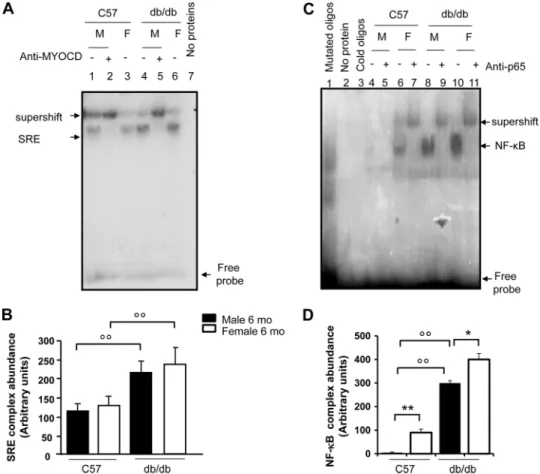
Hearts of Obese db/db Mice Show Evidence for Increased Hypertrophy Signaling—Analysis of cardiac tissues in db/db mice showed significant up-regulation of myocardin as well as MEF2c at the protein level (Fig. 7B, panels a and c). This increased expression was evident after 4 months and was sus- tained after 12 months of diabetes, and was associated with decreased expression of CHIP (Fig. 7B,panel d). Conversely, SRF levels were similar indb/dbmice compared with age and sex-matched C57BL/6J mice (Fig. 7B,panel b). In agreement with our previous report (26), diabetic animals also showed increased heart weight (0.4⫾0.05 gversus0.1⫾0.03 g,n⫽3, p⬍0.05db/dbmiceversusC57 mice) and heart weight to body weight ratio (HW/BW ratio: 0.005⫾0.05versus0.004⫾0.00, n⫽3,p⬍0.05dbd/dbmiceversusC57 mice). To determine SRF activity, gel shift assay with32P end-labeled SRE oligonu- cleotides was conducted using the nuclear proteins isolated
from snap-frozen hearts. As shown in Fig. 8,panels AandB, SRF binding activities were much stronger in the nuclear extracts from age- and sex-matched hearts ofdb/dbmice than those from wild-type controls, indicating their potential for myogenesis. The specificity of the SRF-DNA protein complex formation was verified by competition with cold oligonucleo- tides. The binding activity with the radioactive oligonucleotide probe was abolished in the absence of protein extracts (Fig. 8, panel A,lane 7), indicating the dependence of the shifted radio- active bands on nuclear protein factor binding. To determine if myocardin, an important co-activator of SRF, can interact with SRF in the hearts ofdb/dbmice, supershift analyses were car- ried out by adding anti-myocardin antibody (Fig. 8, panel A, lanes 2 and5). The antibody to MYOCD super-shifted a por- tion of the complexes consisting of nuclear protein binding to DNA, indicating that myocardin was present in the protein- SRE probe complexes. Expression and activity of NF-B were also analyzed in the hearts ofdb/dband C57 mice. In agreement FIGURE 6.The role of NF-B and myocardin in mediating myosin heavy chain-expression in cardiac myoblasts treated with insulin and TNF-␣.Panel A, canine cardiac myoblasts were transfected with a pool of three different anti-myocardin siRNAs (MYOCD-siRNA) and/or anti-NF-B siRNAs or a scrambled (scr) siRNA (negative control), and treated with insulin (10⫺8mol/liter) with or without TNF-␣(10 ng/ml) for 24 h. Following treatments, cells were permeabi- lized and fixed for the staining with an anti-myosin heavy chain (MHC)-antibody. Nuclei were stained with 4⬘,6-diamidino-2-phenylindole. Representative photos show cell size of canine cardiac myoblasts and expression of MHC-.Panels BandD, canine cardiac myoblasts were treated as described for A. After treatment, the expression of MHC-was detected by immunoblotting. Blots shown here are representative of three independent experiments. The results of scanning densitometry (n⫽3 independent experiments) are expressed as arbitrary units inpanels CandD. Columns and bars represent the mean⫾S.D.Panel C, **,p⬍0.01,versusuntreated cells; ˆˆ,p⬍0.01versusinsulin-treated cells; °°,p⬍0.01versusinsulin⫹MYOCD-siRNA-treated cells orversusinsulin⫹NF-B -siRNA-treated cells.Panel E, **,p⬍0.01,versusuntreated cells orversusinsulin⫹TNF-␣-treated cells; ˆ,p⬍0.05 and ˆˆ,p⬍0.01versusinsulin⫹TNF-␣-treated cells; °,p⬍0.05 and °°,p⬍0.01versusinsulin⫹MYOCD-siRNA-treated cells orversusinsulin⫹NF-B -siRNA-treated cells.
at SEMMELWEIS UNIV OF MEDICINE on August 27, 2018http://www.jbc.org/Downloaded from
with our previous reports (26), NF-B binding activities were much stronger in the nuclear extracts from age- and sex- matched hearts ofdb/dbmice than those from the wild-type controls (Fig. 8,panels CandD).
DISCUSSION
A number of clinical studies have indicated a link between insulin resistance/hyperinsulinemia, cardiac hypertrophy and heart failure (6 – 8). Despite this body of evidence, the downstream targets of insulin in the hypertrophy pathways remain poorly understood. In the present study, we demon- strate that myocardin is a modulator of such insulin-induced hypertrophy.
We used insulin at concentrations that would bind and acti- vate insulin receptors in endothelial cells (between 10⫺9and 10⫺7mol/liter) (46). Such concentrations, equivalent to 139 – 13,900U/ml, range from pathophysiological to pharmacolog- ical levels of insulinemia. Concentrations of 10⫺9 and 10⫺8 mol/liter are indeed attainable in fasting and post-prandial states in individuals with insulin resistance (47), indicating that our observations are applicable toin vivosettings where hyper-
insulinemia occurs, with all the caveats of extrapolating fromin vitrofindings.
Myocardin exerts its pro-hypertrophic effect through SRF, which binds to the SRE in the promoter region of genes that are critical for myogenesis (48 –50). Previous studies have shown that insulin may initiate a hypertrophy cascade through Akt, which in turn has been demonstrated to control the function of hypertrophy regulators such as nuclear factor of activated T cells (NFAT), GATA-4, and atrogin-1 (51). Our study shows that the Akt inhibitor LY-294002 attenuates myocardin intra- cellular levels upon insulin treatment. Thus, myocardin can be a downstream target of the insulin/Akt axis in the induction of hypertrophy.
How is myocardin activated in response to insulin signaling?
Based on our data, we suggest that at least two mechanisms are involved: 1) myocardin transcripts and protein levels are increased by insulin, which may account for the increase of myocardin-dependent SRF-SRE binding and subsequent acti- vation of the myogenic program; 2) myocardin activity, which is independent of protein expression level, is induced by insulin signaling, most likely through a post-translational change. Pre- FIGURE 7.Insulin signaling and Western analysis of cardiomyogenic genes in cardiac tissue of C57BL/6 anddb/dbmice.Panel A, levels of IR subunits␣ and, total Akt and the Akt1 isoform, and the phosphorylated isoforms of insulin receptor substrate (IRS)-1, extracellular signal-regulated kinase (ERK)44 and p38 in total protein extracts isolated from male (M) and female (F)db/dbmice compared with sex- and age-matched C57BL/6 control mice. The blot shown here is representative of three independent experiments.Panel B, levels of MYOCD, SRF, MEF 2c, and the CHIP in total protein extracts isolated from male (M) and female (F)db/dbmice compared with sex- and age-matched C57BL/6 control mice. Blots are representative of three independent experiments.Panel C, results of scanning densitometry (n⫽3 mice per group) of experiments as ofpanel Aare expressed as arbitrary units of optical density. Columns and bars represent the mean⫾S.D. **,p⬍0.01, controlversusnon-diabetic control mice.Panel D, Results of scanning densitometry (n⫽3 mice per each group) of experiments as ofpanel Bare expressed as arbitrary units of optical density. Columns and bars represent the mean⫾S.D. **,p⬍0.01, controlversusnon-diabetic control mice.
Insulin, NF- B, and Myocardin Signaling in Cardiac Myoblasts
at SEMMELWEIS UNIV OF MEDICINE on August 27, 2018http://www.jbc.org/Downloaded from
vious studies have shown that CHIP promotes the ubiquitina- tion of phosphorylated myocardin and its degradation by the proteasome, thereby inhibiting myocardin-dependent myo- genic gene expression (14). In this regard, our results show that insulin down-regulates CHIP, which acts as a repressor of myo- cardin expression through protein ubiquitination. Further- more, previous work has shown that MEF2c regulates tran- scriptional expression of myocardin through its enhancer (32).
Our data show that insulin increases levels of MEF2c, which acts as an activator of myocardin expression by enhancing myo- cardin transcription. Thus, it appears that insulin and its down- stream target Akt form an axis with myocardin and its regula- tors, CHIP and MEF2c, participating in inducing cardiac hypertrophy. Our data are therefore consistent with the hypothesis that myocardin transcription and protein expres- sion are increased by insulin, but do not allow us to determine if post-translational modifications are additionally responsible for insulin-dependent myocardin activation. This point remains to be clarified.
In thedb/db mouse model also used by us myocardin was up-regulated without the activation of Akt signaling. This dis- crepancy between thein vitroandin vivosettings might be due to limitations in having used thedb/dbmice as a model of type 2 diabetes, where the well-known inhibitory effect of hypergly- cemia on Akt regulation (52) likely prevails over the effects of hyperinsulinemia. Indeed, unlike other models of type 2 diabe- tes (ob/obmice, Zucker fatty rats), db/db mice are character- ized by an earler and more intense development of hyperglyce- mia relative to hyperinsulinemia (53). Alternatively, the discrepancy may be due to the specific isoform of Akt chroni- cally regulated in vivo by hyperinsulinemia. There is indeed evidence of a divergent regulation of Akt1 and Akt2 in insulin target tissues, where insulin exerts differential effects on these isoforms in a tissue- and species-specific manner (54). Previous studies have shown that chronic hyperinsulinemia may increase myocardial Akt1 activation (55, 56). In the skeletal muscle of obese Zucker rats, Akt2 is reduced, while Akt1 is increased (54).
FIGURE 8.EMSA for myocardin-SRF-SRE and NF-B binding activities in cardiac tissue of C57BL/6 anddb/dbmice.Panel A, SRF activity was determined by EMSA, performed by mixing a32P-labeled oligonucleotide encoding for the consensus sequence of serum response element (SRE)-binding promoter with nuclear proteins from male (M) and female (F)db/dbmouse hearts and sex/age-matched (12-month-old) control hearts (C57BL/6 mice), withn⫽3 mice per group. Specificity of the myocardin-SRF-SRE complex formation was determined by the omission of nuclear proteins and by the presence of a supershift after the addition of an anti-myocardin antibody. The electrophoretic run with nuclear protein extracts from C57BL/6 control hearts is shown inlanes 1–3. The electrophoretic run with nuclear protein extracts fromdb/dbmice is shown inlanes 4 – 6. The autoradiogram shown is representative of three separate gel shift experiments for NF-B.Lane 7shows the electrophoretic run by omitting protein extracts.Panel B, scanning densitometry (n⫽3 mice per each group) of the EMSA gels, expressed as arbitrary units of optical density. Columns and bars represent the mean⫾S.D. °°,p⬍0.01, controlversusdiabetic mice.Panel C, NF-B activity was determined by EMSA, performed by mixing a32P-labeled oligonucleotide encoding for the consensus sequence of the NF-B-binding promoter with nuclear proteins from the hearts of male (M) and female (F)db/dbmice and sex- and age-matched C57BL/6 control mice (3 mice per group). Specificity of NF-B binding activity was evaluated by cold oligonucleotide analysis, the omission of nuclear proteins and by the occurrence of a supershift after the addition of an anti-p65 antibody. The electrophoretic run with nuclear protein extracts fromdb/dbmice is shown inlanes 8 –11. The electrophoretic run with nuclear protein extracts from control hearts (C57BL/6 mice) is shown in lanes 4 –7. This autoradiogram is representative three separate gel shift experiments for NF-B.
Panel D, scanning densitometry (n⫽3 mice per each group) of the EMSA gels, expressed as arbitrary units of optical density. Columns and bars represent the mean⫾S.D. *,p⬍0.05 and **,p⬍0.01, maleversusfemale; °°,p⬍0.01, controlversusdiabetic mice.
at SEMMELWEIS UNIV OF MEDICINE on August 27, 2018http://www.jbc.org/Downloaded from
It is likely that acutely (such as in thein vitromodel of high insulin concentrations) insulin stimulates growth through the same PI3K/Akt pathway by which it mediates glucose uptake.
However, chronically, such as the indb/dbmice, where insulin resistance and decreased glucose uptake through a down-reg- ulation of PI3K/Akt pathway are present, hyperinsulinemia may stimulate growth through an alternative pathway, by increasing myocardial Akt-1 activation.
Increased circulating levels of pro-inflammatory cytokines, such as interleukin (IL)-6 (57) and tumor necrosis factor (TNF)-␣(18), as well as high levels of acute phase reactants (58), have been found in patients with insulin resistance, obesity and type 2 diabetes. Low-grade chronic inflammation may play a role in the pathogenesis of insulin resistance in obesity and type 2 diabetes, as well as in cardiovascular complications of diabe- tes, including cardiac remodeling and heart failure. We have previously shown that TNF-␣synergizes with insulin to induce NF-B activation and expression of adhesion molecules in endothelial cells (37– 40). The present study demonstrates that insulin, alone or in combination with low concentrations of TNF-␣, can activate the transcription factor NF-B in cardiac myoblasts. Previous studies using myoblast cell lines resulted in contradictory results concerning the effect of TNF-␣on the activation of the myogenic program (59). The present study has used primary myoblasts, which replicate the properties of car- diomyocytes more closely than myoblast cell lines (60).
In previous studies, cardiac-specific genetic ablation of NF-B has been shown to attenuate angiotensin II-induced hypertrophy (61), which indicates a key role for NF-B in the induction of cardiac hypertrophy. Here we show that TNF-␣ cooperates with insulin in enhancing SRF-SRE binding and inducing the activation of the myogenic program at levels up to 10 ng/ml, a concentration previously shown to evoke signaling events mediated by TNF-␣ receptor activation in cultured myocytes without inducing necrotic or apoptotic cell death (62). TNF-␣stimulation of the myogenic program and hyper- trophy appears to involve multiple signaling pathways (63).
TNF-␣activates NF-B in cardiomyocytes and myoblasts (22), and in myoblasts in other studies (62, 63). TNF-␣stimulation of the myogenic program through SRF signaling is thus likely to involve mediation by NF-B. We have indeed also shown that the NF-B inhibitor PDTC, as well as NF-B silencing attenu- ate the SRF-SRE binding and the expression of the hypertrophy marker MHC-upon insulin and TNF-␣treatment.
Our experiments were carried out in serum-starved culture conditions (a growth medium supplemented with 2% bovine serum). SRF responds to serum growth factors (64), and the low basal activity of SRF observed in our experiments may reflect the presence of serum growth factors at very low con centrations.
In conclusion, our study reveals that CHIP constitutes an anti-hypertrophy mediator, and can be a target of insulin in the induction of hypertrophy. Insulin cooperates with TNF-␣ in inducing NF-B and SRF-SRE activities. Thus, our data shed new light on the understanding of the molecular mechanism through which insulin induces hypertrophy and remodeling during insulin resistance and hyperinsulinemia in type 2 diabe-
tes. Such studies may help designing strategies to prevent dia- betic cardiomyopathy (65, 66).
Acknowledgment—We thank Harnath Shelat for help in cell cultures.
REFERENCES
1. Frey, N., and Olson, E. N. (2003) Cardiac hypertrophy: the good, the bad, and the ugly.Annu. Rev. Physiol.65,45–79
2. Li, H., Watford, W., Li, C., Parmelee, A., Bryant, M. A., Deng, C., O’Shea, J., and Lee, S. B. (2007) Ewing sarcoma gene EWS is essential for meiosis and B lymphocyte development.J. Clin. Invest.117,1314 –1323 3. Belke, D. D. (2002) Insulin signaling coordinately regulates cardiac size,
metabolism, and contractile protein isoform expression.J. Clin. Invest.
109,629 – 639
4. Samuelsson, A. M., Bollano, E., Mobini, R., Larsson, B. M., Omerovic, E., Fu, M., Waagstein, F., and Holmäng, A. (2006) Hyperinsulinemia:
effect on cardiac mass/function, angiotensin II receptor expression, and insulin signaling pathways.Am. J. Physiol. Heart Circ. Physiol.291, H787–H796
5. Shimizu, I., Minamino, T., Toko, H., Okada, S., Ikeda, H., Yasuda, N., Tateno, K., Moriya, J., Yokoyama, M., Nojima, A., Koh, G. Y., Akazawa, H., Shiojima, I., Kahn, C. R., Abel, E. D., and Komuro, I. (2010) Excessive cardiac insulin signaling exacerbates systolic dysfunction induced by pres- sure overload in rodents.J. Clin. Invest.120,1506 –1514
6. Paolisso, G., Galzerano, D., Gambardella, A., Varricchio, G., Saccomanno, F., D’Amore, A., Varricchio, M., and D’Onofrio, F. (1995) Left ventricular hypertrophy is associated with a stronger impairment of non-oxidative glucose metabolism in hypertensive patients. Eur J. Clin. Invest. 25, 529 –533
7. Hittinger, L., Mirsky, I., Shen, Y. T., Patrick, T. A., Bishop, S. P., and Vatner, S. F. (1995) Hemodynamic mechanisms responsible for reduced subendocardial coronary reserve in dogs with severe left ventricular hy- pertrophy.Circulation92,978 –986
8. Rutter, M. K., Parise, H., Benjamin, E. J., Levy, D., Larson, M. G., Meigs, J. B., Nesto, R. W., Wilson, P. W., and Vasan, R. S. (2003) Impact of glucose intolerance and insulin resistance on cardiac structure and function: sex- related differences in the Framingham Heart Study. Circulation 107, 448 – 454
9. Fonarow, G. C., and Srikanthan, P. (2006) Diabetic cardiomyopathy.En- docrinol. Metab. Clin. North Am.35,575–599
10. Zhang, X., Azhar, G., Chai, J., Sheridan, P., Nagano, K., Brown, T., Yang, J., Khrapko, K., Borras, A. M., Lawitts, J., Misra, R. P., and Wei, J. Y. (2001) Cardiomyopathy in transgenic mice with cardiac-specific overexpression of serum response factor. Am. J. Physiol. Heart Circ. Physiol. 280, H1782—H1792
11. Norman, C., Runswick, M., Pollock, R., and Treisman, R. (1988) Isolation and properties of cDNA clones encoding SRF, a transcription factor that binds to the c-fos serum response element.Cell55,989 –1003
12. Wang, D., Chang, P. S., Wang, Z., Sutherland, L., Richardson, J. A., Small, E., Krieg, P. A., and Olson, E. N. (2001) Activation of cardiac gene expres- sion by myocardin, a transcriptional cofactor for serum response factor.
Cell105,851– 862
13. Ballinger, C. A.,Connell, P., Wu, Y., Hu, Z., Thompson, L. J., Yin, L. Y., and Patterson, C. (1999) Identification of CHIP, a novel tetratricopep- tide repeat-containing protein that interacts with heat shock proteins and negatively regulates chaperone functions. Mol. Cell. Biol. 19, 4535– 4545
14. Xie, P., Fan, Y., Zhang, H., Zhang, Y., She, M., Gu, D., Patterson, C., and Li, H. (2009) CHIP represses myocardin-induced smooth muscle cell differ- entiation via ubiquitin-mediated proteasomal degradation.Mol. Cell. Biol.
29,2398 –2408
15. Bierhaus, A., Schiefofer, S., Schwaninger, M., Andrassy, M., Humpert, P. M., Chen, J., Hong, M., Luther, T., Henle, T., Kloting, I., Morcos, M., Hofmann, M., Tritschler, H., Weigle, B., Kasper, M., Smith, M., Perry, G., Schmidt, A. M., Stern, D. M., Haring, H. U., Schleicher, E., and Nawroth, P. P. (2001) Diabetes-associated sustained activation of the transcription
Insulin, NF- B, and Myocardin Signaling in Cardiac Myoblasts
at SEMMELWEIS UNIV OF MEDICINE on August 27, 2018http://www.jbc.org/Downloaded from