International Journal of
Molecular Sciences
Review
Analytical and Structural Studies for the Investigation of Oxidative Stress in Guanine Oligonucleotides
Györgyi Ferenc1 , Zoltán Váradi2, Zoltán Kupihár2, Gábor Paragi3,4,* and Lajos Kovács2,*
1 Nucleic Acid Synthesis Laboratory, Biological Research Centre, Temesvári krt. 62, H-6726 Szeged, Hungary;
ferenc.gyorgyi@brc.hu
2 Nucleic Acids Laboratory, Department of Medicinal Chemistry, University of Szeged, Dóm tér 8, H-6720 Szeged, Hungary; varadi8008@gmail.com (Z.V.); kupihar.zoltan@med.u-szeged.hu (Z.K.)
3 MTA-SZTE Biomimetic Systems Research Group, Dóm tér 8, 6720 Szeged, Hungary
4 Institute of Physics, University of Pécs, Ifjúságútja 6, 7624 Pécs, Hungary
* Correspondence: paragi@sol.cc.u-szeged.hu (G.P.); kovacs.lajos@med.u-szeged.hu (L.K.);
Tel.:+36-62545145 (L.K.); Fax:+36-62545971 (L.K.)
Received: 31 May 2020; Accepted: 13 July 2020; Published: 15 July 2020 Abstract: DNA damage plays a decisive role in epigenetic effects. The detection and analysis of DNA damages, like the most common change of guanine (G) to 8-oxo-7,8-dihydroguanine (OG), is a key factor in cancer research. It is especially true for G quadruplex structure (GQ), which is one of the best-known examples of a non-canonical DNA arrangement. In the present work, we provided an overview on analytical methods in connection with the detection of OG in oligonucleotides with GQ-forming capacity. Focusing on the last five years, novel electrochemical tools, like dedicated electrodes, were overviewed, as well as different optical methods (fluorometric assays, resonance light scattering or UV radiation) along with hyphenated detection and structural analysis methods (CD, NMR, melting temperature analysis and nanopore detection) were also applied for OG detection. Additionally, GQ-related computational simulations were also summarized. All these results emphasize that OG detection and the analysis of the effect of its presence in higher ordered structures like GQ is still a state-of-the-art research line with continuously increasing interest.
Keywords: oxidative stress; epigenetic; reactive oxygen species; reactive nitrogen species; guanine;
quadruplex; 8-oxo-7,8-dihydro-20-deoxyguanosine; analytical methods; structural investigations;
computational studies
1. Introduction
The presence of an oxidative environment in which aerobic organisms strive constitutes a constant threat. Coping with this condition required the elaboration of distinct mechanisms to deal with the reactive oxygen species (ROS) involved in this process. In our current understanding, ROS have been utilized in upregulation and as messenger molecules during inflammation. The process of this gene regulation is possible due to the special feature of guanine (G) residues in nucleic acids, namely, their easier oxidation compared to other nucleobases and their ability to form tetrads (G4) and quadruplexes (GQs). The oxidation of guanines paired in the quadruplex is twice as fast as oxidation of the same sequence in a duplex context [1,2]. The primary product of G from ROS exposure is 8-oxo-7,8-dihydroguanine (OG (R=H, Scheme1), for the sake of simplicity, the nucleobase term OG is used throughout this review unless the oxidized DNA nucleosides are specifically referred to). It is estimated that≤100,000 OGs are formed per cell daily, and it is the most frequently measured biomarker of ROS-induced DNA oxidation to date [3]. OG is a target for base excision repair initiated by the enzyme OG-glycosylase 1 (OGG1). Traditionally, the formation of OG is considered as a biomarker of
Int. J. Mol. Sci.2020,21, 4981; doi:10.3390/ijms21144981 www.mdpi.com/journal/ijms
Int. J. Mol. Sci.2020,21, 4981 2 of 27
a mutagenetic event [4] but it could be epigenetic by functioning as a transcriptional regulator [5,6] or, in general, as a tool to improve the genome plasticity to redox changes [7]. The formation of OG is either beneficial or detrimental depending on the gene product that is formed from the OG-containing promoter [5]. OG may alter gene expression and its transcriptional impact is dependent on the coding versus template strand of OG occurrence, and it can temporarily stop or even completely abolish mRNA synthesis. The role of guanine lesions is the complex process of epigenetic regulation that has been reviewed recently by several authors [1,5,7–17]. Pertinent to this multifaceted role is the analytical detection, quantification and structural studies of the facilitated formation of OG in G-rich oligonucleotide sequences, in particular, G-quadruplexes and their hydrolysis products (nucleosides, nucleobases), which is the subject of the present review.
Int. J. Mol. Sci. 2020, 21, x FOR PEER REVIEW 2 of 28
is considered as a biomarker of a mutagenetic event [4] but it could be epigenetic by functioning as a transcriptional regulator [5,6] or, in general, as a tool to improve the genome plasticity to redox changes [7]. The formation of OG is either beneficial or detrimental depending on the gene product that is formed from the OG-containing promoter [5]. OG may alter gene expression and its transcriptional impact is dependent on the coding versus template strand of OG occurrence, and it can temporarily stop or even completely abolish mRNA synthesis. The role of guanine lesions is the complex process of epigenetic regulation that has been reviewed recently by several authors [1,5,7–
17]. Pertinent to this multifaceted role is the analytical detection, quantification and structural studies of the facilitated formation of OG in G-rich oligonucleotide sequences, in particular, G-quadruplexes and their hydrolysis products (nucleosides, nucleobases), which is the subject of the present review.
2. Scope and Limitations
The formation of OG is a two-electron oxidation event followed by further oxidative and hydrolytic transformations, resulting in additional products (Scheme 1). The frequency of OG formation is ca. 1 in 106 [18], while that of the four-electron (spiroiminodihydantoin (Sp), 5- guanidinohydantoin (Gh), and imidazolone (Iz)) and six-electron oxidation (5-guanidino- dehydrohydantoin (Ghox)) and hydrolysis products (2,6-diamino-4-hydroxy-5-formamido- pyrimidine (Fapy-G), oxazolone (Z)) is much lower [18,19], therefore our treatment is limited mostly to the analytical and structural studies related to OG (the formation of OG dominates under aerobic conditions, while the yield of Fapy-G increases under anaerobic conditions [18]). The temporal frame of our review encompasses papers from the last five years with occasional reference to earlier papers when justified.
Scheme 1. The oxidative transformation of guanine oligonucleotide derivatives upon reaction with reactive oxygen species (ROS) and reactive nitrogen species (RNS). To have a better overview, not all radical intermediates have been included (adapted from References [5,18–20]).
3. Analytical Detection and Quantitation Methods
The Concentration of ROS, Reactive Nitrogen Species (RNS) and the Characteristic Processes Leading to Their Formation
The typical concentration of oxygen, the source of metabolic ROS, ranges from ∼20 to 100 μM O2 depending on the tissue type [5]. While the partially reduced superoxide radical ion (O2•−) forms Scheme 1.The oxidative transformation of guanine oligonucleotide derivatives upon reaction with reactive oxygen species (ROS) and reactive nitrogen species (RNS). To have a better overview, not all radical intermediates have been included (adapted from References [5,18–20]).
2. Scope and Limitations
The formation of OG is a two-electron oxidation event followed by further oxidative and hydrolytic transformations, resulting in additional products (Scheme1). The frequency of OG formation is ca.
1 in 106 [18], while that of the four-electron (spiroiminodihydantoin (Sp), 5-guanidinohydantoin (Gh), and imidazolone (Iz)) and six-electron oxidation (5-guanidino- dehydrohydantoin (Ghox)) and hydrolysis products (2,6-diamino-4-hydroxy-5-formamido- pyrimidine (Fapy-G), oxazolone (Z)) is much lower [18,19], therefore our treatment is limited mostly to the analytical and structural studies related to OG (the formation of OG dominates under aerobic conditions, while the yield of Fapy-G increases under anaerobic conditions [18]). The temporal frame of our review encompasses papers from the last five years with occasional reference to earlier papers when justified.
3. Analytical Detection and Quantitation Methods
3.1. The Concentration of ROS, Reactive Nitrogen Species (RNS) and the Characteristic Processes Leading to Their Formation
The typical concentration of oxygen, the source of metabolic ROS, ranges from∼20 to 100µM O2 depending on the tissue type [5]. While the partially reduced superoxide radical ion (O2•−
) forms in only 0.1–2% yield, it is still generated in significant amounts. Superoxide has a short cellular half-life (∼1µs) and it is the subject of disproportionation catalyzed by superoxide dismutase (SOD) to furnish
Int. J. Mol. Sci.2020,21, 4981 3 of 27
hydrogen peroxide (H2O2), with a half-life of∼10µs. The steady-state concentration of hydrogen peroxide is∼200 nM and can increase many folds under high metabolic demand or stress. In general, the concentration of ROS is generally high, and its elevated load can overburden the natural defense systems, leading to inflammation. Hydrogen peroxide is a signaling agent for immune cell activation and vascular remodeling. A side reaction of H2O2 is the Fenton reaction to yield the aggressive one-electron oxidant hydroxyl radical (HO•; Ered=2.31 V versus normal hydrogen electrode (NHE)) when H2O2reacts with iron(II) [21] or copper(I) ions [22]. In the presence of physiological bicarbonate concentration (∼25 mM), the Fenton reaction yields primarily carbonate radical anion (CO3•−
) [23].
Reactive nitrogen species (RNS) also contribute to inflammatory responses (there are ca. 80 known DNA defects that can be attributed to ROS or RNS) [11,20]. Among others, peroxynitrite ion (ONOO−), the intracellular concentration of which lies in the nanomolar range [20], reacts with cellular CO2
to yield short-lived nitrosoperoxycarbonate anion (ONOOCO2−
). The ion ONOOCO2−affords, in a homolytic degradation, the one-electron oxidants carbonate radical anion (CO3•−; Ered=1.59 V versus NHE) and nitrogen dioxide radical (•NO2; Ered=1.04 V versus NHE) in ca. 70% yield (the steady-state levels of•NO2 in activated macrophages have been estimated to be in the picomolar range) [20].
The ion ONOOCO2−
preferentially causes oxidation and nitration of G in DNA and probably RNA, it is likely one of the major oxidizing species in vivo due to millimolar concentrations of CO2 in tissues, and the large reaction rate constants of CO2and ONOO−ion [20]. G is the most susceptible to oxidative modification among the canonical nucleobases (Ered=1.29 V versus NHE) [20,24]. It was also demonstrated that the catalytic one-electron oxidation kinetics in poly-GC sequences is particularly rapid [25]. The carbonate radical anion specifically oxidizes G to OG in high yields. Nitrogen dioxide radical is incapable of oxidizing G to OG but it readily oxidizes OG further (0.74 V versus NHE) as an OG is at least 1000-fold more reactive than the parent G toward further oxidation [20]. In addition, unselective one-electron oxidation of duplex DNA can yield an electron hole that can migrate through thousands of base pairs, yielding oxidation at the most sensitive G-rich sites. This feature confers a sacrificial “cathodic protection” character to G tracts to prevent deterioration of other nucleobases [24].
3.2. Electrochemical Methods
The electrochemical methods belong to the most sensitive analytical methods, and therefore their privileged role in the detection of mutagenic or epigenetic OG formation is well motivated owing to its low yet significant occurrence in vivo. Over the years, several sophisticated procedures have been developed to monitor and quantify samples containing the nucleobase OG, its precursor nucleoside 8-oxo-7,8-dihydro-20-deoxyguanosine (o8dGuo, in the literature frequently abbreviated as 8-OHdG as well) or even its presence in ONs or GQs.
3.2.1. Development of New Electrodes
Electrodes Coated with Reduced Graphene Oxide Nanocomposites
Hao et al. [26] have prepared a nanocomposite using reduced graphene oxide (rGO) and ZnO nanoparticles (ZnO@rGO) via an in situ reduction of graphene oxide (GO) with Zn powder and coated glassy carbon electrode (GCE) (Figure1). The ensuing system (ZnO@rGO/GCE), characterized by scanning electron microscopy (SEM) and transmission electron microscopy (TEM), gave significantly enhanced oxidation signals of o8dGuo, compared with unmodified GCE and GO-modified GCE (GO/GCE), as verified by cyclic voltammetry (CV). With the enzyme uricase, the interference of uric acid (UA) was effectively eliminated and accurate sensing of o8dGuo was attained. The linear range of 5 to 5000 nM for the detection of o8dGuo using ZnO@rGO/GCE has been achieved using differential pulse voltammetry (DPV). The limit of detection (LOD) was 1.25 nM.
Int. J. Mol. Sci.2020,21, 4981 4 of 27
Int. J. Mol. Sci. 2020, 21, x FOR PEER REVIEW 4 of 28
Figure 1. ZnO nanoparticles (ZnO@rGO) for the detection of o8dGuo. Reprinted with permission from Reference [26], © 2018, Elsevier.
rGO has also been combined with another metal oxide, dysprosium oxide (Dy2O3) nanoparticles, for the determination of o8dGuo in human blood and urine samples by Manavalan et al. [27].
Dy2O3@rGO was synthesized by a microwave-assisted synthetic route to improve the moderate electrical conductivity; furthermore, the electrocatalytic ability was synergically improved. The electrochemical and interfacial properties were examined by electrochemical impedance spectroscopy (EIS). Under optimum conditions, the electrocatalytic performances of Dy2O3@rGO- modified electrode and control electrodes were analyzed by CV. The Dy2O3@rGO-affixed conventional screen-printed carbon electrode (SPCE) was found to exhibit tremendous electrocatalytic capability toward o8dGuo oxidation. The amperometric detection of o8dGuo worked in the linear range of 50 nM to 315.3 μM, with a LOD of 1.02 nM. The initial sensor response current was still retained after 3000 s. The storage stability was monitored every day for 10 successive days, and no significant deterioration was observed. The method worked well in real human urine and blood serum samples and the results were validated by the HPLC method as well.
GCE Coated with Multi-Walled Carbon Nanotubes (MWCNTs)
Guo et al. [28] have developed an OG sensor based on the multi-walled carbon nanotubes (MWCNTs)-modified GCE characterized by SEM and EIS methods (Figure 2). The linear ranges were 56.3 nM to 6.08 μM and 6.08 μM to 16.4 μM respectively, with the LOD of 18.8 nM (S/N = 3) in CV measurements. UA and dopamine (DA) commonly coexist with o8dGuo in human metabolism and their electrochemical oxidation potentials are close, but with this method, their mixture gave three clear and well-separated oxidation peaks at 0.43 V (o8dGuo), 0.36 V (UA) and 0.22 V (DA), respectively.
Figure 1.ZnO nanoparticles (ZnO@rGO) for the detection of o8dGuo. Reprinted with permission from Reference [26],©2018, Elsevier.
rGO has also been combined with another metal oxide, dysprosium oxide (Dy2O3) nanoparticles, for the determination of o8dGuo in human blood and urine samples by Manavalan et al. [27]. Dy2O3@rGO was synthesized by a microwave-assisted synthetic route to improve the moderate electrical conductivity; furthermore, the electrocatalytic ability was synergically improved. The electrochemical and interfacial properties were examined by electrochemical impedance spectroscopy (EIS). Under optimum conditions, the electrocatalytic performances of Dy2O3@rGO-modified electrode and control electrodes were analyzed by CV. The Dy2O3@rGO-affixed conventional screen-printed carbon electrode (SPCE) was found to exhibit tremendous electrocatalytic capability toward o8dGuo oxidation. The amperometric detection of o8dGuo worked in the linear range of 50 nM to 315.3µM, with a LOD of 1.02 nM. The initial sensor response current was still retained after 3000 s. The storage stability was monitored every day for 10 successive days, and no significant deterioration was observed. The method worked well in real human urine and blood serum samples and the results were validated by the HPLC method as well.
GCE Coated with Multi-Walled Carbon Nanotubes (MWCNTs)
Guo et al. [28] have developed an OG sensor based on the multi-walled carbon nanotubes (MWCNTs)-modified GCE characterized by SEM and EIS methods (Figure2). The linear ranges were 56.3 nM to 6.08µM and 6.08µM to 16.4µM respectively, with the LOD of 18.8 nM (S/N=3) in CV measurements. UA and dopamine (DA) commonly coexist with o8dGuo in human metabolism and their electrochemical oxidation potentials are close, but with this method, their mixture gave three clear and well-separated oxidation peaks at 0.43 V (o8dGuo), 0.36 V (UA) and 0.22 V (DA), respectively.
Int. J. Mol. Sci. 2020, 21, x FOR PEER REVIEW 4 of 28
Figure 1. ZnO nanoparticles (ZnO@rGO) for the detection of o8dGuo. Reprinted with permission from Reference [26], © 2018, Elsevier.
rGO has also been combined with another metal oxide, dysprosium oxide (Dy2O3) nanoparticles, for the determination of o8dGuo in human blood and urine samples by Manavalan et al. [27].
Dy2O3@rGO was synthesized by a microwave-assisted synthetic route to improve the moderate electrical conductivity; furthermore, the electrocatalytic ability was synergically improved. The electrochemical and interfacial properties were examined by electrochemical impedance spectroscopy (EIS). Under optimum conditions, the electrocatalytic performances of Dy2O3@rGO- modified electrode and control electrodes were analyzed by CV. The Dy2O3@rGO-affixed conventional screen-printed carbon electrode (SPCE) was found to exhibit tremendous electrocatalytic capability toward o8dGuo oxidation. The amperometric detection of o8dGuo worked in the linear range of 50 nM to 315.3 μM, with a LOD of 1.02 nM. The initial sensor response current was still retained after 3000 s. The storage stability was monitored every day for 10 successive days, and no significant deterioration was observed. The method worked well in real human urine and blood serum samples and the results were validated by the HPLC method as well.
GCE Coated with Multi-Walled Carbon Nanotubes (MWCNTs)
Guo et al. [28] have developed an OG sensor based on the multi-walled carbon nanotubes (MWCNTs)-modified GCE characterized by SEM and EIS methods (Figure 2). The linear ranges were 56.3 nM to 6.08 μM and 6.08 μM to 16.4 μM respectively, with the LOD of 18.8 nM (S/N = 3) in CV measurements. UA and dopamine (DA) commonly coexist with o8dGuo in human metabolism and their electrochemical oxidation potentials are close, but with this method, their mixture gave three clear and well-separated oxidation peaks at 0.43 V (o8dGuo), 0.36 V (UA) and 0.22 V (DA), respectively.
Figure 2. Glassy carbon electrode (GCE)/multi-walled carbon nanotubes (MWCNT) sensor for the determination of o8dGuo. Reprinted with permission from Reference [28].©2016, Elsevier.
Int. J. Mol. Sci.2020,21, 4981 5 of 27
GCE Coated with Porous Single-Walled Carbon Nanotube (PSWCNT)
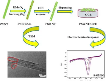
Quantitative measurement of o8dGuo concentration was analyzed by linear sweep voltammetry (LSV) as well, by Shang et al. [29]. First, a porous carbon nanotube was obtained from SWCNT using KMnO4as the etching agent, dropping the aqueous PSWCNT onto a polished GCE surface.
The obtained PSWCNT/GCE-based sensor, characterized by TEM, X-ray diffraction and nitrogen adsorption/desorption isotherm, showed outstanding electrochemical performance for o8dGuo in the measurement ranges 2.99 nM to 3.061µM and 3.061µM to 87.25µM respectively, with an LOD at 1.0 nM. The effects of temperature and time on DNA damage have also been investigated and important dynamical parameters and a kinetic equation with reaction rate constant (k=2.090 min−1) and the apparent activation energy (Ea=30.64 kJ·mol−1) for the OG oxidation have been obtained (Figure3).
Int. J. Mol. Sci. 2020, 21, x FOR PEER REVIEW 5 of 28
Figure 2. Glassy carbon electrode (GCE)/multi-walled carbon nanotubes (MWCNT) sensor for the determination of o8dGuo. Reprinted with permission from Reference [28]. © 2016, Elsevier.
GCE Coated with Porous Single-Walled Carbon Nanotube (PSWCNT)
Quantitative measurement of o8dGuo concentration was analyzed by linear sweep voltammetry (LSV) as well, by Shang et al. [29]. First, a porous carbon nanotube was obtained from SWCNT using KMnO4 as the etching agent, dropping the aqueous PSWCNT onto a polished GCE surface. The obtained PSWCNT/GCE-based sensor, characterized by TEM, X-ray diffraction and nitrogen adsorption/desorption isotherm, showed outstanding electrochemical performance for o8dGuo in the measurement ranges 2.99 nM to 3.061 μM and 3.061 μM to 87.25 μM respectively, with an LOD at 1.0 nM. The effects of temperature and time on DNA damage have also been investigated and important dynamical parameters and a kinetic equation with reaction rate constant (k = 2.090 min−1) and the apparent activation energy (Ea = 30.64 kJ·mol−1) for the OG oxidation have been obtained (Figure 3).
Figure 3. The application of porous single-walled carbon nanotube (PSWCNT) for the determination of o8dGuo. Reprinted with permission from Reference [29]. © 2018, Elsevier.
Voltammetric Sensor for Oxidized DNA Using Ultrathin Films of Osmium and Ruthenium Metallopolymers
In an earlier study, Mugweru et al. [30] assembled films containing the metallopolymers (Os(bpy)2(PVP)10Cl)+ and (Ru(bpy)2(PVP)10Cl)+ (bpy = 2,2’-bipyridine; PVP = poly(vinylpyridines)), layer by layer, on pyrolytic graphite electrodes to obtain sensors that selectively detect oxidized DNA.
Assembly of films was monitored with a quartz crystal microbalance (QCM). The two metallopolymers behaved electrochemically independent of each other in the films. This combination provided a catalytic Os square wave voltammetry (SWV) peak that is mainly selective for OG and the detection of other oxidized nucleobases from the Ru peak. The method is applicable to measurements on DNA in solution or DNA incorporated into films. Using the Os SWV peak, a single oxidized nucleobase out of 6000 nucleotides was detected. A related Os-PVP polymer with higher oxidation potential can generate electrochemiluminescence (ECL) with oligonucleotides containing OG in thin films, providing an alternative method to detect oxidative stress [31]. This purely voltammetric approach is complementary to the ECL method.
Copper-Based Metal Organic Framework Nanoparticles Anchored to Graphite Nanosheets
Cao et al. [32] prepared graphite nanosheets (GN) in a very simple liquid-phase exfoliation of graphite in N,N-dimethylacetamide (DMAc). Ultra-small (less than 5 nm) Cu-based metal organic framework (HKUST-1) nanoparticles were in situ anchored on the surface of GNs with a high degree of dispersion (Figure 4). The synthesized hybrids of graphite nanosheets (HKUST-1/GN) decorated with HKUST-1 nanoparticles, characterized by powder X-ray diffractometry (XRD),
Figure 3.The application of porous single-walled carbon nanotube (PSWCNT) for the determination of o8dGuo. Reprinted with permission from Reference [29].©2018, Elsevier.
Voltammetric Sensor for Oxidized DNA Using Ultrathin Films of Osmium and Ruthenium Metallopolymers
In an earlier study, Mugweru et al. [30] assembled films containing the metallopolymers (Os(bpy)2(PVP)10Cl)+and (Ru(bpy)2(PVP)10Cl)+(bpy=2,2’-bipyridine; PVP=poly(vinylpyridines)), layer by layer, on pyrolytic graphite electrodes to obtain sensors that selectively detect oxidized DNA.
Assembly of films was monitored with a quartz crystal microbalance (QCM). The two metallopolymers behaved electrochemically independent of each other in the films. This combination provided a catalytic Os square wave voltammetry (SWV) peak that is mainly selective for OG and the detection of other oxidized nucleobases from the Ru peak. The method is applicable to measurements on DNA in solution or DNA incorporated into films. Using the Os SWV peak, a single oxidized nucleobase out of 6000 nucleotides was detected. A related Os-PVP polymer with higher oxidation potential can generate electrochemiluminescence (ECL) with oligonucleotides containing OG in thin films, providing an alternative method to detect oxidative stress [31]. This purely voltammetric approach is complementary to the ECL method.
Copper-Based Metal Organic Framework Nanoparticles Anchored to Graphite Nanosheets
Cao et al. [32] prepared graphite nanosheets (GN) in a very simple liquid-phase exfoliation of graphite inN,N-dimethylacetamide (DMAc). Ultra-small (less than 5 nm) Cu-based metal organic framework (HKUST-1) nanoparticles were in situ anchored on the surface of GNs with a high degree of dispersion (Figure4). The synthesized hybrids of graphite nanosheets (HKUST-1/GN) decorated
Int. J. Mol. Sci.2020,21, 4981 6 of 27
with HKUST-1 nanoparticles, characterized by powder X-ray diffractometry (XRD), thermogravimetric analysis (TGA), SEM and TEM, showed excellent electrochemical sensing performance towards the DNA damage biomarker o8dGuo. The DPV concentration measurement was characterized with a fast detection speed (~240 s), wide linear window (10 nM–1µM), high sensitivity (346,857µA mM−1cm−2), low LOD (~2.5 nM) and good reproducibility.
Int. J. Mol. Sci. 2020, 21, x FOR PEER REVIEW 6 of 28
thermogravimetric analysis (TGA), SEM and TEM, showed excellent electrochemical sensing performance towards the DNA damage biomarker o8dGuo. The DPV concentration measurement was characterized with a fast detection speed (~240 s), wide linear window (10 nM–1 μM), high sensitivity (346,857 μA mM−1 cm−2), low LOD (~2.5 nM) and good reproducibility.
Figure 4. The schematic representation of metal organic framework nanoparticles for the measurement of o8dGuo. Reprinted with permission from Reference [32]. © 2019, Elsevier.
3.1.2. Gold Electrodes with o8dGuo-Specific Aptamers
An electrochemical method, developed by Zheng et al. [33], combines o8dGuo-specific aptamer (apt) with metal ion-dependent DNAzymes and exonuclease to achieve high sensitivity with a LOD of 6.82 pM and linearity from 0.01 nM to 7.0 μM applying SWV. The electrochemical sensing platform used a gold electrode modified with substrate DNA of DNAzyme, labelled with methylene blue (MB) redox probe as a working electrode. The state of the surface of the gold electrode was checked employing EIS. During the SWV analysis, reduction of the electrochemical signal is measured thanks to the release of the MB during the cleavage of the substrate DNA by the DNAzyme part of the o8dGuo–apt–DNAzyme complex. In addition, CV was used to verify the experimental principle. The modified gold electrode is stable for a week at 4 °C (Figure 5).
Figure 5. A o8dGuo-specific aptamer construct. Reprinted with permission from Reference [33]. © 20019, Elsevier.
Jia et al. [34] have developed a highly sensitive and selective electrochemical aptasensor by using o8dGuo-specific aptamer hybridized with the capture of DNA immobilized on a gold electrode with a sticky tail left, which initiated the hybridization chain reaction (HCR) through an auxiliary DNA (Aux). The formation of extended double-stranded DNA (dsDNA) structure intercalated a more electroactive species ((Ru(NH3)6)3+), therefore the high sensitivity of the electrochemical method was further increased thanks to the HCR. In the presence of o8dGuo, the aptamer will form a GQ structure that stops the HCR, leading to detection signal decrease. By monitoring the change in the current of the (Ru(NH3)6)3+ ion, the concentration of o8dGuo can be indirectly determined in the sample. The
Figure 4.The schematic representation of metal organic framework nanoparticles for the measurement of o8dGuo. Reprinted with permission from Reference [32].©2019, Elsevier.
3.2.2. Gold Electrodes with o8dGuo-Specific Aptamers
An electrochemical method, developed by Zheng et al. [33], combines o8dGuo-specific aptamer (apt) with metal ion-dependent DNAzymes and exonuclease to achieve high sensitivity with a LOD of 6.82 pM and linearity from 0.01 nM to 7.0µM applying SWV. The electrochemical sensing platform used a gold electrode modified with substrate DNA of DNAzyme, labelled with methylene blue (MB) redox probe as a working electrode. The state of the surface of the gold electrode was checked employing EIS.
During the SWV analysis, reduction of the electrochemical signal is measured thanks to the release of the MB during the cleavage of the substrate DNA by the DNAzyme part of the o8dGuo–apt–DNAzyme complex. In addition, CV was used to verify the experimental principle. The modified gold electrode is stable for a week at 4◦C (Figure5).
Int. J. Mol. Sci. 2020, 21, x FOR PEER REVIEW 6 of 28
thermogravimetric analysis (TGA), SEM and TEM, showed excellent electrochemical sensing performance towards the DNA damage biomarker o8dGuo. The DPV concentration measurement was characterized with a fast detection speed (~240 s), wide linear window (10 nM–1 μM), high sensitivity (346,857 μA mM−1 cm−2), low LOD (~2.5 nM) and good reproducibility.
Figure 4. The schematic representation of metal organic framework nanoparticles for the measurement of o8dGuo. Reprinted with permission from Reference [32]. © 2019, Elsevier.
3.1.2. Gold Electrodes with o8dGuo-Specific Aptamers
An electrochemical method, developed by Zheng et al. [33], combines o8dGuo-specific aptamer (apt) with metal ion-dependent DNAzymes and exonuclease to achieve high sensitivity with a LOD of 6.82 pM and linearity from 0.01 nM to 7.0 μM applying SWV. The electrochemical sensing platform used a gold electrode modified with substrate DNA of DNAzyme, labelled with methylene blue (MB) redox probe as a working electrode. The state of the surface of the gold electrode was checked employing EIS. During the SWV analysis, reduction of the electrochemical signal is measured thanks to the release of the MB during the cleavage of the substrate DNA by the DNAzyme part of the o8dGuo–apt–DNAzyme complex. In addition, CV was used to verify the experimental principle. The modified gold electrode is stable for a week at 4 °C (Figure 5).
Figure 5. A o8dGuo-specific aptamer construct. Reprinted with permission from Reference [33]. © 20019, Elsevier.
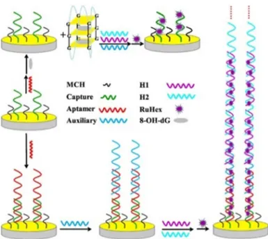
Jia et al. [34] have developed a highly sensitive and selective electrochemical aptasensor by using o8dGuo-specific aptamer hybridized with the capture of DNA immobilized on a gold electrode with a sticky tail left, which initiated the hybridization chain reaction (HCR) through an auxiliary DNA (Aux). The formation of extended double-stranded DNA (dsDNA) structure intercalated a more electroactive species ((Ru(NH3)6)3+), therefore the high sensitivity of the electrochemical method was further increased thanks to the HCR. In the presence of o8dGuo, the aptamer will form a GQ structure that stops the HCR, leading to detection signal decrease. By monitoring the change in the current of the (Ru(NH3)6)3+ ion, the concentration of o8dGuo can be indirectly determined in the sample. The Figure 5. A o8dGuo-specific aptamer construct. Reprinted with permission from Reference [33].
©20019, Elsevier.
Jia et al. [34] have developed a highly sensitive and selective electrochemical aptasensor by using o8dGuo-specific aptamer hybridized with the capture of DNA immobilized on a gold electrode with a sticky tail left, which initiated the hybridization chain reaction (HCR) through an auxiliary DNA (Aux). The formation of extended double-stranded DNA (dsDNA) structure intercalated a more electroactive species ((Ru(NH3)6)3+), therefore the high sensitivity of the electrochemical method was further increased thanks to the HCR. In the presence of o8dGuo, the aptamer will form a GQ structure that stops the HCR, leading to detection signal decrease. By monitoring the change in the current of the (Ru(NH3)6)3+ion, the concentration of o8dGuo can be indirectly determined in the sample.
Int. J. Mol. Sci.2020,21, 4981 7 of 27
The strategy was characterized by EIS measurements. Thus, the resistance continuously increased when the electrode was incubated with capture DNA, 6-mercapto-1-hexanol (MCH), aptamer and Aux. The resistance of aptamer/MCH/capture/electrode heavily decreased in the presence of o8dGuo.
In DPV, the electrochemical signal was mainly implemented by (Ru(NH3)6)3+immobilized on the long-nicked DNA polymers. An impressively wide linear response ranging from 10 pM to 100µM and LOD of 2.5 pM has been attained (S/N=3). The method was successfully applied in human urine samples (0.56 nM concentration of o8dGuo was determined) and interference with UA has been eliminated. The aptasensor was stable for 10 days at 4◦C. Despite the vulnerability of these electrodes, they constitute one of the most specific methods for the quantitation of o8dGuo lesions (Figure6).
Int. J. Mol. Sci. 2020, 21, x FOR PEER REVIEW 7 of 28
strategy was characterized by EIS measurements. Thus, the resistance continuously increased when the electrode was incubated with capture DNA, 6-mercapto-1-hexanol (MCH), aptamer and Aux. The resistance of aptamer/MCH/capture/electrode heavily decreased in the presence of o8dGuo. In DPV, the electrochemical signal was mainly implemented by (Ru(NH3)6)3+ immobilized on the long-nicked DNA polymers. An impressively wide linear response ranging from 10 pM to 100 μM and LOD of 2.5 pM has been attained (S/N = 3). The method was successfully applied in human urine samples (0.56 nM concentration of o8dGuo was determined) and interference with UA has been eliminated.
The aptasensor was stable for 10 days at 4 °C. Despite the vulnerability of these electrodes, they constitute one of the most specific methods for the quantitation of o8dGuo lesions (Figure 6).
Figure 6. An aptamer-based electrochemical biosensor with hybridization chain reaction (HCR) signal amplification for the detection of o8dGuo. Reprinted with permission from Reference [34]. © 2018, Elsevier.
3.1.3. Electrochemiluminescence in a Multiple-mechanism-driven Biosensor
Lv et al. [35] have developed a multiple-mechanism-driven electrochemiluminescent (ECL) biosensor that utilizes competitive catalytic and steric hindrance effects by assembling hemin/GQ on carbon nitride nanosheets (CNNS). A hairpin probe, containing a recognition sequence for the aptamer probe and a “caged” GQ sequence, was conjugated to a hybrid of CNNS and gold nanoparticles (AuNPs; CNNS–AuNPs) on GCE. After the targeted o8dGuo was captured by aptamer probe (o8dGuo-apt), it opened the hairpin structure by hybridizing to it. During the treatment with exonucleases, o8dGuo-apts were protected against them by o8dGuo and they were released after their complementary sequences were digested. Recycling of o8dGuo-apt led to the continuous opening of the hairpin probe and the generation of “active” GQ structures. Finally, by addition of hemin, the liberated GQs were folded into a supramolecular hemin/GQ, that electrocatalytically reduced the H2O2; as a result, its quenching effect was decreased on the ECL of CNNS–AuNPs. A linear dependence of the sensor on the o8dGuo concentration over the range from 10−15 to 10−12 M with a correlation coefficient of 0.998 was obtained. The LOD was calculated to be 38.8 aM (!), which is much lower than those for the best previously reported biosensors. In human serum spiked with o8dGuo, the proposed competitive dual-mechanism-driven biosensor had good accuracy and high precision.
The test of the binding specificity of the method indicated that the aptameric recognition function was retained. Many optimizations are required for greater simplicity, lower cost and for instrumentation use (Figure 7).
Figure 6. An aptamer-based electrochemical biosensor with hybridization chain reaction (HCR) signal amplification for the detection of o8dGuo. Reprinted with permission from Reference [34].
©2018, Elsevier.
3.2.3. Electrochemiluminescence in a Multiple-Mechanism-Driven Biosensor
Lv et al. [35] have developed a multiple-mechanism-driven electrochemiluminescent (ECL) biosensor that utilizes competitive catalytic and steric hindrance effects by assembling hemin/GQ on carbon nitride nanosheets (CNNS). A hairpin probe, containing a recognition sequence for the aptamer probe and a “caged” GQ sequence, was conjugated to a hybrid of CNNS and gold nanoparticles (AuNPs; CNNS–AuNPs) on GCE. After the targeted o8dGuo was captured by aptamer probe (o8dGuo-apt), it opened the hairpin structure by hybridizing to it. During the treatment with exonucleases, o8dGuo-apts were protected against them by o8dGuo and they were released after their complementary sequences were digested. Recycling of o8dGuo-apt led to the continuous opening of the hairpin probe and the generation of “active” GQ structures. Finally, by addition of hemin, the liberated GQs were folded into a supramolecular hemin/GQ, that electrocatalytically reduced the H2O2; as a result, its quenching effect was decreased on the ECL of CNNS–AuNPs. A linear dependence of the sensor on the o8dGuo concentration over the range from 10−15to 10−12M with a correlation coefficient of 0.998 was obtained. The LOD was calculated to be 38.8 aM (!), which is much lower than those for the best previously reported biosensors. In human serum spiked with o8dGuo, the proposed competitive dual-mechanism-driven biosensor had good accuracy and high precision.
The test of the binding specificity of the method indicated that the aptameric recognition function was retained. Many optimizations are required for greater simplicity, lower cost and for instrumentation use (Figure7).
Int. J. Mol. Sci.2020,21, 4981 8 of 27
Int. J. Mol. Sci. 2020, 21, x FOR PEER REVIEW 8 of 28
Figure 7. Electrochemiluminescent (ECL) biosensor developed by Lv et al. [35]. Reprinted with permission from Reference [35]. © 2019, American Chemical Society.
3.2. Optical Methods
3.2.1. Fluorometric Assays
Hu et al. [36] have developed a fluorometric immunoassay, based on strongly photoluminescent and excellently photostable carbon quantum dots (CQDs) and AuNPs, for determining DNA containing oxidatively damaged o8dGuo. The morphology and sizes of AuNPs and CQDs were observed by TEM. In a test experiment, CQDs were modified with glutaraldehyde for conjugation of DNA-o8dGuo, used as the damaged DNA model. AuNPs were functionalized by o8dGuo antibody.
The specific reaction of o8dGuo and o8dGuo antibody brought CQDs and AuNPs into close distance.
When the CQDs were excited by UV light, emission of CQDs was quenched by AuNPs. The target DNA-o8dGuo was detected by recording the quenched fluorescence spectra with an LOD of 7 pM in the 10 pM to 25 μM DNA-o8dGuo concentration range (the paper erroneously gives 700 pM as LOD).
The drawback of the method is that it requires DNA-o8dGuo isolated from urine and anchored to the glutaraldehyde-modified CQD, while urine contains more free OG than DNA-attached o8dGuo (Figure 8).
Figure 8. Fluorometric immunoassay using carbon quantum dots (CQDs) for the detection of DNA containing o8dGuo lesion [36].
Wei et al. [37] developed an ultrasensitive method based on fluorometric determination of o8dGuo by using a three-dimensional (3D) DNA nanomachine. The nanomachine was constructed by assembling hundreds of carboxyfluorescein-labeled single-stranded DNA (ssDNA) oligonucleotides (acting as signal reporter, the latter were quenched by AuNPs until the protecting
Figure 7. Electrochemiluminescent (ECL) biosensor developed by Lv et al. [35]. Reprinted with permission from Reference [35].©2019, American Chemical Society.
3.3. Optical Methods
3.3.1. Fluorometric Assays
Hu et al. [36] have developed a fluorometric immunoassay, based on strongly photoluminescent and excellently photostable carbon quantum dots (CQDs) and AuNPs, for determining DNA containing oxidatively damaged o8dGuo. The morphology and sizes of AuNPs and CQDs were observed by TEM. In a test experiment, CQDs were modified with glutaraldehyde for conjugation of DNA-o8dGuo, used as the damaged DNA model. AuNPs were functionalized by o8dGuo antibody.
The specific reaction of o8dGuo and o8dGuo antibody brought CQDs and AuNPs into close distance.
When the CQDs were excited by UV light, emission of CQDs was quenched by AuNPs. The target DNA-o8dGuo was detected by recording the quenched fluorescence spectra with an LOD of 7 pM in the 10 pM to 25µM DNA-o8dGuo concentration range (the paper erroneously gives 700 pM as LOD).
The drawback of the method is that it requires DNA-o8dGuo isolated from urine and anchored to the glutaraldehyde-modified CQD, while urine contains more free OG than DNA-attached o8dGuo (Figure8).
Int. J. Mol. Sci. 2020, 21, x FOR PEER REVIEW 8 of 28
Figure 7. Electrochemiluminescent (ECL) biosensor developed by Lv et al. [35]. Reprinted with permission from Reference [35]. © 2019, American Chemical Society.
3.2. Optical Methods
3.2.1. Fluorometric Assays
Hu et al. [36] have developed a fluorometric immunoassay, based on strongly photoluminescent and excellently photostable carbon quantum dots (CQDs) and AuNPs, for determining DNA containing oxidatively damaged o8dGuo. The morphology and sizes of AuNPs and CQDs were observed by TEM. In a test experiment, CQDs were modified with glutaraldehyde for conjugation of DNA-o8dGuo, used as the damaged DNA model. AuNPs were functionalized by o8dGuo antibody.
The specific reaction of o8dGuo and o8dGuo antibody brought CQDs and AuNPs into close distance.
When the CQDs were excited by UV light, emission of CQDs was quenched by AuNPs. The target DNA-o8dGuo was detected by recording the quenched fluorescence spectra with an LOD of 7 pM in the 10 pM to 25 μM DNA-o8dGuo concentration range (the paper erroneously gives 700 pM as LOD).
The drawback of the method is that it requires DNA-o8dGuo isolated from urine and anchored to the glutaraldehyde-modified CQD, while urine contains more free OG than DNA-attached o8dGuo (Figure 8).
Figure 8. Fluorometric immunoassay using carbon quantum dots (CQDs) for the detection of DNA containing o8dGuo lesion [36].
Wei et al. [37] developed an ultrasensitive method based on fluorometric determination of o8dGuo by using a three-dimensional (3D) DNA nanomachine. The nanomachine was constructed by assembling hundreds of carboxyfluorescein-labeled single-stranded DNA (ssDNA) oligonucleotides (acting as signal reporter, the latter were quenched by AuNPs until the protecting
Figure 8.Fluorometric immunoassay using carbon quantum dots (CQDs) for the detection of DNA containing o8dGuo lesion [36].
Wei et al. [37] developed an ultrasensitive method based on fluorometric determination of o8dGuo by using a three-dimensional (3D) DNA nanomachine. The nanomachine was constructed by assembling hundreds of carboxyfluorescein-labeled single-stranded DNA (ssDNA) oligonucleotides (acting as signal reporter, the latter were quenched by AuNPs until the protecting DNA were present)
Int. J. Mol. Sci.2020,21, 4981 9 of 27
and tens of swing arms (acting as single-foot DNA walkers) on AuNPs. The activity of this DNA nanomachine was controlled by introducing the protecting oligonucleotides. In the presence of an aptamer against o8dGuo, the protecting oligonucleotides are removed from the swing arms due to DNA strand displacement reaction. In the next step, the detached DNAwalker (DW) hybridizes to the labelled DNA so that the DNA nanomachine becomes activated. Special sequences of signal reporter in the formed duplex can be recognized and cleaved by nicking endonuclease (NEase). This process gives an energy input for the DW to autonomously and progressively move along the surface of the AuNP, to release hundreds of signal reporters causing a rapid increase in green fluorescence. This 3D nanomachine detects o8dGuo in concentrations as low as 4 pM because one aptamer can release hundreds of signal reporters. The linear response range extends from 0.02 to 70 nM. The method worked in the diluted human serum (1:10 ratio) samples spiked with various concentrations of o8dGuo.
This method is not as fast as the previously reported method based on aptamer and unmodified AuNPs [38] because of several incubation procedures, but the LOD of this method is lower by nearly three orders of magnitude than that of earlier methods (Figure9).
Int. J. Mol. Sci. 2020, 21, x FOR PEER REVIEW 9 of 28
DNA were present) and tens of swing arms (acting as single-foot DNA walkers) on AuNPs. The activity of this DNA nanomachine was controlled by introducing the protecting oligonucleotides. In the presence of an aptamer against o8dGuo, the protecting oligonucleotides are removed from the swing arms due to DNA strand displacement reaction. In the next step, the detached DNAwalker (DW) hybridizes to the labelled DNA so that the DNA nanomachine becomes activated. Special sequences of signal reporter in the formed duplex can be recognized and cleaved by nicking endonuclease (NEase). This process gives an energy input for the DW to autonomously and progressively move along the surface of the AuNP, to release hundreds of signal reporters causing a rapid increase in green fluorescence. This 3D nanomachine detects o8dGuo in concentrations as low as 4 pM because one aptamer can release hundreds of signal reporters. The linear response range extends from 0.02 to 70 nM. The method worked in the diluted human serum (1:10 ratio) samples spiked with various concentrations of o8dGuo. This method is not as fast as the previously reported method based on aptamer and unmodified AuNPs [38] because of several incubation procedures, but the LOD of this method is lower by nearly three orders of magnitude than that of earlier methods (Figure 9).
Figure 9. Three-dimensional (3D) nanomachine-assisted detection of o8dGuo lesions [37].
3.2.2. Visual Analysis of Samples without the Use of Expensive Instruments
Ammanath et al. [39] have reported a naked eye detection of o8dGuo by a luminescent paper- based device using a membrane impregnated with an aptamer, as an o8dGuo recognition element, and poly(3-alkoxy-4-methylthiophene (PT)), a dye that changes its optical properties upon interaction with an aptamer in the presence/absence of o8dGuo (Figure 10).
Figure 9.Three-dimensional (3D) nanomachine-assisted detection of o8dGuo lesions [37].
3.3.2. Visual Analysis of Samples without the Use of Expensive Instruments
Ammanath et al. [39] have reported a naked eye detection of o8dGuo by a luminescent paper-based device using a membrane impregnated with an aptamer, as an o8dGuo recognition element, and poly(3-alkoxy-4-methylthiophene (PT)), a dye that changes its optical properties upon interaction with an aptamer in the presence/absence of o8dGuo (Figure10).
The mechanism is that o8dGuo induces a conformational change of the aptamer containing a guanine-rich nucleic acid sequence to form GQ structures. This rigid structure reduces the electrostatic interactions between the PT and the aptamer, thereby leading to fluorescence and color recovery of PT.
Fluorometric and colorimetric monitoring revealed linear responses for o8dGuo concentrations between 500 pM and 500 nM, with LOD of∼300 pM (fluorescence) and∼350 pM (colorimetric), respectively (S/N=3). Colorimetric responses in artificial urine samples allowed rapid, sensitive and selective detection of o8dGuo at clinically relevant (pM to nM) concentration levels. The main advantage of this method is that point-of-care early diagnosis of oxidative stress can be done without instrumentation.
Int. J. Mol. Sci.2020,21, 4981 10 of 27
Int. J. Mol. Sci. 2020, 21, x FOR PEER REVIEW 10 of 28
Figure 10. A luminescent paper-based device for the visual detection of o8dGuo. Reprinted with permission from Reference [39]. © 2018, American Chemical Society.
The mechanism is that o8dGuo induces a conformational change of the aptamer containing a guanine-rich nucleic acid sequence to form GQ structures. This rigid structure reduces the electrostatic interactions between the PT and the aptamer, thereby leading to fluorescence and color recovery of PT. Fluorometric and colorimetric monitoring revealed linear responses for o8dGuo concentrations between 500 pM and 500 nM, with LOD of ∼300 pM (fluorescence) and ∼350 pM (colorimetric), respectively (S/N = 3). Colorimetric responses in artificial urine samples allowed rapid, sensitive and selective detection of o8dGuo at clinically relevant (pM to nM) concentration levels. The main advantage of this method is that point-of-care early diagnosis of oxidative stress can be done without instrumentation.
A similarly sensitive and specific colorimetric method has been elaborated earlier by Wang et al.
[38] for urine sample analysis. The method consists of three steps: (1) the aptamer was adsorbed on the surface of AuNPs which enhances their stability, (2) upon addition of o8dGuo, the conformation of the aptamer changes to form a GQ structure and (3) as a consequence, it loses the ability to protect the nanoparticles and causes a color change from red to blue. The conformational changes were also studied by circular dichroism (CD). The response is linear in the range from 5.6 to 282 nM, LOD is 1.7 nM (Figure 11).
Figure 10. A luminescent paper-based device for the visual detection of o8dGuo. Reprinted with permission from Reference [39].©2018, American Chemical Society.
A similarly sensitive and specific colorimetric method has been elaborated earlier by Wang et al. [38] for urine sample analysis. The method consists of three steps: (1) the aptamer was adsorbed on the surface of AuNPs which enhances their stability, (2) upon addition of o8dGuo, the conformation of the aptamer changes to form a GQ structure and (3) as a consequence, it loses the ability to protect the nanoparticles and causes a color change from red to blue. The conformational changes were also studied by circular dichroism (CD). The response is linear in the range from 5.6 to 282 nM, LOD is 1.7 nM (FigureInt. J. Mol. Sci. 2020, 21, x FOR PEER REVIEW 11). 11 of 28
Figure 11. Gold nanoparticles used in the colorimetric determination of o8dGuo [38].
3.2.3. Resonance Light Scattering Aptasensor Based on Magnetic Nanoparticles
Magnetic nanoparticles (MNPs) were employed as resonance light scattering (RLS) probes [40].
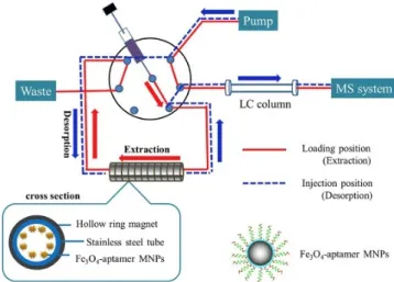
The probe DNA was placed on the surface of MNPs, which produces a rather low RLS signal. If, however, the probe DNA hybridizes with the aptamer against o8dGuo, a sandwich structure will be formed. This results in a significant enhancement of RLS intensity. The aptamer was used as the recognition element to capture o8dGuo. o8dGuo has a stronger affinity for the aptamer than probe DNA, and the conformation of the aptamer therefore switches from a double-stranded to a GQ structure. As a result, MNPs labeled with probe DNA are released, and RLS intensity decreases. The method allows o8dGuo to be detected with a linear response in the 32 pM−12.0 nM concentration range and with a LOD of 11 pM. The MNPs can be reused five times by applying an external magnetic field for recycling. The method was successfully applied to analyze human urine samples for its content of o8dGuo. It was also found that the levels of o8dGuo noticeably increased with the increase of the Air Quality Index, and the method is a viable tool to investigate the relationship between o8dGuo levels and the effect of air pollution. o8dGuo removal is tightly regulated by external and internal stimuli, e.g., ROS accumulation that is affected by air pollution as well (Figure 12) [7].
Figure 11.Gold nanoparticles used in the colorimetric determination of o8dGuo [38].
Int. J. Mol. Sci.2020,21, 4981 11 of 27
3.3.3. Resonance Light Scattering Aptasensor Based on Magnetic Nanoparticles
Magnetic nanoparticles (MNPs) were employed as resonance light scattering (RLS) probes [40].
The probe DNA was placed on the surface of MNPs, which produces a rather low RLS signal. If, however, the probe DNA hybridizes with the aptamer against o8dGuo, a sandwich structure will be formed. This results in a significant enhancement of RLS intensity. The aptamer was used as the recognition element to capture o8dGuo. o8dGuo has a stronger affinity for the aptamer than probe DNA, and the conformation of the aptamer therefore switches from a double-stranded to a GQ structure. As a result, MNPs labeled with probe DNA are released, and RLS intensity decreases.
The method allows o8dGuo to be detected with a linear response in the 32 pM−12.0 nM concentration range and with a LOD of 11 pM. The MNPs can be reused five times by applying an external magnetic field for recycling. The method was successfully applied to analyze human urine samples for its content of o8dGuo. It was also found that the levels of o8dGuo noticeably increased with the increase of the Air Quality Index, and the method is a viable tool to investigate the relationship between o8dGuo levels and the effect of air pollution. o8dGuo removal is tightly regulated by external and internal stimuli, e.g., ROS accumulation that is affected by air pollution as well (Figure12) [7].
Int. J. Mol. Sci. 2020, 21, x FOR PEER REVIEW 11 of 28
Figure 11. Gold nanoparticles used in the colorimetric determination of o8dGuo [38].
3.2.3. Resonance Light Scattering Aptasensor Based on Magnetic Nanoparticles
Magnetic nanoparticles (MNPs) were employed as resonance light scattering (RLS) probes [40].
The probe DNA was placed on the surface of MNPs, which produces a rather low RLS signal. If, however, the probe DNA hybridizes with the aptamer against o8dGuo, a sandwich structure will be formed. This results in a significant enhancement of RLS intensity. The aptamer was used as the recognition element to capture o8dGuo. o8dGuo has a stronger affinity for the aptamer than probe DNA, and the conformation of the aptamer therefore switches from a double-stranded to a GQ structure. As a result, MNPs labeled with probe DNA are released, and RLS intensity decreases. The method allows o8dGuo to be detected with a linear response in the 32 pM−12.0 nM concentration range and with a LOD of 11 pM. The MNPs can be reused five times by applying an external magnetic field for recycling. The method was successfully applied to analyze human urine samples for its content of o8dGuo. It was also found that the levels of o8dGuo noticeably increased with the increase of the Air Quality Index, and the method is a viable tool to investigate the relationship between o8dGuo levels and the effect of air pollution. o8dGuo removal is tightly regulated by external and internal stimuli, e.g., ROS accumulation that is affected by air pollution as well (Figure 12) [7].
Figure 12.Magnetic nanoparticles in the resonance light scattering (RLS) probe for the detection of o8dGuo [40].
3.3.4. UV Radiation
Banyasz et al. [41] have carried out the first study on oxidative damage of human telomere GQs without the mediation of external molecules. The investigation has been performed for GQs formed by folding of heneicosamer d(GGG(TTAGGG)3) single strands in buffered solutions containing Na+ cations (TEL21/Na+). Coupling nanosecond time-resolved spectroscopy and quantum mechanical calculations (TD-DFT), the study focused on the primary species, ejected electrons and guanine radicals, generated upon absorption of UV radiation directly by TEL21/Na+. At 266 nm, corresponding to an energy significantly lower than the guanine ionization potential, the one-photon ionization quantum yield was 4.5× 10⁻³, this value is comparable to that of cyclobutane thymine dimers, the major UV-induced lesions in genomic DNA (quantum yield= (1.1± 0.1)× 10−3). The fate of guanine radicals, generated in an equivalent concentration with that of ejected electrons, has been followed over five orders of magnitude of time. Such a quantitative approach reveals that an important part of radical cation population survives up to a few milliseconds, whereas radical cations produced by chemical oxidants in various DNA systems are known to deprotonate, at most, within a few microseconds. Under the same experimental conditions, neither one-photon ionization nor long-lived radical cations are detected for the telomere repeat d(TTAGGG) in single-stranded configuration, showing that secondary structure plays a key role in these processes. Two types of deprotonated radicals are identified: on the one hand, (G-H2)•radicals (guanine radical deprotonated at the amino group), stable at early times, and on the other hand, (G-H1)•radicals (guanine radical deprotonated at N1, crucially involved in GQ formation), appearing within a few milliseconds and decaying with a
Int. J. Mol. Sci.2020,21, 4981 12 of 27
time constant of∼50 ms (Scheme2). HPLC-coupled mass spectrometry also revealed the presence of o8dGuo; however, o8dGuo is a minor oxidation product in telomeric GQs, the quantum yield of o8dGuo formation ((3.2±0.3)×10−4) is only 7% of the one-photon ionization quantum yield. Still, the quantum yield for o8dGuo formation in telomeric GQs is one order of magnitude higher than that determined for naked genomic DNA [42].
Int. J. Mol. Sci. 2020, 21, x FOR PEER REVIEW 12 of 28
Figure 12. Magnetic nanoparticles in the resonance light scattering (RLS) probe for the detection of o8dGuo [40].
3.2.4. UV Radiation
Banyasz et al. [41] have carried out the first study on oxidative damage of human telomere GQs without the mediation of external molecules. The investigation has been performed for GQs formed by folding of heneicosamer d(GGG(TTAGGG)3) single strands in buffered solutions containing Na+ cations (TEL21/Na+). Coupling nanosecond time-resolved spectroscopy and quantum mechanical calculations (TD-DFT), the study focused on the primary species, ejected electrons and guanine radicals, generated upon absorption of UV radiation directly by TEL21/Na+. At 266 nm, corresponding to an energy significantly lower than the guanine ionization potential, the one-photon ionization quantum yield was 4.5 × 10⁻³, this value is comparable to that of cyclobutane thymine dimers, the major UV-induced lesions in genomic DNA (quantum yield = (1.1 ± 0.1) × 10−3). The fate of guanine radicals, generated in an equivalent concentration with that of ejected electrons, has been followed over five orders of magnitude of time. Such a quantitative approach reveals that an important part of radical cation population survives up to a few milliseconds, whereas radical cations produced by chemical oxidants in various DNA systems are known to deprotonate, at most, within a few microseconds. Under the same experimental conditions, neither one-photon ionization nor long-lived radical cations are detected for the telomere repeat d(TTAGGG) in single-stranded configuration, showing that secondary structure plays a key role in these processes. Two types of deprotonated radicals are identified: on the one hand, (G-H2)• radicals (guanine radical deprotonated at the amino group), stable at early times, and on the other hand, (G-H1)• radicals (guanine radical deprotonated at N1, crucially involved in GQ formation), appearing within a few milliseconds and decaying with a time constant of ∼50 ms (Scheme 2). HPLC-coupled mass spectrometry also revealed the presence of o8dGuo; however, o8dGuo is a minor oxidation product in telomeric GQs, the quantum yield of o8dGuo formation ((3.2 ± 0.3) × 10−4) is only 7% of the one-photon ionization quantum yield. Still, the quantum yield for o8dGuo formation in telomeric GQs is one order of magnitude higher than that determined for naked genomic DNA [42].
Scheme 2. Effect of UV irradiation on G transformations. Adapted with permission from Reference [41]. © 2017, American Chemical Society.
3.3. Hyphenated Techniques
Scheme 2.Effect of UV irradiation on G transformations. Adapted with permission from Reference [41].
©2017, American Chemical Society.
3.4. Hyphenated Techniques
3.4.1. Gas Chromatography–Mass Spectrometry (GC–MS) Analyses
Rozalski et al. [43] have investigated the formation of urinary 5-hydroxymethyluracil (hm5Ura) and OG as potential biomarkers in patients with colorectal cancer using the GC–MS method. These two biomarkers have greater predictive value as a cancer biomarker when used in combination (urinary OG and o8dGuo together with hm5Ura) but are still insufficient as an ideal marker. It is likely that these biomarkers filtered out subjects of the study with higher oxidative stress/chronic inflammation and with a lower ability to remove the deleterious hm5Ura:G mispair. It is also noteworthy that the measurement of DNA lesions in urine can be a good non-invasive biomarker for oxidative stress assessment in colorectal cancer. However, diagnostic performance of this method for early detection is moderate.
3.4.2. G Oxidation Study by EC-LC-MS Chromatography Technique
The EC-LC-MS method integrates electrochemical (EC) oxidation with liquid chromatographic (LC) separation and mass spectrometric (MS) detection. Oberacher et al. [44] have studied the oxidation behavior of nucleic acid derivatives using this approach. It was found that DNA nucleosides were more stable than RNA nucleosides, the electrochemical stability increased in the order guanine≈ acyclovir≈Guo<dGuo≈d(GG)<d(GGG). The mechanistic studies have involved the oxidation of the dinucleotide d(GG) in more detail. In particular, different forms of inter-strand cross-links (d(GG)-H)2, intra-strand cross-links (d(GG)-2H) as well as mono- and di-hydroxylated species ((d(GG)+O), d((G+ O)(G+O)) were detected. d(GG) is first oxidized at a G in a one-electron process to the corresponding radical. Two radicals are combined to form cross-links of the form (d(GG)-H). Intra-strand cross-links were produced at more positive electrochemical potentials than the inter-strand cross-links. In MS/MS,
![Figure 1. ZnO nanoparticles (ZnO@rGO) for the detection of o 8 dGuo. Reprinted with permission from Reference [26], © 2018, Elsevier.](https://thumb-eu.123doks.com/thumbv2/9dokorg/782014.36037/4.892.233.661.823.1074/figure-zno-nanoparticles-detection-reprinted-permission-reference-elsevier.webp)
![Figure 5. A o 8 dGuo-specific aptamer construct. Reprinted with permission from Reference [33]](https://thumb-eu.123doks.com/thumbv2/9dokorg/782014.36037/6.892.169.726.678.930/figure-dguo-specific-aptamer-construct-reprinted-permission-reference.webp)
![Figure 7. Electrochemiluminescent (ECL) biosensor developed by Lv et al. [35]. Reprinted with permission from Reference [35]](https://thumb-eu.123doks.com/thumbv2/9dokorg/782014.36037/8.892.177.720.131.401/figure-electrochemiluminescent-ecl-biosensor-developed-reprinted-permission-reference.webp)
![Figure 9. Three-dimensional (3D) nanomachine-assisted detection of o 8 dGuo lesions [37]](https://thumb-eu.123doks.com/thumbv2/9dokorg/782014.36037/9.892.177.720.434.769/figure-dimensional-d-nanomachine-assisted-detection-dguo-lesions.webp)
![Figure 10. A luminescent paper-based device for the visual detection of o 8 dGuo. Reprinted with permission from Reference [39]](https://thumb-eu.123doks.com/thumbv2/9dokorg/782014.36037/10.892.258.629.131.481/figure-luminescent-device-visual-detection-reprinted-permission-reference.webp)
![Figure 11. Gold nanoparticles used in the colorimetric determination of o 8 dGuo [38]](https://thumb-eu.123doks.com/thumbv2/9dokorg/782014.36037/11.892.151.744.461.673/figure-gold-nanoparticles-used-colorimetric-determination-o-dguo.webp)
![Figure 12. Magnetic nanoparticles in the resonance light scattering (RLS) probe for the detection of o 8 dGuo [40]](https://thumb-eu.123doks.com/thumbv2/9dokorg/782014.36037/12.892.271.619.249.597/figure-magnetic-nanoparticles-resonance-light-scattering-probe-detection.webp)