protocols on oxidative status and DNA damage in red-eared sliders (Trachemys scripta elegans) undergoing endoscopic coeliotomy
MILAN DOŠENOVI C
1, MILENA RADAKOVI C
2p, MILO Š VU CI CEVI C
1, BRANISLAV VEJNOVI C
3, MAJA VASILJEVI C
5, DARKO MARINKOVI C
4and ZORAN STANIMIROVI C
61Department of Equine, Small Animal, Poultry and Wild Animal Diseases, Faculty of Veterinary Medicine, University of Belgrade, Belgrade, Serbia
2Department of Pathophysiology, Faculty of Veterinary Medicine, University of Belgrade, Bulevar oslobodenja 18, 11000 Belgrade, Serbia
3Department of Economics and Statistics, Faculty of Veterinary Medicine, University of Belgrade, Belgrade, Serbia
4Department of Pathology, Faculty of Veterinary Medicine, University of Belgrade, Belgrade, Serbia
5Department of Surgery, Orthopaedics and Ophthalmology, Faculty of Veterinary Medicine, University of Belgrade, Belgrade, Serbia
6Department of Biology, Faculty of Veterinary Medicine, University of Belgrade, Belgrade, Serbia
Received: June 1, 2020 • Accepted: November 16, 2020 Published online: January 27, 2021
ABSTRACT
The aim of this study was to assess how red-eared sliders (Trachemys scripta elegans) respond to anaesthesia itself and coelioscopy. For that purpose, the turtles were anaesthetised with ketamine–
medetomidine or propofol, and the activities of superoxide dismutase (SOD), catalase (CAT) and glutathione S-transferase (GST) and the level of malondialdehyde (MDA) were determined by spec- trophotometry. The possible genotoxic effects of the anaesthetic agents were estimated by comet assay.
A total of 24 turtles were included in this study. The animals were divided into four groups according to the anaesthetic protocol and according to whether endoscopy would be performed. Significantly decreased activities of CAT were found only in the propofol group and in turtles undergoing coelio- scopy. Both anaesthetic protocols induced significantly increased MDA levels, while no differences were observed after the intervention. A significant increase in GST activity was detected in turtles after both anaesthetic protocols, but after coelioscopy significant changes in GST activity were found only in the propofol group. However, no differences in SOD activity and no DNA damages were detected in either group. Thesefindings suggest that ketamine–medetomidine may be more suitable anaesthetic agents in red-eared sliders than propofol.
KEYWORDS
red-eared slider, turtle, anaesthesia, coelioscopy
INTRODUCTION
The red-eared slider (Trachemys scripta elegans) is a semiaquatic turtle belonging to the family Emydidae. Within their natural range, red-eared sliders live in a wide variety of freshwater habitats including rivers, ditches, swamps, lakes and ponds. Red-eared sliders have been the most popular turtles in the pet trade (Global Invasive Species Database, 2020). They
Acta Veterinaria Hungarica
68 (2020) 4, 337–344 DOI:
10.1556/004.2020.00058
© 2021 Akademiai Kiado, Budapest
ORIGINAL RESEARCH PAPER
pCorresponding author.
E-mail:mradakovic@vet.bg.ac.rs, vucicevic@vet.bg.ac.rs
became favoured because of their small size, low price and relatively simple husbandry requirements. Also, the species is widely used as a model in biomedical studies because of its extreme tolerance of oxygen deprivation that supports sur- vival during diving and underwater hibernation (Warren et al., 2006). Therefore, there is an increasing need to anaesthetise these turtles for examinations, diagnostics and surgical procedures (Greer et al., 2001). Endoscopic tech- niques are increasingly used in veterinary medicine (Her- nandez-Divers et al., 2005) because they provide excellent visualisation and are minimally invasive. Coelomic endos- copy (coelioscopy) allows visualisation of almost all internal organs (except the heart and the lungs) in a turtle.
Along with the stress of a surgical trauma, anaesthetic agents are important factors that can disrupt immunological and antioxidant defence systems. Ketamine–medetomidine combinations have been used widely for the chemical re- straint of domestic and non-domestic mammals (Jalanka and Roeken, 1990). Many studies have shown that this combination provides effective anaesthesia to red-eared sliders as well (Greer et al., 2001; Tatli et al., 2016). In addition, the use of propofol has been increasing in veteri- nary medicine for reptiles due to the fast induction and recovery. Studies in mammals have shown that combina- tions of ketamine–medetomidine and propofol have an impact on the oxidant-antioxidant system (de Oliveira et al., 2009; Lee, 2011; Adaramoye et al., 2013). However, the ef- fects of anaesthetics on the oxidative status in turtles have been poorly studied (Valcic et al., 2019). Cells have devel- oped a defence system responsible for neutralising the toxic effects of oxygen radicals, namely antioxidant systems that consist of enzymes such as superoxide dismutase (SOD), catalase (CAT) and glutathione S-transferase (GST) (Halli- well and Gutteridge, 2007). SOD forms the front line of defence that is involved in the detoxification of superoxide anion (O2–) to hydrogen peroxide (H2O2), while CAT is involved in the conversion of H2O2 to water and oxygen.
GST plays a role in detoxification of the exogenous and endogenous electrophiles, as well as the products of oxida- tive stress (Alfadda and Sallam, 2012). Under normal con- ditions, reactive oxygen species (ROS) are maintained at low levels by antioxidants participating in redox homoeostasis.
However, when the generation of ROS overcomes the ca- pacity of the antioxidant defence systems, an excessive level of ROS may interact with macromolecules (proteins, lipids and nucleic acids) and cause damage, which leads to meta- bolic dysfunction and cell death (Halliwell and Gutteridge, 2007). Lipid peroxidation is a major source of cell damage, resulting in cellular injury and death caused by the inter- action of ROS with unsaturated bonds in membrane lipids.
Therefore, the damaged lipids can be detected by the assessment of the produced malondialdehyde (MDA), the final product of lipid peroxidation. Also, during oxidative stress ROS can oxidise DNA, which can lead to several types of DNA damage, including single- and double-strand breaks. These oxidative lesions can be identified using the comet assay, an effective method for detecting DNA damage within cells (Collins, 2014). Therefore, oxidative stress can
be estimated by measuring the activity of key antioxidants and examining oxidation target products, including prod- ucts of lipid peroxidation and DNA oxidation, which pro- vide information about oxidative status and free-radical damage (Allaouchiche et al., 2001; Strasser et al., 2012;
Adaramoye et al., 2013).
For all these reasons, the aim of this study was to assess how red-eared sliders respond to anaesthesia itself and coelioscopy using two anaesthetic protocols (ketamine–
medetomidine versus propofol). The activities of SOD, CAT and GST and the levels of MDA were determined. Moreover, possible DNA-damaging effects of the anaesthetics were estimated by the comet assay. Based on the results obtained, a specific recommendation may be made for a safe anaes- thetic protocol for endoscopy in red-eared sliders.
MATERIALS AND METHODS
Animals
From September 2017 to March 2020, 24 healthy red-eared sliders were admitted to the Clinic for Small Animals, Fac- ulty of Veterinary Medicine, University of Belgrade. Twelve of them were brought in for endoscopy assessment of their reproductive status. The other 12 animals were brought in for a regular clinical check but had to be anaesthetised because they were not co-operative. The turtles were of unknown age, weighed from 1.20 to 2.0 kg. There were 7 males and 17 females. Prior to the examination, the animals were kept under controlled environment conditions in a water tank at 228C water temperature and 21–258C room temperature. The acclimatisation time was 14 days. All an- imals were fasted for 24 h prior to the procedure.
Groups
The study included four groups consisting of 6 animals each.
The first (KMC) and the second (PC) group consisted of individuals anaesthetised with ketamine–medetomidine and propofol, respectively, and subsequently subjected to coe- lioscopy. The third (KM) and fourth (P) group consisted of individuals that were only anaesthetised with ketamine– medetomidine or propofol.
Anaesthetic protocols
Two different anaesthetic protocols were used. The first protocol was applied in Group KMC and Group KM and involved using ketamine (Ketamidor 10% Richter Pharma, Austria) 20 mg/kg and medetomidine (Domitor, Orion Pharma, Finland) 0.2 mg/kg, both mixed in a single syringe and administered intravenously (IV) in the subcarapacial venous plexus. The second protocol was applied in Group PC and Group P and involved the administration of pro- pofol (Diprivan 10%, AstraZeneca UK Limited, United Kingdom) 10 mg/kg IV in the subcarapacial venous plexus.
In Group KMC and Group KM, medetomidine was reversed with atipamezole (Antisedan, Orion Pharma, Finland) 0.2
mg/kg, given intramuscularly (IM) 1 h after the application of the anaesthetics. All animals were given 20 mL of saline subcutaneously in the prefemoral fossa, contralaterally to the incision, meloxicam (Movalis, Boehringer Ingelheim, Ger- many) 0.5 mg/kg IM and enrofloxacin (Baytril, Bayer Ani- mal Health, Germany) 10 mg/kg both IM in the forelimbs postoperatively. The ambient temperature during coelio- scopy was 21–25 8C. Monitoring was based on reflex monitoring and heart rate recording.
Coelioscopy
The depth of anaesthesia was assessed by monitoring the presence or absence of the righting reflex, the head or limb withdrawal reflex and the absence of response to skin inci- sion. After the surgical level of anaesthesia was achieved, the animals were prepared for endoscopy. They were placed in lateral recumbency with one hind limb retracted and secured to expose the prefemoral fossa. Following aseptic prepara- tion of the prefemoral area and the surrounding shell, a 2- to 4-cm-long longitudinal incision was made in the centre of the fossa using surgical scalpel No. 15 (Romed, Holland).
The subcutaneous fat and connective tissues were bluntly dissected with haemostatic forceps (031113, Medgyn Prod- ucts, Illinois, USA). This dissection was continued to the level of the coelomic aponeurosis. Endoscopy was performed using an otoscope for small animal practice of the following dimensions: diameter 5 mm, length 8.5 cm, with a working channel diameter of 5 Fr (67260OSA, Karl Storz Veterinary Endoscopy America Inc., KSVEA) and connected to a xenon light source (201326-20, KSVEA). Recordings were made using a Karl Storz veterinary video camera (692360-20, KSVEA). On completion of the coelioscopy, the coelomic aponeurosis and peritoneum were closed with 2.0 absorbable suture (PolysorbTM, Covidien Ltd., Ireland) in a continuous pattern. Skin closure was done using 2.0 monofilament- polypropylene (SurgiproÔ, Covidien Ltd., Ireland) sutures in a simple interrupted pattern. The cloacal temperature was measured with a thermometer probe (Testo 925, Testo GmbH, Austria).
Blood sampling
One day prior to anaesthesia, blood samples (2 mL) with heparin were collected from the subcarapacial venous plexus in order to determine the baseline levels of oxidative stress indicators and of the total comet score. The second sampling took place 3 h after the anaesthetics had been administered, and approximately 2 h after the coelioscopy had been completed. For the comet assay 100
m
L of whole blood from each turtle was used and the remaining blood was centri- fuged at 1,5003gfor 15 min, and the plasma was extracted.The samples were stored at20 8C until further analysis.
Oxidative stress parameters
The disturbance of oxidative status is most commonly measured via alterations in the activity of CAT, SOD and GST. CAT activity was determined by the rate of
decomposition of H2O2 at 240 nm (Aebi, 1984). Enzyme activity was defined as 1 mmol H2O2 decomposed per minute. CAT activity was expressed in U/ml using molar extinction coefficient (43.6 M1 cm1). The estimation of SOD activity was based on the ability of SOD to inhibit autoxidation of epinephrine to adrenochrome (Misra and Fridovich, 1972). The reaction was measured at 480 nm for 3 min. One unit of SOD was defined as the amount of enzyme required to inhibit the rate of epinephrine autoxidation by 50% at 308C, and the results were expressed in U/mL. GST activity was determined using 1-chloro-2,4-dinitrobenzene (CDNB) as substrate in the presence of reduced glutathione (GSH) (Habig et al., 1974). The change in absorbance was measured at 340 nm for 3 min. The results are expressed in U/mL. To quantify ROS-induced damage, lipid peroxidation was estimated through the concentrations of MDA. The level of MDA in the plasma was determined by measuring the formation of thiobarbituric acid-reactive substances (Girotti et al., 1991). The absorbance of the coloured com- plex was measured by spectrophotometry at a wavelength of 535 nm and expressed in nmoL/g Hb. All the methods used were spectrophotometric and were performed with a Biobase BK-7390 UV-VIS spectrophotometer (Agilent Technologies, USA). The averages of triplicate analyses are reported.
Determination of DNA damage by comet assay
This method identifies the head of the comet as a spherical mass of undamaged DNA, and the damaged DNA (DNA loops around strand breaks) streams out from the head as a comet tail. The alkaline comet assay was performed with slight modifications (Tice et al., 2000). Briefly, blood was diluted in phosphate-buffered saline (PBS) (1:100), embedded in agarose (0.5% low-melting agarose) and layered onto precoated slides (1% normal melting agarose).
The slides were immersed in cold lysis solution (2.5 M NaCl, 100 mM EDTA, 10 mM Tris, 1% Triton X–100, 10% DMSO, at pH 10) overnight at 48C. The slides were then placed in a horizontal gel electrophoresis chamber and submerged in freshly made cold electrophoresis buffer (pH > 13) for 20 min to allow unwinding of DNA. Thereafter, the slides were electrophoresed for 20 min at 25 V and 300 mA. Then, the slides were neutralised with Tris-HCl buffer (pH 7.5) for 15 min. Finally, the slides were stained with ethidium bromide and analysed under afluorescence microscope (AxioImager Z1, Carl Zeiss; excitationfilter, 515–560 nm; emissionfilter, 590 nm). Comets were scored visually and classified intofive categories corresponding to the amounts of DNA in the tails (Anderson et al., 1994). Comet scores were calculated by multiplying the number of damaged cells per specimen by the value of the respective comet class (0–4) and expressed as the total comet score (TCS). Before applying the comet assay, manual cell count and an estimate of cell viability were performed by Trypan blue exclusion test.
Statistical analysis
Depending on the values of coefficients of variation (CV), data for TCS were homogeneous (CV <30%), but data for
SOD, CAT, GST and MDA were heterogeneous (CV >30%), so adequate transformations log10(y þ 1) need to be applied. For all parameters, the groups were compared by two-way ANOVA with repeated measures in one factor, followed by Tukey’s multiple comparison test. Data were presented as mean ± standard deviation (mean ± SD).
Significance was estimated atP < 0.05 andP < 0.01 signif- icance levels. Statistical analysis was done with GraphPad Prism 6 software (GraphPad, San Diego, CA, USA).
RESULTS
A surgical level of anaesthesia was achieved using both protocols. The animals were in total anaesthesia for approximately 30 min (27.1–36 min). After atipamezole had been administered, the animals in the KM and KMC groups awakened from general anaesthesia in 2–10 min, without any complications. The body temperature during coelio- scopy was constant (218C).
Oxidative stress parameters
No differences in SOD activity (P> 0.05) were observed after the administration of propofol and ketamine–medetomidine compared to baseline values (Fig. 1). Also, no changes in SOD activity (P > 0.05) were found in turtles undergoing coelioscopy with either type of protocol applied.
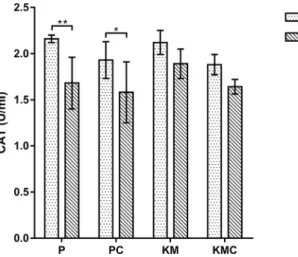
In comparison to the initial values a significant decrease (P< 0.01) in CAT activity was found in thePgroup after the use of the anaesthetic, while in the KM group no differences (P> 0.05) were recorded (Fig. 2). Similarly, in the PC group CAT activity was significantly altered (P < 0.05) and the effect was less profound than in the P group. In the KMC
group there were no significant differences (P > 0.05) in CAT values during the study.
By the analysis of GST activity a significant increment (P
< 0.01) was observed in turtles after the use of propofol and ketamine–medetomidine. GST activity was significantly increased (P < 0.01) in the PC group after the intervention (Fig. 3). However, in turtles that underwent coelioscopy with ketamine–medetomidine no significant differences in GST activity were noticed (P> 0.05).
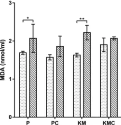
MDA levels were significantly higher (P< 0.05,P< 0.01) in the P and KM groups compared to the initial values (Fig. 4). In contrast, after coelioscopy with two different
Fig. 1.Superoxide dismutase (SOD) activity in the turtles before and after the different anaesthetic protocols. P–propofol (n56);
PC–propofol with coelioscopy (n56); KM–ketamine–mede- tomidine (n56); KMC–ketamine–medetomidine with coelio- scopy (n56). Data are presented as mean±standard deviation
(SD)
Fig. 2.Catalase (CAT) activity in the turtles before and after the different anaesthetic protocols. P–propofol (n56); PC–propofol with coelioscopy (n56); KM–ketamine–medetomidine (n56);
KMC–ketamine–medetomidine with coelioscopy (n56). Data are presented as mean±standard deviation (SD).pP< 0.05,ppP<
0.01 versus initial time
Fig. 3.Glutathione S-transferase (GST) activity in the turtles before and after the different anaesthetic protocols. P–propofol (n56);
PC–propofol with coelioscopy (n56); KM–ketamine–mede- tomidine (n56); KMC–ketamine–medetomidine with coelio- scopy (n56). Data are presented as mean±standard deviation
(SD).ppP< 0.01 versus initial time
protocols, no significant changes in MDA levels were observed (P > 0.05).
Comet assay
In all blood samples, cell viability determined by the Trypan blue exclusion test was acceptable (over 90%), indicating valid conditions for the application of the comet assay in red-eared sliders. The total comet score (TCS), which re- flects the DNA damage, was not significantly altered (P >
0.05) in any of the turtle groups after the treatment as compared to the values at the beginning of the research (Fig. 5).
DISCUSSION
Apart from appropriate equipment and medical expertise, safe anaesthesia implies that anaesthetics are effective and do not produce any unwanted effects on vital functions. Not only the anaesthetic agents but also surgical stress itself can disturb antioxidant defence systems (Kotzampassi et al., 2009). Therefore, to elucidate whether anaesthesia or coe- lioscopy is the main cause of potential oxidative stress, the oxidative status and the level of DNA damage were deter- mined in turtles having undergone anaesthesia with or without intervention. Since there are similar studies on mammals but only one on turtles (Valcic et al., 2019), the effects of ketamine–medetomidine combination and pro- pofol were compared and discussed.
In the current study, SOD activity in the propofol and ketamine–medetomidine groups was not sensitive to expo- sure to the anaesthetics used. Also, no difference in SOD activity was seen after surgery in the groups that underwent coelioscopy. This is in accordance with the study by Allaouchiche et al. (2001)andAdaramoye et al. (2013), who reported no significant changes in SOD activity during the exposure of pigs and rats to propofol. Based on our results for other oxidative stress markers, the possibility of increased levels of some other type of reactive oxygen species cannot be excluded. In line with this, SOD inactivation by hydrogen peroxide was observed (Pigeolet et al., 1990).
Unexpectedly, decreased CAT activity was detected in turtles after exposure to propofol. After coelioscopy with propofol anaesthesia, a less pronounced but significant decrease in CAT activity was detected. In the ketamine–medetomidine groups, no differences in CAT activity were noticed. The results presented above lead to the conclusion that the adaptive antioxidant response of SOD is not accompanied by CAT activity. The decline in CAT activities may have resulted from ROS, especially of H2O2overproduction after propofol administration. We suppose that H2O2occurred as a product of macrophage activation due to oxidative burst after the administration of anaesthetics.Lee (2011)reported that in dogs the plasma total antioxidant status (TAS) levels significantly decreased whereas the plasma total oxidant status (TOS) and oxidative stress index (OSI) levels increased with time in the propofol and thiopental groups.
An investigation estimated the effects of two different anaesthesia protocols (medetomidine–tiletamine and zola- zepam combination versus isoflurane) on oxidative stress in beagle dogs (Choi et al., 2012). The decrease in activities of SOD and CAT compared to baseline values was found in both groups.
In modern anaesthesiology, there is a growing interest in the investigation of GST activity (Mikstacki et al., 2015).
Therefore, increased GST activity points to an adaptive response to the occurrence of oxidative stress. In the present investigation, ketamine–medetomidine increased GST ac- tivity in red-eared sliders. The same effect in GST activity was seen after intervention with propofol anaesthesia.
Adaramoye et al. (2013)reported that propofol significantly Fig. 5.DNA damage expressed as total comet score (TCS) in the
turtles before and after the different anaesthetic protocols. P– propofol (n56); PE–propofol with coelioscopy (n56); KM– ketamine–medetomidine (n56); KMC–ketamine-medetomidine with coelioscopy (n56). Data are presented as mean± standard
deviation (SD)
Fig. 4.Malondialdehyde (MDA) level in the turtles before and after the different anaesthetic protocols. P–propofol (n56); PC– propofol with coelioscopy (n56); KM–ketamine–medetomidine (n56); KMC–ketamine–medetomidine with coelioscopy (n56).
Data are presented as mean±standard deviation (SD).pP< 0.05,
ppP< 0.01 versus initial time
increased GST activity in Wistar rats, which is in agreement with the results obtained in this study. In addition, propofol induced a rise in GST activity in the gills and blood cells of exposedfish (Gressler et al., 2016). However, it was found that prolonged propofol anaesthesia had no effect on GST during and after extended surgery in humans (Murray et al., 1994). In a previous study, GST activity remained unchanged in rats exposed to ketamine with disulfiram, while ketamine alone decreased its activity in heart tissue as compared to the negative control (Cetin et al., 2015).
Therefore, different responses of GST activity indicate its dependency on the type of xenobiotics, species and the tissues that are exposed (Oruç and Uner, 2000; Ferrari€ et al., 2007). We presume that GST activity is induced by anaesthetics, thus intensifying the detoxifying potential in turtles.
In this research, MDA levels significantly increased in the propofol and ketamine–medetomidine group after anaesthesia. However, changes in MDA levels after the intervention with two different anaesthetic protocols were not significantly different from the initial values. This result suggests that coelioscopy did not contribute to the increased lipid peroxidation. We suppose that the elevated MDA level is a consequence of decreased antioxidant activity which could not be reduced due to the disrupted antioxidative mechanism. The results of the present study concur with the findings of an investigation in which the administration of ketamine induced a significant rise in lipid peroxidation in rats (de Oliveira et al., 2009; Gazal et al., 2014; Ahiskalioglu et al., 2015). In another investigation, the application of ketamine alone increased lipid peroxidation, but in combi- nation with thiopental it did not induce detectable changes in MDA levels (Ahiskalioglu et al., 2018). Increased MDA levels were noticed 2 h after surgery with propofol (Han et al., 2015). Alipour et al. (2018)reported that total intra- venous anaesthesia using propofol can increase MDA pro- duction. Contrary to the results obtained in the current study, it was shown in some research that propofol caused a significant reduction in lipid peroxidation (Allaouchiche et al., 2001; Akin et al., 2015). However, other studies revealed no decrease in lipoperoxidation during propofol anaesthesia (Braz et al., 2009; Adaramoye et al., 2013). It was suggested that differences in oxidative stress response be- tween the combination of drugs and anaesthetics given alone are the consequence of the different duration of anaesthesia.
ROS have the ability to react with all components of DNA and cause various lesions such as oxidised bases, abasic sites and strand breaks. Modifications of DNA can lead to mutations and genomic instability, and contribute to carci- nogenesis. In this study the comet assay was used for the detection of DNA damage. There are only a few reports which applied this test on turtles and in all of them their health status was checked (Caliani et al., 2014; Zapata et al., 2016). By comet analysis, an increase in DNA damage was not detected in any of the turtle groups after the adminis- tration of the anaesthetic drugs. Besides, no significant change in the level of DNA damage was detected after anaesthesia followed by coelioscopy with different protocols.
In this regard, other authors reported no damaging effects of ketamine, diazepam, xylazine and isoflurane combination in ponies and horses (Strasser et al., 2012). Also, anaesthesia maintained with propofol did not lead to genotoxicity (Braz et al., 2015). A possible explanation for the absence of DNA damage is that we assessed whole blood cells, which are equipped with powerful antioxidant defence systems (Bas- kurt and Yavuzer, 1994). Therefore, it is possible that, due to sufficient protection, ROS were not able to cause increased DNA damage.
The results obtained in this study indicate that propofol has a more pronounced effect on disrupting the oxidation– reduction balance in red-eared sliders than ketamine– medetomidine does. The analysis of oxidative stress pa- rameters and DNA damage in the groups that underwent coelioscopy leads to the conclusion that the intervention itself is not the main cause of oxidative stress. Significant changes in enzymes (CAT and GST) found in the turtles undergoing coelioscopy could rather be a consequence of the applied anaesthetic agents than a cause of altered antioxi- dant mechanism. Given that both protocols produced a similar effect, it can be assumed that several mechanisms contribute to the increased production of ROS. It was found that propofol and ketamine–medetomidine–atropine induce hyperglycaemia in animals (Zuurbier et al., 2008; Maeda et al., 2018). In the perioperative period, an increase in blood glucose level may occur due to stress hormones and in- flammatory mediators. There is evidence that hyper- glycaemia may result in accelerating the production of ROS (Robertson et al., 2003). Therewith, non-enzymatic glycation of SOD renders the antioxidant inactive (Arai et al., 1987).
Excessive ROS levels may cause a prolonged recovery from anaesthesia and post-anaesthetic complications in the functional ability of vital organs (Brasil et al., 2006). In the current study, altered antioxidant activity was more noticeable in the propofol group than in the ketamine–
medetomidine group.Gnanasekar and Vijayalakshmi (2016) reported that the antioxidant level was significantly higher in ketamine anaesthesia when compared to propofol, revealing a higher production of oxygen free radicals due to lower tissue perfusion and the activation of hepatic cytochrome P450. Another possible explanation might be related to the fact that we applied the combination of ketamine and medetomidine and it is possible that this interaction may have influenced the side effects of both anaesthetics.
Although both of these anaesthetic protocols are widely used in red-eared sliders (Greer et al., 2001; Ziolo and Bertelsen, 2009) with no apparent adverse effects and are therefore considered safe, these findings suggest that ketamine–
medetomidine may be more suitable for anaesthesia in red- eared sliders than propofol, since this combination resulted in a lower degree of oxidative stress.
ACKNOWLEDGEMENTS
The study was supported by the Ministry of Education, Science and Technological Development of the Republic of
Serbia (Grant no. III46002 and Contract no. 451-03-68/
2020-14/200143)
REFERENCES
Adaramoye, O. A., Akinwonmi, O. and Akanni, O. (2013): Effects of propofol, a sedative-hypnotic drug, on the lipid profile, antioxidant indices, and cardiovascular marker enzymes in Wistar rats. ISRN Pharmacol.220, 76–81.
Aebi, H. (1984): Catalasein vitro.Methods Enzymol.105, 121–126.
Ahiskalioglu, A., Ince, I., Aksoy, M., Ahiskalioglu, E. O., Comez, M., Dostbil, A., Celik, M., Alp, H. H., Coskun, R., Taghiza- dehghalehjoughi, A. and Suleyman, B. (2015): Comparative investigation of protective effects of metyrosine and metoprolol against ketamine cardiotoxicity in rats. Cardiovasc. Toxicol.15, 336–344.
Ahiskalioglu, E. O., Aydin, P., Ahiskalioglu, A., Suleyman, B., Kuyrukluyldiz, U., Kurt, N., Altuner, D., Coskun, R. and Suleyman, H. (2018): The effects of ketamine and thiopental used alone or in combination on the brain, heart, and bronchial tissues of rats. Arch. Med. Sci.14, 645–654.
Akin, M., Ayoglu, H., Okyay, D., Ayoglu, F., G€ur, A., Can, M., Yurtlu, S., Hancı, V., K€uç€ukosman, G. and Turan, I. (2015):
Effects of various anesthesia maintenance on serum levels of selenium, copper, zinc, iron and antioxidant capacity. Rev. Bras.
Anestesiol.65, 51–60.
Alfadda, A. A. and Sallam, R. M. (2012): Reactive oxygen species in health and disease. J. Biomed. Biotechnol.2012, 936486.
Alipour, F., Emami, M. R. and Mohri, M. (2018): Endocrine and oxidative stress characteristics in different anesthetic methods during pneumoperitoneum in dogs. Comp. Clin. Pathol.27, 1667–1673.
Allaouchiche, B., Debon, R., Goudable, J., Chassard, D. and Duflo, F. (2001): Oxidative stress status during exposure to propofol, sevoflurane and desflurane. Anesth. Analg.93, 981–985.
Anderson, D., Yu, T. W., Philips, B. J. and Schmezer, P. (1994): The effect of various antioxidants and other modifying agents on oxygen-radical-generated DNA damage in human lymphocytes in the COMET assay. Mutat. Res.307, 261–271.
Arai, K., Maguchi, S., Fujii, S., Ishibashi, H., Oikawa, K. and Taniguchi, N. (1987): Glycation and inactivation of human Cu- Zn superoxide dismutase. J. Biol. Chem.262, 16969–16972.
Baskurt, O. K. and Yavuzer, S. (1994): Some hematological effects of oxidants. In: Nriagu, J. O. and Simmons, M. (eds) Envi- ronmental Oxidants: Advances in Environmental Science and Technology. John Wiley, New York. pp. 405–423.
Brasil, L. J., San-Miguel, B., Kretzmann, N. A., Gomes Do Amaral, J. L., Zettler, C. G., Marroni, N., Gonzalez-Gallego, J. and Tu~non, M. J. (2006): Halothane induces oxidative stress and NF-KB activation in rat liver: protective effective of propofol.
Toxicology227, 53–61.
Braz, M. G., Braz, L. G., Freire, C. M., Lucio, L. M., Braz, J. R., Tang, G., Salvadori, D. M. and Yeum, K. J. (2015): Isoflurane and propofol contribute to increasing the antioxidant status of pa- tients during minor elective surgery: a randomized clinical study. Medicine94(31) e12662015.
Braz, M. G., Magalh~aes, M. R., Salvadori, D. M., Ferreira, A. L., Braz, L. G., Sakai, E. and Braz, J. R. (2009): Evaluation of DNA damage and lipoperoxidation of propofol in patients undergo- ing elective surgery. Eur. J. Anaesthesiol.26, 654–660.
Caliani, I., Campani, T., Giannetti, M., Marsili, L., Casini, S. and Fossi, M. C. (2014). First application of comet assay in blood cells of Mediterranean loggerhead sea turtle(Caretta caretta).
Mar. Environ. Res.96, 68–72.
Cetin, N., Suleyman, B., Altuner, D., Kuyrukluyildiz, U., Ozcicek, F., Coskun, R. and Kurt, N. (2015): Effect of disulfiram on ketamine-induced cardiotoxicity in rats. Int. J. Clin. Exp. Med.
8, 13540–13547.
Choi, K. H., Lee, J. Y., Jeong, S. M. and Kim, M. C. (2012):
Oxidative effects of isoflurane and medetomidine-tiletamine/
zolazepam combination in Beagle dogs. J. Vet. Clin.29, 119–
123.
Collins, A. R. (2014): Measuring oxidative damage to DNA and its repair with the comet assay. Biochim. Biophys. Acta1840, 794–
800.
de Oliveira, L., Spiazzi, C. M. S., Bortolin, T., Canever, L., Petro- nilho, F., Goncalves Mina, F., Dal-Pizol, F., Quevedo, J. and Zugno, A. I. (2009): Different sub-anesthetic doses of ketamine increase oxidative stress in the brain of rats. Prog. Neuro-Psy- chopharmacol. Biol. Psychiatry33, 1003–1008.
Ferrari, A., Venturino, A. and de D’Angelo, A. M. P. (2007): Effects of carbaryl and azinphos methyl on juvenile rainbow trout (Oncorhynchus mykiss)detoxifying enzymes. Pestic. Biochem.
Physiol.88, 134–142.
Gazal, M., Valente, M. R., Acosta, B. A., Kaufmann, F. N., Bra- ganhol, E., Lencina, C. L., Stefanello, F. M., Ghisleni, G. and Kaster, M. P. (2014): Neuroprotective and antioxidant effects of curcumin in a ketamine-induced model of mania in rats. Eur. J.
Pharmacol.724, 132–139.
Girotti, M. J., Khan, N. and McLellan, B. A. (1991): Early mea- surement of systemic lipid peroxidation products in the plasma of major blunt trauma patients. J. Trauma Acute Care Surg.31, 32–35.
Global Invasive Species Database (2020) Species profile:Trachemys scripta elegans. Downloaded from http://www.iucngisd.org/
gisd/speciesname/Trachemysþscriptaþelegans on 04-03-2020.
Gnanasekar, R. and Vijayalakshmi, R. (2016): Response to oxidative stress during surgery under ketamine/propofol anaesthesia in acepromazine-xylazine premedicated horses. IJSRP6, 459–462.
Greer, L. L., Jenne, K. J. and Diggs, H. E. (2001): Medetomidine- ketamine anesthesia in red-eared slider turtles (Trachemys scripta elegans). J. Am. Assoc. Lab. Anim. Sci.40, 8–11.
Gressler, L. T., Sutili, F. J., Loebens, L., Saccol, E. M. H., P^es, T. S., Parodi, T. V., da Costa, S. T., Pavanato, M. A. and Baldisserotto, B. (2016): Histological and antioxidant responses in Rhamdia quelensedated with propofol. Aquacult. Res.47, 2297–2306.
Habig, W. H., Pabst, M. J. and Jakoby, W. B. (1974): Glutathione S- transferases. Thefirst enzymatic step in mercapturic acid for- mation. J. Biol. Chem.249, 7130–7139.
Halliwell, B. and Gutteridge, J. M. C. (2007): Cellular responses to oxidative stress: adaptation, damage, repair, senescence and death. In: Halliwell, B. and Gutteridge, J. M. C. (eds) Free Radicals in Biology and Medicine. ed. Oxford University Press, New York. pp. 187–267.
Han, C., Ding, W., Jiang, W., Chen, Y. U., Hang, D., Gu, D., Jiang, G., Tan, Y., Ge, Z. and Ma, T. (2015): A comparison of the effects of midazolam, propofol and dexmedetomidine on the antioxidant system: a randomized trial. Exp. Ther. Med. 9, 2293–2298.
Hernandez-Divers, S. J., Hernandez-Divers, S. M., Wilson, G. H.
and Scott, S. J. (2005): A review of reptile diagnostic coelio- scopy. J. Herpetol. Med. Surg.15, 16–31.
Jalanka, H. H. and Roeken, B. O. (1990): The use of medetomidine, medetomidine-ketamine combinations, and atipamezole in nondomestic mammals: a review. J. Zoo Wildl. Med.21, 259–
282.
Kotzampassi, K., Kolios, G., Manousou, P., Kazamias, P., Para- mythiotis, D., Papavramidis, T. S. and Eleftheriadis, E. (2009):
Oxidative stress due to anesthesia and surgical trauma:
importance of early enteral nutrition. Mol. Nutr. Food Res.53, 770–779.
Lee, J. Y. (2011): Oxidative stress due to anesthesia and surgical trauma and comparison of the effects of propofol and thio- pental in dogs. J. Vet. Med. Sci.74, 663–665.
Maeda, K., Iwasaki, M., Itou, Y., Iwai, S. and Okano, S. (2018):
Effect of propofol continuous-rate infusion on intravenous glucose tolerance test in dogs. Vet. Sci.5, 43.
Mikstacki, A., Zakerska-Banaszak, O., Skrzypczak-Zielinska, M., Tamowicz, B., Szalata, M. and Slomski, R. (2015): Glutathione S-transferase as a toxicity indicator in general anesthesia: ge- netics and biochemical function. J. Clin. Anesth.27, 73–79.
Misra, H. P. and Fridovich, I. (1972): The role of superoxide anion in the autoxidation of epinephrine and a simple assay for su- peroxide dismutase. J. Biol. Chem.247, 3170–3175.
Murray, J. M., Phillips, A. S. and Fee, J. P. (1994): Comparison of the effects of isoflurane and propofol on hepatic glutathione-S- transferase concentrations during and after prolonged anaes- thesia. Br. J. Anaesth.72, 599–601.
Oruç, E.O. and€ Uner, N. (2000): Combined effects of 2,4-D and€ azinphosmethyl on antioxidant enzymes and lipid peroxidation in liver ofOreochromis niloticus. Comp. Biochem. Physiol.127, 291–296.
Pigeolet, E., Corbisier, P., Houbion, A., Lambert, D., Michiels, C., Raes, M., Zachary, M. D. and Remacle, J. (1990): Glutathione
peroxidase, superoxide dismutase, and catalase inactivation by peroxides and oxygen derived free radicals. Mech. Ageing Dev.
51, 283–297.
Robertson, R. P., Harmon, J., Tran, P. O., Tanaka, Y. and Taka- hashi, H. (2003): Glucose toxicity in beta-cells: type 2 diabetes, good radicals gone bad, and the glutathione connection. Dia- betes52, 581–587.
Strasser, A., K€uhnel, H., Velde, K. and Dadak, A. (2012): Immu- nomodulation during and after castration under inhalation anaesthetic without genotoxic effects on equine lymphocytes.
Res. Vet. Sci.92, 306–310.
Tatli, Z. B., Sen, Z. B. and Gulaydin, A. (2016): Aural abscess in a red-eared slider turtle (Trachemys scripta elegans). Harran Universitesi Veteriner Fak€€ ultesi Dergisi5, 170–172.
Tice, R. R., Agurell, E., Anderson, D., Burlinson, B., Hartmann, A., Kobayashi, H., Miyamae, Y., Rojas, E., Ryu, J. C. and Sasaki, Y.
F. (2000): Single cell gel/comet assay: guidelines forin vitroand in vivogenetic toxicology testing. Environ. Mol. Mutagen.35, 206–221.
Valcic, O., Milanovic, S., Došenovic, M.,Ozvegy, J., Radakovi€ c, M., Vejnovic, B. and Vucicevic, M. (2019): Plasma glutathione peroxidase (GPx3) activity in the freshwater turtleTrachemys scripta elegansafter isoflurane inhalation anesthesia. Vet. Glas.
00, 1–7.https://doi.org/10.2298/VETGL181208005V.
Warren, D. E., Reese, S. and Jackson, D. C. (2006): Tissue glycogen and extracellular buffering limit the survival of red-eared slider turtles during anoxic submergence at 3 8C. Physiol. Biochem.
Zool.79, 736–744.
Zapata, L., Bock, B. C., Orozco, L. Y. and Palacio, J. A. (2016):
Application of the micronucleus test and comet assay in Tra- chemys callirostriserythrocytes as a model forin situgenotoxic monitoring. Ecotoxicol. Environ. Saf.127, 108–116.
Ziolo, M. and Bertelesen, M. (2009): Effects of propofol adminis- tered via the supravertebral sinus in red-eared sliders. J. Am.
Vet. Med. Assoc.234, 390–393.
Zuurbier, C. J., Keijzers, P. J. M., Koeman, A., Van Wezel, H. B. and Hollmann, M. W. (2008): Anesthesia’s effects on plasma glucose and insulin and cardiac hexokinase at similar hemo- dynamics and without major surgical stress in fed rats. Anesth.
Analg.106, 135–142.