Electrospun nanofiber-based niflumic acid capsules with superior physicochemical properties
Norbert Radacsi1,2*, Konstantinos P. Giapis1, George Ovari2, Piroska Szabó-Révész3, Rita Ambrus3
1Division of Chemistry and Chemical Engineering, California Institute of Technology, 1200 E. California Blvd, Pasadena, CA, 91125, USA
2Institute for Materials and Processes, The School of Engineering, The University of Edinburgh, Robert Stevenson Road, Edinburgh, EH9 3FB, UK
3Institute of Pharmaceutical Technology and Regulatory Affairs, University of Szeged, Interdisciplinary Excellence Centre, Eötvös Street 6, H-6720 Szeged, Hungary
*Correspondence to: Norbert Radacsi E-mail: n.radacsi@ed.ac.uk
ABSTRACT
The aim of this study was to assess whether nanofibrous drug mats have potential as delivery systems for poorly water-soluble drugs. Amorphous nanofiber mats from a model poorly water-soluble active pharmaceutical ingredient (API), niflumic acid, together with the polymer excipient, polyvinyl pyrrolidine, were prepared by nozzle-free electrospinning. This technique offers a scalable way for drug formulation, and by increasing the surface area of the drug, the dissolution rate and therefore bioavailability of the API can be improved. In this study, both the amount of the dissolved active ingredient and the dissolution kinetics has been improved significantly when the nanofibrous mats were used in the drug formulation. A 15- fold increase in the dissolved amount of the produced amorphous niflumic acid nanofiber was observed compared to the dissolved amount of the raw drug within the first 15 minutes.
Capsule formulation was made by mixing the electrospun nanofibers with a microcrystalline cellulose filler agent. When comparing the dissolution rate of the capsule formulation on the market with the nanofibrous capsules, a 14-fold increase was observed in the dissolved drug amount within the first 15 minutes.
Keywords: electrospinning, amorphous solid dispersion, poorly water-soluble drug, niflumic acid, physicochemical analysis
1. INTRODUCTION
As the majority of the active pharmaceutical ingredients (APIs) in the pipeline are either insoluble or poorly soluble in water, solving bioavailability problems is crucial for the pharmaceutical industry [1–3]. One way to increase water solubility and/or dissolution rate is to produce the drug in nano-size range, as higher surface area leads to improved solubility and dissolution rate [4]. Another approach for increasing the bioavailability of crystalline drugs is to make the drug into an amorphous form [5].
Niflumic acid (NIF) is a widely used active pharmaceutical ingredient and is the structural analogue of anthranilic acid. NIF has anti-inflammatory activity accompanied by an analgesic effect [8]. It is primarily used to treat different forms of rheumatism and relieve other inflammatory states, the main side-effects being nausea or vomiting [9]. According to the Biopharmaceutics Classification System, NIF belongs to class II, a low water-soluble (26 µg mL-1 at 25 °C), lipophilic and highly permeable compound [10]. To achieve optimal pharmacodynamic properties such as a rapid onset of the drug effect, fast dissolution is important for this type of drug. Previous studies aimed to improve the solubility and dissolution rate of NIF by physical mixing, kneading [6], microwave irradiation [7] and electrospray crystallization [8,9]. These methods used PVP as a stabilizer, as this polymer can prevent the aggregation of the small drug particles and inhibits the crystallization of NIF [10].
Electrospray crystallization is an inexpensive method for the fabrication of submicron-sized NIF, however, it is challenging to scale it up to pilot scales. A similar electrohydrodynamic process, electrospinning, is capable of producing continuous nanofibers at large scales with low costs [11,12]. Electrospinning uses polymeric viscous solutions, from which nanofibers are drawn by high voltage.
In the present paper, nozzle-free electrospinning is investigated for the fabrication of NIF nanofibers in the presence of PVP in the solution. The produced nanofiber drug is then mixed with a filler agent to form capsules. This formulation of nanofiber-based capsules is a novel way of drug development. The effects of the procedure on the product morphology and size distribution were studied by scanning electron microscopy. The structure of the products was investigated using differential scanning calorimetry and X-ray powder diffraction. Wettability
and in vitro dissolution rate profiles of the different samples are presented and compared with each other and with the raw drug.
2. MATERIALS AND METHODS
2.1. Materials
Niflumic acid (NIF) (2-[[3-(trifluoromethyl)phenyl]amino]-3-pyridinecarboxylic acid) was purchased from Sigma-Aldrich. Polyvinylpyrrolidone (PVP, MW=1,300,000) was purchased from Alfa Aesar. The solutions were prepared with 95 % ethanol and 99.8 % anhydrous dimethylformamide (DMF), both purchased from Merck. Pharmacopoeia grade microcrystalline cellulose (D 0,1: 18.721µm, D 0,5: 58.207µm and D 0,9: 144.947µm, where the size (the volume median diameter D 0,5 is the diameter where 50% of the distribution is above and 50% is below. D 0,9), 90% of the volume distribution is below this value. D 0,1, 10% of the volume distribution is below this value) of this excipients was measured by laser diffraction) (Vitacel) was used as a filler agent for capsule formulation.
2.2. Methods
Preparation procedure
Nozzle-free electrospinning technique was employed to fabricate nanostructured NIF-PVP composites from their liquid clear solutions using different concentrations in a home-built setup. The experimental setup has been described in detail elsewhere [12].
The solution was prepared by dissolving NIF and PVP in 2 different ratios (1:1 and 2:1) in ethanol, then 10 vol% DMF was added to the solution to decrease the solution conductivity.
The used concentration was 30 - 30 mg mL-1 for the 1:1 NIF-PVP, and 60 - 30 mg mL-1 for the 2:1 NIF-PVP mixture (Table 1). The solution, containing the dissolved NIF and PVP, was poured into a Teflon (PTFE) bath. A copper-wire coil electrode (with coil spacing = 5 mm and coil diameter = 23 mm) fixed to a metal shaft was rotated through the solution at 3 rpm (see Figure 1A). The electrospinning process was carried out at a voltage of +45 kV applied to the copper-wire coil electrode, and -5 kV potential applied to the collector electrode, creating the potential difference of 50 kV. The electrostatic forces produced by the high voltage increase the surface charge density of the polymeric solution, and a conically-shaped jet, known as the Taylor cone forms [13–15] (Figure 1B). When the applied high voltage is further increased, the accumulated charge density on the surface of the Taylor cone will
generate repulsive electrostatic forces that overcome the surface tension of the polymer solution. The resulting jet that is dispensed from the tip of the Taylor cone experiences electrical instabilities by the applied electric field which causes it to elongate and bend. Upon solidification, nanofibers are produced [16] on the collector mat [17].
The potential difference for the coil electrode was provided by a Spellman SL2000 +60 kV power supply. The collector electrode was a 50 mm long solid stainless-steel rod rotated at 500 rpm by a high torque DC motor. The collector electrode was biased to -5 kV by a -20 kV Glassman WX20N50.0YB2 power supply to increase the collection efficiency. Two different electrode-collector working distances (WD) were studied: 125 and 140 mm (Table 1). All the other process parameters were fixed. The NF1 and NF2 samples were prepared at 140 mm WD, while the WD for the NF3 was lowered to 125 mm. The fabrication was carried out at 22.4 ºC and 54% relative humidity (see Supplementary Fig. 1). The physical mixtures, containing the raw NIF and PVP in the same amount as in the electrospinning experiment, was prepared as a reference by evaporating ethanol at room temperature (designated in the text as PM 1:1 and PM 2:1).
Fig. 1. A. Schematic drawing of the fabrication procedure by the nozzle-free electrospinning method. B. The rotating copper electrode immersed in the solution has a thin liquid film on its
surface, from which the Taylor cones (highlighted by the arrows) emerge.
Sample composition
Two different sample compositions (1:1 and 2:1) were prepared in ethanol-DMF solutions (90% ethanol – 10 % DMF). The used concentration was 30-30 mg mL-1 for the 1:1 NIF- PVP, and 60-30 mg mL-1 for the 2:1 NIF-PVP.
Physical mixtures (PMs) in 1:1 (PM 1:1) and 2:1 (PM 2:1) ratios were prepared by a Turbula Mixer (T2F, Willy A. Bachofen AG, Muttenz, Schwitzerland), using the original or electrospun NIF with PVP (see Table 1).
Table 1. The composition of each sample and their preparation procedures.
Samples NIF PVP Preparation procedure
NIF raw 1 g - Raw industrial form
PM 1:1 1 g 1 g Mixing by Turbula mixer
PM 2:1 2 g 1 g Mixing by Turbula mixer
NF1 60 mg mL-1 30 mg mL-1 Electrospinning, WD=140 mm
NF2 30 mg mL-1 30 mg mL-1 Electrospinning, WD=140 mm
NF3 30 mg mL-1 30 mg mL-1 Electrospinning, WD=125 mm
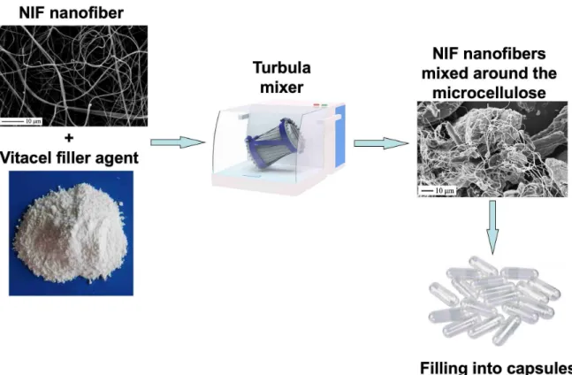
Capsule form was prepared using microcrystalline cellulose (VITACEL A300) as a filling agent. Microcrystalline cellulose promotes the homogeneous distribution of the API, which controls its dosing.
After the pace volume calibration using microcrystalline cellulose of the capsules (transparent, size 00, Capsugel, Lonza Company, Germany), the maximum amount of powder mixture in the applied capsule was determined. 105 mg of powder mixture was used in each capsule and an 14 mg of NIF (according to the dose of the commercial drug) content was used in every sample. NF 1, NF 2 and NF 3 were also mixed with Vitacel ad 105 mg (to be 105 mg of powder mixture). Physical mixtures were prepared by mixing in a Turbula mixer for 10 min at 50 rpm (Figure 2). Reference capsules were also prepared to contain a mixture of cellulose with raw NIF, or PM 1:1, or PM 2:1 (designated in the text as Caps-NIF, Caps-PM 1:1 and Caps-PM 2:1).
Fig. 2. Schematic illustration showing the formulation of the solid dosage form. NIF nanofibers were mixed with the microcrystalline cellulose filler agent in a Turbula mixer.
Then the mixtures were filled into capsules.
Table 2. The composition of NIF-contained capsules prepared by physical mixing and electrospinning.
Samples Compositions
Caps-NIF NIF-VITACEL A300
Caps-PM 1:1 PM 1:1- VITACEL A300 Caps-PM 2:1 PM 2:1- VITACEL A300
Caps-NF1 NF1- VITACEL A300
Caps-NF2 NF2- VITACEL A300
Caps-NF3 NF3 VITACEL A300
2.3. Analysis
Scanning electron microscopy
The morphology and size were analyzed using Scanning electron microscopy (SEM), by a Hitachi S4700 instrument (Hitachi Scientific Ltd., Tokyo, Japan) at 10 kV. The samples were
gold-palladium coated (90 seconds) by a sputter coater (Bio-Rad SC 502, VG Microtech, Uckfield, UK) using an electric potential of 2 kV at 10 mA for 10 minutes. Image processing was done in the ImageJ software environment [18]. Over 150 individual particle measurements were made in at least five different images in order to determine the diameters of the nanofibers accurately.
Differential scanning calorimetry (DSC)
DSC was employed to investigate the crystallization behavior and the melting behavior of the conventional and submicron-sized NIF. The DSC measurements were made with a Mettler Toledo DSC 821e thermal analysis system with the STARe thermal analysis program V9.1 (Mettler Inc., Schwerzenbach, Switzerland). Approximately 2 - 5 mg of the product was examined in the temperature range between 25 °C and 300 °C. The heating rate was 5 °C min-
1. Argon was used as a carrier gas, at a flow rate of 10 L h-1 during the DSC investigation.
X-ray powder diffraction (XRPD)
XRPD was carried out to determine the crystalline form and crystallinity of the produced materials. Samples were analyzed by a Bruker D2 diffractometer (Bruker AXS GmbH, Karlsruhe, Germany). Data collection was carried out at room temperature using monochromatic Cu K α1 radiation (α = 0.154060 nm) in the 2-theta region between 3° and 50°. The samples were placed on a zero-background holder (Si single crystal <510> wafer).
Solubility test
The solubility of NIF was determined in distilled water (pH 5.6) and at simulated gastric media buffer solution (pH 1.2) at 25 °C. 10 mg of the sample was placed into 5 mL of distilled water or buffer solution, and stirred with magnetic stirrer rigorously for 24 hours.
Then the samples were filtered by a filter paper (0.22 µm, FilterBio PES Syringe Filter, Labex Ltd., Budapest, Hungary) to remove any undissolved particles or impurities, and the content of dissolved drug in the liquid was measured spectrophotometrically at 256 nm (Unicam UV/Vis spectrophotometer, Germany).
Wettability studies
Optical contact angle measurement and drop contour analysis were performed using an OCA 20 system (Dataphysics Inc., GmbH, Germany) for measuring the wettability of the capsule products containing NIF. A small amount of powder (150 mg) was compressed under a
pressure of 1 ton by a Specac hydraulic press (Specac Inc., USA). The wetting angles of the pressings were determined after 4.3 µL of distilled water had been dropped onto the surface of the pressed samples. The change in the wetting angle was registered from 1 to 25 s, using the circle fitting method of the OCA system. The method of Wu was applied, in which two liquids with known polar (γlp) and dispersion (γld) components are used for measurement [19].
The solid surface free energy is the sum of the polar (γp) and non-polar (γd) components and is calculated according to equation 1:
p l p s
p l p s d
l d s
d l d s
Θ l
γ γ
γ γ γ
γ γ γ γ
+ +
= +
+ 4( ) 4( )
) cos 1
( (1)
where Θ is the contact angle, γs is the solid surface free energy and γl is the liquid surface tension. The percentage of the polarity can be calculated from the γp and γ values: (γp /γ ) ×
100. The liquids used for our contact angle measurements were double-distilled water (γp = 50.2 mN m-1, γd = 22.6 mN m-1) and diiodomethane (γp = 1.8 mN m-1, γd = 49 mN m-1). The
results of the contact angle measurements using diiodomethane as non-polar component and water as polar component provide information on the surface free energies and polarities of the samples.
In vitro dissolution of nanofibers and formulated capsule products
The dissolution profile of product samples containing equal amounts of drug (14 mg of NIF according to its therapeutic dosage) was determined using the modified paddle method. A buffer solution (50 mL, pH 1.2 ± 0.1) at 37 ± 0.5 ºC was used as a dissolution medium (simulated gastric medium) and the rotation speed of the paddles was 100 rpm. At certain time intervals, 2 mL solution samples were withdrawn and filtered (cut-off 0.2 µm, Minisart SRP 25, Sartorius, Germany) and the amount of dissolved drug was determined spectrophotometrically by an ATI-Unicam UV2-100 UV/VIS spectrophotometer at 256 nm.
The amount of the withdrawn samples was replaced with fresh medium. The NIF content in NF samples and in capsule was measured by UV/Vis spectrophotometry where the standard deviation of concentration was ±10 %.
3. RESULTS AND DISCUSSION
3.1 Characterization of nanofibers containing NIF and PVP
Large nanofiber mats of NIF-PVP composites were produced using the nozzle-free electrospinning technique. This method is capable of producing high yields without any clogging problems, as the fibers are formed from a free surface. In our experiments, a 6 × 7.5 cm mat, with the weight of 100 mg was produced in just 10 minutes (see Supplementary Figure 1).
3.1.1 Micrometric properties
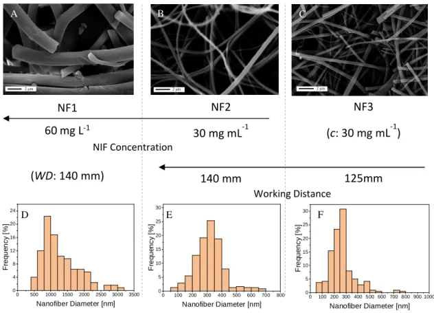
SEM study revealed that the size range of the nanofibers varied between 160 - 1800 nm, depending on the solution concentration parameters. The samples prepared from 1:1 NIF – PVP ratio solution have significantly smaller fiber diameters than the sample prepared from the 2:1 NIF – PVP ratio solution (see Figure 3 and Table 3).
Fig. 3. SEM pictures (A-C) of the three nanofibers prepared and their respective nanofiber diameter distribution histograms (D-F). Their respective niflumic acid concentrations (c) and
working distance (WD) are displayed on the axis between the graphs and pictures.
A B C
NF1 60mg L-1
(WD: 140 mm)
NF2 30 mg mL-1
140 mm
NF3 (c: 30 mg mL-1)
125mm NIF Concentration
Working Distance
2 µm 2 µm 2 µm
0 500 1000 1500 2000 2500 3000 3500 0
4 8 12 16 20 24
Frequency [%]
Nanofiber Diameter [nm]
0 100 200 300 400 500 600 700 800 0
5 10 15 20 25 30
Frequency [%]
Nanofiber Diameter [nm]
0 100 200 300 400 500 600 700 800 900 1000 0
5 10 15 20 25 30
Frequency [%]
Nanofiber Diameter [nm]
A B C
D E F
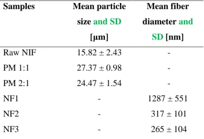
Analysis of the SEM images concluded that by optimizing the electrospinning process parameters, it is possible to reach NIF fibers with a mean diameter of 265 nm (see Table 3). The average size of the manufacturer NIF was around 16 µm, and when adding the polymer, PVP, into the physical mixtures, the average size of the agglomerate can reach 24 - 27 µm. The electrospun samples were stored at room temperature (25 °C) in a desiccator for 1 year, then their morphology and size were investigated again by SEM.
The analysis showed no observable difference in their nanostructure when compared with the fresh samples (see Supplementary Fig. 2-3).
.
Table 3. The Mean particle size and Mean Diameter over Volume (MV) with the Standard Deviation (SD) of the samples
Samples Mean particle size and SD
[µm]
Mean fiber diameter and
SD [nm]
Raw NIF 15.82 ± 2.43 -
PM 1:1 27.37 ± 0.98 -
PM 2:1 24.47 ± 1.54 -
NF1 - 1287 ± 551
NF2 - 317 ± 101
NF3 - 265 ± 104
3. 1. 2 Structural characterization
DSC was employed to investigate the melting points and crystallinity of NIF before and after the crystallization process. Single sharp endothermic peaks were observed (see Supplementary Fig. 4), corresponding to the melting points of the raw NIF (203.25 °C).
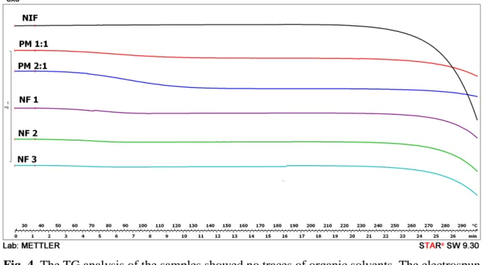
Because of the presence of the PVP, there is a physical interaction between NIF and excipient by heating. It has been previously found that NIF can be dissolved in the melted PVP, resulting in no melting point detected by DSC [20,21]. The TG analysis found no traces of organic solvents, and showed that the prepared samples were thermally stable compared to the raw NIF (Figure 4).
Fig. 4. The TG analysis of the samples showed no traces of organic solvents. The electrospun samples and the physical mixtures with PVP are thermally more stable than the raw NIF.
XRPD was used to examine the crystalline phase of the products. The characteristic peaks of NIF are the 2θ values of 8.2°, 12.88°, 16.16°, 23.18° and 25.4°. Both PMs show the characteristic peaks of NIF, however, all the electrospun nanofibers were amorphous (Figure 5). The XRPD measurement was repeated 1 year after the fabrication procedure, and the results were unchanged.
Fig. 5. XRPD pattern of the NIF, physical mixtures and NF samples. All the electrospun nanofiber samples were amorphous, while the physical mixtures were in a crystalline state.
3.1.3 Solubility test
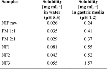
NIF has an acidic character, and its solubility was, therefore, higher at pH 1.2 than at pH 5.5.
The size reduction and presence of PVP, as a wetting agent (Table 4) caused an increase in solubility, especially on the NF3 sample. The PVP prepared by physical mixing resulted in a slight improvement in solubility. However, the electrospun nanofibers with the smallest diameter resulted in a significant, 3-fold increase of solubility in water, and up to 6-fold increase at acidic pH (see Table 4).
Table 4. The solubility data of the produced samples in phosphate buffer at pH 5.5 and gastric media at pH 1.2.
Samples Solubility [mg mL-1]
in water (pH 5.5)
Solubility [mg mL-1] in gastric media
(pH 1.2)
NIF raw 0.026 0.24
PM 1:1 0.035 0.41
PM 2:1 0.029 0.37
NF1 0.081 0.55
NF2 0.043 0.52
NF3 0.055 1.57
3.1.4 Dissolution studies
When the two PMs (where no nanofibers were present) were measured in simulated gastric media (pH 1.2), the dissolution kinetics of NIF was slow, comparable to the raw NIF sample’s dissolution speed. After 120 minutes, only 20% of the drug was liberated. The dissolution rate and dissolved concentration were significantly higher when the amorphous nanofibrous formulations were tested. Both NF1 and NF2 samples demonstrated ~74% dissolved NIF after 15 minutes. The sample, NF3, showed the fastest and highest dissolution behavior, just after 15 minutes, 97% of the drug has been dissolved into the simulated gastric fluid. This means a 5-fold increase in the amount of the dissolved API when compared with the conventional
capsule form in the first 120 minutes (Figure 6). Therefore, our results show that the dissolution behavior can be significantly improved by a nanofabrication method.
Fig. 6. In vitro dissolution of NIF from the nanofiber measured at pH 1.2. The nanofiber- based samples have improved dissolution kinetics, and significantly higher dissolution rates
than the raw NIF or the physical mixtures (PMs).
3.2 Investigation of capsule form containing nanofibers
In general, the produced drug nanofibers could be easily used as dermal formulation (e.g. skin patches). The novelty of this work is the formulation of the solid dosage form capsules, which is significant, as NIF is applied generally in a capsule form [22]. Thus, capsules were also formulated, containing the NIF/PVP nanofiber mixtures and the cellulose filler.
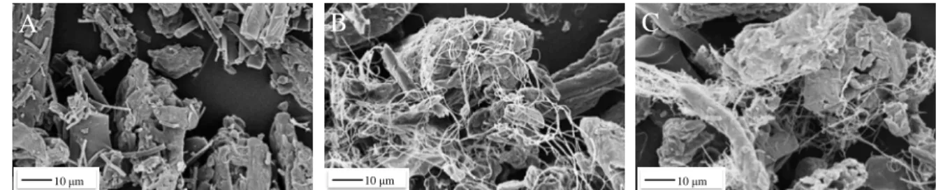
3.2.1 Morphology of the powder mixtures containing nanofibers
During the mixing of the filler agent and nanofiber samples, the uniformity was different. In the case of electrospun fiber (NF3) and capsule formulation (CAPS-NF3) samples (Figure 7), a well-distributed mixture was reached, which can offer improved surface area (see Figure 7C).
A
10 µm
B C
10 µm 10 µm
Fig. 7. SEM images showing the distribution of nanofibers around the filling agent (A. Caps- NF1, B. Caps-NF2 and C. Caps-NF3).
3.2.2 Wettability and dissolution profiles of the powder mixture in the capsule form
The wettability study indicates that the products containing the PVP, Vitacel and nanometer- size range of the drug have a hydrophilic character. Table 5 shows that relatively low contact angles with water were measured for these samples. The contact angle for the manufacturer NIF (~72º) was relatively high, revealing its hydrophobicity. The amorphous nature and the PVP covering a large surface of the NIF particles can be the reasons behind the increased wettability of the electrospun samples. PVP is known to have a surface tension lowering effect, resulting in wetting of the hydrophobic NIF crystalline surface [7,23,24]. The increased specific surface area and the amorphous character can also increase the wettability.
Table 5. Wettability results for the different samples.
Samples
Θwater [˚]
Θdiiodomethane [˚]
γ [mN m-1]
Polarity [%]
NIF 72.5 31.38 51.11 21.91
Caps-NIF 27.80 13.95 75.89 41.34
Caps-PM1 12.5 19.35 79.71 45.76
Caps-PM2 14.76 16.13 79.72 44.79
Caps-NF1 10.5 31.85 77.59 49.49
Caps-NF2 17.26 29.87 76.47 47.79
Caps-NF3 11.09 26.15 78.70 47.65
The hydrophilic character of the samples prepared with electrospun fibers results in a significantly improved dissolution behavior of the active ingredient, NIF. The dissolution results of the nanofiber-based capsules show outstanding kinetics, when compared to the capsules made with the PMs (Figure 8). When the two capsules with no nanofibers present
(Caps-PM1 and Caps-PM2) were measured in simulated gastric media (pH 1.2), the dissolution kinetics of NIF was comparable to the raw NIF sample’s slow dissolution speed, and slightly less drug dissolved out from these samples, when compared with the pure NIF sample’s behavior. After 120 minutes, only ~20% of the drug was dissolved into the gastric fluid. The dissolution rate and dissolved concentration were significantly higher when the capsule formulations with nanofibers were tested. Both Caps-NF1 and Caps-NF2 samples demonstrated ~69% dissolved NIF after 15 minutes. The sample, Caps-NF3, showed the fastest and highest dissolution behavior, just after 15 minutes, 91% of the drug has been dissolved into the simulated gastric fluid. This means a 23-fold increase in the amount of the dissolved NIF, when compared with the conventional capsule form in the first 15 minutes, and a 5-fold increase is observed when compared with the conventional capsule form in the first 120 minutes.
Fig. 8. In vitro dissolution behavior of the prepared capsules measured at pH 1.2. The nanofiber-based capsules show outstanding dissolution kinetics, when compared to the
capsules made with the physical mixtures (PMs).
4. CONCLUSIONS
This study has shown that electrospinning is an effective method to change the physicochemical properties of the pharmaceutical active ingredient, NIF. It can be concluded that decreased size of the drug compound in the nano-size range can be achieved with this
procedure, resulting in a possibility of capsule formulation with nearly 100 % drug release.
According to the structural analysis, the drug was in crystalline form when formulated without the PVP excipient, and in an amorphous form when prepared with PVP. The wettability and dissolution tests revealed the hydrophilic character of NIF produced by electrospinning, while the manufacturer NIF is hydrophobic. Nanofiber formulation of NIF using our method could reduce the gastric irritation of NIF and could facilitate its safe use. A 15-fold increase in the dissolution rate of the produced amorphous niflumic acid nanofiber mats was observed compared to the dissolution rate of the original drug within the first 15 minutes. When the dissolution rate of the original capsule formulation was compared with the nanofibrous capsules, a 23-fold increase was observed in the dissolution kinetics within the first 15 minutes. We conclude that during the pre-formulation and then the final formulation from the lipophilic NIF, we could form a capsule product with good stability due to its amorphous form, and nearly 100 % drug release in the gastric media. It means that the bioavailability may have been improved, therefore the dosage and unwanted side-effects could be decreased. Therefore, our results show that the dissolution behavior of poorly water- soluble drugs can be significantly improved by utilizing the electrospinning nanofabrication method.
ACKNOWLEDGEMENT
The authors would like to acknowledge the Ministry of Human Capacities, Hungary, grant number 20391-3/2018/FEKUSTRAT, for funding.
REFERENCES
[1] A.S. Myerson, S.R. Anderson, R.C. Bennett, D. Green, P. Karpinski, Handbook of industrial crystallization, Butterworth-Heinemann, Woburn, 2002.
https://www.elsevier.com/books/handbook-of-industrial-crystallization/myerson/978-0- 7506-7012-8 (accessed August 6, 2017).
[2] N.J. Babu, A. Nangia, Solubility Advantage of Amorphous Drugs and Pharmaceutical Cocrystals Published as part of the Crystal Growth & Design 10th Anniversary
Perspective, (2011) 2662–2679.
[3] M.N. Patil, A.B. Pandit, Cavitation – A novel technique for making stable nano- suspensions, 14 (2007) 519–530. doi:10.1016/j.ultsonch.2006.10.007.
[4] N. Radacsi, R. Ambrus, Atmospheric Pressure Cold Plasma Synthesis of
Submicrometer-Sized Pharmaceuticals with Improved Physicochemical Properties, Cryst. Growth Des. 12 (2012) 5090–5095.
http://pubs.acs.org/doi/abs/10.1021/cg301026b (accessed April 6, 2013).
[5] R. Ambrus, N.N. Amirzadi, Z. Aigner, P. Szabó-Révész, Formulation of poorly water- soluble Gemfibrozil applying power ultrasound, Ultrason. Sonochem. 19 (2012) 286–
291. doi:10.1016/j.ultsonch.2011.07.002.
[6] R. Ambrus, Z. An, Preparation and Investigation of Inclusion Complexes Containing Nifluminic Acid and Cyclodextrins, (2003) 123–124.
[7] N. Radacsi, J.H. Horst, G.D. Stefanidis, Microwave-Assisted Evaporative Crystallization of Ni fl umic Acid for Particle Size Reduction, (2013) 10–13.
doi:10.1021/cg4010906.
[8] N. Radacsi, R. Ambrus, T. Szunyogh, P. Szabó-Révész, A. Stankiewicz, A. Van Der Heijden, J.H. Ter Horst, Electrospray crystallization for nanosized pharmaceuticals with improved properties, Cryst. Growth Des. 12 (2012) 3514–3520.
doi:10.1021/cg300285w.
[9] R. Ambrus, N. Radacsi, T. Szunyogh, A.E.M. van der Heijden, J.H. ter Horst, P.
Szabó-Révész, Analysis of submicron-sized niflumic acid crystals prepared by electrospray crystallization, J. Pharm. Biomed. Anal. (2013).
doi:10.1016/j.jpba.2012.12.001.
[10] T. Szunyogh, R. Ambrus, Formation of niflumic acid particle size by solvent diffusion and solvent evaporation as precipitation methods, J. Drug Deliv. Sci. Technol. 22 (2012) 307–312. doi:10.1016/S1773-2247(12)50052-3.
[11] A. Greiner, J.H. Wendorff, Electrospinning: A fascinating method for the preparation of ultrathin fibers, Angew. Chemie - Int. Ed. 46 (2007) 5670–5703.
doi:10.1002/anie.200604646.
[12] N. Radacsi, F.D. Campos, C.R.I. Chisholm, K.P. Giapis, Spontaneous formation of nanoparticles on electrospun nanofibres, Nat. Commun. 9 (2018) 4740.
doi:10.1038/s41467-018-07243-5.
[13] M. Geoffrey R., ed., Electrospinning: principles, practice and possibilities, Royal Society of Chemistry, 2015. https://pubs.rsc.org/en/content/ebook/978-1-84973-556-8.
[14] C. Huang, S.J. Soenen, J. Rejman, B. Lucas, K. Braeckmans, J. Demeester, S.C. De Smedt, Chemical Society Reviews, Stimuli-Responsive Electrospun Fibers Their Appl.
40 (2011) 2417–2434. doi:10.1039/c0cs00181c.The.
[15] V. Guarino, L. Ambrosio, Electrofluidodynamic technologies (EFDTs) for biomaterials and medical devices : principles and advances, Woodhead Publishing, 2018.
https://www.elsevier.com/books/electrofluidodynamic-technologies-efdts-for- biomaterials-and-medical-devices/guarino/978-0-08-101745-6.
[16] K. Garg, G.L. Bowlin, Electrospinning jets and nanofibrous structures., Biomicrofluidics. 5 (2011) 13403. doi:10.1063/1.3567097.
[17] Z. Chen, Z. Chen, A. Zhang, J. Hu, X. Wang, Z. Yang, Electrospun nanofibers for cancer diagnosis and therapy, Biomater. Sci. 4 (2016) 922–932.
doi:10.1039/C6BM00070C.
[18] M.D. Abràmoff, P.J. Magalhães, S.J. Ram, Image Processing with ImageJ, Biophotonics Int. 11 (2004) 36–42.
[19] S. Wu, Calculation of interfacial tension in polymer systems, J. Polym. Sci. Part C. 30 (1971) 19–30.
[20] R. Ambrus, Z. Aigner, C. Şoica, C. Peev, P. Szabó-révész, Amorphisation of
nifluminic acid with polyvinylpyrrolidone prepared solid dispersion to reach rapid drug release, Rev. Chim. 58 (2007) 206–209.
[21] R. Ambrus, Z. Aigner, O. Berkesi, C. Şoica, P. Szabó-Révész, Determination of the structural interaction of nifluminic acid-PVP solid dispersions, Rev. Chim. 57 (2006) 1051–1054.
[22] A. V. Egorova, A. V. Anelchyk, I.I. Leonenko, Y. V. Skripinets, V.P. Antonovich, Determination of a series of nonsteroidal anti-inflammatory drugs using the sensitized luminescence of lanthanides, J. Anal. Chem. 70 (2015) 440–449.
doi:10.1134/S1061934815040048.
[23] G. Chawla, A.K. Bansal, Improved dissolution of a poorly water soluble drug in solid dispersions with polymeric and non-polymeric hydrophilic additives, 58 (2008) 257–
274.
[24] A. EL Aferni, M. Guettari, T. Tajouri, Effect of polymer conformation on polymer- surfactant interaction in salt-free water, Colloid Polym. Sci. 294 (2016) 1097–1106.
doi:10.1007/s00396-016-3869-8.