III./1.2. Pathological features, etiology
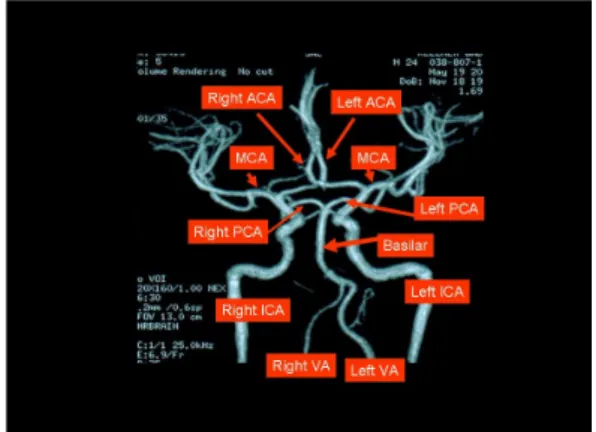
Blood flow to the brain is supplied by four large arteries: the two vertebral and the two internal carotid arteries. The characteristics of this vascular system can be examined by non-invasive techniques (Fig. 10).
Fig. 10: Blood supply to the brain: the cerebral vessels – MR angiogram
The pathological appearance of the most frequent types of stroke is seen in Fig. 11.
Fig. 11: Major types of stroke. Ischemic stroke, intracerebral hemorrhage and subarachnoid hemorrhage
Large vessel (atherothrombotic territorial and borderzone) ischemic stroke is most often caused by atherosclerosis, less frequently by dissection (Fig. 12). Plaques are most likely formed in areas where blood flow is turbulent (e.g. at the branching of the common carotid artery or the middle cerebral artery).
Fig. 12: occlusion of the left vertebral artery and dissection of the basilar artery
A growing atherosclerotic plaque may cause stenosis, and eventually the occlusion of the artery, leading to hypoperfusion of the territory it supplies (Fig. 13). Plaques in the aortic arch or in the carotid artery may cause disturbance of blood flow by another mechanism: small particles detaching from the plaque may result in stroke downstream from these arteries via an embolic mechanism. In embolic strokes, the heart is a frequent
source of emboli (”cardioembolic stroke”). In cardioembolic stroke, fragments may detach from a left atrial thrombus associated with atrial fibrillation or from a left ventricle thrombus associated with myocardial infarction (mural thrombus on the hypokinetic wall) or congestive heart failure. Embolic strokes are characteristically wedge-shaped and affect the cortex as well (Fig. 14).
Fig. 13: CT angiogram. The internal carotid artery is occluded at its origin
Fig. 14: Cardioembolic stroke in atrial fibrillation affecting the left temporal lobe
The focal decrease in cerebral blood flow causes a brain infarct, which may transform into a hemorrhagic infarct over the course of the next few days (more common in cardioembolic strokes). This transformation occurs within the infarcted tissue, therefore it is usually not associated with the progression of clinical signs.
Cerebral small vessel disease is usually associated with hypertension. The lipohyalinic degeneration of the smaller (around 200 m in diameter) vessels results in multiple small (1-2 mm) lacunar lesions that may lead to slowly developing pseudobulbar palsy, dementia, incontinence, and gait disturbance (i.e. not in the form of acute stroke). The typical lacunar syndromes are caused by the arteriolosclerotic occlusion of large (about 800 m) vessels: in such cases, a single larger lacunar infarct is usually responsible for the acute presentation of clinical signs.
Lacunar infarcts were found in 10% of cases in a selected autopsy series.
About 13-25% of acute stroke cases are lacunar infarcts.
Lacunar lesions frequently remain asymptomatic (“silent”).
In everyday neurological clinical practice, the diagnosis of cerebral small vessel disease is used for patients who have specific clinical syndromes associated with lacunar infarcts and/or leukoaraiosis detected by neuroimaging (CT, MRI) (Fig.
15).
Fig. 15: The diagnosis of lacunar infarcts based on clinical signs
In addition to lacunar lesions, cerebral small vessel diseases may have other
consequences as well. The rupture, rather than the occlusion of a small artery within the brain tissue results in an intracerebral hemorrhage (Fig. 3).
3. ábra: Agyállomány vérzés
Developmental disorders of the arteries on the skull base may lead to the formation of aneurysms, as a result of weakening of the arterial wall at certain points. The rupture of an aneurysm causes subarachnoid hemorrhage (Fig. 4).
4. ábra: Subarachnoidealis vérzés