III./3.3.1. Wilson’s disease
Introduction
Wilson’s disease is an autosomal recessive disorder of copper metabolism characterized by excessive copper deposition, predominantly in the liver and the brain. It is a rare disease, its prevalence is 3/100,000 worldwide.
In Hungary, the estimated number of patients with Wilson’s disease is 2-300. Wilson’s disease is fatal without treatment, but most of the symptoms can be eliminated by early diagnosis and prompt initiation of therapy. In case of a fulminant hepatitis, mortality can reach 70%. A comprehensive clinical database is found on the www.eurowilson.org website.
Wilson’s disease was first published in 1912 by Samuel Alexander Kinnier-Wilson in Brain. He described a lethal neurologic disorder associated with progressive lenticular degeneration of the brain and cirrhosis of the liver, which came later to be known as Wilson’s disease, or hepatolenticular degeneration.
Since then, several biochemical and genetic markers have been identified.
Keywords
copper, ceruloplasmin, hepatolenticular degeneration
Contents of the chapter:
III./3.3.1.1. Pathomechanism III./3.3.1.2. Clinical symptoms III./3.3.1.3. Diagnosis and follow up III./3.3.1.4. Therapy
III./3.3.1.1. Pathomechanism
The daily copper requirement of the body is 1-3 mg. In the blood, copper is bound mainly to ceruloplasmin, and to lesser degree to proteins (e.g.
albumin). The concentration of free copper in the blood is 0.1 mg/L. The normal homeostasis of copper is maintained by absorption in the small intestine and excretion into the bile. Forty to seventy percent of an oral dose of copper is absorbed and detected in the blood after 90-150 minutes. The albumin bound copper reaches the liver through the portal vein, where a specific transport system carries it to the hepatocytes.
Absorption of copper is unlimited. The excess of copper is excreted with the bile from the liver via three main routes; one of these is the ATP7B dependent excretion through the Golgi system. In Wilson’s disease, this ATP-ase protein is abnormal and copper cannot be excreted with the bile, but remains stored in hepatocytes. Copper accumulation causes damage to hepatocytes and copper eventually enters the blood. Free copper in the serum becomes elevated, surpassing 0.3 mg/L. Cooper is deposited in many organs, mainly in the liver and the central nervous system. Since it is toxic, it induces oxidation of lipoprotein membranes and generation of free radicals, particularly in the mitochondria.
Wilson’s disease shows an autosomal recessive inheritance. Its causative gene, designated as ATP7B, was mapped to chromosome 13q14.3, and codes a copper transporter ATPase protein with the same name. ATP7B gene is primarily expressed in the liver, but it is also detected in the kidney, brain and placenta in lower levels. In patients with Wilson’s disease from Northern, Eastern or Central European countries, including Hungary, H1069Q point mutation in exon 14 is the most frequent mutation, detected in 64-71% of patients2.
Patients with a homozygous genotype have clinical symptoms, often with neurological manifestations. The number of known mutations within the ATP7B gene exceeds 350, but not all mutations cause Wilson’s disease.
Since clinical symptoms are not always typical for Wilson’s disease and biochemical markers of impaired copper metabolism can be misleading, genetic testing became an important part of the diagnostic work-up.
III./3.3.1.2. Clinical symptoms
Typical clinical symptoms in Wilson’s disease:
a. Secondary chronic liver disease b. Neurological symptoms
c. Psychiatric manifestations d. Ophthalmologic manifestations e. Nephropathy
f. Hemolytic anemia g. Skeletal abnormalities h. Cardiomyopathy i. Skin and nail changes j. Endocrine disturbance
a. Secondary chronic liver disease: Hepatic manifestation is the most common in childhood (8-16 years). Patients exhibit features of acute or chronic liver disease or elevation of liver transaminase enzymes. The type of liver pathology is steatosis and fibrosis in about 24% of the cases, chronic hepatitis in 28%, and cirrhosis in 33%. A subacute, fulminant liver failure may also occur, in which hepatic encephalopathy is characteristic 2-8 weeks after the onset of icterus.
b. Neurological symptoms: Neurological symptoms are the presenting features in 44-88% of patients with Wilson’s disease, especially at the age of 12-35years; copper accumulation may cause neurological manifestations later than hepatopathy3. In 40-50% of the cases, neuropsychiatric features are the first signs.
Frequent neurological symptoms: flapping tremor, postural tremor (53%); parkinsonian syndrome; dysarthria (93%);
dystonia; gait ataxia (79%); ataxia of extremities;
dysdiadochokinesis (100%); pseudobulbar palsy; risus sardonicus; insomnia; epileptic seizure.
c. Psychiatric manifestations: In 20% of patients, the first signs are behavioral changes, irritability, impulsivity, obsessive
behavior, aggression, dementia, depression, maniac-depressive psychosis, and hypersomnia.
d. Ophthalmologic manifestations: In addition to the typical Kayser-Fleischer ring (Fig. 1) and sunflower cataract, abnormal saccades or apraxia of eye opening may occur.
Fig.1: Kayser-Fleischer ring3
e. Nephropathy is an extremely rare initial symptom in Wilson’s disease. In the kidney, copper accumulation can be a hundred times more than normal; its symptoms include proteinuria, glycosuria, phosphaturia, aminoaciduria, microscopic hematuria, hypercalcuria-nephrocalcinosis, and renal osteodystrophy. In fulminant hepatitis, nephropathy is the cause of the liver disease.
f. Hemolytic anemia: Copper acts as a catalyst in hemoglobin metabolism. Elevated copper levels cause a change of membrane stability of erythrocytes and lead to the decrease of the amount of transport enzymes, which is worsened in fulminant liver failure.
In childhood and early adolescence, hemolytic anemia without other apparent causes may be a warning sign of Wilson’s disease, if Coombs test is negative.
g. Skeletal abnormalities: Osteoporosis is observed in 38% of the cases (Hungarian data), but osteomalacia, spontaneous fractures, early osteoarthritis, subchondral cyst formation,
polyarthralgia may also be present. Copper accumulation can also be detected in the joints.
h. Cardiomyopathy: Copper deposition in the myocardium is uncommon. Cardiovascular dysfunction such as congestive heart failure, myocardial hypertrophy, focal myocarditis and
non-specific cardiac arrhythmia may develop.
i. Skin and nail changes: Hyperpigmentation of the anterior lower legs, acanthosis nigricans, and bluish discoloration of the nails may occur in Wilson’s disease.
j. Endocrine disturbance: Primary or secondary amenorrhea, diabetes mellitus, exocrine pancreas insufficiency,
hypoparathyroidism may be due to hepatic failure or copper deposition.
III./3.3.1.3. Diagnosis and follow up
In addition to physical examination and blood biochemistry, cranial MRI examination and genetic testing are important to confirm the diagnosis and to follow the progression of the disease and the efficacy of
pharmacological therapy. The probability level of the diagnosis is defined by a scoring system in Wilson’s disease (Fig. 2).
Fig. 2: Biochemical markers of Wilson’s disease and scoring system for the probability level of the diagnosis1
Important tests:
Biochemical tests , liver function: serum bilirubin, alkaline phosphatase, serum aminotransferase (AST, ALT, GGT) levels 24-hour urinary copper excretion >100 μg in the 65% of the patients; penicillamine provoking test (500 mg of penicillamine intake every 12 hours): 24-hour urinary copper excretion >1600 μg or ten-fold increase compared to baseline.
Serum copper concentration (norm: 70-150 μg/dL):
ceruloplasmin bound and free copper. Copper levels may be low in asymptomatic patients due to the low level of ceruloplasmin or they may be high in active liver failure, where the serum
concentration of free copper is high. The specificity of serum copper level is low.
Free copper (10% of total copper) is bound to albumin and amino acids in the blood. Its concentration is elevated in Wilson’s disease (5-12 μg/dL), but it is higher also in cholestasis.
Serum ceruloplasmin is lower than 20 mg/dL in 95% of the patients. False negative measurements may be caused by infection, pregnancy, estrogen taking. False positive
measurements may be seen in heterozygous carriers of Wilson’s disease (20%), aceruloplasminemia, and severe hepatic
insufficiency.
Hepatic copper content is high ( ≥250 μg/g). The sensitivity of the test is 82%, its specificity is 95%. In untreated patients, level lower than 50 μg/g excludes the diagnosis of Wilson’s disease.
The hepatic copper content is the best biochemical marker of Wilson’s disease.
Slit-lamp examination: the presence of Kayser-Fleischer ring (in 44-62% of the cases) strongly supports the diagnosis, especially in hepatic manifestations. It can be false positive in chronic cholestasis and may be absent in neurological manifestations.
Genetic testing is important in ambiguous cases to screen the family.
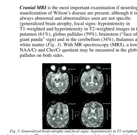
Cranial MRI is the most important examination if neurological manifestation of Wilson’s disease are present, although it is not always abnormal and abnormalities seen are not specific (generalized brain atrophy, focal signs: hypointensity in T1-weighted and hyperintensity in T2-weighted images in the putamen (61%), globus pallidus (59%), brainstem (“face of the giant panda” sign) and in the cerebellum (34%), thalamus and white matter (Fig. 3). With MR spectroscopy (MRS), a low NAA/Cr and Cho/Cr quotient may be measured in the globus pallidus on both sides.
Fig. 3: Generalized brain atrophy and focal signs: hypointensity in T1-weighted and hyperintensity in T2-weighted images in the putamen, globus pallidus and the brainstem
(“face of the giant panda” sign) and in the cerebellum, thalamus and white matter.
III./3.3.1.4. Therapy
Wilson’s disease is fatal without treatment, but most of the symptoms can be eliminated by early initiation of appropriate treatment. Dietary copper should be restricted, its limit is 1.5 mg/day. Patients should avoid
chocolate, sun-dried fruits, liver, veal, ham, spinach, pineapple,
mushroom, and walnut. Soft water can contain high amounts of copper, so distilled water should be used for cooking.
The pharmacological treatment of Wilson’s disease is based on the use of copper chelators.
D-penicillamine
D-penicillamine is a metabolic byproduct of penicillin, thus it is contraindicated in penicillin allergy. A non-toxic copper-penicillamine complex is formed, which enhances the production of metallothionein that can also bind copper. This results in increased renal copper excretion.
The effect of d-penicillamine develops slowly; exacerbation of
neurological symptoms may occur in the early phase. D-penicillamine therapy has to be stopped in 30% of the patients due to side effects (Early side effects: fever, lymphadenopathy, granulo- and thrombocytopenia.
Late side effects: nephrotoxicity, SLE, pemphigus, myasthenia gravis, polymyositis, piridoxin deficiency, disturbance of taste, leuko-, and thrombocytopenia). D-penicillamine therapy leads to the improvement of neurological, psychiatric and hematologic symptoms, and the progression of hepatopathy may halt. Kayser-Fleischer ring may be eliminated, serum free copper concentration decreases, and MRI abnormalities of the brain may disappear. Treatment may also be indicated in patients with
asymptomatic homozygous Wilson’s disease.
Trientine
Trientine is used as an alternative therapy for patients intolerant of
d-penicillamine. It increases the excretion of copper and the concentration of free copper in the serum. Its daily dose is 750-2000 mg/day, given in three divided doses, 30 minutes before meals. It has fewer side effects than d-penicillamine, but it may decrease the concentration of serum iron leading to microcyter anemia. Trientine should not be given with iron, their chelate has a toxic effect.
Zinc
The principal mode of action of zinc is presumed to be the induction of metallothionein in the intestines, erythrocytes and the liver. Generally, 150 mg/day of zinc sulfate or zinc acetate is given, in three divided dosages, 30 minutes before or two hours after meals. Gastric irritation may be a side effect, but it is rare during therapy with zinc acetate.
Microcyter anemia may be a late side effect.
Vitamin E
Vitamin E can be used as antioxidant agent.
Tetrathiomolybdate: investigational drug Liver transplantation:
Liver transplantation is life-saving for patients with fulminant hepatic failure. Hemodialysis may become necessary until liver transplantation is carried out.
The neurological manifestations alone are not an indication for liver transplantation, although some case reports described significant improvement of neurological symptoms after this procedure.
References
Roberts EA, Schilsky ML; American Association for Study of Liver Diseases (AASLD). Diagnosis and treatment of Wilson disease: an update. Hepatology. 2008;47(6):2089-2111.
Folhoffer A, Ferenci P, Csak T, Horvath A, Hegedus D, Firneisz G, Osztovits J, Kosa JP, Willheim-Polli C, Szonyi L, Abonyi M, Lakatos PL, Szalay F. Novel mutations of the ATP7B gene among 109 Hungarian patients with Wilson's disease. Eur J Gastroenterol Hepatol 2007;19(2):105-111.
Ala A, Walker AP, Ashkan K, Dooley JS, Schilsky ML. Wilson's disease. Lancet 2007;369(9559):397-408.