Complex electrophysiological remodeling in postinfarction ischemic heart failure
Bence Hegyia, Julie Bossuyta, Leigh G. Griffithsb,c,d, Rafael Shimkunasa,e, Zana Coulibalya, Zhong Jiana,
Kristin N. Grimsrudf, Claus S. Sondergaardf, Kenneth S. Ginsburga, Nipavan Chiamvimonvata,g,h, Luiz Belardinellii, András Varrój,k, Julius G. Pappj,k, Piero Pollesellol, Jouko Levijokil, Leighton T. Izua, W. Douglas Boydf, Tamás Bányásza,m, Donald M. Bersa, and Ye Chen-Izua,e,g,1
aDepartment of Pharmacology, University of California, Davis, CA 95616;bDepartment of Veterinary Medicine and Epidemiology, University of California, Davis, CA 95616;cDepartment of Cardiovascular Diseases, Mayo Clinic, Rochester, MN 55902;dCollege of Medicine, Mayo Clinic, Rochester, MN 55902;
eDepartment of Biomedical Engineering, University of California, Davis, CA 95616;fDepartment of Surgery, University of California, Davis, Sacramento, CA 95817;gDivision of Cardiovascular Medicine, Department of Internal Medicine, University of California, Davis, CA 95616;hDepartment of Veterans Affairs, Northern California Health Care System, Mather, CA 95655;iDepartment of Clinical Research, Gilead Sciences, Inc., Foster City, CA 94404;
jDepartment of Pharmacology and Pharmacotherapy, Faculty of Medicine, University of Szeged, H-6720 Szeged, Hungary;kMTA-SZTE Research Group of Cardiovascular Pharmacology, Hungarian Academy of Sciences, H-6720 Szeged, Hungary;lCritical Care Proprietary Products, Orion Pharma, FI-02200 Espoo, Finland; andmDepartment of Physiology, Faculty of Medicine, University of Debrecen, H-4012 Debrecen, Hungary
Edited by Richard W. Aldrich, The University of Texas at Austin, Austin, TX, and approved February 23, 2018 (received for review October 17, 2017) Heart failure (HF) following myocardial infarction (MI) is associated
with high incidence of cardiac arrhythmias. Development of thera- peutic strategy requires detailed understanding of electrophysio- logical remodeling. However, changes of ionic currents in ischemic HF remain incompletely understood, especially in translational large- animal models. Here, we systematically measure the major ionic currents in ventricular myocytes from the infarct border and remote zones in a porcine model of post-MI HF. We recorded eight ionic currents during the cell’s action potential (AP) under physiologically relevant conditions usingselfAP-clamp sequential dissection. Com- pared with healthy controls, HF-remote zone myocytes exhibited increased late Na+ current, Ca2+-activated K+ current, Ca2+-acti- vated Cl−current, decreased rapid delayed rectifier K+current, and altered Na+/Ca2+exchange current profile. In HF-border zone myo- cytes, the above changes also occurred but with additional decrease of L-type Ca2+current, decrease of inward rectifier K+current, and Ca2+release-dependent delayed after-depolarizations. Our data re- veal that the changes in any individual current are relatively small, but the integrated impacts shift the balance between the inward and outward currents to shorten AP in the border zone but prolong AP in the remote zone. This differential remodeling in post-MI HF increases the inhomogeneity of AP repolarization, which may enhance the arrhythmogenic substrate. Our comprehensive findings provide a mechanistic framework for understanding why single-channel block- ers may fail to suppress arrhythmias, and highlight the need to con- sider the rich tableau and integration of many ionic currents in designing therapeutic strategies for treating arrhythmias in HF.
ischemic heart failure
|
myocardial infarction|
electrophysiology|
action potential
|
ionic currentsI
schemic cardiomyopathy as a chronic consequence of myo- cardial infarction (MI) represents a leading cause of heart fail- ure (HF). Ischemic HF is characterized by extensive structural and functional remodeling that leads to altered action potential (AP) and increased susceptibility for cardiac arrhythmias (1). Precision therapeutic interventions require in-depth understanding of the electrophysiological changes that promote arrhythmias. However, the changes of ionic currents in ischemic HF remain incompletely understood to date, especially in large-animal models that are needed for translational studies. Emerging evidence suggests differential myocardium remodeling in the infarct border and remote zones, leading to complex changes in ionic currents (2), Ca2+handling (3), and contractility (4). However, most previous studies were conducted either early after MI induction (in 2–7 d) or scar formation (within 8 wk) (2, 5), but not at the clinically rele- vant later stages of HF. Recently, we established a porcine MI model (6) that developed chronic HF (with reduced ejection fraction, EF)over 5 mo, providing a large-animal model for translational study of chronic post-MI HF. In this study, we systematically investigated the changes of ionic currents and AP in ventricular myocytes from the infarct border and remote zones to gain a comprehensive understanding of the electrophysiological remodeling.
The AP of ventricular myocyte is shaped by a constellation of ionic currents that integrate at the cell level. The inward vs. out- ward currents counterbalance instantaneously to determine the AP profile. To gain a comprehensive view of remodeling, we de- veloped an innovativeselfAP-clamp sequential dissection method (7) to record multiple inward and outward ionic currents during the cell’s own AP under physiological conditions.
We found that HF remote-zone myocytes exhibit decreased rapid delayed rectifier K+current (IKr), altered Na+/Ca2+exchange current (INCX), and increases of late Na+ current (INaL), Ca2+- activated K+current [IK(Ca)], and Ca2+-activated Cl−current [ICl(Ca)].
The border-zone myocytes also show the above changes, but with Significance
Cardiac arrhythmias often occur in heart failure (HF) patients, but drug therapies using selective ion channel blockers have failed clinical trials and effective drug therapies remain elusive.
Here we systematically study the major ionic currents during the cardiac action potential (AP) and arrhythmogenic Ca2+re- lease in postinfarction HF. We found that changes in any in- dividual current are relatively small, and alone could mislead as to consequences. However, differential changes in multiple currents integrate to shorten AP in the infarct border zone but prolong AP in the remote zone, increasing AP repolarization inhomogeneity. Our findings help explain why single channel- blocker therapy may fail, and highlight the need to understand the integrated changes of ionic currents in treating arrhyth- mias in HF.
Author contributions: B.H., J.B., L.G.G., W.D.B., T.B., D.M.B., and Y.C.-I. designed research;
B.H., J.B., L.G.G., R.S., Z.J., K.N.G., C.S.S., K.S.G., T.B., and Y.C.-I. performed research; N.C., L.B., A.V., J.G.P., P.P., and J.L. contributed new reagents/analytic tools; B.H., R.S., Z.C., Z.J., L.T.I., T.B., and Y.C.-I. analyzed data; and B.H., N.C., L.B., L.T.I., T.B., D.M.B., and Y.C.-I.
wrote the paper.
The authors declare no conflict of interest.
This article is a PNAS Direct Submission.
Published under thePNAS license.
Data deposition: All relevant data have been deposited and are publicly available at https://doi.org/10.25338/B88593.
1To whom correspondence should be addressed. Email: ychenizu@ucdavis.edu.
This article contains supporting information online atwww.pnas.org/lookup/suppl/doi:10.
1073/pnas.1718211115/-/DCSupplemental.
PHYSIOLOGYPNASPLUS
CaL
ward rectifier K+current (IK1), and arrhythmogenic sarcoplasmic reticulum (SR) Ca2+release-induced delayed after-depolarizations (DADs). An unexpected finding is that the changes in any indi- vidual currents are relatively small, but differential changes in multiple currents integrate at the cell level to shorten AP duration (APD) in the border zone but prolong APD in the remote zone.
Such inhomogeneous remodeling increases the difference in AP polarization between the border and the remote zones that may enhance the arrhythmogenic substrate. The integrated changes in ionic currents and spontaneous Ca2+events increase the propensity for both triggered and reentrant arrhythmias.
The main strategy for antiarrhythmic drug development in re- cent decades has been to identify the culprit ion channels re- sponsible for arrhythmias and develop specific ion channel blockers (or activators) to target individual channels. However, clinical trials of ion channel blockers have met unexpected failure, as summarized by Sanderson (8) in the Editorial on SWORD and CAST II trials:“In few specialties of medicine are new promising
increase mortality.”The present study sheds new light into this important problem by using an innovative approach to measure the complex electrophysiological changes in a large-animal model of chronic post-MI HF. Our findings provide in-depth mechanistic insights into why blocking a single ion channel may fail to suppress arrhythmias and highlights the need for developing more com- prehensive strategies to correct multifaceted electrophysiological changes in ischemic HF.
Results
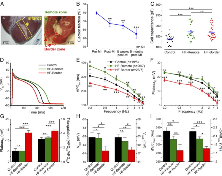
Structural Remodeling and Systolic Dysfunction in Ischemic HF. To provide a clinically relevant large-animal ischemic HF model, we subjected adult Yucatan minipig to microbead embolization of the first diagonal branch of the left anterior descending coronary ar- tery (LAD), which caused transmural MI and a progressive re- duction in the EF. At the time of study (5 mo post-MI) the HF pig hearts exhibited well-healed transmural scars (Fig. 1A), a 36%
reduction in EF (Fig. 1BandTable S1), and dilated cardiomyopathy
Fig. 1. Altered AP morphology in ischemic HF. (A) Photomicrograph of an infarcted heart showing the border zone and the remote zone. (B) LV EF sig- nificantly decreased 5 mo after MI induction (n=12 animals). (C) Cell capacitance increased significantly in failing cardiomyocytes. (D) Representative APs recorded in control, HF-remote, and HF-border cells at 1-Hz pacing frequency (at 36 °C). (E) HF-remote cells have increased APD95while HF-border cells have decreased APD95. (F) Frequency-dependence of Plateau50. (G) Significantly altered Plateau75and AP triangulation in HF-border cells. (H)Vrestis slightly more positive in HF-border cells, in line with decreasedVpeak. (I)dV/dtmaxis decreased only in the HF-border, while−dV/dtmaxis significantly decreased in both HF zones. Mean±SEM,n=19–26 cells/5–7 animals. ANOVA with Bonferroni posttest; n.s., not significant, *P<0.05, **P<0.01, ***P<0.001.
(with 41% increase of end-diastolic volume and 116% increase of end-systolic volume) (Table S1). Post-MI pigs exhibited clinical signs of congestive HF, such as abdominal ascites fluid and pulmonary edema after 5 mo post-MI, but not within 8 wk post-MI, indicating the progression of the disease. Compared with healthy control pig ventricular myocytes, HF myocytes also showed significant hyper- trophy (measured by electrical capacitance) (Fig. 1C), demonstrating structural remodeling at the cellular level.
Heterogenous Changes of AP Morphology in Ischemic HF.HF myo- cytes underwent distinct changes in AP morphology (Fig. 1D), demonstrating functional remodeling. Compared with control, myocytes from the infarct border zone (HF-border) showed short- ened APD (measured at 95% repolarization, APD95), whereas myocytes from the remote zone (HF-remote) showed prolonged APD95(Fig. 1E). The above APD95differences between cell groups remained significant over a range of pacing frequencies (0.2–5 Hz), while APD95 in each cell group progressively decreased with in- creasing pacing frequency (Fig. 1E). These data reveal heteroge- neous remodeling of AP that gives rise to significant APD dispersion from infarct border to remote zones, creating an enhanced substrate for arrhythmias.
Moreover, plateau potentials (Plateau50, Plateau75) were de- creased in the HF-border but remained unchanged in the HF- remote (Fig. 1F), which increased the AP triangulation factor in the HF-border (Fig. 1G). Resting membrane potential (Vrest) was slightly depolarized in the HF-border (Fig. 1H,Left), concomitant with a slight decrease of the AP peak voltage (Vpeak) (Fig. 1H,
Right) and the maximal upstroke velocity (dV/dt) (Fig. 1I,Left). The maximal rate of phase 3 repolarization (−dV/dt) was decreased drastically in the HF-border, and moderately in the HF-remote (Fig. 1I, Right). These data indicate complex and differential changes in the underlying ionic currents that cause APD dis- persion in HF.
Furthermore, the short-term variability (STV) of APD95was also significantly increased in the HF-remote (Fig. S2AandB), with a larger percentage of beats exhibiting more than 5- and 10-ms difference from beat to beat (Fig. S2CandD). This increased temporal variability of APD95 provides an additional substrate for arrhythmias, as previously inferred in human chronic ische- mic HF (9).
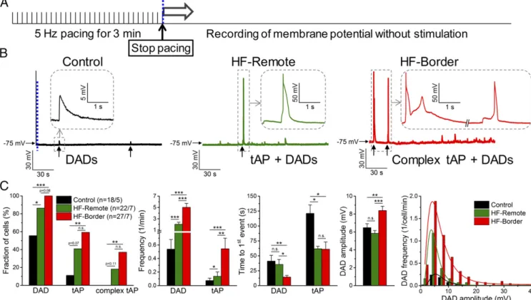
Triggered Activities and Spontaneous SR Ca2+Leak in Ischemic HF.
After-depolarizations are known to occur more frequently in HF (1). Under baseline steady-state pacing conditions, we did not observe early after-depolarizations (EAD). We also evaluated DADs immediately after myocyte pacing at 5 Hz for 3 min (Fig.
2A). We found significant increases of DADs and triggered APs (tAPs) in HF-border and HF-remote cells compared with control (Fig. 2 Band C), with HF-border cells exhibiting DADs with larger amplitudes and tAPs at higher frequency than that of HF- remote (Fig. 2C).
Next, we tested the mechanism of DADs in HF. These DADs were eliminated by inhibitingINCX(Fig. S4A–D), consistent with DADs originating from the inwardINCXinduced by spontaneous SR Ca2+releases (10). Furthermore,IK1density was decreased in
Fig. 2. Arrhythmogenic diastolic activities in ischemic HF. (A) DADs and tAPs were elicited following cessation of 3 min of burst pacing (5 Hz). (B) Repre- sentative records in control, HF-remote, and HF-border cells. Ten of 18 control cells showed a few DADs with small amplitude (enlarged inInset). The last paced beat is shown before the blue dashed line, indicating the cessation of pacing for comparison in magnitude. Nineteen of 22 HF-remote cells showed DADs with a much higher frequency than in control, and among those cells 9 of 22 also showed tAPs (enlarged inInset). All HF-border cells showed DADs and 16 of 27 cells also showed tAPs, often with an EAD superimposed on the tAP repolarization (complex tAP, enlarged inInset). (C) Statistics of the arrhyth- mogenic diastolic events. DAD parameters were assessed only in those events where no tAP occurred subsequently. DAD amplitude data were fitted to a log- normal distribution curve (R2=0.99 in both cases). Mean±SEM,n=18–27 cells/5–7 animals. Fisher’s exact test and ANOVA with Bonferroni posttest; n.s., not significant, *P<0.05, **P<0.01, ***P<0.001.
PHYSIOLOGYPNASPLUS
to cause greater membrane depolarization. Moreover, inhibition of Ca2+–calmodulin-dependent protein kinase II (CaMKII) de- creased DADs to the control level (Fig. S4F–H), implying that pathological CaMKII activation causes SR Ca2+ leak, in agree- ment with prior HF findings in canine (11) and rabbit (12) models.
To directly examine the SR Ca2+ leak in our porcine HF model, we measured diastolic Ca2+sparks, which were markedly increased in frequency and duration in HF-border myocytes (Fig.
S5). Hence, the above data suggest that increased SR Ca2+leak causes DADs and tAPs, especially in the HF-border, which may trigger arrhythmias in ischemic HF.
Intracellular Ca2+Transient and Myocyte Contraction in Ischemic HF.
Given the intertwined nature of electrophysiology and Ca2+sig- naling, we also measured the intracellular Ca2+ concentration ([Ca2+]i) and cell contraction during field-stimulated APs in ven- tricular myocytes (Fig. 3, at 22 °C;Fig. S6, at 36 °C). Notably, [Ca2+]i
transient amplitudes at 0.5 Hz pacing were comparable in both HF zones (Fig. 3B) and in control at room temperature, although the [Ca2+]idecline rate (τ) was slower, and the diastolic [Ca2+]iwas slightly increased in HF (Fig. 3AandC). In contrast, contraction was significantly decreased in both HF zones compared with control (Fig. 3B). Because Ca2+ handling processes are temperature- dependent, [Ca2+]i transients were also compared in the HF- border vs. HF-remote at body temperature. Again, no significant difference was found between the two HF zones (Fig. S6), consis- tent with warming accelerating all Ca2+ transport systems, but without altering relative contributions appreciably (13). This result contrasts with a previous study in sheep showing markedly lower [Ca2+]itransients and cell contractions in border vs. remote zones in 8-wk post-MI myocytes without HF (3). Our 5-mo post-MI porcine hearts may be remodeled during the HF progression in a way that limits differences between HF-border and HF-remote [Ca2+]itran- sients. With little change in [Ca2+]itransient, decreased contraction suggests that a decrease of the myofilament Ca2+responsiveness (in both border and remote zones) might be responsible for reduced EF in this HF model. In the following studies, we focus further on the electrophysiological changes that contribute to arrhythmogenesis.
Remodeling of Inward Currents in Ischemic HF.To measure the ionic currents that shape the AP, we usedselfAP-clamp sequential dis- section for several reasons. First,selfAP-clamp uses each cell’s own steady-state AP as command voltage during ionic current recording;
this enables us to measure currents during that cell’s physiological AP and with preserved [Ca2+]itransients. Second, sequential dis- section of ionic currents (as a specific blocker-sensitive current)
andTable S2); enabling us to measure both inward and outward currents that shape the AP. Third, recording eight different ionic currents in each of a limited number of failing porcine hearts is a unique advantage of ourselfAP sequential dissection approach, much more efficient than conventional voltage-clamp studies of only one ionic current per myocyte. We will first examine the changes in each ionic current, and then investigate how these changes collec- tively reshape the AP and arrhythmogenic substrate in HF.
INaL(recorded as a GS-458967–sensitive current underselfAP- clamp) showed a persistent inward current during AP plateau (Fig. 4A). INaL became larger as a driving force (Vm−ENa) in- creased gradually during AP repolarization. In HF-remote cells, INaLwas increased during the plateau and repolarization phases, causing an increased total Na+entry during the AP cycle. In HF- border cells,INaLalso had increased plateau and peak values, but the integrated total Na+ entry was not different from control because of shortened APD95(Fig. 4).
INCX (measured with a new selective inhibitor, ORM-10962) was outward (Ca2+influx) early in the AP and then turned in- ward during the [Ca2+]itransient (Ca2+efflux), peaking during rapid repolarization in control cells (Fig. 4A). However, in both HF-remote and HF-border cells, outwardINCXwas not observed but was inward throughout the AP (Fig. 4A). InwardINCXdensity increased during the midplateau in HF myocytes, despite exhib- iting similar [Ca2+]itransients as control (Fig. 3A), which drives inwardINCX. This is consistent with an increased functional NCX in HF (10), especially for HF-remote, where midplateauVmwas not different from control (Fig. 1F). However, neither INCX (during phase 3) nor integrated total charge movement through inwardINCXwere significantly different among the three groups (Fig. 4).
ICaL was measured as the nifedipine-sensitive current after blockade of other Ca2+-dependent currents (details inSI Mate- rials and MethodsandFig. S1).ICaLreached its peak early in the AP plateau and gradually decreased during phases 2 and 3 of the AP (Fig. 4A). The peakICaLdensity, the midplateau value, and the total Ca2+charge movement in HF-remote cells were similar to that of the control, but all of these measures were significantly decreased in the HF-border (Fig. 4B).
Remodeling of Outward Currents in Ischemic HF.IKr(measured as an E-4031–sensitive current) was small during the AP plateau, but increased during phase 3 repolarization (Fig. 5A). Peak IKr
density was slightly decreased in both HF zones compared with control (Fig. 5A andB). The slow delayed rectifier K+current (IKs, measured as an HMR-1556–sensitive current) rose gradually
Fig. 3. Changes in [Ca2+]itransient and contraction in ischemic HF. (A) Representative [Ca2+]itransient and simultaneously recorded sarcomere shortening evoked by field stimulation at 0.5-Hz pacing frequency (at 22 °C). (B) No change was found in [Ca2+]itransient amplitude; however, sarcomere shortening significantly decreased in both HF zones. (C) The time constants (τ) of [Ca2+]idecline and relaxation were significantly increased in both HF zones. Mean± SEM,n=16–23 cells/5–7 animals. ANOVA with Bonferroni posttest; n.s., not significant, **P<0.01, ***P<0.001.
during the plateau and declined during repolarization, but IKs density was much smaller thanIKr(Fig. 5A). PeakIKsdensity and the total charge moved were unaltered in either the HF zone vs.
control (Fig. 5B).IK1(measured as a Ba2+-sensitive current) rose rapidly at the end of phase 3 repolarization and had a sustained component during diastole (Fig. 5A). Peak IK1 density was un- changed in the HF-remote, but was significantly decreased in the HF-border (Fig. 5B). The midplateauIK1, but not the diastolicIK1 was increased in the HF-border, consistent with alteredIK1 rec- tification in the HF-border (Fig. 5B). Total charge carried byIK1 was decreased in the HF-border but not in the HF-remote vs.
control (Fig. 5B).
Remodeling of Ca2+-Activated Currents in Ischemic HF.As a signifi- cant advantage, theselfAP-clamp technique allows us to record the dynamic profile of Ca2+-activated currents under the cell’s steady- state AP with preserved [Ca2+]itransient in physiological milieu.
ICl(Ca)(measured as a 9-anthracenecarboxylic acid-sensitive cur- rent) was a large outward current during early repolarization, but declined during the plateau, turning inward during rapid re- polarization (asVmpassed the expected Cl−reversal potential) (Fig. 5A). Both peak outward and inward ICl(Ca) and integrals were higher in the HF-remote and HF-border vs. control (Fig.
5C). Moreover, inward diastolic ICl(Ca)significantly increased in both HF zones vs. control (Fig. 5C).
A smallIK(Ca) (measured as an apamin-sensitive current) was detected during the AP (Fig. 5A), comparable in size to IKsin control. However,IK(Ca)increased significantly in both HF zones, often with a hump coinciding with the larger peak of ICl(Ca)
(Fig. 5).
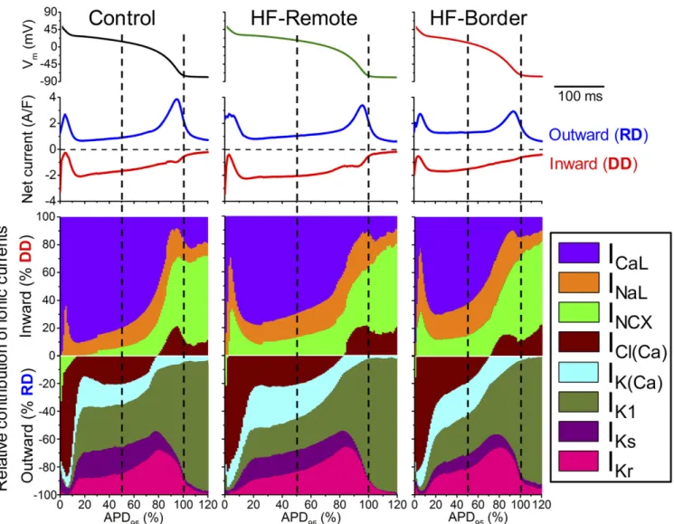
Relative Contributions of Different Ionic Currents in Reshaping APs in Ischemic HF.Vmis determined instantaneously by counterbalancing
inward and outward currents, such that the AP is shaped by in- tegrating all ionic currents. Figs. 4 and 5 show the magnitude and time course of each ionic current during the AP. The contribution of a particular current on the AP at any time point is determined by its relative strength compared with all other currents (Fig. 6 and Fig. S1). To understand the relative contribution of each current to AP shape, we calculated total depolarization drive (DD) as the sum of all inward currents and the total re- polarization drive (RD) as the sum of all outward currents (net currents in Fig. 6). We also express each current as percent of DD or RD at each point in time (Fig. 6, ionic fingerprints).
Among the depolarizing currents, in healthy controls (Fig. 6, Left)ICaL is the largest inward current during the AP plateau phase, much larger thanINaLandINCX. Fast Na+current (INa) during AP upstroke would saturate the amplifier and was not recorded (>50 times larger than peakICaL). However, during late repolarization (phase 3),INaLandINCXgain more influence, andICl(Ca)also becomes inward, contributing to DD. At terminal AP repolarization,INCXbecomes the dominant inward current, and remains the dominant inward current in phase 4. Compared with control, HF-remote myocytes (Fig. 6, Center) show de- creasedICaLbut increasedINaLandINCX. In the HF-border (Fig.
6,Right), the changes in these inward currents follow the same trend but are more pronounced, with the relative contribution of INaL+INCXdoubled at the AP midplateau time point.
Among the repolarizing currents, in healthy controls (Fig. 6, Left, Lower) phase 1 early repolarization of the AP is mainly caused by the outward ICl(Ca) current, with minor contribution from the reverse-modeINCX. In porcine myocytes, transient out- ward K+current (Ito) was not detectable. Hence, in this species, ICl(Ca)rather than Itois the main early repolarizing current. At midplateau of the AP, the relative contribution of several repo- larizing currents [ICl(Ca),IK(Ca),IKr,IKs,IK1] is about equal (∼20%
Fig. 4. Major inward currents during ventricular AP usingselfAP-clamp technique. (A) Representative current traces measured under the cell’s own AP at 1-Hz steady-state pacing in the control, HF-remote, and HF-border. Panels above show the APs used as voltage commands. GS-458967, ORM-10962, and nifedipine were used to record late Na+current (INaL), Na+/Ca2+exchanger current (INCX), and L-type Ca2+current (ICaL), respectively. (B) Peak and midplateau current densities as well as net charges carried by the corresponding ion channels. Mean±SEM,n=6–16 cells/4–6 animals. ANOVA with Bonferroni posttest; n.s., not significant, **P<0.01, ***P<0.001.
PHYSIOLOGYPNASPLUS
of RD). Compared with the control, in HF-Remote (Fig. 6, Center,Lower) the outwardINCXduring phase 1 is diminished;
IK(Ca)is increased during the plateau phase andIK1is decreased.
In the HF-border (Fig. 6,Right,Lower), changes of these outward currents follow the same trend, but the magnitude of changes are more pronounced. Important to the coupling between Ca2+sig- naling and electrical systems, the contribution of Ca2+-activated currents [IK(Ca),ICl(Ca)] to the total repolarizing drive are signifi- cantly increased [IK(Ca)+ICl(Ca)∼50% of RD] at midplateau in both the HF-border and HF-remote cells. The ionic fingerprints in Fig. 6 succinctly convey comprehensive information on the relative power of different ionic currents to shape the AP and the relative contribution of each current to electrophysiological remodeling in ischemic HF.
Discussion
Here we study a large-animal model of chronic ischemic HF to understand increased susceptibility to cardiac arrhythmias. Prior large-animal studies considered earlier stages post-MI (2–7 d) or
relatively early after MI healing (8 wk) (2, 5). Our study on a 5-mo post-MI porcine model with chronic HF is unique in de- termining the long-term remodeling of ionic currents in chronic ischemic HF. We found that the APD is shortened in the infarct border zone but lengthened in the remote zone. Our APD data agree generally with prior findings of shortened APD in the border zone of healed infarct in cat (14), but prolonged remote- zone ventricular APD from rabbit (10), canine (15), and human hearts (16). Importantly, we systematically studied differential changes between border and remote zones, revealing heteroge- neous remodeling in post-MI HF, increasing the inhomogeneity of ventricular AP repolarization. Furthermore, we provide mechanistic insights of the underlying ionic bases for the AP profile changes.
In studying depolarizing currents,ICaL was previously found decreased or unchanged as a result of two opposing effects:
decreased expression and increased phosphorylation of the channel protein in the ventricular myocytes isolated from human HF (16). We found thatICaLdensity and integral during the AP
steady-state pacing in the control, HF-remote, and HF-border. Panels above show the APs used as voltage commands. E-4031, HMR-1556, and Ba2+were used to record rapid and slow delayed rectifier K+currents (IKrandIKs), and inward rectifier K+current (IK1), respectively. Apamin and 9-anthracenecarboxylic acid were used to record Ca2+-activated K+and Cl−currents [IK(Ca)andICl(Ca)], respectively. (B) Peak current densities and net charges carried byIKr,IKs, andIK1, as well as midplateau and diastolic density ofIK1. (C) Peak current densities and net charges carried byIK(Ca), inward and outwardICl(Ca), as well as midplateau and diastolic density ofICl(Ca). Mean±SEM,n=6–16 cells/4–6 animals. ANOVA with Bonferroni posttest; n.s., not significant, *P<0.05,
**P<0.01, ***P<0.001.
was significantly decreased only in HF-border zone but not in HF-remote zone ventricular myocytes.INaLhas been recorded as a tiny Na+ current under voltage-clamp, and was slightly in- creased in HF (15). Here we show that theINaLunder selfAP- clamp is a sustained inward current throughout the AP plateau, shaping the APD and increasing the Na+entry during AP.INaLis markedly increased in HF.
Previously, INCX was found to be up-regulated in HF, which causes larger inwardINCXduring spontaneous SR Ca2+release, thereby increasing DADs and triggered arrhythmia susceptibility (10, 17). Here we used a selective NCX inhibitor ORM-10962 to record the dynamicINCXduring the AP. In control myocytes,INCX
was briefly outward early in the AP, but then predominantly in- ward throughout the remainder of the AP peaking during rapid repolarization. This is similar to prior indirectINCXmeasurements during the AP in rabbit and human ventricular myocytes (18). In post-MI HF, there was no early outwardINCX, and inwardINCX was a sustained current during the plateau (Fig. 4). This finding differs from the above rabbit and human HF results, whereINCX was outward through much of the AP plateau. The difference may be attributable to better maintained [Ca2+]i transients in our failing porcine hearts (Fig. 3) and possibly less rise in [Na+]i, both of which would favor more inwardINCX. The higher inwardINCX
observed here during the plateau (with similar [Ca2+]itransients)
is consistent with functional up-regulation of NCX in our porcine model. Hence, the overall INCX dynamics during AP promotes earlier Ca2+extrusion in HF than in control.
In studying repolarizing currents, reduced repolarization re- serve and altered repolarization currents are hallmarks of HF (19, 20). Previous studies reported that delayed rectifier K+ currents were decreased or unchanged in HF (1). We found no change in basal IKs, which is relatively small in the absence of β-adrenergic stimulation, and only a slight decrease inIKrduring AP in the both HF zones.IK1is reportedly decreased in human and rabbit HF (10, 19). We recorded IK1 dynamics during AP and found that peakIK1density is significantly decreased in the HF-border, but unaltered in the HF-remote. Interestingly, mid- plateauIK1is increased in the HF-border, consistent with altered IK1 rectification, which is attributed to intracellular Mg2+ and polyamines that could be altered in HF (21, 22).IK1represents the dominant outward current in AP repolarization (phase 3) and during diastole (where it stabilizes restingVm).
Na/K-ATPase pump current (INKA, not measured here) con- tributes a smaller diastolic outward current (vs. IK1). INKA is expected to be larger during the AP plateau, and similar in amplitude (∼0.3 A/F) (23) to the four currents measured here [ICl(Ca),IK(Ca),IKr, andIKs] that dominate during the AP plateau (Figs. 5 and 6). Moreover, increases in [Na+]i, which are known
Fig. 6. Dynamical relations of ionic currents during the AP. The contribution of each inward and outward current was normalized to the momentary total outward (RD) or inward (DD) membrane current calculated based on the measured eight ionic current densities. Ionic currents measured in each cell were also normalized to the corresponding APD95. Vertical dashed lines correspond to Plateau50and APD95.
PHYSIOLOGYPNASPLUS
larger outward INKA and outward INCX. Because the overall conductance is low during the AP plateau, relatively small changes in these Na+- and Ca2+-dependent currents can signif- icantly impact repolarization and APD (23).
The Ca2+-activated K+ and Cl− currents have rarely been considered in shaping ventricular APs. Here we recorded IK(Ca) andICl(Ca)underselfAP-clamp with [Ca2+]itransient and demon- strated that these two currents are significant in the control and HF porcine myocytes, and both currents are further increased in HF. Previous studies also reported up-regulation ofIK(Ca)in hu- man cardiomyocytes independent of [Ca2+]ichanges (24).ICl(Ca)
was increased HF in rabbit, but not in human (25). Here we show that ICl(Ca) is increased in porcine HF. Furthermore, our data reveal that the increased outwardICl(Ca)andIK(Ca)counteract the increased inward currents (INaL, INCX) during the AP plateau in HF.
TheselfAP-clamp with sequential current analysis is powerful in showing how multiple ionic currents behave during the physio- logical AP in a single myocyte. Nonetheless, the absolute precision for each individual current may be limited by the cumulative ad- dition of relatively channel-selective agents and time course of experiments. To this end, the low diastolic levels ofIKr,IKs, and ICaLindicated in Fig. 6 are likely close to zero, but are exaggerated graphically because total DD and RD are quite small during di- astole (and that is pressing the precision limits of the method).
Moreover, the main likely diastolic inward currents that balance outwardIK1 +INKAat the stable resting potential are probably INCX, Na+leak, and Ca2+leak. In addition, there is evidence for a TTX-sensitive diastolic leak that is increased in HF (26), and so may be mediated by Na+channels.
HF involves not only remodeling of ionic currents and APs but also diastolic arrhythmogenic events. DADs were frequently ob- served in HF, providing the arrhythmogenic trigger (10, 25). We also found significantly increased DADs and triggered APs. The DADs were mediated byINCX[not byICl(Ca)], similar to previous findings in rabbit and human HF (10, 25). Moreover, we found that the late Na+current, elevated diastolic [Ca2+]i, and CaMKII- dependent SR Ca2+leak all contribute to significantly increasing the frequency of DADs and tAPs, particularly in the infarct bor- der zone. Hence, in porcine chronic ischemic HF, DADs and tAPs are significantly increased and contribute as triggers for initiating arrhythmias, in conjunction with significantly different APD be- tween HF-remote and HF-border cells that may enhance the substrate for reentrant arrhythmias.
Limitations
One limitation of using a porcine model for human translational relevance is our observed lack of Ito in pig myocytes, which contributes to transmural AP differences in the human heart.
Myocardial infarction and remodeling may also affect the transmural regions differently, which could influence the AP dispersion. However, because Itois significantly reduced in HF (1), the lack ofItoin pig may not be a major limitation for dif- ferential repolarization remodeling in infarct border vs. remote zones in HF. The ultimate test would come from studying human cardiac myocytes, which is limited by the availability of adult human HF hearts. Applying the AP-clamp sequential dissection method could aid future human myocyte study. Moreover, a systematic study of Ca2+ handling in different ventricular re- gions and at various pacing rates would further inform the arrhythmogenic mechanisms in ischemic HF. We also did not induce arrhythmias in vivo or in isolated heart optical mapping studies because of the cost of the chronic porcine model, limited number of animals available, and other parallel uses of these pigs. Inducibility of unidirectional block and reentry requires steep APD differences over relatively short distances. Here, the border and remote zones from which myocytes were isolated
spatial detail, and hence cannot comment on APD gradient steepness in vivo. Finally, in relating single-cell electrophysiology to arrhythmogenesis in the heart, one must also consider other im- portant factors, including the electrical coupling between cells, fi- brosis and mechanical load effects, as well as neurohormonal changes that modify cardiac excitation–contraction coupling in HF.
Conclusion and Perspectives
Our data reveal that chronic ischemic HF involves remodeling of a multitude of ion channels and exchangers heterogeneously in the infarct border and remote zones. Even though no individual current shows dramatic changes, the integration of small changes from multiple ionic currents significantly remodel the AP profile.
Differential changes in multiple currents integrate at the cell level to shorten APD in the border zone but prolong APD in the remote zone, giving rise to pronounced dispersion of APD at the tissue level. Such emerging APD dispersions with both long-QT and short-QT substrates create imbalanced electrophysiology landscape to promote arrhythmias in post-MI ischemic HF. Our findings demonstrate that in-depth mechanistic understanding requires comprehensive studies of many ionic currents and sug- gest a framework for precision therapeutics. Indeed, the complex changes of ion channels in HF support the hypothesis that drugs influencing multiple ion channels (e.g., amiodarone, ranolazine) may provide more effective treatment if the integrated effects can rebalance the electrophysiology landscape. Moreover, drugs reducing pathological SR Ca2+ leak (e.g., CaMKII inhibitors) and drugs targeting defective neurohormonal regulations that in- fluence complex changes in multiple ion channels, may also have advantages in suppressing arrhythmias. Given that the strategy of targeting single-ion channels has met failures, the comprehensive analysis that we have done here provide integrated approaches and rich data to inform future development of new strategies for treating arrhythmias in ischemic heart failure.
Materials and Methods
Details are provided inSI Materials and Methods.
Animals.Four- to 6-mo-old adult Yucatan minipigs were subjected to microbead embolization of the first diagonal branch of the LAD, which caused trans- mural MI and progressive reduction in EF over 5 mo (Table S1), at which time left ventricular (LV) myocytes were isolated. The porcine model of chronic MI used is this study was developed to provide a clinically relevant large-animal ischemic cardiomyopathy model, as previously described (6). Cardiomyocytes from the remote or border zone of the infarct were obtained by enzymatic digestion using cannulation and perfusion of the left coronary vasculature.
As control, cardiomyocytes were isolated from the same region of the heart of healthy age-matched sham control minipigs. All animal handling and laboratory procedures followed US National Institutes of Health guidelines and were approved by the Institutional Animal Care and Use Committee of the University of California, Davis.
Electrophysiology. Recordings were performed in isolated ventricular cardiomyocytes using whole-cell patch-clamp with physiological solutions at 36 °C (for ionic composition, seeSI Materials and Methods). APs were evoked in current-clamp experiments where cells were stimulated with short suprathreshold depolarizing pulses at 0.2- to 5-Hz pacing frequencies de- livered via the patch pipette. Ionic currents during the AP were measured usingselfAP-clamp with physiological solutions (at 1-Hz pacing), preserved [Ca2+]icycling, and sequential block of specific ionic currents (Fig. S1andSI Material and Methods) using selective ion channel inhibitors (Table S2), as previously described (7, 27).
[Ca2+]i Transient and Myocyte Contraction. Parallel [Ca2+]i transients and contractions were assessed by Fura-2 fluorescence ratio (F340/F380) and sar- comere length measurements in field-stimulated myocytes (at 22 °C and 36 °C, as indicated, and 0.5-Hz pacing) using an IonOptix system, as pre- viously described (28).
Confocal Imaging of Ca2+Signals.Diastolic Ca2+sparks were detected by Fluo- 4 fluorescence in intact myocytes (at 22 °C) using an Olympus FluoView FV1000 confocal microscope, as previously described (28).
Echocardiography.Averaged data from three consecutive cardiac cycles were obtained in 2D and M-mode assessments using the standard LV outflow tract view. Animals were in dorsal recumbency during imaging with a S5-1 linear probe (Philips Healthcare) under anesthesia (during MI induction) or deep sedation (during follow-up at 8 wk and 5-mo post-MI).
Statistical Analysis.Averaged data are presented as mean±SEM. The number of cells in each experimental group was reported asn=number of cells/
number of animals, and the cells in each group came from three to seven individual animals. Statistical significance of differences was evaluated using
ANOVA to compare multiple groups and Bonferroni posttest was used for pairwise comparisons for continuous variables. Categorical outcomes were evaluated using Fisher’s exact test. A value ofP <0.05 was considered significant.
ACKNOWLEDGMENTS.We thank Matthew L. Stein, Ian P. Palmer, Maximilien Bergman, Maura Ferrero, Lisa Gilardoni, and Mark Jaradeh for their help in animal care, cell isolation, and laboratory tasks. This work was supported by National Institutes of Health Grants R01-HL123526 (to Y.C.-I.), R01-HL90880 (to L.T.I. and Y.C.-I.), P01-HL080101 and R01-HL30077 (to D.M.B.), and R01-HL085727 and R01-HL085844 (to N.C.); VA Merit Review Grants I01 BX000576 and I01 CX001490 (to N.C.); the Hungarian Scientific Research Fund OTKA101196 (to T.B.); California Institute for Regenerative Medicine Grant TR3 05626 Grant (to C.S.S. and W.D.B.); and American Heart Association Grant 14GRNT20510041 (to Y.C.-I.).
1. Tomaselli GF, Marbán E (1999) Electrophysiological remodeling in hypertrophy and heart failure.Cardiovasc Res42:270–283.
2. Dun W, Baba S, Yagi T, Boyden PA (2004) Dynamic remodeling of K+and Ca2+
currents in cells that survived in the epicardial border zone of canine healed infarcted heart.Am J Physiol Heart Circ Physiol287:H1046–H1054.
3. Kim YK, et al. (2002) Altered excitation-contraction coupling in myocytes from re- modeled myocardium after chronic myocardial infarction.J Mol Cell Cardiol34:63–73.
4. Shimkunas R, et al. (2013) Left ventricular myocardial contractility is depressed in the borderzone after posterolateral myocardial infarction.Ann Thorac Surg 95:
1619–1625.
5. Yuan F, et al. (1999) Characteristics of I(K) and its response to quinidine in experi- mental healed myocardial infarction.J Cardiovasc Electrophysiol10:844–854.
6. Hanes DW, et al. (2015) Embolization of the first diagonal branch of the left anterior descending coronary artery as a porcine model of chronic trans-mural myocardial infarction.J Transl Med13:187.
7. Banyasz T, Horvath B, Jian Z, Izu LT, Chen-Izu Y (2011) Sequential dissection of mul- tiple ionic currents in single cardiac myocytes under action potential-clamp.J Mol Cell Cardiol50:578–581.
8. Sanderson J (1996) The SWORD of Damocles.Lancet348:2–3.
9. Piccirillo G, et al. (2007) QT variability strongly predicts sudden cardiac death in asymptomatic subjects with mild or moderate left ventricular systolic dysfunction: A prospective study.Eur Heart J28:1344–1350.
10. Pogwizd SM, Schlotthauer K, Li L, Yuan W, Bers DM (2001) Arrhythmogenesis and contractile dysfunction in heart failure: Roles of sodium-calcium exchange, inward rectifier potassium current, and residual beta-adrenergic responsiveness.Circ Res88:
1159–1167.
11. Johnson DM, et al. (2013) Diastolic spontaneous calcium release from the sarcoplas- mic reticulum increases beat-to-beat variability of repolarization in canine ventricular myocytes afterβ-adrenergic stimulation.Circ Res112:246–256.
12. Ai X, Curran JW, Shannon TR, Bers DM, Pogwizd SM (2005) Ca2+/calmodulin- dependent protein kinase modulates cardiac ryanodine receptor phosphorylation and sarcoplasmic reticulum Ca2+leak in heart failure.Circ Res97:1314–1322.
13. Puglisi JL, Bassani RA, Bassani JW, Amin JN, Bers DM (1996) Temperature and relative contributions of Ca transport systems in cardiac myocyte relaxation.Am J Physiol270:
H1772–H1778.
14. Wong SS, et al. (1982) Dissimilarities in the electrophysiological abnormalities of lateral border and central infarct zone cells after healing of myocardial infarction in cats.Circ Res51:486–493.
15. Valdivia CR, et al. (2005) Increased late sodium current in myocytes from a canine heart failure model and from failing human heart.J Mol Cell Cardiol38:475–483.
16. Chen X, et al. (2002) L-type Ca2+channel density and regulation are altered in failing human ventricular myocytes and recover after support with mechanical assist devices.
Circ Res91:517–524.
17. Hasenfuss G, et al. (1999) Relationship between Na+-Ca2+-exchanger protein levels and diastolic function of failing human myocardium.Circulation99:641–648.
18. Weber CR, Piacentino V, 3rd, Ginsburg KS, Houser SR, Bers DM (2002) Na(+)-Ca(2+) exchange current and submembrane [Ca(2+)] during the cardiac action potential.Circ Res90:182–189.
19. Beuckelmann DJ, Näbauer M, Erdmann E (1993) Alterations of K+currents in isolated human ventricular myocytes from patients with terminal heart failure.Circ Res73:
379–385.
20. Hegyi B, et al. (2018) Altered repolarization reserve in failing rabbit ventricular my- ocytes: Calcium andβ-adrenergic effects on delayed- and inward-rectifier potassium currents.Circ Arrhythm Electrophysiol11:e005852.
21. Zaza A, Rocchetti M, Brioschi A, Cantadori A, Ferroni A (1998) Dynamic Ca2+-induced inward rectification of K+current during the ventricular action potential.Circ Res82:
947–956.
22. Meana C, et al. (2016) Correlation between endogenous polyamines in human cardiac tissues and clinical parameters in patients with heart failure.J Cell Mol Med20:
302–312.
23. Grandi E, Pasqualini FS, Bers DM (2010) A novel computational model of the human ventricular action potential and Ca transient.J Mol Cell Cardiol48:112–121.
24. Yu CC, et al. (2015) Small conductance calcium-activated potassium current is im- portant in transmural repolarization of failing human ventricles. Circ Arrhythm Electrophysiol8:667–676.
25. Verkerk AO, et al. (2001) Ionic mechanism of delayed afterdepolarizations in ven- tricular cells isolated from human end-stage failing hearts.Circulation104:2728–2733.
26. Despa S, Islam MA, Weber CR, Pogwizd SM, Bers DM (2002) Intracellular Na(+) con- centration is elevated in heart failure but Na/K pump function is unchanged.
Circulation105:2543–2548.
27. Chen-Izu Y, Izu LT, Hegyi B, Bányász T (2017) Recording of ionic currents under physiological conditions: Action potential-clamp and‘Onion-Peeling’ techniques.
Modern Tools of Biophysics, ed Jue T (Springer, New York), pp 31–48.
28. Jian Z, et al. (2014) Mechanochemotransduction during cardiomyocyte contraction is mediated by localized nitric oxide signaling.Sci Signal7:ra27.
PHYSIOLOGYPNASPLUS
![Fig. 3. Changes in [Ca 2+ ] i transient and contraction in ischemic HF. (A) Representative [Ca 2+ ] i transient and simultaneously recorded sarcomere shortening evoked by field stimulation at 0.5-Hz pacing frequency (at 22 °C)](https://thumb-eu.123doks.com/thumbv2/9dokorg/1338224.108627/4.877.73.818.818.1028/transient-contraction-representative-transient-simultaneously-sarcomere-shortening-stimulation.webp)