UNCORRECTED PR
OOF
1
Review
2Q1
On the role of 4-hydroxynonenal in health and disease
3Q2
Miklós Csala
a, Tamás Kardon
a, Balázs Legeza
b, Beáta Lizák
a, József Mandl
a, Éva Margittai
c, Ferenc Puskás
d,
4
Péter Száraz
e, Péter Szelényi
a, Gábor Bánhegyi
a,⁎
5 aDepartment of Medical Chemistry, Molecular Biology and Pathobiochemistry, Semmelweis University Budapest, Budapest, Hungary 6 bDepartment of Pediatrics, University of California, San Francisco, CA, USA
7 cInstitute of Human Physiology and Clinical Experimental Research, Semmelweis University, Budapest, Hungary 8 dDepartment of Anesthesiology, University of Colorado, Denver, CO, USA
9 eDepartment of Physiology, University of Toronto, Toronto, Ontario, Canada
a b s t r a c t
1 0 a r t i c l e i n f o
11 Article history:
12 Received 1 October 2014
13 Received in revised form 16 December 2014 14 Accepted 23 January 2015
15 Available online xxxx 16
17 The authors and Valeria Mile dedicate this arti- 18 cle to the pioneer of HNE research, Angelo 19 Benedetti on the occasion of his 70th birthday.
20 Keywords:
21 4-Hydroxynonenal 22 Lipid peroxidation 23 Nrf2
24 Electrophilic stress 25 Proteostasis
Polyunsaturated fatty acids are susceptible to peroxidation and they yield various degradation products, includ- 26 ing the mainα,β-unsaturated hydroxyalkenal, 4-hydroxy-2,3-trans-nonenal (HNE) in oxidative stress. Due to its 27 high reactivity, HNE interacts with various macromolecules of the cell, and this general toxicity clearly contrib- 28 utes to a wide variety of pathological conditions. In addition, growing evidence suggests a more specific function 29 of HNE in electrophilic signaling as a second messenger of oxidative/electrophilic stress. It can induce antioxidant 30 defense mechanisms to restrain its own production and to enhance the cellular protection against oxidative 31 stress. Moreover, HNE-mediated signaling can largely influence the fate of the cell through modulating major cel- 32 lular processes, such as autophagy, proliferation and apoptosis. This review focuses on the molecular mechanisms 33 underlying the signaling and regulatory functions of HNE. The role of HNE in the pathophysiology of cancer, 34 cardiovascular and neurodegenerative diseases is also discussed. 35
36 © 2015 Published by Elsevier B.V.
37 38 39 40
41 1. Synthesis and breakdown of HNE
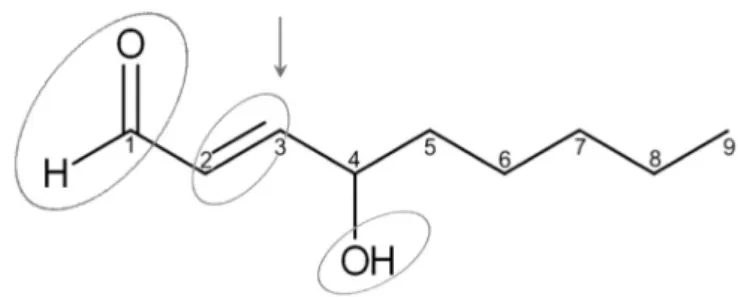
42 4-Hydroxy-2,3-trans-nonenal (4-hydroxynonenal, HNE) is anα,β- 43 unsaturated hydroxyalkenal. The molecule is highly reactive due to its 44 three functional groups: an aldehyde, a double bond (alkene) between 45 carbon C2 and C3, and a secondary alcohol at carbon C4 (Fig. 1). Carbon 46 C1 and C3 are electrophilic sites and carbon C1 is also a redox center.
47 The compound wasfirst described in autooxidized polyunsaturated 48 fatty acids (PUFAs) and triglycerides[1]. Thefirst report on the forma- 49 tion of HNE in a biological system was published Benedetti et al. in 50 Biochim. Biophys. Acta in 1980[2]. This pioneer work investigated the 51 pathological effects of NADPH-Fe induced lipid peroxidation in liver
microsomes, including the defective activity of glucose-6-phosphatase, 52 and identified HNE as the underlying toxic intermediate. 53
1.1. HNE formation 54
Lipid peroxidation is a general term, which refers to different mech- 55 anisms and can be classified as enzymatic, non-enzymatic non-radical 56 and non-enzymatic free-radical mediated peroxidation [3]. Free- 57 radical non-enzymatic peroxidation of PUFAs is the dominant pathway 58 in oxidative stress induced by radiation, heat, free radicals, xenobiotics, 59 metal ions or reactive oxygen or nitrogen species (ROS or RNS). Hydrox- 60 yl radical (OH·), the most powerful initiator of lipid peroxidation can be 61 generated from hydrogen peroxide via the Fenton–and Haber–Weiss 62 reactions, in the presence of free iron or copper ions. Lipid peroxidation 63 can be initiated by a hydroxyl-radical-mediated removal of an H•radical 64 from a lipid (LH), which yields a lipid radical (L•). In the propagation 65 phase, L•reacts with oxygen and forms a lipoperoxyl radical (LOO•). 66 Lipoperoxyl radical in turn reacts with another PUFA to yield a new L• 67 and a lipid hydroperoxyde (LOOH). Thus, one hydroxyl radical can gen- 68 erate a high number of lipid hydroperoxydes until the chain reaction is 69 terminated by a chain-breaking antioxidant (e.g. tocopherol). 70
Lipid hydroperoxydes are regarded as primary products of lipid per- 71 oxidation (Fig. 2). However, these compounds are unstable: they can be 72 Biochimica et Biophysica Acta xxx (2015) xxx–xxx
Abbreviations:AD, Alzheimer's disease; ALDH, aldehyde dehydrogenase; AMI, acute myocardial infarction; ARE, antioxidant response element; CDK, cyclin-dependent kinase;
CHF,chronic heartfailure;DHLA,dihydrolipoicacid;ER,endoplasmicreticulum;GSH,gluta- thione; HL-60, human promyelocytic cell line; HNE, 4-hydroxy-2,3-trans-nonenal; Keap1, Kelch ECH associating protein 1; PC12, rat phaeochromocytoma cell line; pRb, retinoblasto- ma protein; PUFA, polyunsaturated fatty acid; ROS, reactive oxygen species; UPR, unfolded protein response
⁎ Corresponding author at: Department of Medical Chemistry, Molecular Biology and Pathobiochemistry, Semmelweis University Budapest, 1093 Budapest, Tűzoltó utca 37- 47, Hungary. Tel.: +36 1 4591500; fax: +36 1 2662615.
E-mail address:banhegyi.gabor@med.semmelweis-univ.hu(G. Bánhegyi).
BBADIS-64157; No. of pages: 13; 4C:
http://dx.doi.org/10.1016/j.bbadis.2015.01.015 0925-4439/© 2015 Published by Elsevier B.V.
Contents lists available atScienceDirect
Biochimica et Biophysica Acta
j o u r n a l h o m e p a g e :w w w . e l s e v i e r . c o m / l o c a t e / b b a d i s
UNCORRECTED PR
OOF
73 transformed into peroxyl and alkoxyl (LO·) radicals and can be 74 decomposed to secondary products. Alkoxyl radicals are especially 75 prone toβ-scission, which results in the formation of short-chain prod- 76 ucts, including HNE (Fig. 2). Among the end products of lipid peroxida- 77 tion other reactive aldehydes, such as malondialdehyde (MDA) are also 78 present; for more detailed description of the biosynthetic pathways we 79 refer to recent excellent reviews[4,5].
80 The secondary products of lipid peroxidation are reactive, yet rela- 81 tively stable compounds, they can travel remarkable distances from 82 the site of synthesis. HNE, for instance can reach well measurable 83 concentrations in the tissues and in the blood, thus it can be regarded 84 as a biomarker of the oxidative stress. Its physiological concentration 85 is in the submicromolar range (b0.1μM), while in oxidative stress, 86 even micromolar levels can be observed[6].
87 1.2. Biotransformation of HNE
88 In situ lipid peroxidation is not the only source of HNE as it can also 89 be taken up with the food[7]. Thus, HNE is both a xeno- and an endobi- 90 otic substrate for biotransformation. The metabolism of HNE (and other 91 secondary lipid peroxidation products) is rapid and effective, involving 92 all phases of biotransformation. Since the molecule already possesses 93 functional groups suitable for conjugation, the phases I and II of 94 biotransformation can be reversed. It should be noted that the relative 95 contribution of various pathways to HNE biotransformation markedly 96 differs in different species and tissues (see e.g.[8]), which can be in the 97 background of variable toxicity of HNE.
1.2.1. Phase I reactions 98
The aldehyde group is a substrate for oxidoreductases, and it can be 99 reduced to an alcoholic hydroxyl or oxidized to a carboxylic group. The 100 participating enzymes are aldose reductase and aldehyde dehydroge- 101 nase; they form 1,4-dihydroxynonene and 4-hydroxynonenoate, 102 respectively[9,10]. The latter product can undergo a consecutiveβ- 103 oxidation. Several cytochrome P450 isozymes have been also shown 104 to catalyze both the oxidation[11]and the reduction[12]of the alde- 105 hyde group. Cytochome P450s of the CYP4A family are also involved 106 in the oxidative metabolism of HNE, by catalyzing theω- andω-1 oxida- 107 tion of 4-hydroxynonenoate[13,14]. Ketogenic diet upregulatesω- and 108 ω-1 hydroxyation of 4-hydroxynonenoate in rat liver via the induction 109 of CYP4A isozymes[14]. The hydroxyl group of carbon C4 and the 110 double bond between C2 and C3 are also subjects of oxidation or reduc- 111 tion, respectively. 112
1.2.2. Phase II reactions 113
The carbon-carbon double bond of HNE reacts with nucleophilic 114 thiol groups, including that of the tripeptide glutathione (GSH). Michael 115 addition leads to the formation of GSH conjugates. This spontaneous re- 116 action can be highly accelerated by glutathione-S-transferases. The con- 117 jugation reaction is present in most cells and tissues. 118
The glutathione conjugation can be followed by oxidoreductions 119 described above; and thence the glutathione conjugates of 1,4- 120 dihydroxynonane and 4-hydroxynonanate are formed. Aldose reduc- 121 tase has a low micromolar KMtowards the glutathione conjugate of 122 HNE, thus this metabolic pathways seems to be dominant in vivo[15]. 123 It should be noted that the glutathione conjugates of HNE are not in- 124 active product, but potential signal molecules. Mitogenic effect of HNE 125 has been reported to be mediated by the glutathione conjugate reduced 126 by aldose reductase in rat aortic smooth muscle cells [16]. These 127 compounds were also shown to mediate the inflammatory effect of 128 oxidative or glucotoxic stress in adipocytes[17,18]. Inhibition of aldose 129 reductase prevented systemic inflammation and cardiomyopathy upon 130 endotoxin treatment[19]. 131
The oxidized acidic derivatives can be further metabolized by cyto- 132 chrome P4504A, yieldingω-hydroxylated metabolites. The mercapturic 133 acid derivatives of these products are present in the urine and can serve 134 as biomarkers of in vivo lipid peroxidation (for a review see[20]). Glu- 135 tathione and mercapturic acid conjugates of HNE, 1,4-dihydroxynonane 136 and 4-hydroxynonanate are secreted also into the bile. 137
Cysteine can be also a conjugation partner for HNE. In a recent study 138 increased extracellular formation of HNE-cysteine conjugate was ob- 139 served in colon cells with a mutation of the adenomatous polyposis 140 coli gene; the reaction–together with the upregulation of aldehyde de- 141 hydrogenases, glutathione transferase and cystine transporter–confers 142 higher resistance towards HNE in mutant cells[21]. 143
1.2.3. Phase III reactions 144
MRP1 and MRP2 multidrug resistance proteins have been shown to 145 transport glutathione conjugates of HNE and to protect the cell from 146 HNE toxicity[22,23]. Another ATP dependent, but non-ABC transporter, 147 RLIP76 (Ral-interacting GTPase activating protein, also known as Ral- 148 binding protein 1) has high transport activity towards glutathione 149 conjugates of HNE; this protein accounts for the majority of the trans- 150 port[24,25]. Indeed, overexpression of RLIP76 abolished the mitogenic 151 effects of HNE and its glutathione conjugates observed in rat aortic 152 smooth muscle cells, while its downregulation promoted the mitogenic 153 effects[14]. 154
1.3. Adduct formation 155
HNE accumulation and toxicity are counteracted by an efficient and 156 rapid biotransformation. Yet, in spite of these protective efforts, HNE is 157 present in the cells at measurable concentrations, and gives rise to un- 158 desired events. HNE is able to react readily with various cellular 159 Fig. 1.Chemical structure of 4-hydroxy-2,3-trans-nonenal (HNE). Circles indicate the
reactive groups of the molecule; the arrow shows the site of nucleophilic attack.
Fig. 2.Outline of HNE metabolism. HNE is generated as secondary product of lipid perox- idation. It can be detoxified by various reactions of biotransformation; alternatively, it can form macromolecular adducts.
UNCORRECTED PR
OOF
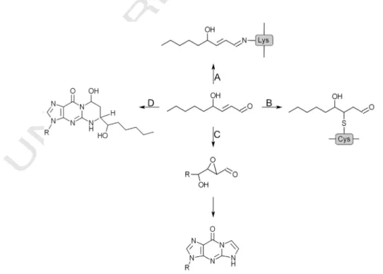
160 components, such as DNA, proteins and other molecules containing nu- 161 cleophilic thiol or amino groups[6,26]. The C2-C3 double bond is re- 162 sponsible for the Michael addition of thiol or amino compounds at the 163 C3 carbon (Fig. 3). The C1 aldehyde group can react with primary 164 amines and form of Schiff bases. While Schiff base formation is relatively 165 slow and reversible, Michael adducts are stable, thus the formation of 166 the latter is preferred in vivo. The cysteinyl, lysyl and histidyl residues 167 of proteins are the main targets of Michael addition. HNE can also 168 react with lipids containing amino groups (e.g. phosphatidyl ethanol- 169 amine, phosphatidyl serine, sphingosine) and with nucleic acids, prefer- 170 ably with the guanin base of DNA (Fig. 3). HNE-DNA adduct formation 171 and the consequent mutagenicity occur in two independent pathways:
172 i, direct interaction of HNE with two nitrogen atoms in guanine 173 moieties, which yields four isomeric propano adducts[27], and ii, oxida- 174 tion of HNE to 2,3-epoxy-4-hydroxy-nonanal[28]and a consecutive 175 formation of exocyclic etheno adducts of guanine, adenine or cytosine 176 moieties[29,30]. The different genotoxicity of HNE in various organs– 177 beside the differences in tissue level of HNE and intensity of DNA repair 178 mechanisms–can be attributed to the tissue- or cell-specific predomi- 179 nance of one of the two pathways, e.g. the latter one is dominant in 180Q3 hepatocytes[31]. (SeeFig. 4.)
181 Protein-HNE adduct formation can lead to alterations in the normal 182 functioning and modified activity of various proteins (for reviews, see 183 [32–34]. Because of its double reactivity (Michael addition and Schiff 184 bases)), HNE can contribute to protein cross-linking and induce a 185 carbonyl stress.
186 2. HNE in signaling
187 Only a few plausible effects can be outlined among the confusingly 188 pleiotropic actions of HNE in cell signaling. Antioxidant, heat shock 189 and ER stress responses are activated by HNE and these responses can 190 be integrated into a unified scheme. The three adaptive pathways 191 serve not only the disposal of HNE and the prevention of its toxic effects, 192 but also the negative regulation of proteostasis and stimulation of pro- 193 tein folding. These latter mechanisms help the cell to enter the lists 194 against adduct formation and misfolding.
2.1. The Keap1–Nrf2–ARE pathway 195
Exogenously added HNE has been shown to interact with practically 196 any signaling pathways in cells (for an extensive list of these interactions 197 see[35]), leading to pathological responses. These effects are due to the 198 covalent interactions of HNE with key proteins of signal transduction 199 pathways; there are no experimental evidences for classic, ligand-type 200 interactions. The main target of HNE as a major endogenous electrophil- 201 ic compound is the Keap1 (Kelch ECH associating protein 1)–Nrf2 202 (nuclear factor erythroid 2-related factor 2)–ARE (antioxidant response 203 element) pathway. This pathway senses prooxidant effects and orga- 204 nizes the cellular response against oxidant or electrophile stress. The 205 extremely cysteine-rich Keap1 under stress-free conditions binds Nrf2 206 and directs it towards ubiquitylation and proteasomal degradation. 207 Cysteine residues of Keap1 are sensitive to electrophilic attacks; while 208 electrophiles in high concentrations form adducts almost completely 209 with cysteinyl thiols, in low concentrations they react with a specific 210 subset of thiols[36,37]. It has been reported that Keap1 directly recog- 211 nizes alkenals (including HNE), NO and Zn2+by three distinct sensors 212 composed by cysteines and basic amino acids[38]. Phylogenetic analy- 213 ses showed that the alkenal sensor is the most ancient one. 214
Independently from the nature of the electrophilic stimuli, it seems 215 that the downstream events are the same; adduct formation or oxida- 216 tion of cysteine residues of Keap1 compromises its potential to recruit 217 the ubiquitin ligase complex, thus Nrf2 can accumulate. The stabilized 218 and accumulated Nrf2, a basic leucine zipper transcription factor, trans- 219 locates to the nucleus, binds to AREs and regulates target genes. The 220 proteins coded by these genes can be divided into different functional 221 groups. 222
NADPH generation is controlled by Nrf2 via glucose-6-phosphate 223 dehydrogenase, phosphogluconate dehydrogenase, malic enzyme 1 224 and isocitrate dehydrogenase 1 expression. NADPH is used by the 225 glutathione and thioredoxin systems as an electron donor. The expres- 226 sion of enzymes of glutathione synthesis, regeneration and utilization 227 (e.g. glutamate–cysteine ligase, glutathione reductase, glutathione-S- 228 transferases and glutathione peroxidase) are also regulated by Nrf2, 229 similarly to those of the thioredoxin system (thioredoxin 1, thioredoxin 230 reductase 1 and peroxiredoxin 1). Finally, the induction of NAD(P)H 231
Fig. 3.Protein and DNA adducts of HNE. Reaction between C1 aldehyde group and primary amines results in Schiff base formation (A). The C2–C3 double bond of HNE can react with cysteinyl, lysyl and histidyl residues of proteins yielding Michael adducts (B). HNE oxidized to an epoxide reacts with different bases in DNA forming etheno adducts (C). HNE can also form propano adducts with guanine bases of DNA (D).
UNCORRECTED PR
OOF
232 quinone oxidoreductase 1, heme oxygenase-1, UDP- 233 glucuronosyltransferases and multidrug-resistance associated 234 proteins completes the antioxidant effects[39]. Thefinal out- 235 comes of the signaling pathway are a reinforced antioxidant 236 defense and a redox shift towards the more reducing conditions.
237 Beside this simplified mechanism, Keap1 and Nrf2 are also targets of 238 protein kinases and phosphatases; many of them are also redox- 239 sensitive. Recentfindings show that the PI3K pathway activates Nrf2 240 [40]. The complexity of the upstream regulation of Nrf2 is evidenced 241 by its huge interactome[41,42].
242 Recent observations suggest that the Keap1–Nrf2–ARE pathway is 243 much more than a signaling system for electrophilic or oxidative stress;
244 it might have a crucial function in the healthy cells too. Some of its 245 signals (e.g. NO) are produced as a second messenger upon various 246 physiological situations. Moreover, ARE-responsive proteins include 247 NADPH generating enzymes, which have prominent role in the interme- 248 diary metabolism. These considerations raise the possibility that Keap1–
249 Nrf2 signaling plays a more general role in redox signaling[36,43].
250 2.2. Endoplasmic reticulum stress—the unfolded protein response
251 Accumulation of unfolded or misfolded proteins in the ER lumen re- 252 sults in ER stress, which provokes an adaptation mechanism called the 253 unfolded protein response (UPR). The UPR moderates protein transla- 254 tion and induces ER chaperones to restore protein folding and luminal 255 homeostasis, i.e. the normal macromolecular crowding, redox condi- 256 tions and Ca2+concentration[44]. HNE and its effect werefirstly de- 257 scribed in rat liver microsomes derived from the ER[2,45–47]; thus, it 258 is not surprising that HNE can interact with the machinery of protein 259 folding, leading to ER stress. Indeed, it has been reported that HNE reacts 260 with the key players of ER folding, protein disulfide isomerase[48,49], 261 and Grp78[48,50]. The ER stress sensor PERK kinase was activated
upon HNE exposure in rat aortic smooth muscle cells[48]. Exogenous 262 HNE provokes ER stress by a mechanism independent from ROS forma- 263 tion or glutathione depletion in endothelial cells[51]. The role of ROS as 264 a possible mediator of HNE-dependent ER stress was also excluded by 265 another study performed in PC12 cells[52]. 266
An important crosstalk between the UPR and the Nrf2 pathway has 267 also been described. It was observed that Nrf2 is a substrate for PERK 268 ER kinase, which is activated in ER stress[53]. Upon phosphorylation 269 the interaction between Keap1 and Nrf2 is weaker and the complex 270 dissociates. Thus, HNE-provoked ER stress amplifies the electrophilic 271 signaling pathway. Interestingly, in de-differentiated cells, a constitu- 272 tive (ER stress independent) PERK-Nrf2 signaling was observed, which 273 protects de-differentiated cells from chemotherapy by increased anti- 274 oxidant defense and drug efflux[54]. 275
2.3. The heat shock response 276
Heat shock response is an adaptive mechanism, which confers resis- 277 tance to various stress conditions caused by heat, electrophiles, xenobi- 278 otics etc. by initiating a transcriptional program primarily regulated by 279 heat shock factor 1 (HSF1). HSF1 upon stress conditions stimulates the 280 expression of various heat shock proteins (HSPs). Similarly to ER chap- 281 erones, their cytosolic counterparts HSP70 and HSP90 are also candi- 282 dates for HNE-dependent modifications[55,56]. Although the exact 283 mechanism is unknown, it can be supposed that modified HSPs lose 284 their repressor functions on HSF1 activation. Thus, HNE can induce 285 heat shock response by an indirect mechanism: binding to HSP70 re- 286 sults in the release of HSF1 from an inactive cytosolic complex and in 287 the increased transcription of the genes of heat shock proteins[33]. In 288 accordance with this mechanism, HSF1 via HSP70 induction prevents 289 the cell from HNE-induced apoptosis[57], while the HSP inhibitor 290 geldanamycin sensitizes the cells towards HNE-dependent cell death 291 Fig. 4.The Keap1–Nrf2–ARE pathway of electrophilic signaling. Under basal conditions, Keap1 binds Nrf2 and promotes its ubiquitylation by Cul3 ubiquitin ligase. Upon elec- trophilic exposure, electrophiles (e.g. HNE) react with thiol groups of Keap1, which results in the dissociation of Cul3 (Cul3 dissociation model) or in a partial detachment of Nrf2 (hinge and latch model). In both cases Nrf2 escapes ubiquitylation, accumulates, translocates into the nucleus, binds to antioxidant response elements (AREs) and in- creases the expression of Nrf2 target genes. Phosphorylation of Nrf2 by protein kinases (including PKC, MAPKs and the ER stress kinase PERK) also promotes the dissociation of the Keap1–Nrf2 complex.
UNCORRECTED PR
OOF
292 [58]. Heat shock response activators are candidates in the therapy of 293 protein conformational diseases; many of the candidate compounds 294 are electrophiles and activators of the Nrf2 pathway too[59,60].
295 2.4. HNE signaling and proteostasis
296 Beside redox/antioxidant regulation, the Keap1–Nrf2–ARE pathway 297 is an important regulator of proteostasis. The autophagy substrate p62 298 interacts with the Nrf2-binding site on Keap1, consequently deficient 299 autophagy results in the accumulation of p62, stabilization of Nrf2 and 300 transcriptional activation of Nrf2 target genes[61]. On the other hand, 301 p62 has been shown to be an ARE-responsive gene product[62], thus 302 p62 creates a positive feedback loop in Nrf2 signaling. Nrf2 dependent 303 mechanisms are also present in the crosstalk between proteasomal 304 and autophagic proteolysis. Increased autophagy was observed in mu- 305 tant mice with proteasomal dysfunction, which was stimulated by the 306 Keap1–Nrf2 pathway[63]. Nrf2 activation is required for the increased 307 level of the proteasomal regulator Pa28αβand for maximal proteolytic 308 capacity of proteasomes[64]. Thus, electrophiles and other Nrf2 activa- 309 tors can regulate stress-induced proteasomal activity and removal of 310 damaged/misfolded proteins[65]. Since HNE-dependent ER stress and 311 the consequent activation of UPR are well documented (see above), 312 the activation of ER associated degradation (ERAD)–a proteasomal 313 pathway–and ER stress dependent autophagy is not surprising[65].
314 Moreover, HNE can also directly interact with the apparatus of transla- 315 tion; e.g. adduct formation with eukaryotic elongation factor 2 results in 316 declined protein synthesis[66].
317 3. HNE in cell life and death decisions
318 As a potent protein-, lipid- and DNA-damaging agent, which triggers 319 ROS generation and directly depletes the antioxidant capacity of the 320 cells, HNE can be expected to cause profound changes in fundamental 321 cellular processes. Similarly to other insults, excessive lipid peroxidation 322 or HNE treatment is also managed by the cells in three ways. The prima- 323 ry response aims adaptation by eliminating the damaged molecules and 324 preventing any further aggravation and more serious consequences.
325 Cell cycle arrest (quiescence or senescence) and activation of autophagy 326 fit nicely into this strategy. A short-lasting mild or moderate challenge 327 (i.e. low concentrations of HNE) can be survived this way. If the com- 328 pensatory attempts fail and the cellular intactness is in jeopardy, pro- 329 grammed cell death still provides a chance to maintain the integrity of 330 the tissue and of the whole organism. However, the energy-requiring, 331 active process of apoptosis can be accomplished only if the cell has 332 enough time and still possesses its basic metabolic features. More severe 333 conditions, when the cell is quickly paralyzed by an overwhelming de- 334 struction (i.e. high concentrations of HNE), lead to necrosis[67]and a 335 more pronounced and expanded tissue loss usually combined with in- 336 flammation. Although an increase in the level of HNE clearly reduces 337 the chance of adaptation in favor of apoptosis or necrosis, the exact 338 HNE doses determining one or the other cell response cannot be uni- 339 formly defined as they vary according to the cell types, duration and 340 several environmental factors.
341 Lipid peroxidation is accelerated in oxidative stress and it is a source 342 of further ROS generation, itself. Therefore, the effects of HNE on au- 343 tophagy, cell proliferation and apoptosis are often indirect and hard to 344 distinguish from those of other ROS- and stress-activated signaling 345 mechanisms. This chapter focuses on the observations that convincingly 346 suggest a direct interaction of HNE with the key components of these 347 cellular processes.
348 3.1. Effect of HNE on autophagy
349 Besides proteasome-dependent proteolysis, autophagy is a major 350 physiological mechanism to degrade intracellular proteins in eukaryotic 351 cells. Autophagy plays an important role in differentiation and normal
growth control, and its enhancement is essential for the survival during 352 starvation. Macro- and microautophagy sequestrate complete regions 353 of the cytosol including organelles, while in chaperone-mediated 354 autophagy contributes to the cellular protein quality-control as it selec- 355 tively removes damaged cytosolic proteins one by one[68]. The pro- 356 teins sequestrated by autophagy are degraded in the lysosomes[69], 357 and hence turn into valuable endogenous nutrient sources. Accordingly, 358 the process is stimulated in energy deficient states by AMP-activated 359 protein kinase and inhibited during the wealth of nutrients by (insu- 360 lin)-Akt-mTOR signaling[70]. The ER can also generate signals to 361 enhance autophagy, in accordance to its function as a nutrient sensor 362 in the cells[71]. There is ample evidence that accumulation of ROS- 363 mediated damages in macromolecules and organelles plays a central 364 role in aging. Elimination of damaged cellular components through 365 autophagy is a major determinant of longevity, and the repeatedly dem- 366 onstrated anti-aging effect of caloric restriction can be at least partly 367 attributed to an enhanced autophagic activity[72,73]. 368
The role of chaperone-mediated autophagy in the removal of HNE- 369 modified proteins was demonstrated in transgenic mice overexpressing 370 LAMP2A receptor. The livers of these animals accumulate fewer dam- 371 aged proteins compared to age-matched wild-type controls due to an 372 enhanced chaperone-mediated autophagy[74]. Activation of autopha- 373 gy in oxidative stress can be considered as an antioxidant defense mech- 374 anism concerning the protective role it plays by eliminating damaged, 375 dysfunctional proteins and organelles[75,76]. Stimulation of autophagy 376 seems to be a pro-survival mechanism in cells undergoing excessive 377 lipid peroxidation. HNE and other lipid peroxidation-derived aldehydes 378 caused a remarkable increase in the number of autophagosomes in cul- 379 tured rat aortic smooth-muscle cells. Enhanced autophagy largely con- 380 tributed to the elimination of damaged proteins, and also prevented 381 cell death[77]. The ER as the organelle specified to protein synthesis, 382 maturation and quality control[78]is also involved in HNE-induced 383 autophagy. Luminal accumulation of damaged polypeptides is the key 384 trigger to the ER stress, which in turn is an important source of signals 385 stimulating autophagy[79]. The contribution of the ER stress to HNE- 386 enhanced autophagy was indeed demonstrated in rat aortic smooth- 387 muscle cells[48]. Sublethal concentration of HNE was also shown to 388 stimulate protective autophagy in differentiated SH-SY5Y neuroblasto- 389 ma cells. Although caspase-3 activation was also observed in the HNE- 390 treated cells, apoptotic cell death intensified only when autophagy 391 had been attenuated through inhibition of glycolysis[80]. 392
A complex and differential regulation of autophagy by HNE during 393 myocardial ischemia and reperfusion has been suggested by a recent 394 study, in which the effect of endogenous HNE was attenuated by over- 395 expression of aldehyde dehydrogenase 2 (ALDH2). ALDH2, one of the 396 major enzymes involved in neutralization of HNE reduced the tissue 397 damage in both ischemia and reperfusion; however its protective effect 398 was due to promotion or inhibition of autophagy in the two phases, re- 399 spectively. The results indicate that AMP-activated protein kinase and 400 Akt-mTOR signaling is compromised by HNE through interference 401 with upstream regulators, such as LKB1 and PTEN[81]. It can be also 402 concluded that HNE-induced autophagy, albeit protective against apo- 403 ptosis, has its own deleterious consequences especially in the myocardi- 404 um. Suppression of HNE-stimulated autophagy in ALDH2-transfected 405 mice has also been reported to ameliorate doxorubicin-induced myo- 406 cardial dysfunction[82]. 407
Intracellular proteins severely damaged by MDA and HNE may be- 408 come undegradable due to their aberrant covalent modifications and 409 their deposition is potentially cytotoxic. Lipofuscin of human retinal pig- 410 ment epithelium was shown to contain injured mitochondrial proteins, 411 which indicates the role of autophagy in the formation of such granules 412 [83]. Further analysis revealed that MDA and HNE interfere with lyso- 413 somal protease activities in these cells both directly and through modi- 414 fications of their substrate peptides[84]. The lysosomal dysfunction 415 results in increased lipofuscinogenesis and reduced autophagy activity 416 in vitro[85]. 417
UNCORRECTED PR
OOF
418 3.2. Effect of HNE on cell cycle and proliferation
419 Protein (Ser/Thr) phosphorylations catalyzed by various cyclin:cyclin- 420 dependent kinase (CDK) dimers are key events at the checkpoints in the 421 eukaryotic cell cycle. For instance, the restriction point is passed when ret- 422 inoblastoma protein (pRb) is hyperphosphorylated by cyclin:CDK dimers 423 accumulating as a result of mitogenic gene expression. The concomitant 424 liberation (and activation) of members of the E2F family of transcription 425 factors prepare the now committed cell to enter the S phase (DNA repli- 426 cation)[86]. CDK activities are controlled by positively and negatively act- 427 ing phosphorylations and can be halted by the association of CDK 428 inhibitors, such as p21 or p16[87]. The latter mechanism contributes to 429 the anti-proliferative action of p53 family transcription factors[88], 430 which in turn is activated by various stress conditions, oncogenic insult 431 and severe DNA-damage[89].
432 3.2.1. Anti-proliferative effects
433 An inverse relationship was found between cell proliferation and 434 lipid peroxidation in various cell types, i.e. it is generally observed that 435 rapid proliferation is accompanied by low level of lipid peroxidation in 436 different tissues including tumors[90]. The phenomenon is at least part- 437 ly due to low level of peroxidizable fatty acids in intensively proliferat- 438 ing cells. Enrichment with arachidonic acid renders tumor cells more 439 susceptible to oxidative stress and to cell cycle arrest through lipid per- 440 oxidation[91,92]. Thesefindings shed light on the potential role of HNE 441 in the modulation of cell cycle control[93]. HNE was found to reduce the 442 proliferative capacity of K562 human erythroleukemic and HL-60 443 human promyelocytic cells[94]. Not only the S phase progression was 444 inhibited, but a concomitant granulocytic differentiation was also initi- 445 ated in HNE-treated HL-60 cells[95]. Diversion from proliferation to- 446 wards differentiation implies the modulation of master regulators of 447 these processes. Indeed, the expression of proto-oncogen c-myc and c- 448 myb transcription factors is transiently suppressed in HL-60 cells after 449 HNE treatment despite unaltered N-ras expression and cAMP levels 450 [96,97]. A remarkable decrease in the c-myc mRNA level was also 451 demonstrated in K562 cells after the addition of HNE at low (1–3μM) 452 concentration[98]. Further investigation revealed that HNE affects cell 453 cycle control around the restriction point by modifying the expression 454 of several genes. It was found to down-regulate D1, D2, and A cyclin ex- 455 pression[99], as well as to induce p21 and reduce E2F4 expression, 456 which leads to a lowered pRb phosphorylation[100]. Although p21 is 457 the major mediator of p53-induced anti-proliferative effects, its ob- 458 served HNE-dependent induction must involve other, yet-unidentified 459 transcription factors in HL-60 cells, which are known to be p53- 460 deficient[101]. The profound alterations in gene expression shift the 461 ratio of DNA-bound activating“free”E2F and suppressing E2F–pRb 462 complexes towards the latter[100]. In line with these observations, 463 phenotypic transformation and immortalization was found in HLE B-3 464 and CCL-75 adherent cells upon transfection with HNE-metabolizing 465 glutathione S-transferase isozymes[102]. The complex pattern of alter- 466 ations in gene expression included the induction of TGFβ, c-myc, CDK2, 467 PKCβII and ERK1/2 as well as the down-regulation of p53, p21, p16, 468 TGFα, and c-jun[102,103]. HNE was also reported to inhibit the prolif- 469 eration of SK-N-BE neuroblastoma cells through an increased expres- 470 sion of p53 family proteins and the consequent up-regulation of p21 471 and down-regulation of cyclin D2, p53 target proteins[104].
472 3.2.2. Proliferative effects
473 It is noteworthy that positive effects of HNE on cell proliferation 474 have also been reported in a few studies. Activation of JNK (but not 475 ERK) isoforms through direct interaction by HNE leading to an increase 476 in c-jun (but not c-fos) mRNA levels and a biphasic increase in AP-1 477 DNA binding was reported in primary human hepatic stellate cells 478 [105]. In this study, these effects were related to the stimulation of 479 procollagen type I gene expression and synthesis rather than to the 480 enhancement of cell proliferation. In fact, HNE has been reported to
reduce the platelet-derived growth factor (PDGF)-induced proliferation 481 through a lowered receptor tyrosine phosphorylation in this cell type 482 [106]. Stimulation of AP-1 DNA binding activity by HNE was also 483 found in rat aortic smooth muscle cells. However, in these cells, some- 484 what controversially, the enhanced cell growth was attributed to the ac- 485 tivation of ERK1 and ERK2 and a consequent induction of c-fos and c-jun 486 protein expression[107]. The anti-proliferative effect of aldehyde dehy- 487 drogenase 3A1 (ALDH3A1), an effective consumer of HNE was shown in 488 primary human corneal epithelial cells and in NCTC-2544 human 489 keratinocyte cell line. However, this phenomenon might not have 490 been directly related to the elimination of HNE and was more likely 491 due to a signaling protein activity of ALDH3A1 involved in mitosis rather 492 than its catalytic function[108]. A striking difference between the 493 effects of HNE on cell proliferation in young and old smooth muscle 494 cells indicates the age-dependence and complexity of the signaling 495 involved. Interestingly the increase in ERK signaling activity, cyclin D1 496 expression and cell growth was more pronounced while the ROS- 497 mediated cytotoxicity was less obvious in young compared to aged 498 smooth muscle cells treated with low concentrations 0.1μM of HNE 499 for 36 h[109]. 500
3.3. HNE-induced apoptotic cell death 501
Apoptosis, the major type of programmed cell death is an ATP- 502 dependent and meticulously regulated process to eliminate disposable 503 and/or incurable cells. The primary cause of apoptosis in physiological 504 conditions is the lack of sufficient survival stimuli, but it can be triggered 505 also by severe stress (e.g. oxidative or organelle stress), DNA-damage, 506 excessive mitogenic signal (oncogenic insult) or by stimulation of plas- 507 ma membrane death receptors (e.g. FAS or TNF receptor) by exogenous 508 ligands (e.g. FAS ligand or TNFα)[110]. Apoptotic caspases, central 509 executors of the program, can be irreversibly activated upon limited 510 proteolysis of their zymogens. Cells are protected against premature 511 or inadequate caspase activation by inhibitors of apoptosis (IAPs), 512 i.e. proteins blocking the active sites of caspases. Initiator procaspases 513 (2, 8, 9 and 10) can be auto-activated within certain multiprotein com- 514 plexes, such as apoptotosome, death inducing signaling complex or 515 PIDDosome[110]. These are assembled upon the release of cytochrome 516 c from the mitochondria, binding of death ligand to its receptor or p53- 517 mediated gene expression, respectively. The release of pro-apoptotic 518 factors from the mitochondria (e.g. cytochrome c, IAP antagonists, 519 endo-DNAse) is a key event in apoptosis. The membrane pore can be 520 formed by pro-apoptotic multidomain members (Bax, Bak) of the Bcl- 521 2 protein family, which are normally restrained by the anti-apoptotic 522 multidomain members (e.g. Bcl-2, Bcl-XL)[111]. Pore formation and 523 thence apoptosis is promoted by the BH3-only members (e.g. Bad, Bid, 524 Bim, Puma, Noxa) of the Bcl-2 family, which are particulate pro- 525 apoptotic factors forwarding various triggers of apoptosis. Bad is kept 526 phosphorylated and inactive by PKB/Akt, and initiates pore formation 527 in the absence of survival signal. Bid is truncated and activated upon ex- 528 ogenous death signals through the death inducing signaling complex. 529 Induction of Bim is part of the ER stress response, while Puma and 530 Noxa are induced by p53[111]. The complex control of apoptosis is af- 531 fected by ROS and by various reactive products of lipid peroxidation in 532 multiple ways. Here we are summarizing the major mechanisms, in 533 which HNE is involved directly; for a more comprehensive overview 534 of the topic see Dalleau et al.[112]. 535
Oxidative stress affects caspase activation and function in multiple 536 ways. Although the maintenance of a certain antioxidant capacity is 537 required for appropriate proteolytic activity of caspases, moderate glu- 538 tathione depletion seems to play an important role in procaspase cleav- 539 age[113]. Two initiator (8 and 9) and the major effector (3) caspases 540 were found to be activated directly by glutathione depletion in HNE 541 treated human T-cell leukemia Jurkat cells. The underlying glutathione 542 depletion was due to oxidation by HNE rather than Fas-mediated gluta- 543 thione release across the plasma membrane[114]. HNE treatment 544
UNCORRECTED PR
OOF
545 caused oxidative stress and glutathione depletion through deregulation 546 of mitochondrial functions and induction of cytochrome P4502E1, 547 which led to an enhanced apoptosis in PC12 rat phaeochromocytoma 548 cells[115]. Stimulated expression of glutathione S-transferase A4-4, a 549 key enzyme of glutathione synthesis in these cells[115]can be consid- 550 ered as a defense mechanism since the protective effect of this enzyme 551 against HNE-induced apoptosis was revealed in human osteoarthritic 552 chondrocytes[116]. The role of glutathione depletion was further sup- 553 ported by the observation that PC12 cells can be protected from HNE- 554 induced cytotoxicity through an increased intracellular glutathione 555 level[117]. Thesefindings do not reveal any direct interaction between 556 HNE and pro-caspase proteins, yet they strongly support the involve- 557 ment of cellular redox state-linked signaling pathways in HNE- 558 induced caspase activation. In line with this, activation of the stress ki- 559 nases JNK and p38 MAPK and simultaneous down-regulation of ERK ac- 560 tivity were demonstrated to play a primary role in the HNE-dependent 561 apoptosis induction in 3 T3 mousefibroblasts[118]. The normal func- 562 tioning of the ER, especially the luminal oxidative folding is largely af- 563 fected by disturbances in the cellular redox state and it is connected to 564 glutathione homeostasis in particular[119,120]. It is therefore likely 565 that the ER stress also contributes to HNE-induced caspase activation.
566 Development of an ER stress in HNE-treated cells has been discussed 567 in relation to autophagy[48]. Several other studies support the ER 568 stressor action of HNE in various cell types[51,52]and it has also been 569 shown to be a link in HNE-induced, redox-mediated apoptosis[121].
570 In addition, the aggravation of the ER stress-induced cardiac dysfunc- 571 tion in ALDH2 knockout mice can be attributed to a similar mechanism 572 [122].
573 Interference of HNE with the survival signal has been found in Jurkat 574 cells[123], in human osteoarthritic chondrocytes[116]and in MG63 575 human osteosarcoma cells[124]. A decrease in the amount of active 576 phosphorylated Akt, which leads to apoptosis induction thorough an in- 577 creased Bax and decreased Bcl-2 protein level, likely contributes to 578 HNE-induced cytotoxicity. However, Akt dephosphorylation and inacti- 579 vation was found to be caspase-3-dependent in HNE-treated Jurkat 580 cells, which contradicts its primary role and also strongly questions its 581 HNE specificity. It still might play a positive feedback role enhancing 582 the HNE-induced primary caspase activation. According to the proposed 583 mechanism, caspases down-regulate Src, and hence decrease the inhib- 584 itory Tyr-phophorylation of protein phosphatase 2A (PP2A), which in 585 turn dephosphorylates Akt for inactivation[123].
586 Activation of p53 has been mentioned as a mechanism leading to cell 587 cycle arrest. This transcription factor is known to act as central guardian 588 of genomic integrity, and as such, it can also stimulate programmed cell 589 death after a severe damage. Accordingly, induction of p53 family gene 590 expression in HNE-treated SK-N-BE neuroblastoma cells not only hin- 591 dered proliferation but also increased the number of apoptotic cells 592 [104]. It should be noted, that p53 activation does not appear to be a 593 key factor in HNE-induced apoptosis. For example, the pro-apoptotic ef- 594 fect of HNE in RAW 264.7 murine macrophage cells has been shown to 595 be dependent on cytochrome c release but not p53 accumulation[125].
596 4. HNE in the pathogenesis of human diseases
597 Oxidative/electrophilic stress and increased lipid peroxidation are 598 important factors in the pathogenesis and progression of several 599 human diseases. Specification of these pathologies has been the topic 600 of numerous excellent recent reviews[35,112,126]. Here we focus on 601 diseases of high incidence, in which the previously described alterations 602 in signaling and pathophysiology can be demonstrated.
603 4.1. HNE and myocardial diseases
604 Heart is the organ characterized by the highest oxygen uptake and 605 consumption in the body due to its constant pumping activity[127].
606 Its energy utilization mainly depends on oxidative phosphorylation by
the myocardial mitochondria. Accordingly, the human ventricular 607 myocyte contains about 7000 mitochondria, occupying approximately 608 25% of the cytoplasmic volume[128,129]. The high myocardial oxida- 609 tive capacity is a constant source of ROS generation and secondary 610 HNE production[130]. While free radicals are short lived, HNE can per- 611 sist and travel from the site of their origin[6]. Under normal conditions, 612 cardiac HNE is neutralized by aldehyde dehydrogenases (ALDHs), gluta- 613 thione S-transferases and aldose reductase[35,131]. However, various 614 pathologic conditions, such as myocardial ischemia reperfusion, heart 615 failure, doxorubicin toxicity and diabetes can overwhelm the metabolic 616 capacity leading to increased HNE formation and consequential 617 myocardial dysfunction[130]. 618
Accordingly myocardial HNE accumulation and toxicity has been im- 619 plicated in several cardiac diseases. It has been demonstrated that oxi- 620 dative stress is elevated in failing human myocardium[132]. HNE- 621 modified protein expression was 5-fold elevated in patients with dilated 622 cardiomyopathy compared to controls, and carvedilol, a beta-blocker 623 with intrinsic antioxidant capacity, reduced HNE levels by 40%, with 624 functional amelioration of heart failure symptoms[132]. Comparison 625 of 8 patients with chronic heart failure (CHF) with 8 age matched pa- 626 tients revealed that patients with CHF and low ejection fraction had sig- 627 nificantly higher unsaturated aldehyde, including plasma HNE levels 628 when compared to healthy controls[133]. Total aldehyde concentration 629 was inversely correlated with +dP/dt left ventricular pressure rise, a 630 well-established indicator of global left ventricular contractility, and 631 cardiac output[133]. In a similar but larger scale study, the blood and 632 plasma levels of protein bound HNE products (HNE-P) were assessed 633 in 61 heart failure (HF) and 71 control patients[134]. All classes of cir- 634 culating fatty acids and potential HNE-P precursors n−6 PUFAs, spe- 635 cifically linoleic acid were also quantified. It was shown that while the 636 circulating levels of HNE-P were similar between heart failure and con- 637 trol patients, HF patients had significantly decreased levels of HNE-P 638 precursor linoleic acid[134]. In addition, a strong association between 639 HNE-P, linoleic acid and HDL-cholesterol was found and it was sug- 640 gested that relative HNE-P increase is associated with HDL-cholesterol 641 decrease in HF patients. HNE-P levels in HF patients were found to pos- 642 itively correlate with New York Heart Association (NYHA) symptom 643 class level[134]. 644
The role of HNE has also been implicated in myocardial ischemia and 645 reperfusion injury. This assumption was tested in isolated perfused 646 hearts from normotensive and spontaneously hypertensive rats with 647 cardiac hypertrophy and signs of heart failure, which were subjected 648 to 30 min global ischemia followed by another 30 min of reperfusion 649 [135]. Generation and release of HNE were demonstrated post- 650 ischemic reperfusion from myocardial effluent in both animal groups. 651 In addition, maximum concentration of HNE in the perfusate correlated 652 with the highest incidence of ventricularfibrillation and maximum con- 653 tractile dysfunction in spontaneously hypertensive rats animals[135]. 654 The effect of ischemia reperfusion on myocardial HNE generation was 655 studied in a rat heart transplantation model[136]. Excised hearts were 656 first subjected to 30, 240, 480 min of cold ischemia. Subsequently they 657 were transplanted into the recipient animal, connected to the abdomi- 658 nal aorta and vena cava and reperfused for 240 min. Immunohisto- 659 chemical staining revealed that cold ischemia did not increase 660 myocardial HNE protein adduct formation. In contrast, transplantation 661 and reperfusion markedly enhanced HNE protein adduct generation 662 by 6-fold, independent of preceding ischemic time[136]. Mitochondrial 663 isoform aldehyde dehydrogenase 2 (ALDH2) was recently identified as 664 an enzyme whose activation by phosphorylation reduced myocardial 665 infarct size by 60% in a rodent model of myocardial ischemia[137]. It 666 was postulated that ALDH2 mediated cardioprotection was likely medi- 667 ated by the elimination of cytotoxic aldehydes particularly HNE. Indeed, 668 pharmacologic activation of ALDH2 led to an in vitro 34% reduction of 669 HNE levels and 60% in vivo reduction in infarct size[137]. The effect of 670 both ALDH2 overexpression and knockout on myocardial ischemia 671 and HNE protein content was further investigated [81]. ALDH2 672
UNCORRECTED PR
OOF
673 overexpression significantly attenuated myocardial infarct size, and 674 prevented the decrease in left ventricular fractional shortening thus 675 left ventricular dysfunction following IR when compared to control an- 676 imals. In controls HNE protein adduct formation was significantly in- 677 creased during ischemia and stayed elevated during reperfusion[81].
678 ALDH2 overexpression depressed HNE rise both during ischemia and 679 reperfusion, while ALDH2 knockout increased HNE protein adduct for- 680 mation during both circumstances. In vitro exogenous HNE treatment 681 significantly compromised cardiomyocyte mechanical function, which 682 in turn was attenuated by ALDH2 overexpression, providing direct evi- 683 dence to HNE toxicity. The effects of chronic pharmacologic ALDH2 acti- 684 vation have been investigated recently in a post-infarction heart failure 685 rat model[138]. Male Wistar rats were subjected to myocardial infarc- 686 tion surgery via the ligation of left anterior descending artery. Heart 687 failure ensured approximately 4 weeks after coronary artery ligation, 688 at this time baseline myocardial parameters were determined, and ani- 689 mals were randomly assigned to chronic Alda-1 treatment, a pharmaco- 690 logic activator of ALDH2 enzyme. 6 weeks later the animals were 691 analyzed and sacrificed. Alda-1 treatment of HF animals significantly 692 improved left ventricular ejection fraction and reversed pathological 693 ventricular remodeling of animal hearts. In the ALDH2 activated HF 694 animals HNE protein adduct levels decreased to sham operated control 695 animal levels[138]. From these experiments one can conclude, that 696 HNE accumulation plays a key role in the pathogenesis of both myocar- 697 dial ischemia reperfusion injury and heart failure, while its removal by 698 ALDH2 enzyme activation is a promising target for pharmaceutical 699 intervention.
700 4.2. HNE and cancer
701 Although the cytopathological effects of HNE have been discovered 702 more than three decades ago[2,139,140], the role of HNE during carci- 703 nogenesis and cancer progression is still debated.
704 Numerous HNE-induced DNA modifications were described, and 705 found to be potentially mutagenic[141–143]. The pathogenic role of 706 these DNA lesions is strongly supported by the observation that HNE- 707 DNA adducts are preferentially formed at the third base of codon 249 708 (−GAGGC/A-) in the most frequently mutated gene in human cancers, 709 the p53[144]. Nevertheless, the greater reactivity of HNE to proteins 710 compared to DNA gave rise to the assumption that modulation of pro- 711 teins involved in DNA repair may contribute to the cytotoxic and carci- 712 nogenic effects of HNE. Indeed, HNE was shown to hinder nucleotide 713 excision repair of DNA damage induced by benzo[a]pyrene diol epoxide 714 (BPDE), as well as DNA damage induced by UV light irradiation in 715 human colon and lung epithelial cells[145]. These results strongly 716 suggest that HNE damages not only the DNA molecule itself but also 717 the DNA repair mechanisms and contributes to human carcinogenesis.
718 Initial studies found low level of HNE in tumor tissues compared 719 with healthy tissues, which was explained by rapid and efficient oxida- 720 tive and conjugative pathways eliminating HNE metabolites from 721 tumor cells[146]and low rate of lipid peroxidation in hepatoma cells 722 [147]. Low levels of HNE were also associated with a decrease in the 723 synthesis and expression of the antiproliferative cytokine, transforming 724 growth factor beta1 (TGF-β1), which repression of TGFβ1 in turn has 725 been shown to correlate with an increase in carcinogenesis progression 726 in human malignant colon tumors[148,149]. Some recent studies have 727 reported controversial observations in several types of human cancers 728 [150–153]. Moreover, other results showed that HNE-protein adduct 729 formation might be implicated in different precarcinogenic stages of 730 hepatitis[154]. The discrepancy of thesefindings can be explained 731 with the heterogeneity of HNE formation and metabolism in various 732 tumor cells. Firstly, the membrane compositions showed different cho- 733 lesterol/polyunsaturated fatty acid ratios, which determine different 734 tendencies to HNE formation[155]. Secondly, certain tumor cells can re- 735 veal a higher expression of detoxification enzymes and antioxidant pro- 736 teins allowing more efficient and rapid HNE metabolism and excretion
[156]. Theα,β-unsaturated aldehydes produced as a result of cellular 737 membrane lipid peroxidation were found to have a dose-dependent 738 promoter activating effect on glutathione S-transferase, the key HNE- 739 metabolizing enzyme, in rat hepatoma cells[157]. 740
As a cancer therapy, anticancer drugs and radiotherapy can induce 741 oxidative stress and trigger cancer cells to apoptosis, however some 742 cells escape the apoptosis through the adaptation to intrinsic oxidative 743 stress, which confers drug resistance. The central role of Nrf2 in the met- 744 abolic regulation in cancer cells were highlighted in recent studies[158, 745 159]. In malignant cells Nrf2 activation provides energetic adaptability 746 and growth advantage; hence increase cancer chemoresistance[160]. 747 Since HNE has direct influence on Keap1–Nrf2–ARE pathway, future 748 studies will be necessary to distinguish physiologic and pathologic 749 roles of HNE, with particular attention to the pro-oxidant anticancer 750 agents and the drug-resistant mechanisms, which could be modulated 751 to obtain a better response to cancer therapy. 752
4.3. HNE and neurodegenerative diseases 753
The sensibility of a living tissue towards oxidative stress has been 754 greatly attributed to its susceptibility to lipid peroxidation, which in 755 turn largely depends on its PUFA levels. While low PUFA levels can pro- 756 vide an increased resistance towards oxidative stress, as in the case of 757 cancer cells, certain cell types including neurons possess PUFA-rich 758 membrane compartments containing high levels of the HNE-precursor 759 omega-6 fatty acids. Combined with the high abundance of metal ions 760 participating in redox transitions and intense oxygen consumption, 761 brain tissue becomes a prime subject of free radicals and HNE-related 762 degenerative disorders[161,162]. 763
4.3.1. Huntington's disease 764
The genetic background of Huntington's disease is well defined, and 765 aggregation of the polyglutamine containing mutant huntingtin protein 766 in the striatum and the cerebral cortex[163]has been extensively inves- 767 tigated. The disease manifests as a late onset neuromotor disorder that 768 has a decade long progress from increasing chorea-like involuntary 769 movements to thefinal stage akinesia. In exploring the pathology of 770 Huntington's on the subcellular level, oxidative stress related scenarios 771 have been examined. Thefindings, in connection with energy metabo- 772 lism and mitochondrial dysfunction[164], oxidative damage in the cen- 773 tral nervous system[165]and in peripheral blood[166], pointed to 774 oxidative stress as a common denominator. Although the trigger has 775 yet to be found, oxidative damage is generally suggested to be an impor- 776 tant part of the pathophysiology of Huntington's disease. In a compre- 777 hensive review by Stack et al., several oxidative modifications of 778 proteins and DNA were identified, and even preventive antioxidant 779 Q10 and creatine treatment was proposed[167]. Evidence of lipid per- 780 oxidation was also found in Huntington's brain tissue in the form of el- 781 evated MDA and HNE levels. Furthermore, HNE was found to colocalize 782 with huntingtin inclusions[168]. Inhibition of proteasomal function by 783 HNE-dependent modifications[169]can lead to accelerated accumula- 784 tion of aggregates. Protein degradation machinery plays an essential 785 role in eliminating folding incompetent proteins. It has also been 786 suggested that the decrease in superoxide dismutase levels and the 787 altered energy metabolism found in Huntington's effected tissue 788 samples can indirectly effect lipid peroxidation and HNE abundance 789 [170]. 790
4.3.2. Parkinson's disease 791
Most commonly known for the symptoms bradykinesia and resting 792 tremors (shaking palsy), Parkinson's disease (PD) is the second most 793 abundant neurodegenerative disease. PD presents with the occurrence 794 of Lewy bodies found in the putamen and substantia nigra of the brain 795 as the histological hallmarks of the disease. A major component of 796 Lewy bodies is the mitochondrial and synaptic vesicle formation related 797 protein:α-synuclein (α-syn). Oxidation and nitration ofα-syn has been 798
UNCORRECTED PR
OOF
799 shown to impact aggregate formation capacity[171]while oxidative 800 modifications by ROS also impair the autophagic machinery, resulting 801 in decreasedα-syn degradation[172]. Lipid peroxidation also leads to 802 the emergence ofα-syn derivatives. HNE-α-syn has a greatly increased 803 oligomerization potential; consequently this adduct formation is 804 suspected to serve as a trigger for aggregation[171]. The involvement 805 of this process in the pathological progress was fortified by thefinding 806 that Lewy bodies stain positive for HNE[173,174]. It has been reported 807 that different electrophiles can initiate the formation ofα-syn oligomers 808 with distinct biochemical, morphological and functional proper- 809 ties[175]. It can be supposed that different oligomers affect the 810 autophagic machinery differently. HNE promotes the formation of 811 seeding-capable oligomers, which distinguished themselves by a 812 cell-to-cell transfer ability[176].
813 Besides the directα-syn relation, HNE was also found in connection 814 with dopamine transport. The binding to the dopamine transporter and 815 inhibiting dopamine uptake further contributes to the loss of dopamine 816 dependent and dopamine secreting neurons[170,177], resulting in 817 further progression of PD.
818 4.3.3. Alzheimer's disease
819 Alzheimer's disease (AD) is the most common and most extensively 820 studied neurodegenerative disorder with about 1000 studies published 821 annually. The related mental pathology from mild cognitive impairment 822 (MCI) to severe late stage Alzheimer's disease is being described in in- 823 creasing details. At the cellular level, the pathomechanism of AD is at- 824 tributed to the impaired processing of amyloid precursor protein by 825 gamma secretases leading to the accumulation of aggregation prone 826 and ROS generating amyloid beta (aβ) peptide. The broad range of stud- 827 ies approach Alzheimer's as a conformational disease[178], a neuro- 828 transmission disorder[179], a response to inflammatory signals[180], 829 and even a metabolic syndrome[181].
830 Increasing evidence has been collected by various groups demon- 831 strating that besides–and in connection with–the well-known senile 832 plaque pathology, increased oxidative stress is common in the central 833 nervous system in patients with AD[182,183]. Since early in vitro stud- 834 ies confirmed the production of hydrogen peroxide and lipid peroxide 835 by increased aβlevels[184], the presence of various lipid peroxidation 836 products has been reported including MDA, F2-isoprostanes[182,185]
837 and HNE[186]. For the last two decades, studies have confirmed the el- 838 evated HNE levels in AD, both in animal models[187]and in patients 839 [188,189]. HNE has been shown to be a member of the amyloid cascade, 840 and contributes to AD pathology as an effector for aβ-induced free 841 radicals[190–192]. The subsequent oxidative modifications affect a 842 wide variety of proteins leading to structural and functional alterations 843 in AD, both at the intracellular and synaptic membrane level[193,194], 844 including enzymes involved in the elimination of aβitself that can even 845 lead to a disease spiral phenomenon[195]. Proteomic analysis of late 846 stage AD samples has shown that while HNE can directly affect inter- 847 neuronal communication via the modification of key proteins such as 848 collapsin response mediator protein 2[194]and glial glutamate trans- 849 porter (GLT-1)[196], its targets are often key enzymes of energy metab- 850 olism: aconitase, aldolase, enolase, and ATP synthase[197], as well as 851 enzymes involved in antioxidant defense: peroxiredoxins, superoxide 852 dismutase, and heme oxygenase[162]. Corollary to the dysfunction of 853 these enzymes, the defenses of neuronal tissue against oxidative 854 damage are further weakened as a result of HNE.
855 Besides the protein modifications, another reported scenario of HNE 856 causing impaired energy metabolism in AD is dihydrolipoic acid (DHLA) 857 consumption via direct adduct formation. In HNE treated brain tissue, 858 both the overall level and enzymatic activity of lipoamide dehydroge- 859 nase (LADH) was found to be decreased as a result of ROS/HNE modifi- 860 cations that led to diminished DHLA production. The combined effect of 861 decreased DHLA synthesis and HNE-lipoic acid conjugate formation 862 leads to a depleted ATP pool[189].
Due to its multiple targets and undeniable effects, lipid peroxidation 863 and HNE adduct formation should attract increased attention and lead 864 to further investigations in thefield of neurodegenerative disorders. 865
5. Concluding remarks 866
HNE emerged 34 years ago as a deleterious, cytotoxic product 867 of lipid peroxidation. During the time elapsed countless pathological 868 effects have been attributed to HNE toxicity. However, the intensive 869 research in thefield also revealed less destructive and more specific 870 effects of the molecule. Recent observations on the role of HNE in signal- 871 ing have shed light on adaptive cytoprotective responses too. It became 872 evident that activation of HNE sensors prepare the cells for antioxidant 873 defense, stimulate the elimination of damaged cellular components or 874 even serve the protection of the organism by inducing apoptosis in 875 severely injured cells[112,198]. Although excessive HNE formation 876 is obviously a pathogenic factor, activation of these protective mech- 877 anisms deserves a great scientific interest because it might reveal 878 new molecular targets of medical significance[36,199]. Further stud- 879 ies on HNE metabolism and signaling can contribute to the better un- 880 derstanding of cardiovascular and neurodegenerative diseases. 881 Moreover, HNE research can largely promote the development of 882 new medical interventions, e.g. the exploration and improvement 883 of electrophile therapeutics. 884
Acknowledgment 885
This work was supported by the Hungarian Scientific Research Fund 886 (OTKA) grant nos. 100293, 105416, 105246, 106060 111031 and 887 111899 and by the Hungarian Research and Technological Innovation 888 Fund (KMR_12-1-2012-0074). É.M. was supported by the János Bolyai 889 scholarship of the Hungarian Academy of Sciences, B.L. was supported 890 by grant P2BSP3 148469 from the Swiss National Science Foundation, 891 G.B. was supported by a MEDinPROT grant of the Hungarian Academy 892 of Sciences. 893
References 894
[1] E. Schauenstein, H. Esterbauer, G. Jaag, M. Taufer, Über die Wirkungen von 895 Aldehyden auf gesunde und maligne Zellen, 1. Mitt.: Hydroxy-octenal, ein neuer 896 Fettaldehyd, Monatsh. Chem. 95 (1964) 180–183. 897
[2] A. Benedetti, M. Comporti, H. Esterbauer, Identification of 4-hydroxynonenal as a 898 cytotoxic product originating from the peroxidation of liver microsomal lipids, 899 Biochim. Biophys. Acta 620 (1980) 281–296. 900
[3] E. Niki, Y. Yoshida, Y. Saito, N. Noguchi, Lipid peroxidation: mechanisms, inhibition, 901 and biological effects, Biochem. Biophys. Res. Commun. 338 (2005) 668–676. 902 [4] F. Gueraud, M. Atalay, N. Bresgen, A. Cipak, P.M. Eckl, L. Huc, I. Jouanin, W. Siems, K. 903
Uchida, Chemistry and biochemistry of lipid peroxidation products, Free Radic. Res. 904 44 (2010) 1098–1124. 905
[5] C.M. Spickett, The lipid peroxidation product 4-hydroxy-2-nonenal: advances in 906 chemistry and analysis, Redox Biol. 1 (2013) 145–152. 907
[6] H. Esterbauer, R.J. Schaur, H. Zollner, Chemistry and biochemistry of 4- 908 hydroxynonenal, malonaldehyde and related aldehydes, Free Radic. Biol. Med. 11 909 (1991) 81–128. 910
[7] M.D. Guillen, E. Goicoechea, Toxic oxygenated alpha, beta-unsaturated aldehydes 911 and their study in foods: a review, Crit. Rev. Food Sci. Nutr. 48 (2008) 119–136. 912 [8] R. Zheng, A.C. Dragomir, V. Mishin, J.R. Richardson, D.E. Heck, D.L. Laskin, J.D. 913
Laskin, Differential metabolism of 4-hydroxynonenal in liver, lung and brain of 914 mice and rats, Toxicol. Appl. Pharmacol. 279 (2014) 43–52. 915
[9] D.P. Hartley, J.A. Ruth, D.R. Petersen, The hepatocellular metabolism of 4- 916 hydroxynonenal by alcohol dehydrogenase, aldehyde dehydrogenase, and 917 glutathione S-transferase, Arch. Biochem. Biophys. 316 (1995) 197–205. 918 [10] S. Srivastava, B.L. Dixit, J. Cai, S. Sharma, H.E. Hurst, A. Bhatnagar, S.K. Srivastava, 919
Metabolism of lipid peroxidation product, 4-hydroxynonenal (HNE) in rat erythro- 920 cytes: role of aldose reductase, Free Radic. Biol. Med. 29 (2000) 642–651. 921 [11] I. Amunom, L.J. Stephens, V. Tamasi, J. Cai, W.M. Pierce Jr., D.J. Conklin, A. 922
Bhatnagar, S. Srivastava, M.V. Martin, F.P. Guengerich, R.A. Prough, Cytochromes 923 P450 catalyze oxidation of alpha, beta-unsaturated aldehydes, Arch. Biochem. 924 Biophys. 464 (2007) 187–196. 925
[12] I. Amunom, L.J. Dieter, V. Tamasi, J. Cai, D.J. Conklin, S. Srivastava, M.V. Martin, F.P. 926 Guengerich, R.A. Prough, Cytochromes P450 catalyze the reduction of alpha, beta- 927 unsaturated aldehydes, Chem. Res. Toxicol. 24 (2011) 1223–1230. 928
[13] F. Gueraud, J. Alary, P. Costet, L. Debrauwer, L. Dolo, T. Pineau, A. Paris, In vivo 929 involvement of cytochrome P450 4A family in the oxidative metabolism of the 930
![smallintestinalepithelialcellline,wheretheinhibitorsMI432andMI460decreasedthe ],antirheumatic[ ]orhepcidin-loweringeffects[ ].Thekeyroleofmatriptase reducethegrowthandinvasionpotentialoftumorcells[ , ],andmaypossessantivi-ral[ ],inflammation[ ],virus-cellf](data:image/gif;base64,R0lGODlhAQABAIAAAP///wAAACH5BAEAAAAALAAAAAABAAEAAAICRAEAOw==)