0139–3006 © 2019 Akadémiai Kiadó, Budapest DOI: 10.1556/066.2019.48.3.14
PHYTOCHEMICAL PROFILE, ANTIOXIDANT AND
ANTIPROLIFERATIVE ACTIVITIES OF OLIVE LEAF EXTRACTS FROM AUTOCHTHONOUS TUNISIAN CULTIVARS
H. ESSAFIa, b*, N. TRABELSIa, C. BENINCASAc, A. TAMAALLIa, E. PERRIc and M. ZARROUKa aLaboratory of Biotechnology of Olive, Center of Biotechnology of BorjCedria, BP 901, 2050, Hammam-Lif.
Tunisia
bUniversity of Tunis El Manar, Faculty of Sciences of Tunis, Campus University, Tunis 1060. Tunisia
cCREA Research Centre for Olive, Citrus and Tree Fruit, C. da Li Rocchi, 87036 Rende (CS). Italy.
(Received: 5 January 2019; accepted: 21 February 2019)
The aim of the present study was to investigate the biological activities of Tunisian olive leaf extracts and to correlate these activities to their phytochemical composition. The phenolic profi le of four Tunisian autochthonous cultivars Chemlali, Sayali, Neb jmel, and Meski was determined using LC/MS-MS. The antioxidant activity of olive leaf extracts was evaluated using DPPH test. The antiproliferative effect was also investigated using MTT assay. The phytochemical screening showed that phenolic content and phenolic class repartition were signifi cantly affected by olive leaf cultivars. Twenty-one components were identifi ed and oleurpein, luteolin 4-glucoside, luteolin 7-glucoside, and apigenin 7-glucoside were the major phenolic components. Among all extracts, Sayali exhibited the strongest antioxidant and antiproliferative activities (IC50 41.36 μg ml–1 / EC50 147.11 μg ml–1). The MTT result showed that olive leaf extract reduced MCF-7 cell viability in a dose-dependent manner. Western blot analysis demonstrated that olive leaf extracts exhibited antiproliferative activity through apoptosis induction.
Keywords: olive leaves, biological activities, free radical scavenging, antiproliferative activity, apoptosis
Olive leaves represent an abundant and unexploited source of bioactive compounds (TALHAOUI
et al., 2018). Indeed, fi nding new applications for olive leaves is of great value to the economy, to the environment, and to the human health (RODRIGUES et al., 2015). Several studies have shown that olive leaf exhibit a large number of biological activities including antimicrobial, anti-inflammatory, antioxidant, and anti-carcinogenic activities (SABRY, 2016). All these benefi cial effects have been related mainly to the presence of bioactive phenolic compounds.
The phenolic composition of olive leaf is quite complex and strongly infl uenced by several factors, such as geographical origin, extraction procedure, and cultivar (ABAZA et al., 2015).
Recently, there has been great interest in olive leaf as a source of antioxidants, which can signifi cantly delay or prevent oxidation of targeted substrate by scavenging free radicals (KHALIQ et al., 2015). Moreover, olive leaves have shown anticancer effects on various tumor cell lines including breast cancer cells (SAMET et al., 2014). These mentioned health properties have promoted active research on methods to identify and quantify olive leaves extract. In this regard, the aim of this study was to investigate the chemical composition and the biological potentials of Tunisian olive leaf cultivars.
* To whom correspondence should be addressed.
Phone/fax: +216 71 430 934; e-mail: hanen_safi @yahoo.fr
1. Materials and methods
1.1. Sample preparation
Olive leaves of autochthonous cultivars (Chemlali, Sayali, Neb jmel, and Meski) were collected in mid-March from an olive orchard located in Mahdia (center of Tunisia, 35 30′ N, 11° 04′ E). The climate of this area is moderate. Olive trees were grown under natural rainfall, without application of pesticides. Fresh leaves were dried at room temperature (25 °C), ground to a fi ne powder, and conserved at –80 °C until analysis.
1.2. Extraction and phenolic analysis
Phenolic extraction was evaluated according to the procedure described by ABAZA and co- workers (2011). The powdered tissue was added to a mixture of methanol and water (80:20, v/v). The mixture was extracted in the ultrasonic bath for 15 min at room temperature and centrifuged at 5000 r.p.m. for 25 min. The fi ltrates were evaporated in rotary evaporator (Buchi, Switzerland) below 40 °C.
The samples were analysed by an API 4000 Q-Trap mass spectrometer equipped with a HPLC 1200 series system (Agilent Technologies). The LC-MS was operated in the negative ion mode using multiple reaction monitoring. The chromatographic separation of the analytes was achieved by reverse phase chromatography using an Eclipse XDB-C8-A HPLC column (5 μm particle size, 50 mm length and 4.6 mm i.d.). A binary mobile phase was made of 0.1%
aqueous formic acid (A) and methanol (B). The total elution time was 20 min per injection (BEN MOHAMED et al., 2018)
1.3. Determination of the DPPH radical scavenging activity
The capacity to scavenge the free radical DPPH was monitored according to the method employed by ABAZA and co-workers (2011) with some modifi cations. Briefl y, 0.1 ml of olive leaf extract at different concentrations was added to 3.9 ml of methanolic DPPH solution (6×10⁻⁵ M). The mixture was shaken and left to stand in the dark for 30 min. The radical scavenging activity (% RSA) was calculated according to the formula:
% RSA=[(Abs C−Abs S)/Abs C]×100
where Abs C is the absorbance of the control and Abs S is the absorbance of the sample
1.4. Cells culture and treatments
MCF-7 breast cancer cells obtained from American Type Culture Collection (ATCC) were maintained at 37 °C in a humidifi ed atmosphere of 95% air and 5% CO2. The phenolic extract was dissolved in dimethylsulfoxide (DMSO) at a concentration ranging from 31.25 μg ml–1 to 500 μg ml–1 and diluted in DMEM/F12 medium supplemented with 1% DCC-FBS.
1.5. Assessment of cell viability
MCF-7 cells were seeded on 48 well plates (0.2×10⁵ cells/well) and grown for 24 h in complete medium. The antiproliferative effect was measured using MTT assay. Seventy-two hours after treatments, fresh MTT was added. After 2 h incubation, cells were lysed with 200 μl of DMSO. The optical density was measured at 570 nm in a spectrophotometer.
386
1.6. Western blot analysis
Blots were incubated overnight at 4 °C with anti-poly (ADP-ribose) polymerase1 (PARP-1) antibody, anti-cyclin D1. Membranes were stripped and incubated overnight with an anti- glyceraldehyde 3-phosphate dehydrogenase antibody, and immune-reactive bands were visualized with the Western blotting detection system (SIRIANNI et al., 2012).
1.7. Statistical analyses
ANOVA analysis was used to evaluate the phenolic compounds as function of cultivar. The Tukey’s multiple comparisons test was used to compare groups. P<0.05 was considered statistically signifi cant. Pearson’s correlation and linear regression were used to evaluate the relationship between phenolic composition/antioxidant activity/growth inhibition of MCF-7 cells.
2. Results and discussion
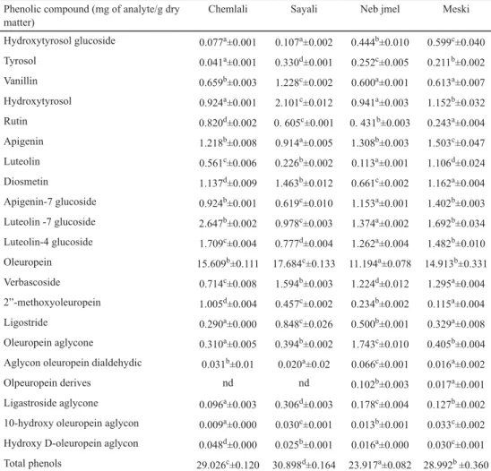
The phenolic extracts of Chemlali, Sayali, Neb jmel, and Meski cultivars were screened for the range of phenolic compounds. All extracts were found to contain high and varying amounts of phenolic compounds. In our study, significant differences were observed for total phenolic contents in extracts. As shown in Table 1, the highest amount was found in Sayali (30.89 mg g–1), while the lowest was registered in Neb jmel extract (23.91 mg g–1). Our results reported that olive leaves represent a potential source of phenolic compounds.
In the present study, a total of 21 compounds were identifi ed including oleuropeosides, flavonoids, alcohol phenols, and simple phenols. Quantitatively a difference in the distribution of phenolic compounds and their corresponding groups was observed. The results from ANOVA showed signifi cant quantitative differences in phenolic compounds according to the cultivar (P<0.05). The obtained result revealed that the phenolic profi le of olive leaves is strongly affected by genotype factor. Our results are in accordance with those reported in the literature (KONTOMINAS, 2010; ABAZA et al., 2015).
In all analysed olive leaf cultivars, secoiridoids were by far the main abundant group of phenolic compounds. This dominance of secoiridoids in the phenolic fraction of olive leaves is also reported in the literature (KHALIQ et al., 2015). The main compound in this group was oleuropein. The registered values were 17.68 mg g−1 (Sayali); 15.61 mg g−1 (Chemlali); 14.93 mg g−1 (Meski), and 11.19 mg g−1 (Neb jmel). Several authors, by the characterization of the phenolic composition of Greek olive leaves (GOULAS et al., 2009) and Tunisian olive leaves (TAAMALLI et al., 2012) have observed that oleuropein is the main abundant compound in olive leaves of all cultivars.
Furthermore, our results showed that apigenin-7–O-glucoside was present at concentrations ranging from 0.62 mg g−1 in Sayali extract to 1.40 mg g−1 in Meski extract.
Apigenin and luteolin were detected in all samples analysed. Luteolin was found in mean concentration ranging from 0.22 mg g−1 (Sayali) to 1.11 mg g−1 (Meski). These results are similar to those reported by several authors. MEIRINHOS and co-workers (2005) have reported luteolin-diglucoside and luteolin 4-O-glucoside the most frequent fl avonoids in Portuguese olive cultivars. Diosmetin was detected and quantifi ed in all cultivars. The concentration of tyrosol ranged from 0.041 mg g−1 in Chemlali to 0.33 mg g−1 in Sayali cultivars. The amounts
of hydroxytyrosol varied from 0.92 to 2.10 mg g−1 for Chemlali and Sayali cultivars, respectively. The concentration of hydroxytyrosol glucoside in Meski was the highest among all cultivars.
Table 1. Quantifi cation of individual phenolic compounds in the leaves of different cultivars Phenolic compound (mg of analyte/g dry
matter)
Chemlali Sayali Neb jmel Meski
Hydroxytyrosol glucoside 0.077a±0.001 0.107a±0.002 0.444b±0.010 0.599c±0.040
Tyrosol 0.041a±0.001 0.330d±0.001 0.252c±0.005 0.211b±0.002
Vanillin 0.659b±0.003 1.228c±0.002 0.600a±0.001 0.613a±0.007
Hydroxytyrosol 0.924a±0.001 2.101c±0.012 0.941a±0.003 1.152b±0.032
Rutin 0.820d±0.002 0. 605c±0.001 0. 431b±0.003 0.243a±0.004
Apigenin 1.218b±0.008 0.914a±0.005 1.308b±0.003 1.503c±0.047
Luteolin 0.561c±0.006 0.226b±0.002 0.113a±0.001 1.106d±0.024
Diosmetin 1.137d±0.009 1.463b±0.012 0.661c±0.002 1.162a±0.004 Apigenin-7 glucoside 0.924b±0.001 0.619c±0.010 1.153a±0.001 1.402b±0.003 Luteolin -7 glucoside 2.647b±0.002 0.978c±0.003 1.374a±0.002 1.692b±0.034 Luteolin-4 glucoside 1.709c±0.004 0.777d±0.004 1.262a±0.004 1.482b±0.010 Oleuropein 15.609b±0.111 17.684c±0.133 11.194a±0.078 14.913b±0.331 Verbascoside 0.714c±0.008 1.594b±0.003 1.224d±0.012 1.295a±0.004 2”-methoxyoleuropein 1.005d±0.004 0.457c±0.002 0.234b±0.002 0.115a±0.004 Ligostride 0.290a±0.000 0.848c±0.026 0.500b±0.001 0.329a±0.008 Oleuropein aglycone 0.310a±0.005 0.394b±0.002 1.743c±0.010 0.405b±0.004 Aglycon oleuropein dialdehydic 0.031b±0.01 0.020a±0.02 0.066c±0.001 0.016a±0.002
Olpeuropein derives nd nd 0.102b±0.003 0.017a±0.001
Ligastroside aglycone 0.096a±0.003 0.306d±0.003 0.178c±0.004 0.127b±0.002 10-hydroxy oleuropein aglycon 0.009a±0.000 0.030c±0.001 0.013b±0.001 0.033c±0.002 Hydroxy D-oleuropein aglycon 0.048d±0.000 0.025b±0.001 0.016a±0.000 0.030c±0.001 Total phenols 29.026c±0.120 30.898d±0.164 23.917a±0.082 28.992b ±0.360 Results are expressed as mean ± standard deviation; signifi cant differences in the same row are showed by different letters (P<0.05); nd: not detected
The obtained results revealed that Sayali extract showed the highest antioxidant potential (IC50 value 41.56 μg ml–1). Moreover, all olive leaf extracts were able to scavenge free radicals. These results were in agreement with the report by SALAH and co-workers (2012), who showed that olive leaf extracts present a potent antioxidant activity.
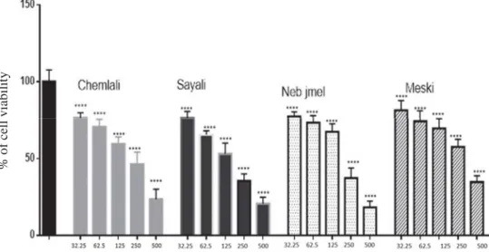
Moreover, all extracts induced the decrease of MCF-7 growth in a dose-dependent manner (Fig. 1). As shown in Table 2, olive leaf extracts from Sayali have shown the highest inhibitory effect against MCF-7 cells with a CE50 of 196 μg ml–1, followed by Neb jmel,
388
Chemlali, and Meski. These results are in agreement with fi ndings of FU and co-workers (2010). The authors have estimated the CE50 values of olive leaves ranging from 200 to 300 μg ml–1.
Table 2. Antioxidant and antiproliferative activities of various extracts of the Tunisian olive leaf cultivars by DPPH and MTT assays
Olive leaf extract DPPH (IC50) μg ml–1 MTT Test (CE50) μg ml–1
Chemlali 50.17±1.28 213.05±2.13
Sayali 41.56±0.41 147.77±2.78
Neb jmel 58.16±1.21 183.61±1.07
Meski 63.52±0.47 280.07±2.55
The results were given as half maximum effective concentration (IC50 and CE50); IC50 and CE50 values represent the means ± standard deviation of three replications (P<0.05).
% of cell viability
Fig. 1. Effects of different concentration of olive leaf extracts (Chemlali, Sayali, Neb jmel, and Meski) on MCF-7 cell proliferation
Columns are means of three independent experiments each performed with triplicate samples expressed as percent of basal, bars: SD; xxxx: signifi cant difference at P<0.0001 compared with basal
Western blotting analysis (Fig. 2) revealed the presence of cleaved form of PARP-1 in MCF-7 treated cells. Our result demonstrated that olive leaf extracts induce a slightly upregulated expression of cyclin D1 in treated MCF-7 cells when compared to control cells.
The obtained result showed that treatment of MCF-7 cells with olive leaf extracts stimulated the apoptosis effi ciently.
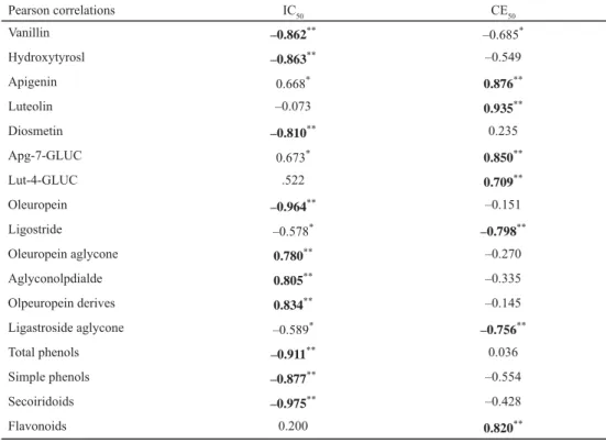
As shown in Table 3, a positive linear correlation between the antioxidant capacity values (IC50) and total phenol content was found. The antioxidant activity of olive leaf extracts could be attributed to their high amounts of total phenol (R2=0.911) and total secoiridoids (R2= –0.975). Moreover, antioxidant activity could be attributed to the higher oleuropein amount (R2= –0.964), as well.
Fig. 2. Effects of olive leaf phenolic extracts on PARP-1 and cyclin D1 activation. Blots are representative of three independent experiments with similar results. (C: Basel; 6: Chemlali; 7: Sayali; 8: Neb jmel; 9: Meski; GAPDH:
glyceraldehyde 3-phosphate dehydrogenase antibody)
Table 3. Pearson correlations
Pearson correlations IC50 CE50
Vanillin –0.862** –0.685*
Hydroxytyrosl –0.863** –0.549
Apigenin 0.668* 0.876**
Luteolin –0.073 0.935**
Diosmetin –0.810** 0.235
Apg-7-GLUC 0.673* 0.850**
Lut-4-GLUC .522 0.709**
Oleuropein –0.964** –0.151
Ligostride –0.578* –0.798**
Oleuropein aglycone 0.780** –0.270
Aglyconolpdialde 0.805** –0.335
Olpeuropein derives 0.834** –0.145
Ligastroside aglycone –0.589* –0.756**
Total phenols –0.911** 0.036
Simple phenols –0.877** –0.554
Secoiridoids –0.975** –0.428
Flavonoids 0.200 0.820**
*: P<0.05; **: P<0.01
From this study, it was shown that phenolic compounds largely contribute to the antioxidant activities of olive leaf. Our results are in agreement with those reported in the literature. BENAVENTE-GARCÍA and co-workers (2000) reported that the radical scavenging potential of olive leaf extract depends mainly on its amount of phenolic compounds. On the other hand, the obtained results of correlation of phenolic content and CE50 have shown that the anti-proliferative effect in MCF-7 breast cancer cells does not correlate with phenolic content or with antioxidant capacity.
390
3. Conclusions
The results reveal the richness of Tunisian olive leaves in phenolic compounds. Furthermore, phenolic profi le could be used as a fi ngerprint to discriminate olive leaves by cultivar.
On the other hand, the obtained results showed that Sayali extract exhibited the strongest antioxidant and antiproliferative potentials. In the light of these fi ndings, Sayali extract could be recommended as a source of antioxidant components for therapeutic industries.
References
ABAZA, L., BEN YOUSSEF, N., MANAI, H., MAHJOUB HADDADA, F., METHENNI, K. & ZARROUK, M. (2011): Chétoui olive leaf extracts: Infl uence of the solvent type on phenolics and antioxidant activities. Grasas Aceites, 62, 96–104.
ABAZA, L., TAAMALLI, A., NSIR, H. & ZARROUK, M. (2015): Olive tree (Olea europeae L.) leaves: importance and advances in the analysis of phenolic compounds. Antioxidants (Basel), 4, 682–698.
BEN MOHAMED, M., GUASMI, F., BEN, S., RADHOUANI, F., FAGHIM, J., … & BENINCASA, C. (2018): The LC-MS/MS characterization of phenolic compounds in leaves allows classifying olive cultivars grown in South Tunisia.
Biochem. Syst. Ecol., 78, 84–90.
BENAVENTE-GARCÍA, O., CASTILLO, J., LORENTE, J., ORTUÑO, A. & DEL RIO, J.A. (2000): Antioxidant activity of phenolics extracted from Olea europaea L. leaves. Food Chem., 68, 457–462.
FU, S., ARRÁEZ-ROMAN, D., SEGURA-CARRETERO, A., MENÉNDEZ, J.A., MENÉNDEZ-GUTIÉRREZ, M.P., … & FERNÁNDEZ- GUTIÉRREZ, A. (2010): Qualitative screening of phenolic compounds in olive leaf extracts by hyphenated liquid chromatography and preliminary evaluation of cytotoxic activity against human breast cancer cells. Anal.
Bioanal. Chem., 397, 643–654.
GOULAS, V., VASSILIKI, E., ANASTASSIOS, N., TROGANIS, E.P., FOTSIS, T. & GEROTHANASSIS, E.B. (2009): Phytochemicals in olive-leaf extract and their antiproliferative activity against cancer and endothelial cells. Mol. Nutr. Food Res., 53, 600–608.
KHALIQ, A., SABIR, S.M., AHMAD, S.D., BOLIGON, A.A., ATHAYDE, M.L., … & KHAN, A. (2015): Antioxidant activities and phenolic composition of olive (Olea europaea) leaves. J. Appl. Bot. Food Qual., 21, 16–21.
KONTOMINAS, M. (2010): Composition and antioxidant activity of olive leaf extracts from greek olive cultivars. J.
Am. Oil Chem. Soc., 87, 369–376.
MEIRINHOS, J., SILVA, B.M., VALENTÃO, P., SEABRA, R.M., PEREIRA, J.A., … & FERRERES, F. (2005): Analysis and quantifi cation of fl avonoidic compounds from Portuguese olive (Olea europaea L.) leaf cultivars. Nat. Prod.
Res., 19, 189–195.
RODRIGUES, F., PIMENTEL, F.B. & OLIVEIRA, M. (2015): Olive by-products: Challenge application in cosmetic industry.
Ind. Crop. Prod., 70, 116–124.
SABRY, O. (2016): Benefi cial health effects of olive leaves extracts. J. Nat. Sci. Res., 4, 1–9.
SALAH, M., BEN ABDELMELEK, H. & ABDERRABA, M. (2012): Study of phenolic composition and biological activities assessment of olive leaves from different varieties grown in Tunisia. Med. Chem., 2, 107–111.
SAMET, I., HAN, J., JLAIEL, L., SAYADI, S. & ISODA, H. (2014): Olive (Olea europaea) leaf extract induces apoptosis and monocyte/macrophage differentiation in human chronic myelogenous leukemia K562 cells : Insight into the underlying mechanism. Oxid. Med. Cell. Longev. 2014, 1–16
SIRIANNI, R., CAPPARELLI, C., CHIMENTO, A., PANZA, S., CATALANO, S., ... & ANDÒ, S. (2012): Molecular and cellular endocrinology nandrolone and stanozolol upregulate aromatase expression and further increase IGF-I- dependent effects on MCF-7 breast cancer cell proliferation. Mol. Cell. Endocrinol., 363, 100–110
TALHAOUI, N., TRABELSI, N., TAAMALLI, A. & VERARDO, V. (2018): Olea europaea as potential source of bioactive compounds for diseases prevention. -in: ATTA-UR RAHMAN (Ed.): Studies in Natural Products Chemistry, 57, Elsevier, pp. 389–411.
TAAMALLI, A., ARRÁEZ-ROMÁN, D., ZARROUK, M., VALVERDE, J., SEGURA-CARRETERO, A. & FERNÁNDEZ-GUTIÉRREZ, A.
(2012): The occurrence and bioactivity of polyphenols in Tunisian olive products and by-products. J. Food Sci., 77, 1–11.