7
Comparison of Methods for the
Determination of Nuclear Spin as Applied to Radioactive Nuclei
VICTOR W. COHEN
Brookhaven National Laboratory, Upton, Long Island, New York.
Introduction
The role of angular momentum in the theories of atomic nuclei is one of paramount importance. While it was postulated initially by Goudsmit and Back to explain optical hyperfine structure it has assumed the role of a fundamental nuclear property, along with charge and mass, to enter into theories, such as nuclear composition, beta-ray disintegration and parity conservation as well as electromagnetic inter- actions with atomic electrons. The fact that the spin and related moments, i.e. magnetic dipole, electric quadrupole, and higher moments may enter into interactions of different types, permits us to make inter-related measurements and clear up ambiguities in either theory or the experimental techniques. It was this multiplicity of related phenomena that permitted the early clarification of our under- standing of nuclear spin by studies of the alkali atoms in the early
1930's.1'2'3'4'5 The work on sodium and cesium pointed out that our interpretation of hyperfine structure and the Breit-Rabi interaction were basically correct and that there were errors in the measurements made by optical spectroscopy.
In the study of nuclear spin properties the atomic beam technique has been of extreme importance because of the clear cut nature of its data. When one deals with a monatomic gas at low pressure, interactions involving inter- or intra-molecular forces are completely absent. The other techniques of study of nuclear spin which involve study of gases of greater density, liquids or solids involve forces between the particles which can be evaluated by approximate methods but which cannot be eliminated. It is the purpose of this discussion to compare the various techniques for measurement of spin properties as
i 121
122 VICTOR W. COHEN
they apply in particular to radioactive nuclei, to discuss their relative merits and limitations to show how they supplement each other. The various methods considered will be (1) optical spectroscopy, (2) mole- cular gas microwave spectroscopy, (3) nuclear resonance, (4) para- magnetic resonance and (5) atomic beams. The rf spectroscopy of pure quadrupole transitions is a fruitful technique for study of quadru- pole information but there seems little chance of its being of importance to the radioactive nuclei because of the large amount of material re- quired for the investigation.
In this discussion we will not consider those naturally occurring nuclides such as K40 and Rb87 which possess very weak radioactivity.
Techniques applicable to stable nuclei apply equally well to them.
The problems of measurement of spins of unstable nuclei differ from those associated with the corresponding stable ones in three ways: (1) usually only small quantities of material are available, (2) isotopic abundances vary from a few percent to one in 107, and (3) the radio- activity of the material under study. This last property makes available a means of detection of the presence of minute amounts of material.
Material Available
The first problem of the radioactive species is that of sample prepara- tion. In general, the amounts of material available are small by standards of the normal techniques. Because of the potential health hazard associated with the radiations from radioactive nuclei it has been necessary in a few cases to provide extensive shielding and remote handling of the apparatus. In most cases, however, the handling prob- lem is simplified by the small amount of material required.
In the past ten years techniques have been developed which make possible the determination of nuclear spin properties using amounts ranging from 1010 to 1017 atoms.
Processes of production of a particular radioactive nucleus may be by one of two general types, first, such as («, γ) in which the product is chemically identical with the starting material, and second, such as
(n, p) or (d, n) products in which the product has a different atomic number from its parent. The (n, γ) product cannot be separated chemi- cally from its parent except in unusual cases, as for example, a Szilard- Chalmers process. This means that with ordinary neutron capture cross- sections and lifetimes of the order of days or weeks the isotopic purity of the nuclide of interest may be of the order of one part per million. As we shall show later, this definitely rules out the possibility of certain techniques of investigation. In cases in which the capture cross-section for thermal neutrons is large, such as Co59 and Eu151 a pile irradiation
COMPARISON OF METHODS FOR R A D I O A C T I V E N U C L E I 1 2 3
of a year may build up a concentration of the radioactive isotope of well over 1%.
In working with pile produced n, y products using techniques in which isotopic dilution is not important, one has considerable latitude in choice of amount both of the stable and of the radioactive species.
Such a choice may be based upon considerations of convenience.
For the case of isotopes produced by such processes as (/z, p) or (d, «), where the product differs chemically from its parent, it can, at least in principle, be separated chemically into a state which is isotopically pure. The great difficulty entering into this problem lies in the fact that the parent material or the reagents used in the separation may contain as trace impurities more of the stable counterpart than of the radio- active product. Normal cyclotron irradiations may produce concentra- tions less than 1 part per million of the product. It is easy to see how impurities greater than this magnitude might be introduced in the process of chemical separation.
The general subject of the chemistry of preparation of carrier-free nuclides is a difficult one and each one can represent an appreciable part of a research on spin determinations.6 To prepare a radionuclide with an isotopic purity higher than five per cent may represent a real accomplishment in chemical technique.
Optical Spectroscopy
The field of optical spectroscopy has been widely used to study nuclear spins by hyperfine structure. In all, more than 75 elements have studied over the past thirty years by this method.
A complete analysis of an optical spectrum may yield data on the following nuclear properties :
(a) Nuclear spin,
(b) Nuclear-electronic magnetic interaction energy (ΔΡΚ), (c) Nuclear electric quadrupole interaction energy (if the nuclear
spin is greater than \).
From the fine structure it is possible to compute the mean value of 1/r3 for the electronic wave function near the nucleus. This quantity enters into computation of the nuclear electric quadrupole moment from this or other experiments involving nuclear quadrupole inter- action.
The multiplicity of an optical electronic energy state will be given by either 2] +1 or 2/ + 1, depending upon whether / is less than or greater than /. If the value of/is to be inferred from an observed optical splitting, the levels must be sufficiently well identified to know whether die splitting is due to / o r / . There have been several cases where the
124 V I C T O R W. C O H E N
interpretation of hyperfine structure has lead to uncertain or to in- correct values of /.
The magnitude of the hyperfine structure splitting is a function of the value of the nuclear magnetic moment. The calculation of the moment involves a knowledge of the electronic wave function at the nucleus which, at present, in most cases is not known with sufficient precision to give values comparable to those obtained by nuclear resonance.
In optical hyperfine structure studies one is faced with two problems : (1) obtaining of sufficient light intensity for proper recording, and (2) keeping the spectral lines narrow enough for adequate resolution of closely spaced multiplets. Working with radioactive nuclei one is forced to use minute samples so that the light source must be miniaturi- zed. For efficiency of light utilization the nearest to an ideal source would be one shaped so as to serve as the usual slit of the spectroscope.
Sources have been used by several investigators which approach this size and shape.
The phenomena of "cleanup" in a gas discharge tube is a process whereby ions bury themselves in the walls of the tube and are no longer available in the discharge. This of course is very serious if onjy a minute gas sample is available. In the electrodeless high frequency dis- charge the ions do not receive enough energy to be removed. Such sources containing about 1016 atoms have operated over lifetimes long enough for extensive research without appreciable loss. It should be possible to extend this to even smaller tubes, perhaps less than 0.5 cc, which at a pressure of 0.1 mm would contain only 2 x 10¹⁵ atoms.
The hollow-cathode discharge tube cooled with liquid nitrogen or helium has been used extensively for high resolution spectroscopy. It has the advantage of keeping the velocity of the emitting atoms down to an effective temperature near that of the cathode. Such low velocity results in a reduced Doppler broadening. If the voltage across the tube is kept low, the "cleanup" is not great and this type of tube can be miniaturized to an effective volume of one cc or thereabouts.
The choice between the hollow cathode tube and the electrodeless discharge seems to depend upon the preferences of the experimenter as well as the properties of the material under investigation.
In utilizing these types of light sources several investigators have gone to an electronic scanning system which obviates some of the diffi- culties of photographic methods. A combination of a high resolution grating and a Fabry-Perot interferometer is used. The interferometer spacing is maintained constant in distance but the instrument is in a gas-tight enclosure in which the pressure is changed slowly and continu- ously. The light intensity at the grating focal plane is received on a
liquid nitrogen-cooled photomultiplier and the spectral intensity is recorded graphically.
Optical hyperfine structure investigations are limited to material with an isotopic purity of at least a few per cent. In the light of the discussion in the previous section this is a severe limitation. The method is quite applicable to such nuclides as A37 and Kr85 which would be very difficult to study by any other means.
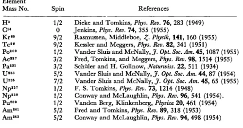
Table I includes the spins of those radioactive nuclei that have been determined by optical methods. In a few cases they have also been studied by other techniques. H3 and C14 have been studied by the method of alternating intensities of the rotational bands of the molecular spectrum.
TABLE 1
SPINS OF RADIOACTIVE NUCLEI MEASURED BY OPTICAL SPECTROSCOPY, NOVEMBER 1, 1957
Element
Mass No. Spin References
Dieke and Tomkins, Phys. Rev. 76, 283 (1949) Jenkins, Phys. Rev. 74, 355 (1955)
Rasmussen, Middleboe, £. Physik, 141, 160 (1955) Kessler and Meggers, Phys. Rev. 82, 341 (1951)
Vander Sluis and McNally, J. Opt. Soc. Am. 45,1087 (1955) Fred, Tomkins, and Meggers, Phys. Rev. 98, 1514 (1955) Schüler and H. Gollnow, Naturwiss. 22, 511 (1934) Vander Sluis and McNally, J. Opt. Soc. Am. 44, 87 (1954) Vander Sluis and McNally, J. Opt. Soc. Am. 45, 65 (1955) F. S. Tomkins, Phys. Rev. 73, 1214 (1948)
Gonway and McLaughlin, Phys. Rev. 96, 541 (1954).
Vanden Berg, Klinkenberg, Physica 20, 461 (1954) Fred and Tomkins, Phys. Rev. 89, 318 (1953) Gonway and McLaughlin, Phys. Rev. 94, 498 (1954) H3
C"
Kr85 T c "
po2 09 Ac»7 Pa231 U233 XJ235 Np237 Np239 pu230 Am241 Am243
1/2 0 9/2 9/2 1/2 3/2 3/2 5/2 7/2 1/2 1/2 1/2 5/2 5/2
Gas Microwave Spectroscopy
Microwave spectroscopy has been applied to a study of the pure rotational spectrum of gas molecules in the ground electronic state and generally in the lowest vibrational state. The rotational frequencies of many molecules are such that transitions can be observed in the fre- quency range between a few thousand and a hundred thousand mega- cycles. These transitions may be split by an interaction between the nuclear electric quadrupole moment and the electric field gradient at the nucleus. From the multiplicity of the resulting spectrum, the relative
126 V I C T O R W. C O H E N
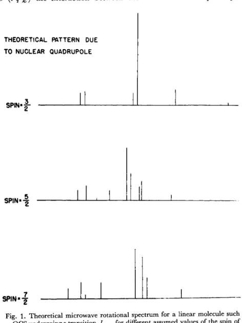
spacing and the rough relative intensities, one can infer the nuclear spin.7 Fig. 1 indicates the theoretical pattern to be expected for a typical linear molecule undergoing a / = 1 <-> 2 transition for different assumed values of the spin.
The scale factor of the separation of the components is proportional to (eqQ) the interaction between the nuclear electric quadrupole
THEORETICAL PATTERN DUE TO NUCLEAR QUADRUPOLE
SPIN«|-
SPIN»-§
SP,N.2
1—L—I LU 1
Fig. 1. Theoretical microwave rotational spectrum for a linear molecule such as OGS undergoing a transition /1<f^2for different assumed values of the spin of
one of the nuclei.
moment Q and the gradient of the electric field, q. Fig. 2 is an experi- mental curve obtained with OCS35 indicating good agreement with the theoretical pattern for / = f.8
V / 2
QUADRUPLE COMPONENTS OF J = l ^ 2 TRANSITION IN OCS
Fig. 2. Experimental curve of the spectrum of the / i ^ .2 line for OCS3 5. The rela-
/ tive positions of the components calculated for an assumed spin of |- are shown below.
Unfortunately the value of q cannot in general be computed with precision, so that no accurate value of Q can be obtained. It is possible, however, to compute a rough approximation of q on the basis of atomic wave functions and the type of chemical bonds involved. The errors in this type of calculation, together with uncertainties concerning the polarization of the inner electron shells, may result in an overall un- certainty as high as 50%.
In the case of two isotopes of the same element, the electronic states are practically identical so that the ratio of the respective electric quadrupole moments will be proportional to their interaction constants.
This gives us at least some precise information on electric quadrupole moments.
There are a number of very basic limitations on the applicability of this method :
(a) The element in question must be incorporated into a molecule which has :
(1) a permanent electric dipole moment.
(2) a simple structure such as linear, symmetric top, (in some cases a simple asymétrie rotor is analyzable).
128 V I C T O R W. C O H E N
(3) It must be vaporizable to a pressure of at least a few microns, at a temperature attainable in a wave guide system.
(4) The moment of inertia must be such that the rotational states involve low quantum numbers.
(b) The isotopic purity must be not less than about 1 part per 1000.
(c) The total number of atoms required for any spin determination is of the order of 1014 to 1015, the limitation depending to some extent on the character of the spectrum.
The limitation on isotopic purity is very severe in that it may prevent the utilization of all the available material. One is limited in the total gas pressure which can be admitted into the spectroscope. An excess will produce pressure broadening of the line. The situation may arise in which only a small fraction of the material available can be utilized.
The Zeeman effect of a microwave line may be studied by placing the wave guide in a magnetic field. Such an experiment may yield information about the nuclear and molecular magnetic moments. Since this type of experiment involves the splitting of one of the components of the normal spectrum, the resulting intensity will be considerably reduced. This means that the isotopic purity required for the Zeeman effect will be higher than that required for a spin determination.
Table 2 contains data on spins and magnetic moments of radioactive nuclei as obtained by microwave spectroscopy.
T A B L E 2
SPINS AND MOMENTS OF RADIOACTIVE N U C L E I M I C R O W A V E SPECTROSCOPY NOVEMBER 1, 1957
Nucleus Spin μ References
C1 4 0 A. Roberts, Phys. Rev. 73, 1405 (1948) S35 3/2 1.00 ± 0 . 0 4 Cohen et al., Phys. Rev. 76, 703 (1949) ; Burke et al., Phys. Rev. 93, 193 (1954) Gl3 e 2 1.32 ± 0 . 0 8 Townes a n d Aamodt, Phys. Rev. 76, 691
(1949); Aamodt a n d Fletcher, Phys. Rev.
98, 1317 (1955)
Se7 9 7/2 - 1 . 0 1 5 ±0.015 H a r d y et al., Phys. Rev. 85, 494 (1952) ; H a r d y et al., Phys. Rev. 9 2 ,1 5 3 2 (1953) I125 5/2 Fletcher and Amble, Bull. Am. Phys. Soc. 2,
30 (1957)
I1 2 9 7/2 Livingston et al., Phys. Rev. 76, 149 (1949)
I131 7/2 Livingston et al., Phys. Rev. 92, 1271 (1953)
Nuclear Magnetic Resonance
Nuclear magnetic resonance is an extremely accurate method of measuring ratios of magnetic moments. Unfortunately the quantity of material required, considerably greater than that required by other methods, is of the order of 1018 to 1020 atoms, depending upon the value of the magnetic moment. Iso topic purity of the order of 1/10 to 1%
is required. A few measurements have been made on radioactive nuclei of long half-life which for non-scientific reasons are available in gram
quantities.
The interaction between a nucleus and an external magnetic field may be complicated by the existence of an electric quadrupole inter- action between the nucleus and a non-cubic electric field due to mole- cular or crystalline environment. If this interaction is small it may result merely in a broadening of the line. If the interaction is large compared to the magnetic spin-lattice interaction, the line may be so broadened as to be undetectable. Some care must therefore be exercised in a choice of compound of the nuclide under study.
In view of the above difficulties and the developments on other techniques it is doubtful whether much work will be done in this field.
The results obtained to date by nuclear resonance are shown in Table 3. The fourth column indicates roughly the number of atoms required for the measurement.
TABLE 3
Nucleus H3 Gl3e T c "
J129
M 2.97876 1.2838
5.65724 2.60304
±0.00003 0.0003
0.00040 0.0003
No. of atoms used 10"
2 X 10¹⁹ 2 X 10¹⁹
Ref.
(a) (b) (c) (d)
(a) Bloch, Graves, Packard, and Spence, Phys. Rev. 71, 55 (1947).
(b) Sogo and Jeffries, Phys. Rev. 98, 1316 (1955).
(c) Walchli, Livingston, and Martin, Phys. Rev. 85, 479 (1955).
(d) Walchli, Livingston, and Hebert, Phys. Rev. 82, 97 (1951.) Paramagnetic Resonance
The phenomenon of paramagnetic resonance furnishes a means for studying the Zeeman levels of a paramagnetic system. This system can be ionic, atomic, or molecular. It may be in a gas, crystal, or solution.
The requirement that a system be paramagnetic is that it have one or more electrons which are magnetically unpaired. As examples of such
130 VICTOR W. COHEN
systems we may cite free atoms in non-singlet states, ions of the iron or rare earth groups, and molecules known as free radicals which are electrically neutral. The method has been used to obtain information about the ground state of ions and the nature of the crystalline fields.
However, the electron may interact with a nuclear magnetic moment and thus give us information about which we are here concerned.
There have been several excellent review articles about the inter- actions of ions in crystals.9»10 We shall present here only a brief discussion of the principles.
In considering the Zeeman effect of paramagnetic ions it is possible to approximate the ion as a free electron, the orbital contribution being, in some respects, "quenched" by the crystalline field. The splitting factor, g, is therefore very nearly 2.
Let us enumerate the most important interactions to which the un- paired electron is subjected in a crystal.
(a) If the crystalline symmetry is non-cubic there will be a Stark splitting of the ground state of the ion in the absence of a magnetic field.
(b) Magnetic dipole-dipole interaction between adjacent para- magnetic ions.
(c) Magnetic interaction between the electron and an externally applied magnetic field.
(d) Magnetic interaction, similar to optical hfs, between the electron and the magnetic moment of the nucleus upon which the electron is bound. This will clearly give a multiplicity to the energy levels of 21 +1 which gives us our means of determining /.
(e) Interaction between the nuclear magnetic moment and the ex- ternal applied magnetic field.
The magnetic dipole-dipole interaction (b) is of the order of μο/r3 where μ o is the magnetic moment of the electron and r the interionic distance. By placing the ion in a diamagnetic lattice with a dilution of 1/100 to 1/1000 this interaction may be reduced below a significant value. Term (e) above is very small in all magnetic fields generally used, 3-10 kg. Fig. 3 illustrates the three remaining terms (a), (c) and (d), for a typical case of V+ + diluted in a Zn Tutton salt11. The zero field splitting is due to the Stark effect. In a weak field the splitting due to the magnetic field is, to a first approximation, the Zeeman effect. In the strong field or Paschen-Back region the nuclear interaction results in a multiplicity of 2/ + l.The scale factor of the strong field splitting is determined by the bW term or the hfs interaction energy.
In a paramagnetic resonance experiment the specimen is placed in a resonant cavity in the region where the microwave magnetic field is a
maximum and perpendicular to the externally applied magnetic field.
The radiation frequency is maintained constant and equal to the natural frequency of the cavity. The external magnetic field is varied slowly
h9,000Mc/sec
*4&
h23,000Mc/sec
Fig. 3. Schematic Zeeman energy level diagram of the V+ + ion in a Tutton salt. The spectrum for the hyperfine components of the m,(è<—>~ i) transi-
tion is shown below.
and as the energy difference between the two levels for which a transi- tion is allowed satisfies the condition.
\E2-Ei\ = hv one observes an absorption of microwave energy.
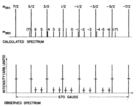
A typical spectrum for a mixture of two stable isotopes with different spins, V50 (/ = 6, 20% of total V) and W (/ = | , 80%) is shown in Fig. 4.11 The magnetic moments as well as the spins are different, giving rise to a complex spectrum. Where certain lines overlap, obviously the weaker one cannot be resolved. If the abundance of the weaker one had been very much less, clearly the lines would have been lost in the tails of the stronger lines. The lowest practical isotopic con- centration for a spin determination is in the vicinity of 1%.
Here, as in optical hyperfine structure, the splitting is a function of Δν and the nuclear magnetic moment cannot be evaluated directly from the experiment. If one can observe a resonance spectrum for two isotopes the ratio of the two Av's can be related to the ratio of magnetic moments to within an accuracy at best of about 1 part in 1000.
132 VICTOR W. COHEN
The efficiency of utilization of a rare isotope is severely limited if one is using a single crystal. On growing a single crystal from solution one must necessarily leave a major fraction of the solute still in solution.
m(5l) 7/2 5/2 3/2 1/2 -1/2 -3/2 -5/2 -7/2
"(δθ) (7)
I I
I
6 5
1_L
4 3 2
_LL
I 0 -I
LJJ
-2 -3
J_L
- 4 - 5 -6
l_LL
(-7)
I I
_L_
CALCULATED SPECTRUM
±± + +I++ l-n- ±±
-670 GAUSS OBSERVED SPECTRUM
Fig. 4. Theoretical and observed spectrum of V+ + ion with V50 and V51 present in a ratio of about 1 to 4.
One may avoid some of the problems of working with single crystals by going to solutions. Several elements have exhibited hyperfine structure in paramagnetic resonance spectra in dilute solutions of organic complexes. This is true for B, V, N, C, and Be.12 Aqueous solutions of inorganic ions can be studied in the region of 9,000 Mc.
At 25,000 Mc water has a very high dielectric absorption so that it can- not be used in this region of frequency. Phosphorus, arsenic and anti- mony dissolved in solid silicon in a concentration of about 10~4 and at temperatures of a few degrees absolute, are in the atomic state and show paramagnetic resonance. Hyperfine structure patterns of these ele- ments can thus be studied.13
It is difficult to specify the lower limit of material necessary for a spin determination. The sensitivity will depend in part upon the number of states into which the ground state is split. This may be quite appreci- able for the case of V50, for example, with a nuclear spin of 6 giving rise to 13 hfs components for each electronic state, which in this case of the
COMPARISON OF METHODS FOR R A D I O A C T I V E N U C L E I 133
Tutton salt is 3 for each of two equivalent ions, giving rise to a total of 78 states. The Boltzmann factor relating population of the two Te- states shows that by going to low temperatures an appreciable gain in sensitivity is available. Such a gain is quite possible for crystals but will not be available for liquid solutions. Solutions, on the other hand, enable one to utilize all of the material which may be available. In practice results have been obtained with crystals using samples of 1015 atoms and in favourable cases one may hope to do better.
Feher14 has developed a modification of paramagnetic resonance in which the electron resonance line is enhanced by a simultaneous application of a radio frequency field corresponding to the energy inter- val of the hyperfine splitting. This double resonance applied near
1°K makes possible a determination not only of the spin and Δν but also gi. By use of this method he has measured both the spin and moment of p32 u sing a sample of 4 x 10¹⁴ atoms in a solid solution in Si15. This technique, while more complex than simple paramagnetic resonance, should prove a valuable tool because of its extra sensitivity and the prospect of obtaining additional information.
A further variation of paramagnetic resonance available for radio- active nuclei with spins of 1 or greater has been developed by Jeffries16 and by Pipkin and Culvahouse.17 At low temperature, a partial nuclear polarization can be induced by the application of a microwave field. This polarization is detected by the anistropy in the emitted y radiations. This has the very valuable property of being independent of the presence of the stable isotope of the element under investigation.
It has been successfully applied to Mn52, Co60(16), and As76.(17)
The spin data of radioactive nuclei which have been measured by paramagnetic resonance methods are listed in Table 4.
Atomic Beams
For the past 25 years the atomic beam technique has been a powerful tool for determining the spin properties of nuclei. Because of the simplicity of the ground state of the atoms studied and the absence of environmental interactions the results have been clearly interpreted.
As each advance in beam detection was developed a new group of ele- ments was attacked. After the initial detection by the deposit of a visible film, the surface ionization detector opened up extensive work on the alkalis. The mass spectrometer, especially when combined with an electron ionizer, opened up research to a wide variety of elements such as B, Sb, Au, Ag, and Pr with, undoubtedly, many more to follow.
The use of the radiations from radioactive nuclei furnishes a powerful means of detection. Since its first application by Smith and Bellamy in
1 3 4 VICTOR W . COHEN
TABLE 4
SPINS AND MOMENTS OF RADIOACTIVE NUCLEI BY PARAMAGNETIC RESONANCE OCTOBER 1, 1958
Nucleus
p32
Mn53 Go56 Go57 Co58 Co60 As76 Ce141 Nd147 Eu152 Eu154
TJ233 XJ236 N p 2 3 7 pu2 3 9
Pu241
Spin 1 7/2
4 7/2
2 5 2 7/2 5/2
3 3 5/2 7/2 5/2 1/2 5/2
μ
0.2523 (3) 5.050 (7) 3.855 (7) 4.6 (2) 4.052 (11)
3.800 (7) -0.906 (4) 0.89 (9) 0.56 2.0 2.1 0.5 0.3 6.0 0.4
References
Feher et al, Phys. Rev. 107, 1463 (1957) Dobrowolsky, Phys. Rev. 104, 1378 (1956) Jones et al, Phys. Rev. 102, 738 (1956)
Baker et al, Proc. Phys. Soc. A 66, 305 (1953) Dobrov and Jeffries, Phys. Rev. 108, 60 (1957) Dobrowolsky et al., Phys. Rev. 101, 1001 (1956) Pipkin and Gulvahouse, Phys. Rev. 106, 1102
(1957)
Kedzie et al., Phys. Rev. 108, 54 (1957) Kedzie et al., Phys. Rev. 108, 54 (1957) Abraham et al., Phys. Rev. 108, 58 (1957) Abraham et al, Phys. Rev. 108, 58 (1957) Dorain et al., Phys. Rev. 105, 1307 (1957) Hutchinson et al, Phys. Rev. 102, 292 (1956) Bleaney et al, Phys Mag. 45, 992 (1954) Bleaney et al, Phil. Mag. 45, 773, 991 (1954) Bleaney et al, Phil. Mag. 45, 991 (1954) Note : Probable errors are given in parentheses and the figures refer to the last signi-
ficant figure of the value of μ. Where no probable error is given it may range from 25 to 5 0 % .
195119 it has been applied to a very wide range of nuclei and has given new impetus to the field of measurement of nuclear spins.
At the time of this writing there are least nine laboratories throughout the world equipped to study atomic beams by this method.20 The detection technique consists simply of allowing the beam to strike a solid surface upon which the atoms will stick. After a standard deposi- tion time, the target is removed and its radioactivity is measured, giving an index of the beam intensity averaged over the time of deposit. One deposition is made for each condition of applied rf or magnetic field.
This constitutes a slow procedure compared to the continuously reading detector, but it is very effective.
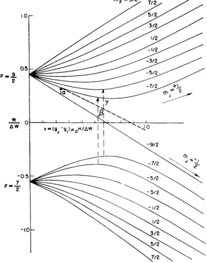
The schematic features of most of the experiments may be described by reference to the Breit-Rabi equation21 for the hyperfine energy of an atom in a 2S± state with nuclear spin 7,
AW AW 4m
WA + im) = + giuoHm + (1 + x+xA 2)*
~ * ' 2(27+1) ***** - 2 V 27+1 ;
where the + of the ± sign refers to the state for F = / + \ and the — refers to / — £, and the field parameter
Figure 5 illustrates graphically the behavior of W as a function of
Fig. 5. Zeeman energy levels for an atom in a 2S± state with a nuclear spin of 4 as calculated by the Breit-Rabi formula.
x for an arbitrarily chosen value for / of 4. In the strong field the states are characterized by two groups, nij = +£ and mj —\. In a non-
136 V I C T O R W . C O H E N
uniform magnetic field those atoms in the +£ state will be deflected toward the weak field while those in the - J state will be deflected in the opposite direction.
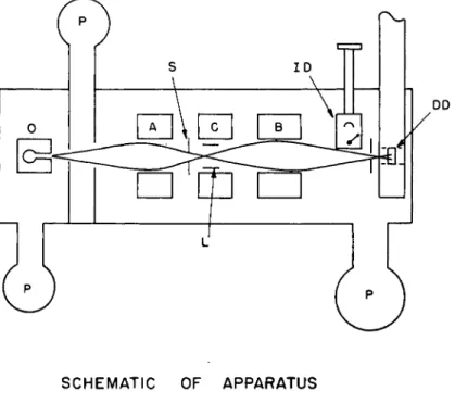
The general schematic arrangement of the apparatus is shown in Fig. 6. We see here an oven, non-uniform magnetic fields A and B, a uniform magnetic field region C on which the oscillatory magnetic field
SCHEMATIC OF APPARATUS
Fig. 6. A typical schematic arrangement of a magnetic resonance flop-in type of apparatus: O—oven, P—pumps, S—defining slit; A, B, G refer to the A, B, and G fields; L—Oscillatory field loop; ID—the surface ionization detector for monitoring the beam with a stable isotope; DD—deposition detector for radio-
active atoms.
can be superimposed, and the slit which defines« that portion of the beam which strikes the detecting target. In the A field region the atoms in the two mi states will be deflected in opposite directions. The gradients of both the A and B fields are oriented perpendicular to the beam axis and in the same direction. For either state the deflection produced by the B field will be in the same direction as that produced by the A field as long as the atoms pass adiabatically over the entire trajectory. If now in the C region they make any transition from one group to the
COMPARISON OF METHODS FOR R A D I O A C T I V E N U C L E I 1 3 7
other the sign of the deflection will change and they will be focused to the detecting slit and strike the target.
The transition a in Fig. 5 will satisfy the condition for an atom to be
"flopped", i.e. to undergo a transition in which m changes by ± 1 . In this type of experiment the detector receives no beam material unless the oscillatory field is in resonance with the energy of a transition.
The transition a in Fig. 5 has the convenient property that in the weak field region, i.e. where the energy states for the various m values are approximately linear in H, the transition frequency v at a given H is a function only of/, not of Δι>( = LWJh)
_ 2μρΗ
V~ A(2/+l)#
In measuring the spin of a particular nucleus one knows initially whether its spin will be integral or half-integral according to whether the mass number is even or odd. One may then compute the values of the transition frequency for all possible assumed values of/and a known H. Each of these frequencies is applied in turn and one of them must satisfy the flop4n condition. In principle, then, only a few observations should be necessary to establish the value of the spin.
Once / is determined, the next quantity of interest is Δν, the hfs interaction constant. This constant is, in effect, the scale factor of the abscissa in Fig. 5 and can be evaluated with fair precision by determin- ing the curvature of the energy level diagram in the intermediate field region. Once the frequency is determined at a known field in the linear region as described above, one increases the field by a small amount and then searches for a resonance at a frequency slightly higher than that value extrapolated from the weak field data. From this departure from a linear extrapolation one may make a very rough estimate of Δν.
Using this value of Δι> one uses the Breit-Rabi formula to calculate the frequency expected for a slightly higher value of the field. The neigh- borhood of this value is searched and from the observed resonance fre- quency a more precise value of Δν may be computed. This procedure is repeated successively at higher fields until the desired accuracy is obtained. If Δν can be evaluated to better than one megacycle the zero field transition can be looked for and the optimum precision for Δν can be obtained. Precision of the order of 1 part in 105 is not at all difficult.
Where precision measurements of both Δν and gi have been made it has been found that the Fermi-Segrè22 formula, which relates àW, / and gi
AW=ir 7tf/(2/+1)0(0)2
K
138 V I C T O R W. C O H E N
is not quite accurate. This formula treats the nuclear-electronic magnetic interaction as though the nucleus acted as a point dipole.
For a more precise treatment, considering the penetration of the elec- tron inside the nuclear volume, one must consider the nuclear magneti- zation to result from a combination of nucléon intrinsic magnetic moments and orbital contributions. Over the nuclear volume, therefore the interactions in two isotopes of the same element will differ.
The departure from the Fermi-Segrè formula has been expressed for the ratio of two AW^'s for two isotopes by defining a correction, Δ, known as the hfs anomaly, by the equation
Δ ^ ι=^ ( 2 Ζ α ) + 1 ) / \
&W2 £/<2>(2/(2)+l) \ /
The significance of the hfs anomaly in nuclear theory has been dis- cussed by Bohr and Weisskopf.23 One might expect that as different models of nuclear composition are proposed this data may be of value in support or contradiction with theory.
The hfs anomaly has been evaluated for several sets of isotopes where the gi could be determined with sufficient accuracy. The values of Δ for various combinations has ranged from about 1 x 10⁻⁴ to 3 x 10~3.
In view of the small values of Δ, gi must be determined with high precision. With materials available in macroscopic quantities this is quite simple by nuclear resonance methods. To determine gi from atomic beam experiments requires an experiment of a high order of precision.
If one examines, for example, the two transitions labelled ß, y in Fig. 5, they will be approximately equal but will differ by a small term 8v = (g^oH/h). The two components of this apparent doublet must be resolved and the separation measured with precision of 1 part in 103 or better in order that Δ may be evaluated.
In a typical example, this splitting may be of the order of 200 kc as compared to a mean frequency of a few thousand megacycles. In order to measure Δ, therefore, one must measure the resonance fre- quencies to a precision of at least 1 part in 107. There are other possible transitions which may be used to determine gi but they all require very sharp and precisely determined resonances. Measurements of this kind have been made for Cs134, Cs135 and Cs137 by Stroke24 using the surface ionization detector and mass spectrometer. It appears quite feasible to measure Δ by radioactive detection and, in fact, the problem is being actively pursued in at least two laboratories.
A significant step in the development of the technique of radio- active detection was made by the group at the University of California
at Berkeley by taking advantage of the fact that the area of beam depo- sition on the target is extremely small. The radioactivity of the target is detected with a scintillation crystal whose area is merely large enough to cover the deposit. Such a small crystal produces a small background due to natural contamination so that significant data can be obtained with very small amounts of material.
The problem of radioactive detection requires that the beam material should have a high probability of sticking to the target surface on the first impact. The sticking properties of gas atoms on solid surfaces are not well understood, partly because most surfaces in an atomic beam apparatus are, strictly speaking, not "pure". The effects of adsorbed films from the atmosphere or from various vapors present inside the vacuum system result in a surface which may not be accurately repro- ducible or may not be the same as that of another investigator using the same nominal materials. A few surfaces have been found which empirically seem to work with high efficiency. These are lampblack, sulfur, copper cooled with liquid nitrogen, and, with slightly lower efficiency, tungsten. Among the elements which have been detected by this means, are: Na, K, Cs, Cu, Ag, Au, In, Tl, Al, Sn, I, Bi, Am.
In detecting by radioactive deposition the presence of the stable isotope is not a drawback. In fact, an excess of stable over unstable atoms may be advantageous from the standpoint of being easier to manipulate. Problems such as adsorption on the oven walls are vastly reduced if the oven load is macroscopic rather than microscopic.
Chemical preparation of products obtained by reactions such as cyclo- tron (d,n) or pile {n,p) are greatly facilitated by addition of large amount of inert carrier.
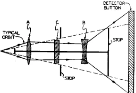
An interesting variation of the atomic beam geometry which permits greater collective efficiency was developed independently by Paul at the University of Bonn25 and by Hamilton at Princeton.26 This consists of using for A and B fields a multipolar system shown in Fig. 7. Such a field has the property that the gradient is everywhere directed toward the beam axis in spite of the fact that the value of the field itself varies in a complex manner. Geometrically the effect of such a field on a beam which originates from a point-source oven is similar to that of a thin lens operating on rays of light. The schematic arrangement of the beam system is shown in Fig. 8. Atoms in one nij group of magnetic states will see the A field as a converging lens, while those in the opposite nij group will see it as diverging. After passing through the C field region the B field acts in a similar manner to the A. One cannot change the orientation of the field gradient. An atom can impinge on the ring collector only if it undergoes a transition induced by the
140 V I C T O R W. C O H E N
oscillatory field in the C field region so that its trajectory avoids the beam stops as indicated. This technique effectively focuses in two dimensions while the conventional A and B fields focus only in one dimension. The
Fig. 7. Cross-section of the 6-pole deflecting magnets used by Hamilton and co-workers. The field gradient is directed toward the axis and is proportional to
the distance from the axis.
result is that the solid angle of trajectories which leave the oven and can be received by the detector is very much higher than in the con- ventional system. One can therefore use a smaller charge of active material in the oven.
Fig. 8. Ray diagram of the optical analogue of the radial focusing system.
In addition to the extensive data obtained by radioactive deposition there has been some excellent work done on several radioactive nuclei
by methods which do not utilize the activity. The prime example of this is the work on Cs134, Cs135, Cs137 by Stroke, Jaccarino, Edmonds, and Weiss,24 who used surface ionization plus mass spectrometer detection.
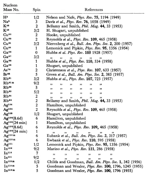
Table 5 presents a summary of results obtained up to October 1, 1958, by atomic beam methods.
TABLE 5
SPINS OF RADIOACTIVE NUCLEI MEASURED BY ATOMIC BEAMS, OCTOBER 1, 1958
Nucléon
Mass No. Spin References Nelson and Nafe, Phys. Rev. 75, 1194 (1949)
Davis et al, Phys. Rev. 76, 1058 (1949) Bellamy and Smith, Phil Mag. 44, 33 (1953) H. Shugart, unpublished
Hooke, unpublished
Reynolds et al, Phys. Rev. 109, 465 (1958)
Nierenberg et al., Bull. Am. Phys. Soc. 2, 200 (1957) Lemonick and Pipkin, Phys. Rev. 95, 1356 (1954) Hubbs et al. Phys. Rev. 105 1928 (1957)
Hubbs etal, Phys. Rev. 110, 534' (1958) Shugart, unpublished
Christenson et al.9 Phys. Rev. 107, 633 (1957) Green et al., Bull. Am. Phys. Soc. 2, 383 (1957) Hubbs et al., Phys. Rev. 107, 723 (1957)
Bellamy and Smith, Phil. Mag. 44, 33 (1953) Hamilton, unpublished
Reynolds et al., Phys. Rev. 109, 465 (1958) Shugart, unpublished
Hamilton, unpublished Hamilton, unpublished
Reynolds et al., Phys. Rev. 109, 465 (1958)
55 55 55 55 55 55
Eubank et al, Bull. Am. Phys. Soc. 2, 317 (1957) Ewbank et al, Phys. Rev. 110, 595 (1958) Lemonick and Pipkin, Phys. Rev. 95, 1356 (1954) Marino et al, Phys. Rev. I l l , 286 (1958)
H3 Na*
K 4 2 K 4 S
Gueo Gueo Gue l Cu64 Gae e Ga87 G a ·8 Ga8 8 As76 BrM Rb8 1 Rbeim R bM Rb8 3 Rb8 4 R b8 e Agio*
A g1 0 4 A g1 0 6
Agloe(8.6d) Agloe(24min) Agloe(8.6d) Agloe(24 min)
Ag1 1 0 m A g i i o m
A gi n
In109 In110 In111 lni i s m Ini i 4 m
I ni i e m
1/2 3 2 3/2
2 2 3/2
1 0 3/2
1 1 2 5 3/2 9/2 5 5/2
2 2 2 2 1/2
6 1 6 1 6 6 1/2 9/2 7 9/2 1/2 5 5
Childs and Goodman, Bull. Am. Phys. Soc. 1, 342 (1956) Goodman and Wexler, Phys. Rev. 100, 1796, 1245 (1955) Goodman and Wexler, Phys. Rev. 100, 1796 (1955)
142 V I C T O R W. C O H E N
T A B L E 5 (CONTD.)
Nucléon
Mass No. Spin References
Lipworth, Bull. Am. Phys. Soc. 2, 316 (1957) Garvin et al, Bull. Am. Phys. Soc. 2, 383 (1957) Sherwood, unpublished
Garvin et al, Phys. Rev. Let. 1, 292 (1958) Garvin et al, Phys. Rev. I l l , 534 (1958) Lipworth, unpublished
Silsbee et al, Bull Am. Phys. Soc. 2, 30 (1957) Nierenberg et al, Phys. Rev. 104, 1380 (1956) Bellamy and Smith, Phil Mag. 44, 33 (1953) Nierenberg et al, Bull. Am. Phys. Soc. 1, 343 (1956) Stroke et al, Phys. Rev. 105, 590 (1957)
Goodman and Wexler, Cohen and Gilbert, Phys. Rev. 97, 243 (1955)
Stroke et al, Phys. Rev. 105, 590 (1957)
Eubank'J* al, Bull Am. Phys. Soc. 2, 383 (1957)
J123 J124 J128 J130
£131 J181
Cs1 2 7 Cs1 2 9 Cs1 3 0 Cs1 8 1 Cs1 3 2 Cs1 3 4
Cs134 m
Cs1 3 6 Cs1 3 7 A u1 0 1 A u1 9 2 A u1 9 8 Au1 9 4 A u1 9 6 A u1 9 8 A u1 9 9
^ 1 9 7
^1198111 1 ^ 1 · ·
rJ,p04
Bi2 0 8 Bi2 0 4 Bi2 0 5 Bi2 o e Bi2 1 0
A t2 1 1
N p2 3 8 N p2 3 9 P u2 3 9 Pu2 3 9 A m2 4 1 C m2 4 2
5/2 2 1 5 7/2 7/2 1/2 1/2 1 5/2
2 4 8 7/2 7/2 3/2 1 3/2
1 2 2 3/2 1/2 7 1/2
2 9/2
6 9/2
6 1 9/2 2 5/2 1/2 1/2 5/2 0
Hooke et al, Bull Am. Phys. Soc. 2, 317 (1957) Shugart, unpublished
Christenson et al, Phys. Rev. 101, 1389 (1956) Shugart,"Brink et al, Phys. Rev. 107, 189 (1957)
Lindgren and Johansen (to be published in Nuclear Physics)
„ „ „ „ „ „ ; Shugart, unpublished K. F. Smith, Physica 18, 989 (1952)
Garvin et al, Phys. Rev. Let. 1, 74 (1958) Hubbs and Marrus, Phys. Rev. 110, 287 (1958) Albridge et al, Phys. Rev. I l l , 1137 (1958) Hubbs et al, Phys. Rev. 109, 390 (1958) Hubbs et al, Bull. Am. Phys. Soc. 2, 316 (1957) Hubbs, unpublished
Hubbs et al, Phys. Rev., to appear March 1959
In the interest of simplicity only the spins are listed in this table.
Since interpretation of magnetic moments data depends upon the method of measurement and the various theoretical corrections which
must be applied to the experimental data, the original references must be consulted where precise values of the moments are required. Exten- sive magnetic moment data is listed in some previously published tables.27
Conclusion
The use of radioactive detection is a powerful tool in the method of atomic beams for nuclear spin and moment determinations. If one is willing to make extensive tests to obtain the proper target for deposition, using liquid He if necessary, this detection method should be applicable to nearly all radioactive nuclides.
Among the difficulties of extending the atomic beam field is the problem of producing a beam of atoms of refractory elements. Some significant progress has been made recently by the Berkeley group in working with Np and Am.28 Such materials as the Pt group might conceivably be vaporized by plating on a tungsten wire and then flash- ing it. In this manner the wire would replace the oven and one would probably require a fresh wire for each observation.
The atomic beam method is not readily applicable to elements for which the ground state is a singlet. In principle one might use very strong gradient A and B fields, such as have been used in beam studies on molecules in which the net magnetic moment is of the order of a nuclear magneton. Such an apparatus would necessarily be much less efficient in the use of material because of the narrower slits and longer beam. No one has yet attempted such an experiment.
The technique of atomic beams is extremely productive, having yielded approximately 50 spins, most of them in the past two years.
This method is still not universal, and one must recognize that the methods of optical spectroscopy as well as paramagnetic resonance must be utilized further in the study of ground state moments of the radio- active nuclei.
REFERENCES
1 G. Breit and I. I. Rabi, Phys. Rev. 38, 2082 (1931).
2 I. I. Rabi and V. W. Cohen, Phys. Rev. 43, 582 (1933).
8 V. W. Cohen, Phys. Rev. 46, 713 (1934).
* H. Kopfermann, £ . Physik 73, 437 (1932).
5 Jackson, Proc. Roy. Soc. A 143, 455 (1933).
6 W. M. Harrison and J. G. Hamilton, Chem. Rev., Oct. 1, 1951.
7 C. H. Townes and A. L. Schawlow, "Microwave Spectroscopy," McGraw-Hill, New York, 1955.
8 Burke, Strandberg, Cohen, and Koski, Phys. Rev. 93, 193 (1954).
» C. Kikuchi and R. D. Spence, Am. J. Phys. 18, 167 (1950).
144 V I C T O R W. C O H E N
10 B. Bleaney and K. W. S. Stevens, Reports on Progress in Physics, XVI (1953).
11 Kikuchi, Sirvetz and Cohen, Phys. Rev, 92, 109 (1953).
12 Pake, Weissman, and Townsend, Discuss, Faraday Soc, No. 19, p. 147, 1955.
18 R. G. Fletcher, W. A. Yager, G. L. Pearson and F. R. Merritt, Phys. Rev. 95, 844 (1954).
14 G. Feher, Phys. Rev. 103, 834 (1956).
15 G. Feher, Fuller, and Gere, Phys. Rev. 107, 1463 (1957).
l e G. D.Jeffries, Phys. Rev. 106, 164 (1957).
17 Pipkin and Culvahouse, Phys. Rev, 106, 1102 (1957).
18 Abraham, Kedzie, and Jeffries, Phys. Rev. 106, 165 (1957).
19 Smith and Bellamy, Phil. Mag. 44, 33 (1953).
20 In England: Oxford, Cambridge.
In Germany: University of Bonn.
In Sweden : University of Upsala.
In United States : Argonne National Laboratory, Brookhaven National Labora- tory, Oak Ridge National Laboratory, Princeton University, University of California.
21 The equation generally referred to as the Breit-Rabi equation was first given by Kusch, Millman, and Rabi, Phys. Rev, 57, 765 (1940) and represents the original Breit-Rabi equation of Reference 1 with added terms in g{,
22 E. Fermi and E. Segrè, £ . Physik 60, 320 (1930).
28 A. Bohr and V. W. Weisskopf, Phys. Rev. 77, 94 (1950).
24 Stroke, Jaccarino, Edmonds, and Weiss, Phys. Rev. 105, 590 (1957).
25 H . Friedburg and W. Paul, Naturwiss. 38, 159 (1951).
26 Lemonick and Pipkin, Phys. Rev. 95, 1356 (1954).
27 N. F. Ramsey, "Nuclear Moments," Wiley, New York, (1953); H. E. Walchli,
"A Table of Nuclear Moment Data," Oak Ridge National Laboratory, Oak Ridge, Tenn., Index No. ORNL 1469 (1953), and Supp. No. 2 (1955); W. A.
Nierenberg, Annual Rev. of Nuclear Science, 1958 (in press).
28 J . C. Hubbs, Bull. Am. Phys. Soc. II, 2, 316 (1957) and private communication on Am.