MobiDB 3.0: more annotations for intrinsic disorder, conformational diversity and interactions in proteins
Damiano Piovesan
1,†, Francesco Tabaro
1,2,†, Lisanna Paladin
1, Marco Necci
1,3,4, Ivan Mi ˇceti ´c
1, Carlo Camilloni
5, Norman Davey
6,7, Zsuzsanna Doszt ´anyi
8,
B ´alint M ´esz ´aros
8,9, Alexander M. Monzon
10, Gustavo Parisi
10, Eva Schad
9,
Pietro Sormanni
11, Peter Tompa
9,12,13, Michele Vendruscolo
11, Wim F. Vranken
12,13,14and Silvio C.E. Tosatto
1,15,*1Department of Biomedical Sciences, University of Padua, via U. Bassi 58/b, 35131 Padua, Italy,2Institute of Biosciences and Medical Technology, Arvo Ylp ¨on katu 34, 33520 Tampere, Finland,3Department of Agricultural Sciences, University of Udine, via Palladio 8, 33100 Udine, Italy,4Fondazione Edmund Mach, Via E. Mach 1, 38010 S. Michele all’Adige, Italy,5Department of Biosciences, University of Milan, 20133 Milano, Italy,6Conway Institute of Biomolecular & Biomedical Research, University College Dublin, Belfield, Dublin 4, Ireland,7UCD School of Medicine
& Medical Science, University College Dublin, Belfield, Dublin 4, Ireland,8MTA-ELTE Lend ¨ulet Bioinformatics Research Group, Department of Biochemistry, E ¨otv ¨os Lor ´and University, 1/c P ´azm ´any P ´eter s ´et ´any, H-1117, Budapest, Hungary,9Institute of Enzymology, Research Centre for Natural Sciences, Hungarian Academy of Sciences, PO Box 7, H-1518 Budapest, Hungary,10Structural Bioinformatics Group, Department of Science and Technology, National University of Quilmes, CONICET, Roque Saenz Pena 182, Bernal B1876BXD, Argentina,
11Department of Chemistry, University of Cambridge, Cambridge CB2 1EW, UK,12Structural Biology Brussels, Vrije Universiteit Brussel (VUB), Brussels 1050, Belgium,13VIB-VUB Center for Structural Biology, Flanders Institute for Biotechnology (VIB), Brussels 1050, Belgium,14Interuniversity Institute of Bioinformatics in Brussels, ULB/VUB, 1050 Brussels, Belgium and15CNR Institute of Neuroscience, via U. Bassi 58/b, 35131 Padua, Italy
Received September 22, 2017; Revised October 13, 2017; Editorial Decision October 16, 2017; Accepted October 19, 2017
ABSTRACT
The MobiDB (URL: mobidb.bio.unipd.it) database of protein disorder and mobility annotations has been significantly updated and upgraded since its last ma- jor renewal in 2014. Several curated datasets for in- trinsic disorder and folding upon binding have been integrated from specialized databases. The indirect evidence has also been expanded to better capture information available in the PDB, such as high tem- perature residues in X-ray structures and overall con- formational diversity. Novel nuclear magnetic reso- nance chemical shift data provides an additional ex- perimental information layer on conformational dy- namics. Predictions have been expanded to provide new types of annotation on backbone rigidity, sec- ondary structure preference and disordered bind- ing regions. MobiDB 3.0 contains information for the complete UniProt protein set and synchroniza- tion has been improved by covering all UniParc se- quences. An advanced search function allows the
creation of a wide array of custom-made datasets for download and further analysis. A large amount of information and cross-links to more specialized databases are intended to make MobiDB the cen- tral resource for the scientific community working on protein intrinsic disorder and mobility.
INTRODUCTION
The protein structure-function paradigm is a cornerstone of molecular biology, offering a mechanistic understanding of processes ranging from enzyme catalysis, signal transduc- tion to molecular recognition and allosteric regulation. Un- derlying this paradigm is the assumption that proteins be- come functional by assuming a well-defined structure, typ- ically described by the coordinates of all its atoms. A solid foundation of this view is provided by the 130 000 struc- tures of proteins and complexes in the Protein Data Bank, PDB (1). However, it is increasingly recognized that many proteins do not obey this rule. Intrinsically disordered pro- teins (IDPs) or regions (IDRs) are devoid of order in their native unbound state (2–4). Intrinsic disorder is prevalent
*To whom correspondence should be addressed. Tel: +39 49 827 6269; Fax: +39 49 827 6363; Email: silvio.tosatto@unipd.it
†These authors contributed equally to the paper as first authors.
C The Author(s) 2017. Published by Oxford University Press on behalf of Nucleic Acids Research.
This is an Open Access article distributed under the terms of the Creative Commons Attribution License (http://creativecommons.org/licenses/by-nc/4.0/), which permits non-commercial re-use, distribution, and reproduction in any medium, provided the original work is properly cited. For commercial re-use, please contact
in the human proteome (5), appears to play important sig- naling and regulatory roles (2) and is frequently involved in disease (6). The discovery of intrinsic disorder and its prevalence and functional importance is transforming the field of molecular biology. As intrinsic disorder is emerging as a general phenomenon, databases are collecting and pre- senting disorder related data in a systematic manner. Mo- biDB has been a major contributor by providing consensus predictions and functional annotations for all UniProt pro- teins, driving the field ahead (7,8). The MobiDB upgrade we present in this paper is essential for several reasons.
There is a rapid advance in the functional understand- ing of intrinsic disorder. The functional classification of IDPs/IDRs is becoming ever more elaborate, with sev- eral newly recognized functional mechanisms (9). For ex- ample, the central role of intrinsic disorder in the forma- tion of membraneless organelles, such as nucleoli and stress granules, by liquid-liquid phase separation has been char- acterized recently (10–13). A wide range of experimen- tal observations on the structure-function relationship of IDPs/IDRs is furthering our understanding of disordered states and of the manners in which they function (14–16).
These developments have also played a central role in the re- cent update of the DisProt database (17), the central repos- itory of experimentally characterized IDPs and IDRs. The re-curated version of this database contains experimental observations of disorder for more than 800 protein entries and a renewed functional ontology schema. The experimen- tal evidence on which it rests has also been significantly aug- mented to include a broad range of biophysical techniques.
DisProt is the basis for most developments in disorder pre- dictors (18,19), and its recent update is a major motivation for a new version of MobiDB.
Additional developments in the field make this release timely. A major source of intrinsic disorder is the identifi- cation of residues with missing atomic coordinates in the PDB, which can now be augmented by cryo-electron mi- croscopy (cryo-EM) data. This is having a tremendous im- pact on structural biology (20,21). Structural descriptions of IDPs and IDRs under physiological conditions have also greatly advanced and are starting to appear in dedicated databases such as IDEAL (22), DIBS (23) and MFIB (24).
IDPs and IDRs can perform key roles in molecular recogni- tion by folding upon binding of short linear motifs (SLiMs) covered in the ELM database (25). Generally, the full func- tional characterization of IDPs and IDRs requires the de- scription not just of their free (disordered) states (26,27), but also of their residual dynamics in the bound states (28).
Fuzzy (disordered) complexes can be found in FuzDB (29) and structural ensembles describing the free form (30) in the protein ensemble database (PED (31)). Techniques such as in-cell Nuclear magnetic resonance (NMR) spectroscopy (32,33) and single-molecule fluorescence (34) will soon help study these structures in the physiological state. In reflec- tion of all these developments, we are now launching a sig- nificantly updated version of our database, MobiDB 3.0.
The new version incorporates additional curated data from specialized databases. Novel annotation features include disorder derived from publicly available NMR chemical shift data (35) and an extended list of predictors. Database
Figure 1.Overview of different annotation data types (A) and levels of accuracy (B) in MobiDB 3.0.
searches are facilitated by an improved search algorithm, pre-calculated data and new sections in the database.
DATABASE DESCRIPTION
MobiDB 3.0 is intended to be a central resource for large- scale intrinsic disorder sequence annotation. This new ver- sion is organized by both type of disorder annotation and quality of disorder evidence (Figure1). Disorder informa- tion is grouped in three different sections: disorder, linear interacting peptides (LIPs) and secondary structure pop- ulations. The latter represents the conformational hetero- geneity of IDPs and IDRs as the ability to populate differ- ent secondary structure populations in solution. LIPs are structure fragments that interact with other molecules pre- serving an elongated structure or folding upon binding. The data in MobiDB is organized hierarchically. The top tier is formed by manually curated data from external databases and represents the highest quality annotations. Annotations derived from experimental data such as X-ray and NMR chemical shifts are indirect but far more abundant. At the bottom, predictions provide disorder annotation at lower confidence than experimental evidence. The main disorder definition in MobiDB is provided by a consensus combin-
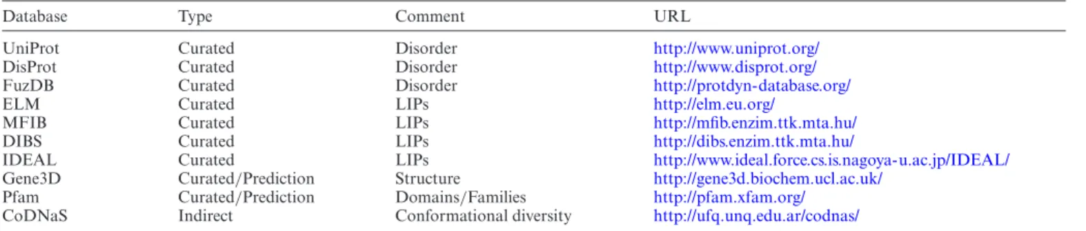
Table 1. Overview of databases integrated into MobiDB 3.0
Database Type Comment URL
UniProt Curated Disorder http://www.uniprot.org/
DisProt Curated Disorder http://www.disprot.org/
FuzDB Curated Disorder http://protdyn-database.org/
ELM Curated LIPs http://elm.eu.org/
MFIB Curated LIPs http://mfib.enzim.ttk.mta.hu/
DIBS Curated LIPs http://dibs.enzim.ttk.mta.hu/
IDEAL Curated LIPs http://www.ideal.force.cs.is.nagoya-u.ac.jp/IDEAL/
Gene3D Curated/Prediction Structure http://gene3d.biochem.ucl.ac.uk/
Pfam Curated/Prediction Domains/Families http://pfam.xfam.org/
CoDNaS Indirect Conformational diversity http://ufq.unq.edu.ar/codnas/
ing all available sources prioritizing curated and indirect evidences over predictions in analogy to the previous ver- sion (8). In the following, we will describe the main re- cent improvements since the previous release. The database schema, web interface and server have been completely re- designed and the underlying technology updated. The fea- ture viewer showing sequence annotations is now fully dy- namic and allows the generation of high quality images for publications with a click. Where available, MobiDB anno- tation is projected directly onto the structure and shown in a new 3D viewer. The look and feel and organization of the page and loading latency were also improved.
New curated data
MobiDB 3.0 includes different sources of manually curated disorder annotations (Table1). These annotations fall into two categories: disorder and LIPs. LIPs are binding regions presumed or demonstrated to be intrinsically disordered that fold upon binding. These come under different names such as SLiMs or MoREs (molecular recognition elements) in the literature. The IDEAL database calls them ‘protean’
segments (ProS) (22). MobiDB includes both ‘verified’ and
‘possible’ ProS from IDEAL, where verified means disorder has been experimentally observed in the isolated molecule.
The Database of Disordered Binding Sites (DIBS, (23)) col- lects cases where a disordered region folds upon binding with a globular domain and the Mutual Folding Induced by Binding (MFIB, (24)) database includes disordered re- gions that fold upon binding with another disordered re- gion. ELM (25) provides SLiM annotations involved in binding and post-translational modifications. General dis- order annotation, i.e. without any knowledge about tran- sition driven by interactions, is collected from UniProtKB (36), DisProt (17) and FuzDB (29). UniProtKB provides manually curated disorder annotations under the region field in the features section. FuzDB collects cases of fuzzy complexes, where conformational diversity has a functional role in the regulation and formation of protein complexes or higher-order assemblies. DisProt has been recently re- vamped and MobiDB now propagates DisProt disordered regions by homology transfer. Regions homologous to ex- perimentally characterized IDRs are mapped across ho- mologs obtained from GeneTree alignments (37). Regions with identity and similarity>80% and an alignment of at least 10 residues are retained as homologous IDRs. Gene3D (38) contributes complementary order annotation to the MobiDB consensus calculation, while Pfam (39) is used
to highlight protein domains. Lastly MobiDB also maps CoDNaS information to highlight conformation diversity in globular regions. CoDNaS measures structural differ- ences among conformers of the same protein (40).
New indirect annotations
Previous releases of MobiDB provided indirect annotations from the PDB through missing residues in X-ray struc- tures and mobile regions from NMR ensembles as calcu- lated with the Mobi software (41). In the current release, this annotation has been complemented with additional indirect information from experimental data in the PDB and chemical shifts from the Biological Magnetic Reso- nance Data Bank (BMRB) (35). The new Mobi 2.0 software (42) is used to extract LIPs and disorder information from PDB files. Disorder is encoded by three different parame- ters: high-temperature, missing and mobile residues. High- temperature residues are detected from B-factor regions for X-ray and cryo-EM structures using a threshold propor- tional to the resolution of the structure. Missing residues are available for all experimental types and obtained com- paring the experimental sequence (i.e. PDB SEQRES en- tries) with the observed residues in the structure (i.e. PDB ATOM entries). A mobility estimate is provided for NMR structures by comparingCαdisplacement and local confor- mations in different aligned models (41). LIPs are identified by comparing intra- versus inter-chain contacts calculated using RING (43). The closest atoms between two residues are used to establish a contact which is then distinguished by chemical type (e.g. hydrogen bond, salt bridge,−stack).
LIPs are identified as any region where the number of inter- chain contacts is at least two times the number of intra- chain contacts (42).
MobiDB 3.0 better exploits the power of NMR spec- troscopy to probe the structural properties of proteins in solution, as well as their dynamics on a wide range of timescales (44). Chemical shifts quantify structural fluctu- ations of proteins up to the millisecond timescale and are relatively easy to measure. Using chemical shifts to ob- tain information about the statistical populations of dif- ferent structural motifs allows for a more comprehensive structural description of proteins in solution than static structures or binary definitions such as ‘ordered’ and ‘dis- ordered’ (44). MobiDB 3.0 uses chemical shift data from BMRB directly as reported without applying chemical shift re-referencing methods. The software packages ␦2D (45) and Random Coil Index (RCI) (46) are used to calculate
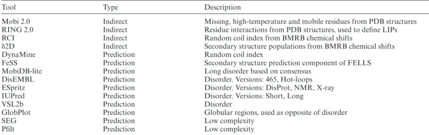
Table 2. Overview of tools used into MobiDB 3.0
Tool Type Description
Mobi 2.0 Indirect Missing, high-temperature and mobile residues from PDB structures
RING 2.0 Indirect Residue interactions from PDB structures, used to define LIPs
RCI Indirect Random coil index from BMRB chemical shifts
␦2D Indirect Secondary structure populations from BMRB chemical shifts
DynaMine Prediction Random coil index
FeSS Prediction Secondary structure prediction component of FELLS
MobiDB-lite Prediction Long disorder based on consensus
DisEMBL Prediction Disorder. Versions: 465, Hot-loops
ESpritz Prediction Disorder. Versions: DisProt, NMR, X-ray
IUPred Prediction Disorder. Versions: Short, Long
VSL2b Prediction Disorder
GlobPlot Prediction Globular regions, used as opposite of disorder
SEG Prediction Low complexity
Pfilt Prediction Low complexity
two-dimensional ensembles in terms of secondary structure populations (44) and backbone flexibility. Secondary struc- ture populations are calculated only for residues with at least three atom types with measured chemical shifts, as us- ing fewer chemical shifts results in less accurate mappings of the populations (45). MobiDB 3.0 reports the experimen- tal conditions at which the chemical shifts were measured as the structural properties of some proteins can change drastically between different conditions (e.g. binding part- ners, lipids, pH) and these can help elucidate protein func- tion (44). When an entry in MobiDB is associated to mul- tiple chemical shifts, an overview of the predominant sec- ondary structure conformation is provided in a consensus track. This can be expanded in the feature viewer to show experimental conditions such as pH, temperature, binding partners, molecular state, sample information and the title of the corresponding BMRB entries.
New predictors
MobiDB 3.0 includes the same set of disorder predictors used in the previous release: ESpritz (47), IUpred (48), Dis- EMBL (49) and VSL2b (50). Consensus generation is han- dled by MobiDB-lite (51), which uses a stronger majority threshold and enforces at least 20 consecutive disordered residues to provide highly specific predictions. This is com- pleted by a continuous representation of the fraction of methods predicting disorder for each residue. DynaMine (52), Anchor (53) and FeSS (54) are now also part of the an- notation pipeline. DynaMine (52) predicts backbone flexi- bility where 1.0 means complete order (stable conformation, i.e. rigid) and 0 means fully random bond vector movement (highly dynamic, i.e. flexible). Anchor predicts binding re- gions located in disordered proteins, providing LIP annota- tions for all proteins in the database. FeSS is a component of the FELLS method (54) providing three-state (helix, sheet, coil) secondary structure propensity. FeSS prediction confi- dence can be interpreted similarly to the dynamic behavior measured by␦2D in chemical shifts, i.e. a propensity to re- main in a given state of secondary structure. The complete list of tools is available in Table2.
The MobiDB-lite version used in MobiDB 3.0 has been extended to provide a structural characterization of the dis- order regions that can help interpret their functional role. It distinguishes different types of disordered regions by mea-
suring the fraction of charged residues and net charge ac- cording to a previous classification (55). The different types are: positive polyelectrolites (D PPE), negative polyelectro- lites (D NPE), polyampholites (D PA) and weak polyam- pholites (D WC). A statistical analysis of the different dis- order flavours was already performed on the MobiDB 2.0 data (8).
Usage and annotated data
MobiDB now contains all sequences from UniParc, the most comprehensive non-redundant set of protein se- quences. Entries are identified also by UniProtKB (36) ac- cession numbers and can be retrieved by organism, taxon- omy and other identifiers provided by UniProtKB. Predic- tion results are combined with indirect disorder evidences derived from PDB data (using Mobi 2) and data extracted from manually curated third party databases. MobiDB an- notations are used by DisProt (17) curators to guide the an- notation of disorder regions. MobiDB data is made avail- able to the public via a web interface allowing extensive search functionalities and RESTful services for program- matic access. MobiDB 3.0 includes a pre-calculated con- sensus for all entries allowing real-time statistics and down- load of entire datasets in different formats directly from the web interface. The new database schema makes it possible to perform complex search queries and to generate custom datasets, for example retrieving all entries with manually cu- rated annotations. The MobiDB update has been automa- tized and is scheduled every three months due to the high computational cost of generating predictions for new se- quences.
DISCUSSION
MobiDB 3.0 improves on previous releases by adding de- scriptions of conformational diversity and disorder-related functions, both in terms of experimental data and pre- dictions. A particular field where it may have a signifi- cant impact is the establishment of a long-awaited disor- der sequence-function relationship schema. The most reli- able proxy to this goal is to assess the function of a pro- tein by homology transfer, i.e. transferring functional an- notation based on sequence similarity. Aligning IDR se- quences is complicated by their high evolutionary variabil-
ity and often limits evolutionary analysis (56,57). New func- tional terms introduced in the DisProt update (17), repre- sent non-canonical functions probably only characteristic of IDPs which are not incorporated in functional classifica- tion schemes such as GO (58). A large-scale analysis of IDP functional annotations will be necessary to find adequate boundaries for transferring IDP functions by homology. As sufficient data is now available in MobiDB 3.0, we expect a rapid advance in the field of sequence-function correlations of IDPs.
For proteins with sufficient NMR data, MobiDB now features quantitative annotations incorporating structure and equilibrium dynamics in a unified framework. These large-scale quantitative annotations will help understand the biological role of order and disorder, and serve as a basis to construct predictive models. As NMR measurements of proteins in their native complex environments, such as in- side living cells, are becoming more common (59), we will be able to address fundamental biological questions with greater physiological relevance (60).
MobiDB is widely used by scientific community and by third party services such as DisProt (17) and ProViz (61).
It has recently joined the InterPro consortium to provide disorder annotation alongside protein domains and fami- lies (62). MobiDB is becoming a thematic hub for IDPs in- side the European sustainable bioinformatics infrastructure (ELIXIR) and we encourage contributions of novel predic- tors and datasets. Future work will focus on including IDP annotations into core data resources such as UniProt.
ACKNOWLEDGEMENTS
We acknowledge ELIXIR-IIB (elixir-italy.org), the Ital- ian Node of the European ELIXIR infrastructure (elixir- europe.org), for supporting the development and mainte- nance of MobiDB.
FUNDING
COST Action [BM1405 NGP-net]; FIRC Research Fel- lowship [16621 to D.P.]; Hungarian Academy of Sciences
‘Lend ¨ulet’ Grant [LP201418/2016 to Z.D.]; Hungarian Sci- entific Research Fund [OTKA K 108798 to Z.D.]; Hungar- ian Academy of Sciences Postdoctoral Fellowship [to B.M.];
Agencia de Ciencia y Tecnolog´ıa [PICT-2014–3430 to G.P.];
Universidad Nacional de Quilmes [1402/15]; OTKA Grant [PD-OTKA 108772 to E.S.]; Research Foundation Flanders (FWO) Odysseus Grant [G.0029.12 to P.T.]; FWO project [G032816N to W.F.V.]; AIRC IG Grant [17753 to S.T., in part]. Funding for open access charge: COST Action [BM1405].
Conflict of interest statement.None declared.
REFERENCES
1. Burley,S.K., Berman,H.M., Kleywegt,G.J., Markley,J.L.,
Nakamura,H. and Velankar,S. (2017) Protein Data Bank (PDB): the single global macromolecular structure archive.Methods Mol. Biol.
Clifton NJ,1607, 627–641.
2. Wright,P.E. and Dyson,H.J. (2015) Intrinsically disordered proteins in cellular signalling and regulation.Nat. Rev. Mol. Cell Biol.,16, 18–29.
3. Habchi,J., Tompa,P., Longhi,S. and Uversky,V.N. (2014) Introducing protein intrinsic disorder.Chem. Rev.,114, 6561–6588.
4. Tompa,P. (2012) Intrinsically disordered proteins: a 10-year recap.
Trends Biochem. Sci.,37, 509–516.
5. Pancsa,R. and Tompa,P. (2012) Structural disorder in eukaryotes.
PLoS One,7, e34687.
6. Uversky,V.N., Oldfield,C.J. and Dunker,A.K. (2008) Intrinsically disordered proteins in human diseases: introducing the D2 concept.
Annu. Rev. Biophys.,37, 215–246.
7. Di Domenico,T., Walsh,I., Martin,A.J.M. and Tosatto,S.C.E. (2012) MobiDB: a comprehensive database of intrinsic protein disorder annotations.Bioinformatics,28, 2080–2081.
8. Potenza,E., Di Domenico,T., Walsh,I. and Tosatto,S.C.E. (2015) MobiDB 2.0: an improved database of intrinsically disordered and mobile proteins.Nucleic Acids Res.,43, D315–D320.
9. van der Lee,R., Buljan,M., Lang,B., Weatheritt,R.J.,
Daughdrill,G.W., Dunker,A.K., Fuxreiter,M., Gough,J., Gsponer,J., Jones,D.T.et al.(2014) Classification of intrinsically disordered regions and proteins.Chem. Rev.,114, 6589–6631.
10. Hyman,A.A., Weber,C.A. and J ¨ulicher,F. (2014) Liquid-liquid phase separation in biology.Annu. Rev. Cell Dev. Biol.,30, 39–58.
11. Shorter,J. (2016) Membraneless organelles: phasing in and out.Nat.
Chem.,8, 528–530.
12. Toretsky,J.A. and Wright,P.E. (2014) Assemblages: functional units formed by cellular phase separation.J. Cell Biol.,206, 579–588.
13. Brangwynne,C.P., Tompa,P. and Pappu,R.V. (2015) Polymer physics of intracellular phase transitions.Nat. Phys.,11, 899–904.
14. Berlow,R.B., Dyson,H.J. and Wright,P.E. (2017) Hypersensitive termination of the hypoxic response by a disordered protein switch.
Nature,543, 447–451.
15. Mylona,A., Theillet,F.-X., Foster,C., Cheng,T.M., Miralles,F., Bates,P.A., Selenko,P. and Treisman,R. (2016) Opposing effects of Elk-1 multisite phosphorylation shape its response to ERK activation.Science,354, 233–237.
16. Bah,A., Vernon,R.M., Siddiqui,Z., Krzeminski,M., Muhandiram,R., Zhao,C., Sonenberg,N., Kay,L.E. and Forman-Kay,J.D. (2015) Folding of an intrinsically disordered protein by phosphorylation as a regulatory switch.Nature,519, 106–109.
17. Piovesan,D., Tabaro,F., Miˇceti´c,I., Necci,M., Quaglia,F., Oldfield,C.J., Aspromonte,M.C., Davey,N.E., Davidovi´c,R., Doszt´anyi,Z.et al.(2017) DisProt 7.0: a major update of the database of disordered proteins.Nucleic Acids Res.,45, D1123–D1124.
18. Walsh,I., Giollo,M., Di Domenico,T., Ferrari,C., Zimmermann,O.
and Tosatto,S.C.E. (2015) Comprehensive large-scale assessment of intrinsic protein disorder.Bioinformatics,31, 201–208.
19. Necci,M., Piovesan,D., Dosztanyi,Z., Tompa,P. and Tosatto,S.C.E.
(2017) A comprehensive assessment of long intrinsic protein disorder from the DisProt database.Bioinformatics,
doi:10.1093/bioinformatics/btx590.
20. Callaway,E. (2015) The revolution will not be crystallized: a new method sweeps through structural biology.Nature,525, 172–174.
21. Cheng,Y. (2015) Single-Particle Cryo-EM at Crystallographic Resolution.Cell,161, 450–457.
22. Fukuchi,S., Amemiya,T., Sakamoto,S., Nobe,Y., Hosoda,K., Kado,Y., Murakami,S.D., Koike,R., Hiroaki,H. and Ota,M. (2014) IDEAL in 2014 illustrates interaction networks composed of intrinsically disordered proteins and their binding partners.Nucleic Acids Res.,42, D320–D325.
23. Schad,E., Fich ´o,E., Pancsa,R., Simon,I., Doszt´anyi,Z. and M´esz´aros,B. (2017) DIBS: a repository of disordered binding sites mediating interactions with ordered proteins.Bioinformatics, doi:10.1093/bioinformatics/btx640.
24. Fich ´o,E., Rem´enyi,I., Simon,I. and M´esz´aros,B. (2017) MFIB: a repository of protein complexes with mutual folding induced by binding.Bioinformatics, doi:10.1093/bioinformatics/btx486.
25. Dinkel,H., Van Roey,K., Michael,S., Kumar,M., Uyar,B.,
Altenberg,B., Milchevskaya,V., Schneider,M., K ¨uhn,H., Behrendt,A.
et al.(2016) ELM 2016–data update and new functionality of the eukaryotic linear motif resource.Nucleic Acids Res.,44, D294–D300.
26. Arai,M., Sugase,K., Dyson,H.J. and Wright,P.E. (2015) Conformational propensities of intrinsically disordered proteins influence the mechanism of binding and folding.Proc. Natl. Acad.
Sci. U.S.A.,112, 9614–9619.
27. Borcherds,W., Theillet,F.-X., Katzer,A., Finzel,A., Mishall,K.M., Powell,A.T., Wu,H., Manieri,W., Dieterich,C., Selenko,P.et al.(2014)
Disorder and residual helicity alter p53-Mdm2 binding affinity and signaling in cells.Nat. Chem. Biol.,10, 1000–1002.
28. Tompa,P. and Fuxreiter,M. (2008) Fuzzy complexes: polymorphism and structural disorder in protein-protein interactions.Trends Biochem. Sci.,33, 2–8.
29. Miskei,M., Antal,C. and Fuxreiter,M. (2017) FuzDB: database of fuzzy complexes, a tool to develop stochastic structure-function relationships for protein complexes and higher-order assemblies.
Nucleic Acids Res.,45, D228–D235.
30. Tompa,P. and Varadi,M. (2014) Predicting the predictive power of IDP ensembles.Structure,22, 177–178.
31. Varadi,M., Kosol,S., Lebrun,P., Valentini,E., Blackledge,M., Dunker,A.K., Felli,I.C., Forman-Kay,J.D., Kriwacki,R.W., Pierattelli,R.et al.(2014) pE-DB: a database of structural ensembles of intrinsically disordered and of unfolded proteins.Nucleic Acids Res.,42, D326–D335.
32. Theillet,F.-X., Binolfi,A., Bekei,B., Martorana,A., Rose,H.M., Stuiver,M., Verzini,S., Lorenz,D., van Rossum,M., Goldfarb,D.et al.
(2016) Structural disorder of monomeric␣-synuclein persists in mammalian cells.Nature,530, 45–50.
33. Felli,I.C., Gonnelli,L. and Pierattelli,R. (2014) In-cell13C NMR spectroscopy for the study of intrinsically disordered proteins.Nat.
Protoc.,9, 2005–2016.
34. Sakon,J.J. and Weninger,K.R. (2010) Detecting the conformation of individual proteins in live cells.Nat. Methods,7, 203–205.
35. Ulrich,E.L., Akutsu,H., Doreleijers,J.F., Harano,Y., Ioannidis,Y.E., Lin,J., Livny,M., Mading,S., Maziuk,D., Miller,Z.et al.(2008) BioMagResBank.Nucleic Acids Res.,36, D402–D408.
36. Consortium,The UniProt (2017) UniProt: the universal protein knowledgebase.Nucleic Acids Res.,45, D158–D169.
37. Vilella,A.J., Severin,J., Ureta-Vidal,A., Heng,L., Durbin,R. and Birney,E. (2009) EnsemblCompara GeneTrees: Complete,
duplication-aware phylogenetic trees in vertebrates.Genome Res.,19, 327–335.
38. Lees,J., Yeats,C., Perkins,J., Sillitoe,I., Rentzsch,R., Dessailly,B.H.
and Orengo,C. (2012) Gene3D: a domain-based resource for comparative genomics, functional annotation and protein network analysis.Nucleic Acids Res.,40, D465–D471.
39. Finn,R.D., Coggill,P., Eberhardt,R.Y., Eddy,S.R., Mistry,J., Mitchell,A.L., Potter,S.C., Punta,M., Qureshi,M.,
Sangrador-Vegas,A.et al.(2016) The Pfam protein families database:
towards a more sustainable future.Nucleic Acids Res.,44, D279–D285.
40. Monzon,A.M., Rohr,C.O., Fornasari,M.S. and Parisi,G. (2016) CoDNaS 2.0: a comprehensive database of protein conformational diversity in the native state.Database (Oxford),2016, baw038.
41. Martin,A.J.M., Walsh,I. and Tosatto,S.C.E. (2010) MOBI: a web server to define and visualize structural mobility in NMR protein ensembles.Bioinformatics,26, 2916–2917.
42. Piovesan,D. and Tosatto,S.C.E. (2017) Mobi 2.0: an improved method to define intrinsic disorder, mobility and linear binding regions in protein structures.Bioinformatics,
doi:10.1093/bioinformatics/btx592.
43. Piovesan,D., Minervini,G. and Tosatto,S.C.E. (2016) The RING 2.0 web server for high quality residue interaction networks.Nucleic Acids Res.,44, W367–W374.
44. Sormanni,P., Piovesan,D., Heller,G.T., Bonomi,M., Kukic,P., Camilloni,C., Fuxreiter,M., Dosztanyi,Z., Pappu,R.V., Babu,M.M.
et al.(2017) Simultaneous quantification of protein order and disorder.Nat. Chem. Biol.,13, 339–342.
45. Camilloni,C., De Simone,A., Vranken,W.F. and Vendruscolo,M.
(2012) Determination of secondary structure populations in disordered states of proteins using nuclear magnetic resonance chemical shifts.Biochemistry,51, 2224–2231.
46. Berjanskii,M.V. and Wishart,D.S. (2005) A simple method to predict protein flexibility using secondary chemical shifts.J. Am. Chem. Soc., 127, 14970–14971.
47. Walsh,I., Martin,A.J.M., Di Domenico,T. and Tosatto,S.C.E. (2012) ESpritz: accurate and fast prediction of protein disorder.
Bioinformatics,28, 503–509.
48. Doszt´anyi,Z., Csizmok,V., Tompa,P. and Simon,I. (2005) IUPred:
web server for the prediction of intrinsically unstructured regions of proteins based on estimated energy content.Bioinformatics,21, 3433–3434.
49. Linding,R., Jensen,L.J., Diella,F., Bork,P., Gibson,T.J. and Russell,R.B. (2003) Protein disorder prediction: implications for structural proteomics.Structure,11, 1453–1459.
50. Peng,K., Radivojac,P., Vucetic,S., Dunker,A.K. and Obradovic,Z.
(2006) Length-dependent prediction of protein intrinsic disorder.
BMC Bioinformatics,7, 208.
51. Necci,M., Piovesan,D., Doszt´anyi,Z. and Tosatto,S.C.E. (2017) MobiDB-lite: fast and highly specific consensus prediction of intrinsic disorder in proteins.Bioinformatics,33, 1402–1404.
52. Cilia,E., Pancsa,R., Tompa,P., Lenaerts,T. and Vranken,W.F. (2013) From protein sequence to dynamics and disorder with DynaMine.
Nat. Commun.,4, 2741.
53. M´esz´aros,B., Simon,I. and Doszt´anyi,Z. (2009) Prediction of protein binding regions in disordered proteins.PLoS Comput. Biol.,5, e1000376.
54. Piovesan,D., Walsh,I., Minervini,G. and Tosatto,S.C.E. (2017) FELLS: fast estimator of latent local structure.Bioinformatics,33, 1889–1891.
55. Das,R.K. and Pappu,R.V. (2013) Conformations of intrinsically disordered proteins are influenced by linear sequence distributions of oppositely charged residues.Proc. Natl. Acad. Sci. U.S.A.,110, 13392–13397.
56. Brown,C.J., Johnson,A.K., Dunker,A.K. and Daughdrill,G.W. (2011) Evolution and disorder.Curr. Opin. Struct. Biol.,21, 441–446.
57. Csizmok,V., Felli,I.C., Tompa,P., Banci,L. and Bertini,I. (2008) Structural and dynamic characterization of intrinsically disordered human securin by NMR spectroscopy.J. Am. Chem. Soc.,130, 16873–16879.
58. Ashburner,M., Ball,C.A., Blake,J.A., Botstein,D., Butler,H., Cherry,J.M., Davis,A.P., Dolinski,K., Dwight,S.S., Eppig,J.T.et al.
(2000) Gene ontology: tool for the unification of biology. The Gene Ontology Consortium.Nat. Genet.,25, 25–29.
59. Plitzko,J.M., Schuler,B. and Selenko,P. (2017) Structural Biology outside the box-inside the cell.Curr. Opin. Struct. Biol.,46, 110–121.
60. Gierasch,L.M. and Gershenson,A. (2009) Post-reductionist protein science, or putting Humpty Dumpty back together again.Nat. Chem.
Biol.,5, 774–777.
61. Jehl,P., Manguy,J., Shields,D.C., Higgins,D.G. and Davey,N.E.
(2016) ProViz-a web-based visualization tool to investigate the functional and evolutionary features of protein sequences.Nucleic Acids Res.,44, W11–W15.
62. Finn,R.D., Attwood,T.K., Babbitt,P.C., Bateman,A., Bork,P., Bridge,A.J., Chang,H.-Y., Doszt´anyi,Z., El-Gebali,S., Fraser,M.et al.
(2017) InterPro in 2017-beyond protein family and domain annotations.Nucleic Acids Res.,45, D190–D199.