Eco- and genotoxicity profiling of a rapeseed biodiesel using a battery of bioassays 1
Bettina Eck-Varanka1, Nora Kováts1*, Eszter Horváth1, Árpád Ferincz2, Balázs Kakasi3, 2
Szabolcs Tamás Nagy 4, Kornélia Imre 5, Gábor Paulovits6 3
1 University of Pannonia, Institute of Environmental Sciences, Egyetem str. 10, 8200 4
Veszprém, Hungary 5
2 Department of Aquaculture, Szent István University, Páter K. str. 1, 2100 Gödöllő, Hungary 6
3 University of Pannonia, Research Institute of Biomolecular and Chemical Engineering, 7
Egyetem str. 10, 8200 Veszprém, Hungary 8
4University of Pannonia, Georgikon Faculty, Department of Animal Sciences, Deák Ferenc 9
str. 16, 8360 Keszthely, Hungary 10
5MTA-PE Air Chemistry Research Group, Egyetem str. 10, 8200 Veszprém, Hungary 11
6 Balaton Limnological Institute, Centre for Ecological Research, Hungarian Academy of 12
Sciences, Klebelsberg Kunó str. 3, 8237 Tihany, Hungary 13
14
* Corresponding author: Nora Kováts, e-mail: kovats@almos.uni-pannon.hu; Tel: +36 88 15
626116 16
17
Abstract 18
19
Biodiesel is considered an important renewable energy source but still there is some 20
controversy about its environmental toxicity, especially to aquatic life. In our study, the toxicity 21
of water soluble fraction of biodiesel was evaluated in relatively low concentrations using a 22
battery of bioassays: Vibrio fischeri bioluminescence inhibition, Sinapis alba root growth 23
inhibition, Daphnia magna immobilization, boar semen live/dead ratio and DNA fragmentation 24
and Unio pictorum micronucleus test. While the S. alba test indicated nutritive (stimulating) 25
effect of the sample, the biodiesel exerted toxic effect in the aquatic tests. D. magna was the 26
most sensitive with EC50 value of 0.0226%. For genotoxicity assessment, the mussel 27
micronucleus test (MNT) was applied, detecting considerable genotoxic potential of the 28
biodiesel sample: it elucidated micronuclei formation already at low concentration of 3.3%.
29
Although this test has never been employed in biodiesel eco/genotoxicity assessments, it seems 30
a promising tool, based on its appropriate sensitivity, and representativity.
31 32
Keywords: biodiesel; aquatic toxicity; bioluminescence inhibition; flow cytometry; Daphnia 33
immobilization test; micronucleus test 34
Introduction 35
36
Biofuel is regarded as a renewable energy source and considered a clean, economically 37
efficient possibility to substitute fossil fuels (Ji, 2016). The European Directive 2009/28/CE 38
sets a target to establish a 10% biofuel share in the motor fuel market by 2020 (Escobar et al., 39
2014).
40
However, the environmental hazard of biodiesel in comparison to fossil fuels has not 41
been assessed unambiguously. In most cases, toxicity hazards are evaluated within the 42
framework of Life Cycle Assessment, that is, toxic impact generated during production of either 43
biofuels or fossil fuels are quantified (e.g. Yang, 2013). When the environmental hazard of the 44
product is addressed, most studies report on the toxicity (either cyto- or genotoxicity) of diesel 45
exhaust produced by combustion of biodiesel. Steiner et al. (2013) compared the in vitro 46
toxicity of diesel exhaust produced by bio- and fossil diesel combustion in human lung cells 47
and found that compared to exhausts from fossil diesel, exhaust from pure rapeseed methyl 48
ester decreased oxidative stress but increased pro-inflammatory responses, while the blend of 49
20% rapeseed-methyl ester (RME) and 80% fossil diesel decreased both oxidative stress and 50
pro-inflammatory responses. On the other hand, Turrio-Baldassarri et al. (2004) found that 51
diesel and biodiesel blend emissions showed similar mutagenic potency and genotoxic profile 52
assessed by the Salmonella typhimurium and mammalian microsome assays. Kooter et al.
53
(2011) assessed the environmental performance of biodiesel and pure plant oil after combustion 54
in comparison to conventional fuels and reported that biofuels resulted in lower PM mass, but 55
also concluded that they should be treated with caution due to potentially increased toxicity.
56
Liu et al. (2009) evaluated the extracts of gaseous emissions of a biodiesel blend (B10, 10%
57
palm fatty acid methyl ester) and a diesel. Samples were collected at different loading modes 58
(idling, 10%, 33%, and 55%) and it was concluded that the addition of biodiesel increased the 59
toxicity for all operation modes.
60
In aquatic environments, Rosen et al. (2014) compared the ecotoxicity of two biofuels 61
(one derived from Camelina sativa (wild flax) seeds and the other derived from algae) to that 62
of a jet fuel and a ship diesel. For ecotoxicity assessments, acute and chronic/sublethal tests 63
were conducted on four standard marine species: topsmelt larvae (Atherinops affinis), mysid 64
shrimp (Americamysis bahia), purple sea urchin (Strongylocentrotus purpuratus) and 65
Mediterranean mussel (Mytilus galloprovincialis). Alternative fuels proved significantly less 66
toxic to marine organisms. In order to assess potential risk of fuel spills in aquatic ecosystems, 67
Khan et al. (2007) compared ecotoxicity of diesel, neat biodiesel (B100) and biodiesel blends 68
(B50, B20, and B5) on two freshwater organisms, Daphnia magna (water flea) juveniles and 69
Oncorhynchus mykiss (rainbow trout) fry. Diesel was found to have the highest toxicity both 70
expressed as mortality rate and EC50 while B100 exerted the lowest toxicity. In general, the 71
more diesel fraction was added, the higher toxicity was experienced.Bluhm et al. (2012) give 72
a comprehensive review on aquatic toxicity testing of different biodiesel blends.
73
Though all studies which assess the environmental risk of biodiesels on aquatic 74
ecosystems agree that biodiesels exert lower toxicity than fossil fuels, there is some indication 75
that the risk of biodiesels is far from negligible. In the study of Khan et al. (2007), though diesel 76
exerted higher toxicity than biodiesel, Daphnia LC50 of neat biodiesel was 4.65 ppm, while that 77
of fossil fuel was 1.78. Nogueira et al. (2011) found that pure biodiesel and biodiesel blends 78
triggered biochemical responses in Nile tilapia (Oreochromis niloticus) after short-term 79
exposure. Another study conducted on armored catfish (Pterygoplichthys anisitsi) gave similar 80
results (Nogueira et al., 2013).
81
The main aim of the study was to provide a comprehensive eco- and genotoxicological 82
profile for a Hungarian blend biodiesel, including a wide range of available test organisms and 83
end-points:
84 85
Method Test organism End point
ISO 21338:2010 Vibrio fischeri bioluminescence inhibition
ISO 11269-1:2012 Sinapis alba root growth inhibition
OECD Guideline No. 202. Daphnia magna immobilization
Flow cytometry Boar semen live/dead ratio and DNA
fragmentation
Micronucleus test Unio pictorum micronuclei number
86 87
Of the selected bioassays, the Daphnia immobility test and the Vibrio fischeri 88
bioluminescence inhibition test have already been used for assessing the toxicity of different 89
biodiesels (e.g. Khan et al., 2007; Hollebone et al. 2008). Also, the V. fischeri bioassay has been 90
found sensitive to characterize traffic-related emissions (Lin and Chao, 2002; Liu et al., 2009;
91
Vouitsis et al., 2009; Kováts et al., 2013).
92
The Sinapis alba root growth inhibition assay was selected to represent the toxic effect 93
of biodiesel to terrestrial plants. Though this bioassay has not been directly used in biodiesel 94
toxicity assessment, it has been proven to be an appropriate test organism for assessing PAH 95
(Polycyclic Aromatic Hydrocarbons) contaminated soils (Sverdrup et al., 2003).
96
In addition to characterization of this biodiesel blend by the given bioassays, the study 97
was aimed at assessing the applicability and sensitivity of two additional tests which have not 98
been used in previous biodiesel studies.
99
The boar sperm bioassay was developed by Andersson et al. (1998, 2004) as a 100
mammalian cell model. Boar sperm can be obtained non-invasively therefore it does not require 101
the sacrifice of laboratory animals and represents multiple modes of action of different 102
chemicals which interfere with mitochondrial activity (Vicente-Carrillo et al., 2015). It has been 103
mostly used for detecting the toxicity of bacterial and fungal toxins (e.g. Andersson et al., 2010;
104
Rasimus et al., 2012; Mikkola et al., 2015) and was recently adapted to flow cytometry to 105
measure different end points like plasma membrane integrity or mitochondrial transmembrane 106
potential changes (Ajao et al., 2015).
107
The mussel micronucleus test is a non-invasive and relatively easy-to-perform tool to 108
detect the effect of any kind of genotoxic compounds in aquatic environments. Micronuclei 109
formation indicates chromosomal DNA damage occurring as a result of either chromosome 110
breakage or mitotic chromosome mis-segregation (Bolognesi et al. 2012). It can be used for 111
metal pollution (Guidi et al., 2010, Falfushynska et al., 2012), to determine the genotoxic effect 112
of PAH compounds (Woznicki et al., 2004, Michel et al., 2013) or in in situ environmental 113
status assessments (Kolarevic et al., 2009, Stambuc et al., 2009).
114 115
Materials and methods 116
117
Biodiesel 118
Sample used was a rapeseed-based biodiesel, kindly provided by Rossi Biofuel Co., 119
Komárom, Hungary. According to the safety data sheet, the composition of the biodiesel was 120
99.7% FAME (Fatty Acid Methyl Ester) and 0.3% methanol, pH=7 and its density was 0.875- 121
09 g/cm3. 122
Because the main goal was to investigate the biodiesel effect on the aquatic 123
environment, a stock solution was made by adding water to the sample in 1:1 ratio. The solution 124
was shaken at 130 rpm at 20°C for 24 hours, then it was allowed to settle for 30 min. The 125
aqueous phase was separated from the oily phase in a separatory funnel.
126 127 128
Vibrio fischeri bioluminescence inhibition test 129
The test was made according to ISO 21338:2010: Water quality - Kinetic determination 130
of the inhibitory effects of sediment, other solids and colored samples on the light emission of 131
Vibrio fischeri (kinetic luminescent bacteria test). The kinetic reading allows the measurement 132
of highly turbid or colored samples (Lappalainen et al. 1999, 2001).
133
The freeze-dried photobacteria were rehydrated with the reconstitution solution and 134
stabilized at 15°C for 15 minutes before the measurement. For the assay the Ascent 135
Luminometer (marketed by ABOATOX Co.) was used. After the sample was added to the 136
bacterial suspension, bioluminescence intensity was continuously recorded for the first 30 sec.
137
After the pre-set exposure time, 30 min in our case, luminescence intensity was read again. The 138
light output of the unstressed bacteria (the first 30 sec) was used as a reference in calculating 139
the results.
140
EC50 and EC20 values were calculated from the light inhibition percentages by the 141
Aboatox software provided with the Ascent Luminometer. The light inhibition (INH%) was 142
calculated based on the following equations:
143
𝐾𝐹 =𝐼𝐶30 𝐼𝐶0 144
𝐼𝑁𝐻% = 100 − 𝐼𝑇30
𝐾𝐹 × 𝐼𝑇0× 100 145
where KF is the correction factor, IC0 and IC30 are the luminescence intensities of the control 146
at the beginning and after 30 min, IT0 and IT30 are the luminescence intensities of the sample 147
at the beginning and after the 30 min contact time.
148
From the inhibition data of each concentration the software calculates Gamma using the 149
equation below:
150
𝐺𝑎𝑚𝑚𝑎 = 𝐼𝑁𝐻%
100 − 𝐼𝑁𝐻%
151
and the inhibition that belongs to the Gamma=1 value gives the EC50. 152
153
Sinapis alba root growth inhibition test 154
The root growth inhibition test was performed according to ISO 11269-1:2012 Soil 155
quality - Determination of the effects of pollutants on soil flora - Part 1: Method for the 156
measurement of inhibition of root growth. The test assesses toxic effects on seedlings and early 157
growth of higher plants following exposure to the test substance in the soil or aqueous solution.
158
The test was run in two replicates, in 4 concentrations. Filters were put in petri dishes 159
then 5-5 cm3 sample/control were poured on each filter. When the filters got completely wet, 160
25-25 seeds were placed at equal distance from each other in every petri dish and the dishes 161
were covered. The samples were stored in a dark place at 20-22°C for 72 hours. After the 162
exposure time, root length of each plant was measured. Root length inhibition was calculated 163
using the following equation:
164
𝑋 =𝐾 − 𝑀
𝐾 × 100 165
where X is the root length inhibition (%) for each concentration, K is the root length of the 166
control plants (mm), and M is the root length of the plants in each concentration (mm).
167 168
Daphnia magna immobilization test 169
This is an acute immobilization test that was carried out by the OECD Guideline 170
No. 202. For the 48 hour immobilization test not more than 24 hour old daphnids were used, 171
bred under accredited GLP conditions. The stock solution was made from the biofuel sample 172
with aerated, stale tap water then it was ultrasonicated (Branson Sonifier; 3x1 min, 30%
173
amplitude). After a range finding test we adjusted a dilution series of bisecting dilution from 174
0.1% to 0.0008% biofuel concentration. The test was made in 3 replicates, each with 10 animals 175
per dilution. After 48 hours the immobile animals were counted and a log-logistic model was 176
fitted on the concentration-immobility data from which the EC50 value was calculated (R 177
software, drc package).
178 179
Flow cytometry (FC) 180
Boar semen was obtained from a local pig farm. The sperm was transferred to the lab 181
immediately after collection and extended with a commercial semen extender (BTS - Minitube) 182
to approximately 30 million spermatozoa per ml. Cell concentrations were measured with a 183
Minitube SDM-1 photometer, calibrated for porcine sperm. The sperm samples were used for 184
testing within a few days after collection.
185
For the flow cytometric boar sperm assay, 200 μl extended boar semen was exposed to 186
5 l of test substance (biodiesel sample) for 30 minutes at room temperature in the dark to 187
monitor short term cellular effects (Andersson et al., 2004). For long term effects, 20 µl 188
biodiesel was added into 2 ml extended boar sperm and incubated for 1 day (Hoornstra et al., 189
2003). Methanol was used as control the same way according to the applied exposure time.
190
When the incubation time expired each sample was extended further with PBS 191
(phosphate buffered saline, P4417-Sigma) to one million sperm cells per ml, the optimal cell 192
concentration for the applied Beckman Coulter FC500 flow cytometer (Beckman Coulter, Inc., 193
Brea, CA, USA). The cytometer was equipped with a 488 nm 20 mW Ar ion laser. The proper 194
alignment of the flow cytometer was monitored daily with FlowCheck fluorospheres (6605359, 195
Beckman Coulter). Acquisitions were automatically stopped after 300 sec or 20 000 events.
196
Data files were stored as list mode (LMD) files and were analyzed with Flowing Software 197
(Version 2.5.1, http://www.flowingsoftware.com).
198
LIVE/DEAD® Sperm Viability Kit (L-7011, Life Technologies) was used to determine 199
the live/dead cell ratio. The labelling protocol followed the manual of the kit, supplied by the 200
manufacturer. Briefly, 1 µl SYBR14 (0.1 mM solution in DMSO) and 5 µl of PI (2.4 mM 201
solution in distilled water) were added to each sperm suspension, then incubated in the dark at 202
room temperature for 10 minutes.
203
The DNA fragmentation was measured as the quick method described in Riccardi and 204
Nicoletti (2006). Sperm suspensions were washed once with PBS (400 × g, 10 min). After that 205
1 ml of propidium iodide (PI) fluorochrome solution was added to the samples and incubated 206
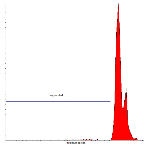
at 4 °C for an hour in the dark then measured directly. PI histograms were used to determine 207
cellular DNA content. In case of DNA fragmentation, DNA fragments may leak out of the cells 208
hence the remaining DNA content represent lower intensity peaks below the main PI peak 209
(Figure 1.) 210
Results were compared to controls using Yates corrected Chi-square test. The statistical 211
analysis was performed using GraphPad QuickCalcs software.
212
213 214
Figure 1. DNA fragmentation based on propidium iodide fluorescence intensities 215
216
Micronucleus (MN) test 217
Although no standardized test method is available for the mussel MN test, there are well 218
described, step-by-step test protocols published. Our assay was performed based on the protocol 219
given by Wozniczki et al. (2004), with some modifications. Treatments were performed in 3 220
replicates for each concentration and for the control. 10 individuals were kept in aquaria of 3 L 221
volume. In the aquaria Lake Balaton water was used. The mussels were not fed during the 222
experiment, aquaria were constantly aerated, and the temperature was set at 22°C. Organisms 223
were exposed for 4 days, and the sample was renewed after 2 days. As test organism, the 224
freshwater bivalve Unio pictorum was selected as it already proved to have high sensitivity for 225
a wide range of environmental contaminants (Vuković-Gačić et al., 2014).
226
After 4 days, hemolymph was taken from the posterior adductor muscle using an 227
improved non-lethal technique based on the method described by Gustafson et al (2005). 1 ml 228
hemolymph sample was mixed with 0.3 ml 10% acetic acid in methanol as a fixative and 229
centrifuged at 1000 rpm for 5 minutes. The supernatant was discarded and the rest was fixed in 230
1 ml 80% ethanol, thus the sample can be kept refrigerated for a few weeks. For processing the 231
samples, refrigerated samples were centrifuged again at 1000 rpm for 5 minutes and the 232
supernatant was discarded. The pellet which contained the hemolymph cells in a more 233
concentrated form, was smeared onto a microscope slide and allowed to dry. After that the 234
slides were fixed in 80% methanol, air dried again and stained with 5% Giemsa in distilled 235
water for 20 minutes.
236
Photos of the cells were taken by a Zeiss AxioScope A1 microscope with an AxioCam 237
ICC1 camera and Zen 2011 program at 400x magnification. For each animal 1000 cells were 238
counted, micronuclei frequency was identified according to Fenech (1992).
239
One-way ANOVA with Tukey post hoc test was used to compare the mean MN numbers 240
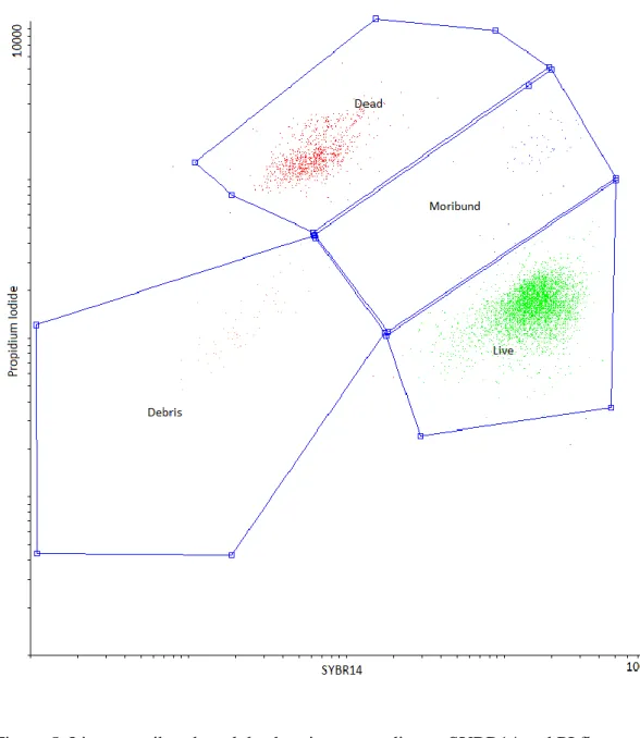
between the treatments. To use the ANOVA test the following assumptions were met: each 241
group has approximately normal distribution (Shapiro-Wilk normality test: W = 0.9732, 242
p = 0.3099), all groups have a common variance (Bartlett’s test: Bartlett's K-squared = 3.1215, 243
df = 4, p = 0.5377), independence of observations and all groups has equal sample number. In 244
each group there were 15 individuals but for the statistical analysis the 10 most undoubtable 245
were used (where the color and the quality of the pictures were the best). No transformations 246
were applied on the data.
247 248
Results 249
250
Vibrio fischeri bioluminescence inhibition test 251
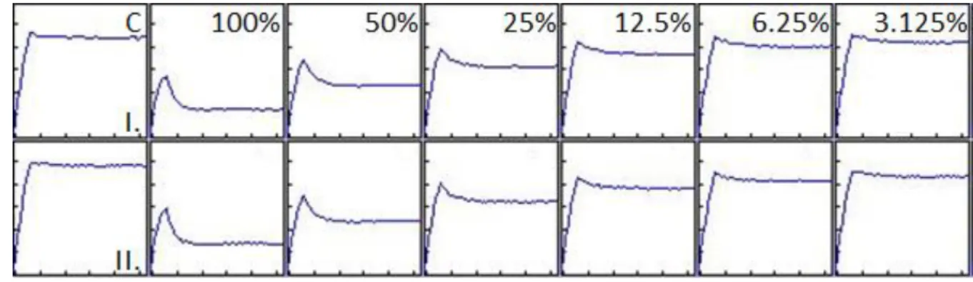
Figure 2 shows the bioluminescence reading for the first 30 sec. An immediate decrease 252
in the light output after adding the bacterial suspension to the sample already gives an indication 253
on the toxicity of the sample (Mortimer et al., 2008). After 30 minutes of exposure, calculated 254
EC50 was 12.52% and EC20 was 1.90%.
255 256
257
Figure 2. Light output during the first 30 secs of the 30 minutes exposure. I and II depict the 258
two replicates. C: Control. The peak shows the maximum light output of the bacteria, which 259
immediately starts to diminish after the test bacteria get in contact with the sample.
260 261
Sinapis alba growth inhibition test 262
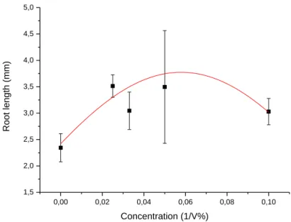
The measured root length of the treated seeds was greater than in the control in every 263
concentration but no clear trend could be noticed as Figure 3 shows below. Due to the 264
stimulating effect on the seeds neither EC50 values nor inhibition was calculated. This pattern 265
can be experienced for samples which contain plant nutrients: in this case nutrients might mask 266
the toxic effect in low concentrations (USEPA, 2000).
267 268
0,00 0,02 0,04 0,06 0,08 0,10
1,5 2,0 2,5 3,0 3,5 4,0 4,5 5,0
Root length (mm)
Concentration (1/V%)
269 270
Figure 3. Concentration-response curve for the Sinapis alba test.
271 272
Daphnia magna immobilization test 273
Of the conducted tests, the D. magna immobilization test appeared to be the most 274
sensitive. After a few range finding test the adjusted concentration was between 0.001% and 275
0.1%, calculated EC50 value was 0.0226%.
276 277
Flow cytometry 278
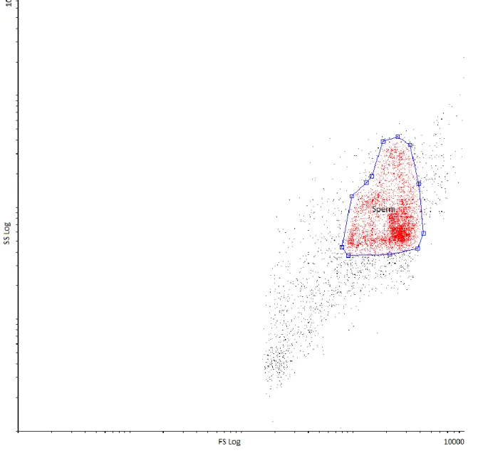
A well distinguishable sperm region was established according to forward scatter versus 279
side scatter properties (Figure 4.). This sperm region was gated to SYBR14 vs. PI dot plots, 280
where distinct living, dead and moribund populations were discriminated. Moribund cells were 281
included in the dead category during data analysis (Figure 5.).
282
283 284
Figure 4. Differentiation of sperm population based on forward scatter versus side scatter 285
properties 286
287 288
Figure 5. Live-, moribund- and dead regions according to SYBR14 and PI fluorescence 289
290
The results show that live cell ratio was around 82-83% after 30 minutes exposition and 291
the samples did not differ from controls significantly (p= 0.0547). After one day exposure, the 292
biodiesel treated samples showed statistically significant (from 83% to 77%; p<0.0001) 293
decrease in live cell ratio.
294
After 30 minutes, the biodiesel samples indicated only a few percent (less, than 2%) of 295
spermatozoa with DNA fragmentation similarly to the control and the percentage of cells with 296
fragmented DNA did not change after one day exposition.
297 298 299 300
Micronucleus test 301
The genotoxic response was expressed as the number of micronuclei/1000 cells. Figure 302
6 shows typical micronucleus formation, a concentration-effect curve is given in Figure 7. The 303
data of the test were not suitable for calculating EC50 values so statistical analysis was 304
performed. The result of the one way ANOVA was p=0.00025 (F=6.7152, df=4) so the effect 305
of each concentration could be separated from each other. To determine the difference between 306
the control and the treatments, a two sample t-test was carried out. The results show that the 307
control and the most diluted concentration do not differ significantly (p=0.882), but for the 308
other concentrations (3.3%, 5% and 10%) statistically significant difference could be 309
established (p=0.009; p=0.019 and p=0.0003, respectively).
310
311
Figure 6. Typical micronucleus formation (A-E) and normal agranular hemolymph cells (F-J) 312
from Unio pictorum Giemsa painted hemolymph.
313 314
Control 2,5% 3,3% 5% 10%
0 2 4 6 8 10
Concentration (%)
MN number (MN/1000 cell)
315
Figure 7. Concentration-effect relationship for the micronucleus test.
316
Discussion 317
318
The biodiesel impact on water resources is composed of several factors. Biofuel 319
production demands a great volume of water that can be replaced with seawater or wastewater 320
in a certain amount (Wu et al. 2009). The spilled biofuel (as well as any other type of fuel) 321
forms a non-aqueous phase layer on the water surface damaging sea birds and other animals 322
that try to pass through. Biodiesel has a low solubility in water but the intensive waving and 323
water flow cause some degree of mixing. We used a similar mix for the tests that appeared to 324
exert highly toxic and genotoxic effect on the aquatic life.
325
Terrestrial ecosystems were represented by the standardized S. alba seedling emergence 326
and seedling growth test. The results of this test showed no sign of toxicity. Moreover, in this 327
test lower concentrations seemed to exert stimulating effect. This is a typical concentration- 328
response relationship in cases where the sample contains plant nutrients (USEPA, 2000).
329
However, these negative results do not necessarily imply that biodiesel should be 330
completely safe for terrestrial ecosystems. On one hand, several studies have been targeted to 331
assess biodegradability of biodiesel or different biodiesel-fossil fuel blends. These studies 332
support that biodiesel can be biodegraded considerably faster than diesel both under aerobic 333
(e.g. Lapinskienė et al., 2006, Yassine et al., 2013) and anaerobic conditions (e.g. Wu et al., 334
2015).
335
On the other hand, seed germination tests showed that biodegradation products might 336
pose actual risk. Tamada et al. (2012) followed biodegradation of biodiesel and vegetable oils 337
for a period of 180 days. Seed germination tests revealed an increasing toxicity of biodiesel 338
metabolites as bacterial decomposition went by. In the same study, using the earthworm 339
(Eisenia foetida) test, biodiesel was the only contaminant that proved to be toxic. A similar 340
study was conducted by Cruz et al. (2013a, b). Cucumis sativus and Brassica oleracea seed 341
germination inhibition showed that after two months of biodegradation, biodiesel was the most 342
toxic contaminant in comparison to diesel and waste lubricant oil. Phytotoxicity of metabolites 343
was also demonstrated in the study of Hawrot–Paw and Izwikow (2015) using garden cress 344
(Lepidium sativum) and spring barley (Hordeum vulgare).
345
In order to assess the potential ecotoxicity of different biodiesel blends on aquatic life, 346
two standard and widely used assays were used in our study, the Daphnia magna immobility 347
assay and the Vibrio fischeri bioluminescence inhibition bioassay. The V. fischeri 348
bioluminescence inhibition bioassay detected considerable toxicity with the EC50 of 12.52%.
349
This assay was used in a study of Yassine et al. (2012) to assess the toxicity of the water 350
accommodated fraction (WAF) of six commercial soybean biodiesel/petrodiesel blends at 351
different oil loads. These results can provide a good basis for comparison with our results, as in 352
the preparation of WAF, oils were introduced to water with the highest load of 1:1. V. fischeri 353
EC50s for WAFs of B20, B40, B60, B80 and B100 blends fall very close to each other, app.
354
5%. In our test, only neat biodiesel was assessed, test results showed slightly lower ecotoxicity.
355
Differences might have been caused by different biodiesel types: while in the study of Yassine 356
et al. soybean-methyl ester biodiesel was used, our sample was a rapeseed methyl ester. In a 357
comparative study of Hollebone et al. (2008) three different biodiesels (two based on vegetable 358
oils of canola and soy, and one animal-source waste fry oil) were assessed using Microtox. The 359
soy-based biodiesel exerted the highest toxicity on the test bacterium.
360
In addition, although the same test organism, V. fischeri was used in both assessments, 361
test protocols differed. In our study a kinetic protocol was followed, which was developed 362
especially for the assessment of turbid and/or colored samples. As light output in the sample is 363
assessed independently from the control, false toxicity readings caused by turbidity and/or color 364
of the sample can be avoided (Lappalainen et al., 2001).
365
D. magna showed extreme sensitivity with EC50 value of 0.0226%. Though the Daphnia 366
bioassay is the most frequently used test in biodiesel ecotoxicity assessments, results given by 367
different studies are rather difficult to compare with each other due to different sample 368
preparation protocols (oil in water dispersion, OWD vs. water accommodated fraction, WAF) 369
or differences in test protocols (e.g. different exposure regimes) (Bluhm et al., 2012). Khan et 370
al. (2007) in a comparative study used OWD of biodiesels derived from recycled cooking oils 371
and fats, employing daphnids and rainbow trouts (Oncorhynchus mykiss) as test organisms.
372
Daphnids were found more sensitive: EC50 in the D. magna assay was 4.65 ppm, while O.
373
mykiss EC50 was 455.28 ppm (after 24 hour exposure in the D. magna test and 96 hour exposure 374
in the O. mykiss test). Acute Daphnia EC50 value determined by Tjarinto et al. (2014) fall very 375
close, 3.157 ppm.
376
Hollebone et al. (2008) suggest that OWD sample might not be representative when 377
ecotoxicity on daphnids is to be evaluated. When OWD of different biodiesels were 378
investigated, higher toxicity was detected than in WAFs of the same biodiesels. The possible 379
explanation was that in OWDs oil layers formed which might have caused either physical 380
smothering or trapping of daphnids, enhancing mortality rate. Based on these findings, the WAF 381
of our biodiesel was further ultrasonicated to avoid such possible physical effects, therefore the 382
experienced low EC50 must have reflected actual toxicity.
383
Literature studies reveal that apart from standard bioassays, tests conducted using other 384
test organisms also support the potential risk of biodiesels on different elements of aquatic 385
ecosystems, both freshwater and marine. A study of Leite et al. (2011) determined the toxicity 386
of the water-soluble fractions (WSF) of three different biodiesel fuels to two marine organisms, 387
the sea urchin Echinometra lucunter and the microalga Tetraselmis chuii. A non-lethal bioassay 388
was conducted by Gauthier (2012) using behavioral alterations of the crayfish Orconectes 389
rusticus and found that biodiesel and crude oil had equal negative effects on chemosensory 390
behavior of the crayfish. Gorcharoenwat et al. (2017) evaluated the effects of the water soluble 391
fraction of palm biodiesel on Macrobrachium rosenbergii, the giant freshwater prawn, which 392
is an economically important native aquatic organism in Southeast Asia living in freshwater to 393
brackish water. It was found that histologically abnormal alterations appeared in the gills of 394
tested larvae. Some freshwater plant species such as duckweed (Lemna minor) or water milfoil 395
(Myriophyllum spicatum) were seriously affected by biodiesel in a comprehensive study of 396
Birchall et al. (1995).
397
Considering genotoxicity/mutagenicity, in most cases biodiesel exhaust emission has 398
been evaluated (reviewed by Bluhm et al., 2012, Claxton, 2015). Direct genotoxicological 399
assessments of biodiesel samples have been carried out much less often, these literature studies, 400
however, indicate the genotoxic nature of biodiesel samples.
401
Leme et al. (2012) carried out spill simulations with neat diesel and biodiesel. In their 402
study, water soluble fraction of the biodiesel exerted mutagenic and genotoxic effects in the 403
Salmonella/microsome preincubation assay and the in vitro Chinese hamster ovary cell MN 404
test. The authors attributed these effects to the presence of potentially toxic compounds in the 405
biodiesel derived from the raw material source used in the production chain.
406
Cavalcante et al. (2014) found that biodiesel can cause cytotoxic, biochemical and 407
genotoxic alterations in the hepatocyte cell line of Danio rerio (ZFL), depending on the 408
production route: methylic (BdMt) route producing biodiesel with more intense effect than 409
ethylic (BdEt) route.
410
In our study, the mussel micronucleus test (MNT) was applied, detecting considerable 411
genotoxic potential of the biodiesel sample: it elucidated micronuclei formation already at low 412
concentration of 3.3%. This test has never been employed in biodiesel eco/genotoxicity 413
assessments, however, it seems promising. It shows appropriate sensitivity, and moreover, 414
mussels are a representative group when ecological risk to aquatic ecosystems is to be 415
addressed. Different mussel biomarkers have been used for example to assess or monitor 416
ecological impacts of oil spills (e.g. Pérez-Cadahía et al., 2004, Laffon et al., 2006).
417
The mechanism of micronuclei formation is relatively well discussed. Acentric 418
chromosome or chromatid fragments from misrepaired or unrepaired DNA double-strand 419
breaks can lead to MN formation (Savage, 1988). MN can also originate from broken 420
nucleoplasmic bridges during telophase when chromosome fragments fail to be included in the 421
daughter nuclei (Dianov et al., 1991). Lagging whole chromosomes at anaphase also can create 422
MN. This can happen in the centromeric and pericentromeric DNA repeat sequence by the 423
hypomethylation of cytosine or by the defects of the kinetochore protein or the mitosis check 424
point (Pironon et al., 2010). Abnormal centrosome amplification and some spindle dysfunction 425
could also be the cause of MN formation (Gisselsson, 2008).
426
As the results of the flow cytometric boar sperm test indicated, biodiesel has a slight 427
cytotoxic effect.
428 429
Conclusions 430
431
A battery of bioassays was employed to provide complex information on the eco- and 432
genotoxicity of a rapeseed biodiesel, including the Sinapis alba root growth inhibition, the 433
Daphnia magna immobilization, Vibrio fischeri bioluminescence inhibition, boar semen 434
live/dead ratio as well as DNA fragmentation and the Unio pictorum micronucleus tests.
435
The sample exerted significant effect on aquatic test organisms, D. magna being far the 436
most sensitive with EC50 value of 0.0226%. The V. fischeri bioluminescence inhibition bioassay 437
also detected considerable toxicity with the EC50 of 12.52%. These results raise environmental 438
concern about biodiesel, especially in case of accidental oil spills.
439
On the other hand, no acute toxicity was shown by the terrestrial S. alba test.
440
The mussel micronucleus test, using the freshwater Unio pictorum detected considerable 441
genotoxic potential of the biodiesel sample: it elucidated micronuclei formation already at low 442
concentration of 3.3%. It was the first time this bioassay has been employed in biodiesel 443
genotoxicity assessment and one of the aims of the study was to evaluate its applicability for 444
such samples. It seems to be an appropriate tool, based on its representativity, sensitivity and 445
cost effectiveness.
446 447
Acknowledgement 448
This work was supported by the BIONANO_GINOP-2.3.2-15-2016-00017 project.
449
Árpád Ferincz was supported by the Bolyai János Fellowship of the Hungarian Academy of 450
Sciences. Special thanks go to Ms Lana Wolmarans for language editing.
451
References 452
of various 453
1. Ajao, C., Andersson, M.A., Teplova, V.V., Nagy, S., Gahmberg, C.G., Andersson, 454
L.C., et al., 2015. Mitochondrial toxicity of triclosan on mammalian cells. Toxicol.
455
Rep. 7(2), 624-637. http://dx.doi.org/10.1016/j.toxrep.2015.03.012 456
2. Andersson, M.A., Mikkola R., Helin J., Andersson M.C., Salkinoja-Salonen M., 1998.
457
A novel sensitive bioassay for detection of Bacillus cereus emetic toxin and related 458
depsipeptide ionophores. Appl. Environ. Microbiol. 64(4), 1338-43.
459
3. Andersson, M.A., Jaaskelainen, E.L., Shaheen, R., Pirhonen, T., Wijnands, L.M., 460
Salkinoja-Salonen, M.S., 2004. Sperm bioassay for rapid detection of cereulide- 461
producing Bacillus cereus in food and related environments. Int. J. Food Microbiol.
462
94, 175-183. doi:10.1016/j.ijfoodmicro.2004.01.018 463
4. Andersson, M.A., Mikkola, R., Rasimus, S., Hoornstra, D., Salin, P., Rahkila, R., et 464
al., 2010. Boar spermatozoa as a biosensor for detecting toxic substances in indoor 465
dust and aerosols. Toxicol. in Vitro 24, 2041–2052. doi:10.1016/j.tiv.2010.08.011 466
5. Birchall, C., Newman, J.R., Greaves, M.P., 1995. Degradation and phytotoxicity of 467
biodiesel oil. lnstitute of Arable Crops Research, Report, Long Ashton Research 468
Station, Bristol, UK, pp. 50 469
6. Bluhm, K., Heger, S., Seiler, T.B., Hallare, A.V., Schaeffer, A., Hollert, H., 2012.
470
Toxicological and ecotoxicological potencies of biofuels used for the transport 471
sector—a literature review. Energy Environ. Sci. 5, 7381–7392.
472
doi:10.1039/c2ee03033k 473
7. Bolognesi, C., Fenech, M., 2012. Mussel micronucleus cytome assay. Nat. Protoc. 7, 474
1125–1137. doi:10.1038/nprot.2012.043 475
8. Cavalcante, D.G.S.M., da Silva, N.D.G., Marcarini, J.C., Mantovani, M.S., Marin- 476
Morales, M.A., Martinez, C.B.R., 2014. Cytotoxic, biochemical and genotoxic effects 477
of biodiesel produced by different routes on ZFL cell line. Toxicol. in Vitro 28, 1117–
478
1125. http://dx.doi.org/10.1016/j.tiv.2014.05.008 479
9. Claxton, L.D., 2015. The history, genotoxicity and carcinogenicity of carbon-based 480
fuels and their emissions: Part 4 – Alternative fuels. Mut. Res. 763, 86–102.
481
http://dx.doi.org/10.1016/j.mrrev.2014.06.003 482
10. Cruz, J.M., Lopes, P.R.M., Montagnolli, R.N., Tamada, I.S., Silva, N.M.M.G., Bidoia, 483
E.D., 2013a. Toxicity assessment of contaminated soil using seeds as bioindicators. J.
484
Appl.Biotechnol. 1, 1–10. doi: 10.5296/jab.v1i1.3408 485
11. Cruz, J.M., Lopes, P.R.M., Montagnolli, R.N., Tamada, I.S., Silva, N.M.M.G., Bidoia, 486
E.D., 2013b. Phytotoxicity of soil contaminated with petroleum derivatives and 487
biodiesel. Ecotoxicol. Environ. Contam. 8(1), 49-54. doi: 10.5132/eec.2013.01.007 488
12. Dianov, G. L., Timchenko, T. V., Sinitsina, O. I., Kuzminov, A. V., Medvedev, O. A., 489
Salganik, R. I., 1991. Repair of uracil residues closely spaced on the opposite strands 490
of plasmid DNA results in doublestrand break and deletion formation. Mol. Gen.
491
Genet. 225, 448–452.
492
13. Escobar, N., Ribal, J., Clemente, G., Sanjuán, N., 2014. Consequential LCA of two 493
alternative systems for biodiesel consumption in Spain, considering uncertainty. J.
494
Clean. Prod. 79, 61-73. http://dx.doi.org/10.1016/j.jclepro.2014.05.065 495
14. Falfushynska, H.I., Gnatyshyna, L.L., Stoliar, O.B., 2013. Effect of in situ exposure 496
history on the molecular responses of freshwater bivalve Anodonta anatina 497
(Unionidae) to trace metals. Ecotox. Environ. Safe. 89, 73–83.
498
http://dx.doi.org/10.1016/j.ecoenv.2012.11.024 499
15. Fargašová, A., 1998. Root growth inhibition, photosynthetic pigments production, and 500
metal accumulation in Sinapis alba as the parameters for trace metals effect 501
determination. Bull. Environ. Contam. Toxicol. 61, 762–769.
502
doi:10.1007/s001289900826 503
16. Fenech, M., Neville, S., 1992. Conversion of excision-repairable DNA lesions to 504
micronuclei within one cell cycle in human lymphocytes. Environ. Mol. Mutagen. 19, 505
27–36. doi: 10.1002/em.2850190106 506
17. Gauthier, S.J., 2012. Biodiesel and crude oil effects on foraging capacity of crayfish 507
Orconectus rusticus. Thesis, Graduate College of Bowling Green State University, 508
pp.41 509
18. Gisselsson, D., 2008. Classification of chromosome segregation errors in cancer.
510
Chromosoma, 117, 511–519. https://doi.org/10.1007/s00412-008-0169-1 511
19. Gorcharoenwat, P., Piyatiratitivorakul, S., Puanglarp, N., Pengprecha, S., 2017. Effect 512
of water soluble fractions of diesel and biodiesel on larvae of the giant freshwater 513
prawn, Macrobrachium rosenbergii, under different thermal conditions. Walailak J.
514
Sci. Tech. 14(11), 837-848.
515
20. Guidi, P., Frenzilli, G., Benedetti, B., Bernardeschi, M., Falleni, A., Fattorini, D., et 516
al., 2010. Antioxidant, genotoxic and lysosomal biomarkers in the freshwater bivalve 517
(Unio pictorum) transplanted in a metal polluted river basin. Aquat. Toxicol. 100, 75–
518
83. https://doi.org/10.1016/j.aquatox.2010.07.009 519
21. Gustafson, L. L., Stoskopf, M. K., Bogan, A. E., Showers, W., Kwak, T. J., Hanlon, 520
S., Levine, J. F., 2005. Evaluation of a nonlethal technique for hemolymph collection 521
in Elliptio complanata, a freshwater bivalve (Mollusca: Unionidae). Dis. Aquat.
522
Organ. 65, 159–165.
523
22. Hawrot–Paw, M., Izwikow, M., 2015. Ecotoxicological effects of biodiesel in the soil.
524
J. Ecol. Eng. 16, 34–39. doi: 10.12911/22998993/60451 525
23. Hollebone, B.P., Fieldhouse, B., Landriault, M., Doe, K., Jackman, P., 2008. Aqueous 526
solubility, dispersibility and toxicity of biodiesels. Int. Oil Spill Conf. Proc. 929–936.
527
24. Hoornstra, D., Andersson, M.A., Mikkola, R., Salkinoja-Salonen, M.S., 2003. A new 528
method for in vitro detection of microbially produced mitochondrial toxins. Toxicol.
529
in Vitro 17, 745-751. doi:10.1016/S0887-2333(03)00097-3 530
25. Ji, X., Long, X., 2016. A review of the ecological and socioeconomic effects of 531
biofuel and energy policy recommendations. Renew. Sust. Energ. Rev. 61, 41–52.
532
http://dx.doi.org/10.1016/j.rser.2016.03.026 533
26. Khan, N., Warith, M.A., Luk, G., 2007. A comparison of acute toxicity of biodiesel, 534
biodiesel blends, and diesel on aquatic organisms. J. Air Waste Man. 57, 286–296.
535
doi: 10.1080/10473289.2007.10465333 536
27. Kolarević, S., Knežević-Vukčević, J., Paunović, M., Tomović, J., Gačic, Z., Vuković- 537
Gačić, B., 2009. The anthropogenic impact on water quality of the River Danube in 538
Serbia: microbiological analysis and genotoxicity monitoring. Archives of Biological 539
Science. Belgrade 63(4), 1209-1217. https://doi.org/10.2298/ABS1104209K 540
28. Kooter, I.M., van Vugt, M.A.T.M., Jedynska, A.D., Tromp, P.C., Marc M.G.
541
Houtzager, M.M.G., et al., 2011. Toxicological characterization of diesel engine 542
emissions using biodiesel and a closed soot filter. Atmos. Environ. 45, 1574-1580.
543
doi:10.1016/j.atmosenv.2010.12.040 544
29. Kováts, N., Ács, A., Ferincz, Á., Kovács, A., Horváth, E., Kakasi, B., et al., 2013.
545
Ecotoxicity and genotoxicity assessment of exhaust particulates from diesel-powered 546
buses. Environ. Monit. Assess. 185(10), 8707-8713. doi: 10.1007/s10661-013-3206-3 547
30. Laffon, B., Rábade, T., Pásaro, E., Méndez, J., 2006. Monitoring of the impact of 548
Prestige oil spill on Mytilus galloprovincialis from Galician coast. Environ. Int. 32, 549
342 – 348. doi:10.1016/j.envint.2005.07.002 550
31. Lapinskienė, A., Martinkus, P., Rėbždaitė, V., 2006. Eco-toxicological studies of 551
diesel and biodiesel fuels in aerated soil. Environ Pollut. 142, 432-437.
552
doi:10.1016/j.envpol.2005.10.023 553
32. Lappalainen, J., Juvonen, R., Vaajasaari, K., Karp, M., 1999. A new flash method for 554
measuring the toxicity of solid and colored samples. Chemosphere 38(5), 1069-1083.
555
https://doi.org/10.1016/S0045-6535(98)00352-X 556
33. Lappalainen, J., Juvonen, R., Nurmi, J., Karpm M., 2001. Automated color 557
correction method for Vibrio fischeri toxicity test. Comparison of standard and 558
kinetic assays. Chemosphere 45, 635-641. https://doi.org/10.1016/S0045- 559
6535(00)00579-8 560
34. Leite, M.B.N.L., de Araújo, M.M.S., Nascimento,I.A., da Cruz, A.C.S., Pereira, S.A., 561
do Nascimento, N.C., 2011. Toxicity of water-soluble fractions of biodiesel fuels 562
derived from castor oil, palm oil, and waste cooking oil. Environ. Toxicol. Chem.
563
30(4), 893–897. doi: 10.1002/etc.444 564
35. Leme, D.M., Grummt, T., de Oliveira, D.P., Sehr, A., Renz, S., Reinel, S., et al., 565
2012. Genotoxicity assessment of water soluble fractions of biodiesel and its diesel 566
blends using the Salmonella assay and the in vitro MicroFlow® kit (Litron) assay.
567
Chemosphere 86, 512–520. doi:10.1016/j.chemosphere.2011.10.017 568
36. Lin, T-C., Chao, M-R., 2002. Assessing the influence of methanol-containing additive 569
on biological characteristics of diesel exhaust emissions using Microtox and Mutatox 570
assays. Sci. Tot. Env. 284, 61-74. https://doi.org/10.1016/S0048-9697(01)00866-X 571
37. Liu, Y.Y., Lin, T.C., Wang, Y.J., Ho, W.L., 2009. Carbonyl compounds and toxicity 572
assessments of emissions from a diesel engine running on biodiesels. J. Air Waste 573
Manage. 59(2), 163-171. http://dx.doi.org/10.3155/1047-3289.59.2.163 574
38. Michel, C., Bourgeault, A., Gourlay-Francé, C., Palais, F., Geffard, A, Vincent- 575
Hubert, F., 2013. Seasonal and PAH impact on DNA strand-break levels in gills of 576
transplanted zebra mussels. Ecotox. Environ. Safe. 92, 18–26.
577
https://doi.org/10.1016/j.ecoenv.2013.01.018 578
39. Mikkola, R., Andersson, M.A., Hautaniemi, M., Salkinoja-Salonen, M.S., 2015.
579
Toxic indole alkaloids avrainvillamide and stephacidin B produced by a biocide 580
tolerant indoor mold Aspergillus westerdijkiae. Toxicon 99, 58-67.
581
http://dx.doi.org/10.1016/j.toxicon.2015.03.011 582
40. Mortimer, M., Kasemets, K., Heinlaan, M., Kurvet, I., Kahru, A., 2008. High 583
throughput kinetic Vibrio fischeri bioluminescence inhibition assay for study of 584
toxic effects of nanoparticles. Toxicol in Vitro, 22, 1402-1417.
585
doi:10.1016/j.tiv.2008.02.011 586
41. Nogueira, L., Sanches, A.L.M., da Silva, D.G.H., Ferrizi, V.C., Moreira, A.B., de 587
Almeida, E.A., 2011. Biochemical biomarkers in Nile tilapia (Oreochromis niloticus) 588
after short-term exposure to diesel oil, pure biodiesel and biodiesel blends.
589
Chemosphere, 85, 97–105. doi:10.1016/j.chemosphere.2011.05.037 590
42. Nogueira, L., da Silva, D.G.H., Oliveira, T.Y.K., da Rosa, J.M.C. Felício, A.A., de 591
Almeida, E.A., 2013. Biochemical responses in armored catfish (Pterygoplichthys 592
anisitsi) after short-term exposure to diesel oil, pure biodiesel and biodiesel blends.
593
Chemosphere, 93, 311–319. http://dx.doi.org/10.1016/j.chemosphere.2013.04.083 594
43. Pérez-Cadahía, B., Laffon, B., Pásaro, E., Méndez, J., 2004. Evaluation of PAH 595
bioaccumulation and DNA damage in mussels (Mytilus galloprovincialis) exposed to 596
spilled Prestige crude oil. Comp. Biochem. Physiol. Part C 138, 453–460.
597
doi:10.1016/j.cca.2004.08.001 598
44. Pironon, N., Puechberty, J., Roizés, G., 2010. Molecular and evolutionary 599
characteristics of the fraction of human alpha satellite DNA associated with CENP-A 600
at the centromeres of chromosomes 1, 5, 19, and 21. BMC Genomics, 11, 195–203.
601
https://doi.org/10.1186/1471-2164-11-195 602
45. Rasimus, S., Mikkola, R., Andersson, M.A., Teplova, V.V., Venediktova, N., Ek- 603
Kommonen, C., Salkinoja-Salonen, M., 2012. Psychrotolerant Paenibacillus tundrae 604
isolates from barley grains produce new cereulide-like depsipeptides (paenilide and 605
homopaenilide) that are highly toxic to mammalian cells. App. Environ. Microb.
606
78(10), 3732–3743. doi:10.1128/AEM.00049-12 607
46. Riccardi, C., Nicoletti, I., 2006. Analysis of apoptosis by propidium iodide staining 608
and flow cytometry. Nat. Protoc. 1, 1458-1461. doi:10.1038/nprot.2006.238 609
47. Rosen, G., Dolecal, R.E., Colvin, M.A., George, R.D., 2014. Preliminary ecotoxicity 610
assessment of new generation alternative fuels in seawater. Chemosphere 104, 265–
611
270. http://dx.doi.org/10.1016/j.chemosphere.2013.11.023 612
48. Savage, J. R., 1988. A comment on the quantitative relationship between micronuclei 613
and chromosomal aberrations. Mut. Res., 207, 33–36.
614
49. Štambuc, A., Pavlica, M., Vignjević, G., Bolarić, B., Klobučar, G.I.V., 2009.
615
Assessment of genotoxicity in polluted freshwaters using caged painter’s mussel, Unio 616
pictorum. Ecotoxicology 18, 430–439. doi: 10.1007/s10646-009-0297-2 617
50. Steiner, S., Czerwinski, J., Comte, P., Popovicheva, O., Kireeva, E., Müller, L., et al., 618
2013. Comparison of the toxicity of diesel exhaust produced by bio- and fossil diesel 619
combustion in human lung cells in vitro. Atmos. Environ. 81, 380-388.
620
http://dx.doi.org/10.1016/j.atmosenv.2013.08.059 621
51. Sverdrup, L.E., Krogh, P.H., Nielsen, T., Kjær, C., Stenersen, J., 2003. Toxicity of 622
eight polycyclic aromatic compounds to red clover (Trifolium pratense), ryegrass 623
(Lolium perenne), and mustard (Sinapsis alba). Chemosphere 53, 993–1003.
624
doi:10.1016/S0045-6535(03)00584-8 625
52. Tamada, I.S., Montagnolli, R.N., Lopes, P.R., Bidoia, E.D., 2012. Toxicological 626
evaluation of vegetable oils and biodiesel in soil during the biodegradation process.
627
Braz. J. Microbiol. 43, 1576–81.
628
53. Tjarinto, R., Rachmatiah, I., Salami, S., 2014. Toxicity test of water-soluble fractions 629
of waste vegetable oil-based biodiesel and biodiesel/diesel blends on Daphnia magna 630
and Allium cepa. Proceedings of the 3rd Applied Science for Technology Innovation, 631
ASTECHNOVA International Energy Conference, Yogyakarta, Indonesia, pp. 294- 632
633 301
54. Turrio-Baldassarri, L., Battistelli, C.L., Conti, L., Crebelli, R., De Berardis, B., 634
Iamiceli, A.L., et al., 2004. Emission comparison of urban bus engine fueled with 635
diesel oil and ‘biodiesel’ blend. Sci. Total Environ. 327, 147–162.
636
doi:10.1016/j.scitotenv.2003.10.033 637
55. USEPA, 2000. Method Guidance and Recommendations for Whole Effluent Toxicity 638
(WET) Testing (40 CFR Part 136). EPA 821-B-00-004. U.S. Environmental 639
Protection Agency, Office of Water 640
56. Vicente-Carrillo, A., Edebert, I., Garside, H., Cotgreave, I., Rigler, R., Loitto, V., et 641
al., 2015. Boar spermatozoa successfully predict mitochondrial modes of toxicity:
642
Implications for drug toxicity testing and the 3R principles. Toxicol. in Vitro 29, 582–
643
591. http://dx.doi.org/10.1016/j.tiv.2015.01.004 644
57. Vouitsis, E., Ntziachristos, L., Pistikopoulos, P., Samaras, Z., Chrysikou, L., Samara, 645
C. et al., 2009. An investigation on the physical, chemical and ecotoxicological 646
characteristics of particulate matter emitted from light-duty vehicles. Environ. Pollut.
647
157, 2320–2327. https://doi.org/10.1016/j.envpol.2009.03.028 648
58. Vuković-Gačić, B., Kolarević, S., Sunjog, K., Tomović, J., Knežević-Vukčević, J., 649
Paunović, M., Gačic, Z., 2014. Comparative study of the genotoxic response of 650
freshwater mussels Unio tumidus and Unio pictorum to environmental stress.
651
Hydrobiologia 735, 221–231. doi: 10.1007/s10750-013-1513-x 652
59. Yang, Y., 2013. Life cycle freshwater ecotoxicity, human health cancer, and 653
noncancer impacts of corn ethanol and gasoline in the U.S. J. Clean. Prod. 53, 149- 654
157. http://dx.doi.org/10.1016/j.jclepro.2013.04.009 655
60. Wozniczki, P., Lewandowska, R., Brzuzan, P., Ziomek, E., Bardega, R., 2004. The 656
level of DNA damage and the frequency of micronuclei in haemolymph of freshwater 657
mussels Anodonta woodiana exposed to benzo[a]pyrene. Acta Toxicol. 12, 41–45.
658
61. Wu, M., Mintz, M., Wang, M., Arora, S., 2009. Water consumption in the production 659
of ethanol and petroleum gasoline. Environ. Manage. 44, 981–997.
660
doi:10.1007/s00267-009-9370-0 661
62. Wu S., Yassine M.H., Suidan, M.T., Venosa, A.D., 2015. Anaerobic biodegradation of 662
soybean biodiesel and diesel blends under methanogenic conditions. Water Res. 87, 663
395-402. http://dx.doi.org/10.1016/j.watres.2015.09.024 664
63. Yassine, M.H., Wu, S., Suidan, M.T., Venosa, A.D., 2012. Microtox aquatic toxicity of 665
petrodiesel and biodiesel blends: The role of biodiesel's autoxidation products. Environ.
666
Toxicol. Chem. 31, 2757–2762. doi: 10.1002/etc.2001 667
64. Yassine, M.H., Wu, S., Suidan, M.T., Venosa, A.D., 2013. Aerobic biodegradation 668
kinetics and mineralization of six petrodiesel/soybean-biodiesel blends. Environ. Sci.
669
Technol. 47 (9), 4619-4627. doi: 10.1021/es400360v 670
671