J Gastrointestin Liver Dis, September 2017 Vol. 26 No 3: 305-308 1) 1st Department of Internal
Medicine of Semmelweis University, Budapest;
2) Department of Pathology of the University of Pécs, Pécs, Hungary
Address for correspondence:
Dániel Németh MD 1st Department of Internal Medicine of Semmelweis University;
Korányi Sándor Street 2/a, Budapest, Hungary nemeth.daniel@med.
semmelweis-univ.hu
Received: 29.07.2017 Accepted: 25.08.2017
Cholangiocarcinoma in Wilson’s Disease – a Case Report
Dániel Németh1, Anikó Folhoffer1, Gábor Smuk2, Béla Kajtár2, Tamás Tornóczky2, Ferenc Szalay1
INTRODUCTION
Wilson’s disease (WD) caused by ATP7B mutations is a rare autosomal recessive disorder of toxic copper accumulation resulting in any type of liver disease and/or neurological- psychiatric symptoms or Coombs negative hemolytic anemia [1].
The Kayser-Fleischer ring in the cornea is a characteristic but not a specific finding causing no alteration of the eye function [2]. Hepatocellular carcinoma (HCC) was suggested to be less frequent in WD than in liver diseases of other origin [3, 4]. The protective role of copper against malignancies is debated. Only a few cases of cholangiocarcinoma (CCC) in WD have been published. Here we report on a case with rapidly
CASE REPORT
ABSTRACT
It has been suggested that hepatobiliary carcinomas are less frequent in Wilson’s disease (WD) than in liver diseases of other etiology. However, the protective role of copper against malignancies is debated. Only a few cases of cholangiocarcinoma (CCC) in WD have been published. Here we report on a case of a 47-year- old male H1069Q homozygous, Kayser-Fleischer ring positive WD patient with a low ceruloplasmin level who was followed up and treated with chelating agents throughout nine years. The patient presented with neurological symptoms and liver cirrhosis at diagnosis. Clinical symptoms regressed after the treatment initiation. Rapidly developed tumour metastases were found in the bones, lung and liver (without jaundice).
Autopsy revealed cholangiocarcinoma as the primary tumour confirmed by strong CK7 positivity and glypican-3 negativity. The curiosity of the presented case is the very rapid development of CCC despite continuous chelating agent therapy.
Key words: copper chelating agents – Wilson’s disease – cholangiocarcinoma – hepatolenticular degeneration – liver cirrhosis.
Abbreviations: CCC: cholangiocarcinoma; HCC: hepatocellular carcinoma; WD: Wilson disease.
Available from: http://www.jgld.ro/wp/archive/y2017/n3/a16/
DOI: http://dx.doi.org/10.15403/jgld.2014.1121.263.nem
developed CCC in a 47-year-old male WD patient who had been followed up and treated with chelating agents throughout nine years.
CASE REPORT
This Caucasian male patient presented at the age of 29 years movement disorders, considered as disharmonic vestibular function. Two years later, abnormal liver function tests were identified; however, the proper diagnosis of WD was established only six years thereafter. The diagnosis was based on neuro-psychiatric disorders, the presence of Kayser-Fleischer ring, the low ceruloplasmin level (0.11 g/L) and the detection of H1069Q homozygous mutation, which is the most frequent gene mutation of the ATP7B gene in Hungary. At this time he had severe depression, dysarthria, hand tremor and dystonia.
D-penicillamine treatment was initiated, but after seven years it had been changed to trientine due to thrombocytopenia.
During chelating therapy the neurological symptoms regressed and liver tests became normal. The regular ultrasound monitoring did not identify focal liver lesions.
At 46-years old he was admitted to the hospital because of rapidly developed severe back pain. The ultrasound in concordance with the physical finding revealed an 80x36x66
306 Németh et al
J Gastrointestin Liver Dis, September 2017 Vol. 26 No 3: 305-308
mm painful paravertebral mass on the right side. The physical status was otherwise normal. The X-ray showed no bone fracture, but osteolytic lesions in the hip. The laboratory tests showed moderate transaminase elevation (AST 55 IU/L, ALT 73 IU/L), normal alkaline phosphatase (304 IU/L), increased gamma-GT (272 IU/L) and CRP (53.1 mg/L) levels. Serum bilirubin and blood counts were normal. The tumor markers as AFP and CEA were in normal range, but CA19-9 level was very high (2996 U/ml).
Fine needle aspiration biopsy revealed an adenocarcinoma.
Immunohistochemistry was positive for CK and CK7 and negative for Vim and TTF1. CT examination revealed multiplex foci in the liver and the lungs, and osteolytic metastases in the hip. The pancreas was normal and the primary tumor was not identified.
The condition of the patient rapidly deteriorated, jaundice developed and he died ten days after the admission.
The autopsy showed tumor metastases in the liver, in the lungs and bones. The liver was enlarged, filled with greyish necrotic tumor mass occupying a large part of the liver parenchyma (Fig. 1). The pancreas, bowels, urinary tract were normal.
The histology proved CCC as a primary tumor. Neoplastic glands, epithelial proliferation and strong CK7 staining in the tumor cells (Figs. 2-4) were identified. Glypican-3 expression was undetectable.
The coronal slides of the brain showed bilateral cavitation and softening of the putamen (Fig. 5). The histology identified gliosis and many Alzheimer type II cells at the region of the softened areas, but no Opalski cells characteristic of WD were seen (Fig. 6).
DISCUSSION
The first report on hepatobiliary malignancy in Wilson’s disease was published in 1968 [5]. Many reports have been published thenceforth; however, most of them dealt with Fig. 1. Slice of the liver: cirrhotic liver parenchyma
with a central hilar tumor mass and multiple peripheral tumor nodules are apparent on the slice of the liver. Yellowish areas indicate necrosis. Greenish color corresponds to severe cholestasis.
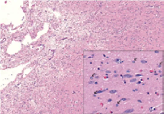
Fig. 2. Microscopic examination of the liver (H&E, x20): neoplastic glands (arrows) are evident embedded in abundant, dense, collagenous stroma.
Marked nuclear atypia is visible. The non-neoplastic liver parenchyma is present (left side).
Fig. 3. Microscopic examination of the liver tumor (H&E, x20): hypercellular neoplastic epithelial cell proliferation. The lesion forms papillary structures with little stroma. Intracytoplasmatic mucin (arrow) is detectable in few cells.
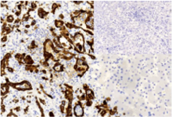
Fig. 4. Immunohistochemistry examination of the liver tumor: CK7 positivity, intense cytoplasmic staining in tumor cells (left). Insert: No glypican-3 expression was detected (neoplastic glands are at the lower part of the picture).
Fig. 5. The coronal slice of the brain: bilateral softening of the putamen with cavitation.
Cholangiocarcinoma in Wilson’s disease 307
J Gastrointestin Liver Dis, September 2017 Vol. 26 No 3: 305-308
HCC carcinoma in WD patients. Cholangiocarcinoma has been published mainly as case reports; there are only two retrospective cohort studies on abdominal/hepatobiliary malignancies [6, 7].
However, HCC seems to be less frequent in WD patients than in cirrhotic patients with other etiology [3, 4]. On the other hand, effective chelating therapy may also prevent the deterioration of liver disease and the development of cirrhosis and HCC [8].
There are conflicting theories regarding the low incidence of HCC in WD patients. Formerly it has been suggested that copper might have a protective role against HCC in these patients [9]. On the contrary, recent data showed that excessive amount of copper may cause cell damage via the production of reactive oxygen species (ROS) resulting in genetic and epigenetic alterations [10]. An elevated level of copper has been found in many human malignancies such as colon, lung, prostate and breast cancers [11]. Copper may also play a central role in the BRAF associated tumorigenesis in melanomas, thyroid cancers and hairy cell leukaemias [12]. Thus, chelating agents might have an antitumor effect and be effective as an anticancer therapy [8, 11, 13].
In our patient CCC developed despite nine years of chelating agent therapy (seven years with penicillamine and two years with trientine). The patient presented with cirrhosis and neurological symptoms at the time of diagnosis.
His symptoms regressed, and his cirrhosis was compensated during the treatment. It is remarkable that CCC developed very rapidly, since the regular abdominal ultrasound did not identify any sign of malignancy, which became obvious at eight months after the last examination.
Several neuropathological findings characteristic of WD were present and were confirmed by the autopsy in our patient such as bilateral softening of the putamen and the presence of Alzheimer type II cells [14]. However, Opalski cells, said to be pathognomonic of copper toxicity in the brain, were absent, in concordance with a former publication [15]. In our patient, CCC developed on a cirrhotic liver, similarly to HCC, as previously published [6, 16].
Fig. 6. Microscopic examination of the putamen (H&E, x4): the lesion had ill-defined borders, inflammatory cells were not present. Near the softened areas, scattered Alzheimer type II cells (insert, x40, arrows) were seen. Opalski cells characteristic of Wilson’s disease were not found.
CONCLUSION
The presented case argues that cholangiocarcinoma may develop in well-treated Wilson’s disease patients, on a cirrohic liver, similarly to hepatocellular carcinoma. The curiosity of this case is the very rapid development of cholangiocarcinoma despite continuous chelating agent therapy.
Conflicts of interest: The authors declare that there is no potential conflict of interest relevant to this article.
Authors’ contribution: F. S., A. F. and D. N. managed the patient and contributed to the diagnosis, follow-up and the writing of the manuscript. G. S. and T. T. performed the autopsy, the histological work up and the photo documentation. B. K. was the neuropathologist who provided the neuropathological images and review. All authors contributed to the writing of the manuscript and read the revised one.
REFERENCES
1. Petrukhin K, Fischer SG, Pirastu M, et al. Mapping, cloning and genetic characterization of the region containing the Wilson disease gene. Nat Genet 1993;5:338-343. doi:10.1038/ng1293-338
2. Wilson SA. Kayser-Fleischer Ring in Cornea in two Cases of Wilson’s Disease (Progressive Lenticular Degeneration). Proc R Soc Med 1934;27:297-298.
3. van Meer S, de Man RA, van den Berg AP, et al. No increased risk of hepatocellular carcinoma in cirrhosis due to Wilson disease during long- term follow-up. J Gastroenterol Hepatol 2015;30:535-539. doi:10.1111/
jgh.12716
4. Beinhardt S, Leiss W, Stättermayer AF, et al. Long-term outcomes of patients with wilson disease in a large Austrian cohort.
Clin Gastroenterol Hepatol 2014;12:683-389. doi:10.1016/j.
cgh.2013.09.025
5. Girard PF, Vachon A, Tommasi M, Paliard P, Rochet M, Barthe J.
Hepatolenticular degeneration and primary cancer of the liver. Lyon Med 1968;219:1395-1400.
6. Walshe JM, Waldenström E, Sams V, Nordlinder H, Westermark K. Abdominal malignancies in patients with Wilson’s disease. QJM 2003;96:657-662. doi:10.1093/qjmed/hcg114
7. Pfeiffenberger J, Mogler C, Gotthardt DN, et al. Hepatobiliary malignancies in Wilson disease. Liver Int 2015;35:1615-1622.
doi:10.1111/liv.12727
8. Yoshii J, Yoshiji H, Kuriyama S, et al. The copper-chelating agent, trientine, suppresses tumor development and angiogenesis in the murine hepatocellular carcinoma cells. Int J Cancer 2001;94:768-773.
doi:10.1002/ijc.1537
9. Wilkinson ML, Portmann B, Williams R. Wilson’s disease and hepatocellular carcinoma: possible protective role of copper. Gut 1983;24:767-771.
10. Linder MC. The relationship of copper to DNA damage and damage prevention in humans. Mutat Res 2012;733:83-91. doi:10.1016/j.
mrfmmm.2012.03.010
11. Tisato F, Marzano C, Porchia M, Pellei M, Santini C. Copper in diseases and treatments, and copper-based anticancer strategies. Med Res Rev 2010;30:708-749. doi:10.1002/med.20174
308 Németh et al
J Gastrointestin Liver Dis, September 2017 Vol. 26 No 3: 305-308
12. Brady DC, Crowe MS, Turski ML, et al. Copper is required for oncogenic BRAF signaling and tumorigenesis. Nature 2014;509:492- 496. doi:10.1038/nature13180
13. Moriguchi M, Nakajima T, Kimura H, et al. The copper chelator trientine has an antiangiogenic effect against hepatocellular carcinoma, possibly through inhibition of interleukin-8 production. Int J Cancer 2002;102:445-452. doi:10.1002/ijc.10740
14. Taly AB, Prashanth LK, Sinha S. Wilson’s disease: An Indian perspective.
Neurol India 2009;57:528-540. doi:10.4103/0028-3886.57789 15. Meenakshi-Sundaram S, Mahadevan A, Taly AB, Arunodaya GR, Swamy
HS, Shankar SK. Wilson’s disease: A clinico-neuropathological autopsy study. J Clin Neurosci 2008;15:409-417. doi:10.1016/j.jocn.2006.07.017 16. Iwadate H, Ohira H, Suzuki T, et al. Hepatocellular Carcinoma
Associated with Wilson’s Disease. Intern Med 2004;43:1042-1045.
doi:10.2169/internalmedicine.43.1042