Review Article
Biomarkers in Urachal Cancer and Adenocarcinomas in the Bladder: A Comprehensive Review Supplemented by Own Data
Henning Reis ,
1Ulrich Krafft,
2Christian Niedworok,
2Orsolya Módos,
3Thomas Herold,
1Mark Behrendt,
4Hikmat Al-Ahmadie,
5Boris Hadaschik,
2Peter Nyirady,
3and Tibor Szarvas
2,31Institute of Pathology, University Hospital Essen, University of Duisburg-Essen, Hufelandstr 55, 45147 Essen, Germany
2Department of Urology, University Hospital Essen, University of Duisburg-Essen, Hufelandstr 55, 45147 Essen, Germany
3Department of Urology, Semmelweis University, Üllői út 78/b, 1082 Budapest, Hungary
4Department of Urology, The Netherlands Cancer Institute - Antoni van Leeuwenhoek Hospital, Plesmanlaan 121, 1066 CX Amsterdam, Netherlands
5Department of Pathology, Memorial Sloan Kettering Cancer Center, 1275 York Avenue, New York, NY 10065, USA
Correspondence should be addressed to Henning Reis; henning.reis@t-online.de Received 25 July 2017; Accepted 6 February 2018; Published 12 March 2018
Academic Editor: Tilman Todenhöfer
Copyright © 2018 Henning Reis et al. This is an open access article distributed under the Creative Commons Attribution License, which permits unrestricted use, distribution, and reproduction in any medium, provided the original work is properly cited.
Urachal cancer (UrC) is a rare but aggressive cancer. Due to overlapping histomorphology, discrimination of urachal from primary bladder adenocarcinomas (PBAC) and adenocarcinomas secondarily involving the bladder (particularly colorectal adenocarcinomas, CRC) can be challenging. Therefore, we aimed to give an overview of helpful (immunohistochemical) biomarkers and clinicopathological factors in addition to survival analyses and included institutional data from 12 urachal adenocarcinomas. A PubMed search yielded 319 suitable studies since 1930 in the English literature with 1984 cases of UrC including 1834 adenocarcinomas (92%) and 150 nonadenocarcinomas (8%). UrC was more common in men (63%), showed a median age at diagnosis of 50.8 years and a median tumor size of 6.0 cm. No associations were noted for overall survival and progression-free survival (PFS) and clinicopathological factors beside a favorable PFS in male patients (p= 0 047). The immunohistochemical markers found to be potentially helpful in the differential diagnostic situation are AMACR and CK34βE12 (UrC versus CRC and PBAC), CK7,β-Catenin and CD15 (UrC and PBAC versus CRC), and CEA and GATA3 (UrC and CRC versus PBAC). Serum markers like CEA, CA19-9 and CA125 might additionally be useful in the follow-up and monitoring of UrC.
1. Introduction
The urachus is a remnant of the fetal structure connecting the allantois and the fetal bladder. During early fetal develop- ment, the urachus usually regresses to form an obliterated
fibromuscular canal, known as the median umbilical liga- ment [1
–4]. Failure of complete luminal obliteration has been described in up to one-third of adults and can rarely lead to various anomalies including cysts,
fistulas, and diver-ticula or rarely malignant transformation [2, 5].
Our understanding of urachal cancer (UrC) has evolved since the seminal studies by Begg [6] in the 1930’s following the
first report by Hue and Jacquin [7] in 1863 and earlierworks of Cullen in 1916 [8]. UrC is a very rare but highly malignant tumor entity with an incidence of
<1% of all blad-der cancers [1, 9, 10]. Establishing the diagnosis of UrC can be challenging for the urologist, pathologist, and radiologist and usually requires a multidisciplinary approach. In terms of histopathology, many overlapping features with the main di
fferential diagnostic entities exist. While the diagnostic
Volume 2018, Article ID 7308168, 21 pages https://doi.org/10.1155/2018/7308168
criteria adapted and established by Sheldon et al. [11] are most widely used, Gopalan and colleagues [1] modified these criteria and Paner and colleagues proposed diagnostic cri- teria for nonglandular type UrC [12].
Recently, Paner and colleagues also gave a review on the diagnosis and classi
fication of urachal epithelial neoplasms [13]. To furthermore give a current overview of the clinical and therapeutical implications of UrC, we have recently con- ducted a meta-analysis of the literature including 1010 cases of UrC [14].
Histologically, urachal adenocarcinomas (which are the most common carcinomas of urachal origin) overlap with their main differential diagnostic entities, that is, primary bladder adenocarcinomas and colorectal adenocarcinomas.
The present work therefore aims to provide an overview and summary of the immunohistochemical biomarkers in UrC and their potential role in the diagnosis of such tumors. It is combined with clinicopathological evidence and its data is collected from the published literature since 1930. Additionally, it is supported by our own data of immunohistochemical expression in 12 UrC cases with 11 different antibodies including the report of GATA3 expres- sion in this disease.
2. Literature Review and Statistics
A PubMed search was conducted using the string [“urachus carcinoma
”OR
“urachus cancer
”OR
“urachal carcinoma
”OR
“urachal cancer
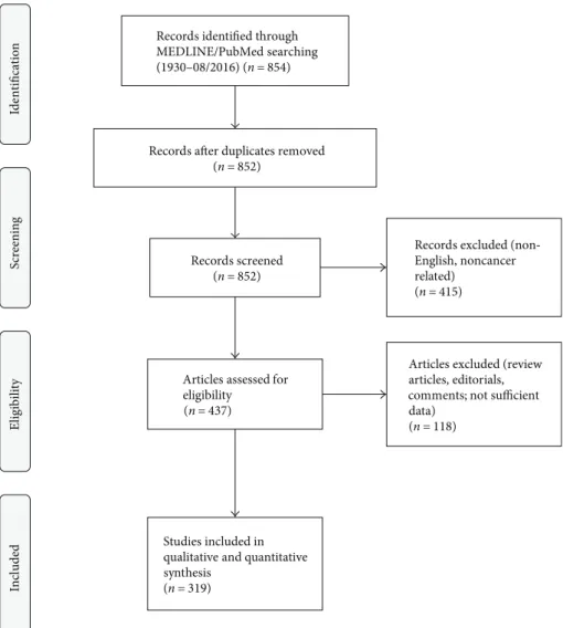
”] which returned 854 results (end of data acquisition: 08/2016). The algorithm of study selection is illustrated in Figure 1. Information was extracted from whole papers written in English language and from English abstracts in case of other primary language. In case of differ- ent entities in the papers, only information regarding UrC was extracted. When available, survival data was recorded for both overall survival (OS) and progression-free survival (PFS). For statistical analyses, SPSS (v23; IBM, Armonk, USA) was used. Pearson correlation analysis was conducted when appropriate. Survival analyses were conducted using the Kaplan Meier method with the log-rank test and univari- able Cox analysis. When appropriate, continuous variables were dichotomized at their median level for analysis of their impact on survival.
3. Additional Data from Our Own Institution The clinicopathological data of our cohort has been pub- lished previously [15]. Immunohistochemical studies were performed on formalin-fixed and paraffin-embedded urachal adenocarcinoma tissue using a BenchMark ULTRA System (Ventana Medical Systems, Tucson, USA) following manu- facturer’s instructions. A total of 11 different antibodies were performed on 12 cases of urachal adenocarcinomas from the University Hospital of Essen including
β-Catenin, CD15, CDX2, CEA, CK7, CK20, GATA3, MLH1, MSH2, MSH6, and PMS2 (Supplementary Table 1). The study was approved by the ethic committee of the University Hospital of Essen (16-6902-BO, 04.28.2016).
Further details on used antibodies, protocol information, and results of the immunohistochemical analyses are dis- played in Supplementary Table 1.
4. General Results of the Literature Review Three hundred and nineteen studies were identified that con- tained su
fficient information on cases of UrC. The number of publications has increased rapidly in the recent years with 169 (53%) publications since the year 2000 and 75 (24%) studies from 2011
–2016.
A total of 2154 cases of UrC were identi
fied, with infor- mation on UrC histology available in 1984 cases (92%), of which 1834 (92%) were adenocarcinomas. The majority of studies with information on UrC cases were case reports (74%), while contributing only a minor part to the total num- ber of cases (16%). In 1491 cases, gender information was available with evidence showing that most UrC cases occurred in men (63%) compared to women (37%). The mean and median age were 48.6 and 50.8 years, respectively (range: 0.3
–86.0 years), and tumor size 7.1 cm and 6.0 cm, respectively (range: 0.5
–25.0 cm). Data on tumor grades were sporadic and inconsistent and could not be further analyzed.
Survival data were available in 76 cases (adenocarcinomas:
n= 60, nonadenocarcinomas:n= 16) with a median follow-
up of 12 months in the total cohort (range: 1–62 months).
The median OS for the entire cohort was 46.8 months (ade- nocarcinomas: 42.7 months) with a 1-year survival of 86%
(adenocarcinomas: 86%), 3-year survival of 63% (adenocarci- nomas: 59%), and a 5-year survival of 41% (adenocarci- nomas: 35%). The median PFS for the entire cohort was 46.6 months (adenocarcinomas: 41.1 months) and 75% at 1-year (adenocarcinomas: 72%), 60% at 3 years (adenocarci- nomas: 55%), and 39% at 5 years (adenocarcinomas: 33%). It is important to note that some of the survival data derives from older papers with different treatment strategies that might have affected the outcome analysis. In fact, recent epidemiological studies demonstrate higher survival rates (5-year overall survival of approximately 50%) due to advances in the surgical and medical management of this dis- ease [16].
Detailed clinicopathological data are listed in Table 1.
Data on UrC adenocarcinomas were collected from these ref- erences [1, 3, 4, 9
–11, 15
–251].
5. Specific Review Data: Adenocarcinomas
Histopathologically, both primary adenocarcinomas of the
bladder and urachal adenocarcinomas show similar subtypes
although their distribution differs [21, 252, 253]. In invasive
urachal adenocarcinoma, the following four subtypes are
described in the 2016 WHO classification:
mucinous(colloi-
dal) type with preponderance of extracellular mucin and
malignant epithelia
floating within,
enteric (intestinal)type
with preponderance of malignant strati
fied epithelium
resembling colorectal adenocarcinomas,
mixedtype with nei-
ther a mucinous nor an enteric pattern prevailing,
not other specified (NOS)type with a pattern not easily identifiable as
mucinous or enteric type, and
signet ring celltype with signet ring cell morphology prevailing.
It is important to note that the concept of mucinous cys- tic tumors as recently proposed by Amin et al. [17] was not applied for this study due to the lack of such a classification in the older literature. In their work, Amin et al. described a distinct subgroup of urachal neoplasms with predominant cystic appearance in analogy to similar neoplasms in the ovary. This includes mucinous cystadenomas, mucinous cys- tic tumor of low malignant potential (MCTLMP), and mucinous cystadenocarcinoma with microinvasion or frank invasion. However, although in cox analyses tumor size was not associated with survival, not larger but smaller tumor size exhibited a higher hazard ratio for OS possibly giving support to the concept of favorable prognosis of mucinous cystic lesions of urachal origin [17].
In a population-based study from Wright and colleagues including 151 UrC adenocarcinomas and 1374 primary blad- der adenocarcinomas, the mucinous/colloid pattern was detected in 48% of UrC adenocarcinomas [16]. Also in our analysis, the mucinous type represented the most common special type of urachal adenocarcinomas (57%) (Table 1,
Figure 2(a)). In Wright and colleagues
’work, the second most common pattern was the NOS type (39%), which in our analysis accounted for 14%. However, in their analysis, the enteric type was not explicitly mentioned, which com- prised 15% in our study. Additionally, they described the sig- net ring cell type and the mixed type in 7% each, which comprised 6% and 8% in our analysis, respectively. The prog- nostic value of these histopathologic features, however, remains to be established while a more favorable clinical course for UrC adenocarcinomas as compared to primary bladder adenocarcinomas was found [16]. Additionally, pres- ence of signet ring cell morphology and higher tumor grade were identi
fied of unfavorable prognostic value in some [10, 21, 157, 192, 221] but not all series [24] of UrC adenocarci- nomas. Regarding signet ring cell morphology, this may result from the differing definitions and lack of consistent cut-off on the amount of signet ring cells across the different studies. In our survival analysis from cases of the literature, we could not detect an influence of type of UrC adenocarci- noma on OS, but a borderline in
fluence on PFS in terms of a survival bene
fit for intestinal type UrC. However, this
find- ing was not consistent in further (Kaplan Meier) analyses,
Records identified through MEDLINE/PubMed searching (1930–08/2016) (n= 854)
ScreeningIncludedEligibilityIdentification
Records after duplicates removed (n= 852)
Records screened (n= 852)
Records excluded (non‑ English, noncancer related)
(n= 415)
Articles assessed for eligibility
(n= 437)
Articles excluded (review articles, editorials, comments; not sufficient data)
(n= 118)
Studies included in qualitative and quantitative synthesis
(n= 319)
Figure1: PRISMAflow diagram. The diagram illustrates the phases and selection criteria used for study selection in this work.
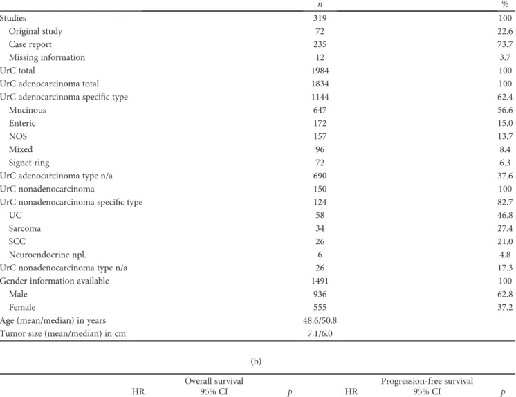
Table1: Review data of the cohort (a) and in association with survival data (b). UrC: urachal cancer; NOS: not otherwise specified; n/a: data not available; UC: urothelial carcinoma; SCC: squamous cell carcinoma; npl.: neoplasms; HR: hazard ratio; CI: confidence interval; ref.:
reference group.∗Data analysis was not feasible due to insufficient number of cases;^Cut-offvalues were chosen after partition tests.
(a)
n %
Studies 319 100
Original study 72 22.6
Case report 235 73.7
Missing information 12 3.7
UrC total 1984 100
UrC adenocarcinoma total 1834 100
UrC adenocarcinoma specific type 1144 62.4
Mucinous 647 56.6
Enteric 172 15.0
NOS 157 13.7
Mixed 96 8.4
Signet ring 72 6.3
UrC adenocarcinoma type n/a 690 37.6
UrC nonadenocarcinoma 150 100
UrC nonadenocarcinoma specific type 124 82.7
UC 58 46.8
Sarcoma 34 27.4
SCC 26 21.0
Neuroendocrine npl. 6 4.8
UrC nonadenocarcinoma type n/a 26 17.3
Gender information available 1491 100
Male 936 62.8
Female 555 37.2
Age (mean/median) in years 48.6/50.8
Tumor size (mean/median) in cm 7.1/6.0
(b)
Overall survival Progression-free survival
HR 95% CI p HR 95% CI p
UrC type
Adenocarcinoma 2.031 0.452–9.117 0.355 4.782 0.626–36.506 0.131
Nonadenocarcinoma ref. ref.
UrC adenocarcinoma
Mucinous 0.888 0.192–4.095 0.879 0.736 0.203–2.667 0.641
Nonmucinous ref. ref.
UrC nonadenocarcinoma
UC n/a∗ n/a∗ n/a∗ n/a∗ n/a∗ n/a∗
Non-UC ref. ref.
Gender
Male 0.629 0.276–1.430 0.268 0.197 0.039–0.981 0.047
Female ref. ref.
Age^
<45 years 1.880 0.769–4.598 0.167 1.534 0.652–3.610 0.327
>45 years ref. ref.
Tumor size^
<7.0 cm 2.644 0.502–13.923 0.251 2.943 0.592–14.622 0.187
>7.0 cm ref ref.
thus preventing further conclusions. Data density was too low for any tumor grading-related analyses.
Regarding the gender distribution, our review data is sim- ilar to epidemiological studies with preponderance of male UrC patients (62.8%) [16]. While no signi
ficant in
fluence of gender was detectable on OS in our accumulated data from the literature, male gender was associated with improved PFS (p
= 0 047), an effect which was mainly related to the ade-nocarcinoma part of the cohort (p
= 0 009; SupplementaryFigure 1). This effect has not yet been mentioned in the liter- ature while its cause remains to be elucidated. It seems not to be related to median tumor size or age at diagnosis as these factors were not associated with gender. Additionally, no sig- ni
ficant prognostic associations were noted for these two fac- tors, neither in the total cohort nor in subgroup analyses.
6. Specific Review Data:
Nonadenocarcinoma Neoplasms
In addition to UrC adenocarcinomas, nonglandular ura- chal tumors are included in the recent WHO 2016 classi-
fication. These are urothelial, squamous, neuroendocrine, and mixed-type neoplasms, which are stated to account for 4% to 27% of cases [12, 13, 253, 254]. These neo- plasms are histologically and immunophenotypically simi- lar to their counterparts elsewhere in the body [253].
Our analysis yielded 124 (of 150) cases of nonadenocar- cinoma UrC with further classified histology. Urothelial car- cinomas (UC) represented the largest group (n
= 58, 47%)(Table 1, Figure 2(b)) [3, 10–12, 19, 23, 46, 71, 82, 85, 127, 223, 255–267]. The second largest group was the group of sarcomas (
n= 34, 27%), however, with a large variety of dif- ferent entities including childhood rhabdomyosarcoma (embryonal, alveolar and NOS types) [268
–272], leiomyo- sarcomas [273
–276],
fibrosarcomas [277, 278], and also some cases without further specification of the type of entity
[3, 11, 47, 279–281]. Attention has to be paid to the fact that the reported sarcomas derive from a broad timespan with di
fferent knowledge levels, respectively. Therefore, some of these neoplasms would today be classi
fied di
fferently. In addition to mesenchymal lesions, our analysis showed 26 cases of squamous cell carcinomas (SCC; 21%) [11, 71, 85, 105, 223, 282–291], followed by neuroendocrine neoplasms including small cell carcinomas with 6 cases (5%) [9, 10, 12, 201]. In the remaining cases, information on nonadeno- carcinoma entity was at least partly missing [3, 11, 19, 71, 85, 141, 292, 293] or included other entities such as yolk sac tumors [187, 294, 295] or a neuroblastoma [296].
In addition to malignant urachal tumors, several other intermediate and benign tumors or conditions of the urachus have been reported some mimicking urachal cancer and thus posing a di
fferential diagnostic problem. Tumors or condi- tions rated as intermediate include in
flammatory myo
fibro- blastic tumors (IMT) [297–300], a solitary
fibrous tumor(SFT) [301], desmoid
fibromatoses [302, 303], a hemangio-pericytoma [304], and a Castleman’s disease [305], while benign tumors and conditions include dermoid cysts [301, 306], teratomas [307, 308], leiomyomas [309, 310], (fibrous) hamartomas [311, 312], a hemangioma [313], a
fibroade- noma [314], malakoplakia [315], abscesses [316
–318], a xanthogranulomatous urachitis [319], a urachal tuberculosis [320], actinomycosis [321
–323], an endometriosis [324], a perforated colonic diverticulitis [325], and even a
fishbone within an urachal cyst [326].
7. Biomarkers in Urachal
Cancer: Immunohistochemistry
Given the extensively overlapping histopathological features of adenocarcinomas of urachal and primary bladder origin on the one hand and secondary adenocarcinomas from different sites on the other, biomarkers for differential
n = 647 57%
n = 172 15%
n = 157 14%
n = 96 8%
n = 72 6%
Mucinous Enteric NOS Mixed Signet ring
(a)
UC Sarcoma SCC NE npl.
n = 58 47%
n = 34 27%
n = 26 21%
n = 6 5%
(b)
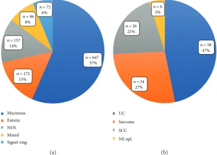
Figure2: Distribution of the different types of UrC (a) in urachal adenocarcinomas with available information of special type and (b) in nonadenocarcinoma UrC with information of special type. UrC: urachal cancer; NOS; not otherwise specified; UC: urothelial carcinoma;
SCC: squamous cell carcinoma; NE npl.: neuroendocrine neoplasms.
diagnostic purposes are required. The most important differ- ential diagnostic problems with significant impact on thera- peutic decisions may be categorized as follows:
(1) Differentiation between invasion/metastasis of colo- rectal adenocarcinomas and urachal adenocarci- nomas. Exclusion of a possible invasion of this cancer (to the bladder) is a necessary step for the de
finitive diagnosis of UrC and of therapeutic relevance.
(2) Distinguishing urachal adenocarcinomas from those of primary bladder origin has also a direct clinical impact on the surgical treatment. In localized disease, primary bladder adenocarcinomas are usually treated with complete cystectomy while urachal adenocarci- nomas mostly require partial cystectomy with
en blocremoval of the umbilical ligament and umbilicus (radical versus partial cystectomy) with significantly different impact on quality of life [4, 85].
(3) Identification of the origin of a (mucinous) adenocar- cinoma of unknown primary is also important as ura- chal adenocarcinomas frequently metastasize to various organs, such as the bone, lung, and liver.
Identi
fication of urachal origin of a (mucinous) ade- nocarcinoma can have a direct therapeutic conse- quence [4].
An overview of the immunohistochemical markers assessed in urachal adenocarcinomas is provided in Table 2 and a representative example is illustrated in Figure 3. Further detailed information is provided in Supplementary Table 2.
The immunohistochemical markers most often employed in the work up of adenocarcinomas of different sites usually include Cytokeratin 20 (CK20) and CK7. In our analysis of a total of 116 urachal adenocarcinomas, only 4 cases were negative for CK20—an overall positive rate of 97% [1, 17, 23, 26, 27, 31, 36, 48, 56, 60, 62, 67, 74, 77, 86, 95, 99, 124
–126, 128, 148, 156, 163, 164, 216, 217, 220, 240, 245, 251, 327]. Considering the robust CK20 expression in adenocarci- nomas of sites of di
fferential diagnostic interest, CK20 has no signi
ficant value in this setting.
In contrast, expression of CK7 in these tumors is widely variable. In urachal adenocarcinomas, CK7 exhibited a pooled reactivity rate of 51%, compared to considerable lower rates in colorectal cancer (0–38%, Table 2, Supplementary Table 2) [1, 17, 23, 26, 27, 31, 36, 48, 56, 60, 67, 74, 77, 86, 99, 124, 125, 156, 163, 164, 216, 217, 220, 240, 251, 255, 292, 327, 328]. However, similar to urachal adenocarcinomas, pri- mary bladder adenocarcinomas constantly exhibited rela- tively high CK7 reactivity rates (33%
–70%), thus limiting the value of CK7 in the discrimination between these two entities [23, 329].
Additionally, CK20 and CK7 were the only markers with sufficient data for survival analyses. However, no significant influence of the CK20/CK7 expression profile on OS or PFS was noted.
As a rather specific nuclear marker for intestinal epithe- lia and corresponding adenocarcinomas, CDX2—a homeo- box gene coding for a transcription factor with intestine
specificity—has been proposed for differential diagnostic considerations. However, nuclear CDX2 reactivity was evident in the majority of urachal adenocarcinomas (90%) [1, 17, 23, 26, 31, 60, 99, 125, 126, 128, 216, 217, 240, 245, 251, 327, 330] and many primary bladder adenocarcinomas (13%–83%) [329, 331]. In addition to its reactivity in almost all colorectal adenocarcinomas, CDX2 immunopositivity has been detected in considerable numbers in several adenocar- cinomas from di
fferent sites such as the gastrointestinal tract, pancreas, and ovary [330, 332]. Furthermore, CDX2 reactivity has been described in cystitis glandularis and intestinal metaplasia of the bladder and glandular epithelia of urachal remnants [23, 333–335]. Taken together, CDX2 is not helpful in the differential diagnosis of adenocarci- nomas in the urinary bladder.
Another plausible biomarker in this context is
β-Catenin, a protein involved in cell-cell adhesion and gene transcrip- tion regulation [336]. In normal cells,
β-Catenin staining is restricted to the membrane/cytoplasm, while in colorectal adenocarcinomas,
β-Catenin exhibits nuclear accumulationdue to mutation or loss of the adenomatous polyposis coli (APC) gene then acting as a transcriptional activator [337].
While in colorectal adenocarcinomas nuclear
β-Cateninexpression can be found in the majority of cases, nuclear
β- Catenin reactivity was detected in a low rate of primary blad- der adenocarcinomas (0%
–17%) [329, 338]. Similarly, in ura- chal adenocarcinomas, nuclear
β-Catenin expression was a rare event. In our summary analysis, any type of nuclear
β- Catenin was detected in 9 of 63 cases (14%) [1, 17, 23, 26, 125, 216, 220, 328]. APC mutations, however, can be found in urachal adenocarcinomas slightly more often than the immunohistochemical results propose [20, 204]. From a dif- ferential diagnostic point of view, nuclear
β-Catenin expres-sion may be useful in distinguishing primary bladder and urachal adenocarcinomas from secondary bladder involve- ment by colorectal adenocarcinomas. However,
β-Catenin is of no use in the di
fferentiation of primary bladder from urachal adenocarcinomas as both entities exhibit comparable
β-Catenin staining characteristics.
Further potential markers include Claudin-18 and Reg IV, however, with no available data in primary bladder ade- nocarcinomas. Claudin-18 has been reported to be of diagnos- tic value especially in pancreatic and gastric cancer, but is rarely expressed in colorectal adenocarcinomas [339–341].
Although exhibiting a positivity rate of 53% in the total num-
ber of urachal adenocarcinomas cases, it was found to have
only a low positivity rate (27%) in enteric type urachal adeno-
carcinomas, thus limiting its usefulness in UrC diagnostics
with regard to the largest group of intestinal-di
fferentiated
colorectal adenocarcinomas [23]. Reg IV is associated with
the cellular phenotype of the intestine and expressed in vari-
ous cancers with intestinal differentiation such as gastric and
colorectal cancer [342]. In urachal adenocarcinomas, Reg
IV expression was detected in 85% of cases arguing against
its potential use in the differential diagnostics of adenocarci-
nomas detected in the bladder [23]. Both markers addition-
ally failed to demonstrate diagnostic value in signet ring cell
UrC compared to signet ring cell carcinoma of colorectal
origin [23].
Further, possibly useful biomarkers of urachal adenocarci- nomas with data of at least 10 cases are alpha-methylacyl-CoA racemase (AMACR, p504s), CD15 (Leu-M1), carcinoem- bryonic antigen (CEA), CK34
βE12 (high-molecular weight cytokeratin), GATA binding protein 3 (GATA3), mucin 2 (MUC2), and mucin 5 AC (MUC5AC).
In urachal adenocarcinomas, AMACR was found to be positive in a low number of cases (17%), while in colorectal
and primary bladder adenocarcinomas, a significantly higher number (>66%) of cases exhibited AMACR- reactivity [17, 77, 343, 344]. In contrast, CK34βE12 was more frequently positive (67%) in urachal adenocarcinomas, while being variably expressed in primary bladder or colorectal adenocarcinomas [1, 26, 77, 125, 255, 345]. A comparable distribution was detected for MUC2 and MUC5AC with high positivity rates in urachal adenocarcinomas (100% and 92%)
(a) (b) (c) (d)
(e) (f) (g) (h)
(i) (j)
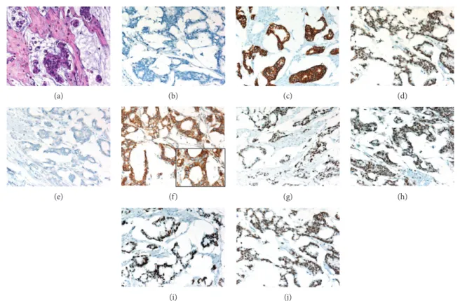
Figure3: A representative case of mucinous urachal adenocarcinoma. (a) Atypical cellsfloating in extracellular mucin. Focal signet ring cell morphology is noticeable (H&E staining). The case exhibited a typical profile in further immunohistochemical studies with no reactivity for CK7 (b) but positive reactivity for CK20 (c) and CDX2 (d, nuclear). As in all analyzed cases, no GATA3 reactivity was noted (e). In the β-Catenin immunohistochemistry, a strong membranous and cytoplasmic but no nuclear reactivity was noted (f; inlay magnification 600x).
The immunohistochemical reactions against the MMR proteins all were positive, that is, MLH1 (g), PMS2 (h), MSH2 (i), and MSH6 (j).
Table2: Useful immunohistochemical antibodies in the differential diagnosis of urachal adenocarcinoma (UrC), colorectal adenocarcinoma (CRC), and primary bladder adenocarcinoma (PBAC). Loss of MMR proteins (MLH1, MSH2, MSH6, and PMS2) additionally favors colorectal over urachal adenocarcinomas. For more details on reactivity rates, number of cases, and references, please refer to Supplementary Table 2. Please note that data density is low for most antibodies limiting significance.“Highest”data quality is available for CK7, β-Catenin, and CEA.−: negative (0% positive); (−): mostly negative (1–25% positive); +/−: some positive (26–50% positive); (+):
mostly positive (51–75% positive); +: positive (76–100% positive).
Reactivity Differential diagnosis
Biomarker (IHC) UrC CRC PBAC UrC versus CRC and PBAC UrC and PBAC versus CRC UrC and CRC versus PBAC
AMACR (p504s) (−) + (+) + − −
CK34βE12 (HMWCK) (+) (−) +/− + − −
CK7 (+) +/− (+) − + −
β-Catenin (nuclear) (−) + (−) − + −
CD15 (Leu-M1) + +/− (+) − + −
CEA + + (+) − − +
GATA3 − − (+) − − +
and lower rates in colorectal and primary bladder adenocar- cinomas, however, with significant overlap [86, 95, 100, 125, 128, 251, 335, 346, 347].
In contrast, CD15 was detected in high rates of both ura- chal and primary bladder adenocarcinomas (86% and 73%, resp.) compared to colorectal adenocarcinomas with a lower reactivity rate (
<50%) [21, 27, 124, 125, 348]. CEA was opposingly found to be positive in all analyzed cases of ura- chal adenocarcinomas and in a similarly high rate of colorec- tal adenocarcinomas but lower rates in primary bladder adenocarcinomas (29–67%) [21, 27, 48, 60, 67, 77, 86, 100, 106, 124, 163, 165, 245, 255, 348–353].
Finally, GATA3 was not found to be expressed in urachal adenocarcinomas and colorectal adenocarcinomas but in approximately half of cases of primary bladder adenocarci- nomas [335, 354, 355]. In addition, nuclear GATA3 reactivity might be useful in the di
fferential diagnosis of bladder adeno- carcinomas with signet ring morphology [355].
The example of GATA3 in particular illustrates the need of rigorous case selection of primary bladder and/or urachal adenocarcinomas in the studies. Inclusion of UC with glandular differentiation or plasmacytoid UC could significantly weaken the validity of such a study and there- fore its conclusions.
The distribution of the DNA mismatch repair (MMR) proteins, that is, MutL homolog 1 (MLH1), MutS homolog 2 (MSH2), MutS homolog 6 (MSH6), and PMS1 homolog 2 (PMS2), in the di
fferent entities might in addition also be of di
fferential diagnostic and pathogenetic interest. While no data is available for primary bladder adenocarcinomas, spo- radic colorectal adenocarcinomas exhibit a loss of MMR pro- teins in 10–15% in total with emphasis on MLH1 [356]. In urachal adenocarcinomas, some tumors with microsatellite instability characterized by immunohistochemistry were described [25]. We, however, detected no loss of MMR pro- teins by immunohistochemistry in our own institutional cases (
n= 12). In additional preliminary molecular analyses, we also did not detect evidence of microsatellite instability (unpublished data). This seems to point to molecular di
ffer- ences in adenocarcinomas of urachal and colorectal origin.
Further important biomarkers in differential diagnostic considerations of adenocarcinomas in general are hormone receptors. In our review data, urachal adenocarcinomas did not express estrogen and progesterone receptors by immu- nohistochemistry, which might be of particular interest in the discrimination of a metastasis of urachal adenocarci- nomas to the ovary and vice versa [17, 56, 67]. In this set- ting, the immunonegativity of urachal adenocarcinomas for cancer antigen 125 (CA125) might also be of value, how- ever, with the limitation of only 8 cases reported in the liter- ature [27, 60, 86].
Low numbers of cases and no differential diagnostic value regarding the discrimination of urachal adenocarcinomas from primary bladder and colorectal adenocarcinomas were detected for
α-fetoprotein (AFP), carbohydrate antigen 19-9(CA19-9), cluster of differentiation 10 (CD10), CK19, Das-1, E48, E-Cadherin, gross cystic disease
fluid protein15 (GCDFP15), mucin 1 (epithelial membrane antigen) (MUC1 (EMA)), mucin 6 (MUC6), Thrombmodulin,
thyroid transcription factor 1 (TTF1), Uroplakin III, Villin, and Vimentin [27, 48, 60, 86, 100, 106, 125, 126, 172, 198, 215, 217, 245, 292, 330].
However, this might not be the case for some rare and special differential diagnostic considerations as for example in the discrimination of ductal prostate cancer and the enteric type of urachal adenocarcinomas, in which immuno- histochemistry for prostate specific acid phosphatase (PAP) and prostate-speci
fic antigen (PSA) (both negative in urachal adenocarcinomas and positive in ductal prostate cancer) might be useful [21, 27, 74, 77, 100, 245, 255, 357].
In addition to the di
fferential diagnostic context, immu- nohistochemical markers might be of further clinical value.
We recently assessed the expression and prognostic rele- vance of six immunohistochemical markers (Ki67, p53, biglycan (BGN), receptor for hyaluronan-mediated motility (RHAMM), and insulin-like growth factor II mRNA binding protein 3 (IMP3)) in urachal adenocarcinomas. RHAMM, IMP3, Ki67, and p53 were found to be increased in urachal adenocarcinomas. However, none of the analyzed markers exhibited any prognostic information [15].
Although immunohistochemical biomarkers are widely used in di
fferential diagnostic considerations, their interpre- tation is per se subjective. This applies in every situation in which these markers are used and therefore also in the immunohistochemical differential diagnosis of UrC. A fur- ther limitation of the collected data might also be the thresh- old at which the authors of the different source studies called an immunohistochemical marker positive or negative. Often- times, this information is missing while it can be very impor- tant. For example, the decision to call a case positive for nuclear
β-Catenin might depend only on a few stained tumor nuclei but with important di
fferential diagnostic implications [23, 329].
These considerations and also the partly overlapping positivity rates of the di
fferent immunohistochemical biomarkers make it di
fficult to recommend a step wise biomarker-guided approach. This could create a false sense of sensitivity and specificity of the biomarkers in the different situations. From our experience, immunohistochemical staining of a panel of antibodies, which depends on the differ- ential diagnostic setting (Table 2), is the best way to come to a conclusion in this setting. This process might of course also include the use of further antibodies in addition to the core panel and always lies in the expertise of the diagnostic histopathologist.
8. Biomarkers in Urachal Cancer:
Serum Markers
The (histomorphological) parallels between urachal and
colorectal adenocarcinomas furthermore gave the rationale
to test colorectal tumor markers in serum samples of
patients with urachal adenocarcinomas, especially CEA,
CA19-9, and CA125. In CRC, these markers are elevated
in approximately one third (CA125), half (CA19-9), and
two-thirds (CEA) of patients with a considerable variance
depending on tumor size and other variables [358]. In pri-
mary bladder adenocarcinomas, however, only sporadic
data is available with reports of elevated serum levels of these markers [359, 360].
In urachal adenocarcinomas, 44 studies including data on serum parameters were available, including 7 original studies and 37 case reports with a total of 140 patients.
Siefker-Radtke and colleagues reported on the largest cohort and found elevated (
>3 ng/ml) CEA serum levels in 59% of patients with urachal adenocarcinomas (median:
36 ng/ml) [24]. In 5 cases, CEA also decreased in response to chemotherapy, suggesting the potential utility of CEA test- ing in monitoring (or follow-up) of UrC. When analyzing the literature, elevated CEA serum levels were reported in 55.7%
(59/106) of patients at the time of diagnosis [24, 26, 33, 42, 60, 67, 79, 80, 86, 88, 89, 95, 99, 106, 118, 128, 131, 149, 156, 163
–165, 167, 179, 196, 200, 207, 208, 212, 214, 240, 241, 244, 246, 251]. In our analyses, elevated CEA levels at diagnosis were associated with worse OS (
p= 0 008) and PFS (
p= 0 009) in dichotomized analyses (elevated versusnormal), however, with only sparse survival data.
Additionally, elevated serum levels of CA19-9 and CA125 were reported in 50.8% (31/61) and 51.4% (19/37), respectively [24, 26, 33, 42, 76, 79–81, 86, 88, 89, 95, 99, 104, 114, 128, 149, 168, 200, 207, 213, 240, 244, 246, 251].
As with CEA, elevated levels of CA19-9 exhibited a trend towards worse OS and PFS (both
p= 0 09). No prognostic association was noted for CA125. In addition, elevated serum levels of CA125 did not correlate with negative immunohis- tochemical tissue expression, however, with a low case num- ber (n
= 8).Other serum biomarkers reported in low case numbers of urachal adenocarcinomas include lactate dehydrogenase (LDH) [80, 199], cancer antigen 15-3 (CA15-3) [26, 114, 156], AFP [16, 42, 95, 106, 156] with one case in a seven- month-old infant with a yolk sac tumor of the urachus [295], and neuron-speci
fic enolase (NSE) [156] including one case of a neuroblastoma in a six-month-old child [296].
In summary, measurement of serum biomarkers might be useful in the follow-up and disease monitoring of UrC.
9. Conclusions
We identified a total of 1984 cases of UrC from 319 suitable studies with sufficient data from the English literature with overall 1834 adenocarcinomas (92%). While only minor var- iations in clinicopathological factors such as gender distribu- tion (male preponderance), age at diagnosis, tumor size, and adenocarcinoma subtypes were noted, none of these factors were associated with overall survival. However, regarding progression-free survival, an advantage for male patients especially in the adenocarcinoma cohort was noted, while no such association was observed for nonglandular neo- plasms of urachal origin.
The summary of existing evidence on immunohisto- chemical markers supplemented with our own data highlighted a di
fferential diagnostic role for AMACR, CK34
βE12, CK7,
β-Catenin, CD15, and CEA (Table 2) which can be helpful in the routine di
fferential diagnostic workup of adenocarcinomas in the bladder. Also, GATA3 might be helpful in the differentiation of urachal from
primary bladder adenocarcinomas, with data presented almost exclusively derived from our institutional cohort. In addition, serum markers such as CEA, CA19-9, and CA125 might be useful in the follow-up and monitoring of UrC while CEA and CA19-9 may also be of prognostic value.
Conflicts of Interest
The authors declare that they have no conflicts of interest.
Acknowledgments
Tibor Szarvas was supported by János Bolyai Research Scholarship of the Hungarian Academy of Sciences. Gabriele Ladwig and Isabel Albertz are thanked for their skillful work.
Supplementary Materials
Supplementary Table 1(a): Detailed information on used antibodies and protocol in the cohort of the UrC register of the University Hospital of Essen. Supplementary Table 1(b): IHC results in the analyzed cohort of the UrC register of the University Hospital of Essen Detection of any immu- noreactivity in the tumor cells was assessed as positive and complete lack of immunoreactivity as negative. For
β-Catenin, the nuclear (nuc.) and membranous/cytoplasmic (m/c) reactivities were analyzed separately. The DNA mis- match repair proteins MLH1, MSH2, MSH6, and PMS2 were evaluated as positive if more than 10% of tumor cells exhib- ited a nuclear immunoreactivity. Specific immunostaining was cytoplasmic in case of CD15, CEA, CK7, and CK20 and nuclear in case of CDX2 and GATA3. Supplementary Table 2: detailed information on immunohistochemical bio- marker expression in urachal adenocarcinomas (UrC ADC) including own data (
∗) in comparison to
figures from the lit- erature for colorectal (CRC) and primary bladder adenocar- cinomas (PBAC). This table includes information about markers of potential di
fferential diagnostic usefulness in UrC versus CRC and PBAC, UrC and PBAC versus CRC, and UrC and CRC versus PBAC. ^ is positive in hepatoid carcinomas of the urinary bladder [329]; ^^ is usually pos- itive in clear cell PBAC [329]; ^^^Figures for sporadic colorectal cancer (nonhereditary), total loss of MMR pro- teins is evident in 10
–15% of sporadic colorectal cancers [356]
∗Own data from the UrC register of the University Hospital of Essen is included to increase data quality (all:
n= 12, but CK7: n= 11). [15] UrC: urachal cancer; ADC:
adenocarcinoma; AFP:
α-fetoprotein; AMACR: alpha-methylacyl-CoA racemase; mem/cyt: membranous/cyto- plasmic immunoreactivity;
β-Catenin: β-Catenin; CA19-9:carbohydrate antigen 19-9; CA125: cancer antigen 125;
CD10: cluster of differentiation 10; CD15: cluster of differ- entiation 15; CDX2: caudal-type homeobox protein 2; CEA:
carcinoembryonic antigen; CK7: cytokeratin 7; CK19: cyto- keratin 19; CK20: cytokeratin 20; CK34βE12 (HMWCK):
cytokeratin 34 (1,5,10,14) (high-molecular weight cytokera- tin); ER: estrogen receptor; GATA3: GATA binding protein 3; GCDFP15: Gross cystic disease
fluid protein 15; HER2:human epidermal growth factor receptor 2; MMR: DNA
mismatch repair proteins; MLH1: mutL homolog 1;
MSH2: mutS homolog 2; MSH6: mutS homolog 6; MUC1 (EMA): mucin 1, cell surface associated (epithelial membrane antigen); MUC2: mucin 2, oligomeric mucus/gel-forming;
MUC5AC: mucin 5AC oligomeric mucus/gel-forming;
MUC6: mucin 6, oligomeric mucus/gel-forming; PR: proges- terone receptor; PSAP: prostatic specific acid phosphatase;
PSA: prostate-specific antigen; PMS2: PMS1 homolog 2;
RegIV: Regenerating gene IV; TTF1: thyroid transcription factor 1 (NK2 homeobox 1). Supplementary Figure 1:
progression-free survival in urachal adenocarcinomas regard- ing gender [1, 15, 17, 21, 23, 25
–27, 31, 36, 48, 56, 60, 62, 67, 74, 77, 86, 95, 99, 100, 106, 124
–126, 128, 148, 156, 163
–165, 172, 198, 215
–217, 220, 221, 240, 245, 251, 255, 292, 327
–335, 338
–355, 361
–388].
(Supplementary Materials)References
[1] A. Gopalan, D. S. Sharp, S. W. Fine et al.,“Urachal carci- noma: a clinicopathologic analysis of 24 cases with outcome correlation,” The American Journal of Surgical Pathology, vol. 33, no. 5, pp. 659–668, 2009.
[2] V. Upadhyay and A. Kukkady, “Urachal remnants: an enigma,” European Journal of Pediatric Surgery, vol. 13, no. 6, pp. 372–376, 2003.
[3] J. R. Molina, J. F. Quevedo, A. F. Furth, R. L. Richardson, H. Zincke, and P. A. Burch,“Predictors of survival from ura- chal cancer: a Mayo Clinic study of 49 cases,” Cancer, vol. 110, no. 11, pp. 2434–2440, 2007.
[4] R. A. Ashley, B. A. Inman, T. J. Sebo et al.,“Urachal carci- noma: clinicopathologic features and long-term outcomes of an aggressive malignancy,” Cancer, vol. 107, no. 4, pp. 712–720, 2006.
[5] G. E. Schubert, M. B. Pavkovic, and B. A. Bethke-Bedurftig,
“Tubular urachal remnants in adult bladders,” The Journal of Urology, vol. 127, no. 1, pp. 40–42, 1982.
[6] R. C. Begg,“The Urachus: its anatomy, histology and devel- opment,”Journal of Anatomy, vol. 64, Part 2, pp. 170–183, 1930.
[7] L. Hue and M. Jacquin,“Cancer colloide de la lombille et de paroi abdominale anterieure ayant envahi la vessie,”Union Med de la Siene-Inf Rouen, vol. 6, pp. 418–420, 1863.
[8] T. B. Cullen, Embryology, Anatomy, and Diseases of the Umbilicus Together with Diseases of the Urachus, W. B.
SAUNDERS COMPANY, Philadelphia, PA, USA, 1916.
[9] D. E. Johnson, G. B. Hodge, F. W. Abdul-Karim, and A. G.
Ayala, “Urachal carcinoma,” Urology, vol. 26, no. 3, pp. 218–221, 1985.
[10] J. H. Pinthus, R. Haddad, J. Trachtenberg et al.,“Population based survival data on urachal tumors,”The Journal of Urol- ogy, vol. 175, no. 6, pp. 2042–2047, 2006.
[11] C. A. Sheldon, R. V. Clayman, R. Gonzalez, R. D. Williams, and E. E. Fraley,“Malignant urachal lesions,”The Journal of Urology, vol. 131, no. 1, pp. 1–8, 1984.
[12] G. P. Paner, G. A. Barkan, V. Mehta et al.,“Urachal carcino- mas of the nonglandular type: salient features and consider- ations in pathologic diagnosis,” The American Journal of Surgical Pathology, vol. 36, no. 3, pp. 432–442, 2012.
[13] G. P. Paner, A. Lopez-Beltran, D. Sirohi, and M. B. Amin,
“Updates in the pathologic diagnosis and classification of epi- thelial neoplasms of urachal origin,”Advances in Anatomic Pathology, vol. 23, no. 2, pp. 71–83, 2016.
[14] T. Szarvas, O. Modos, C. Niedworok et al.,“Clinical, prognos- tic, and therapeutic aspects of urachal carcinoma—a compre- hensive review with meta-analysis of 1,010 cases,”Urologic Oncology, vol. 34, no. 9, pp. 388–398, 2016.
[15] C. Niedworok, M. Panitz, T. Szarvas et al.,“Urachal carci- noma of the bladder: impact of clinical and immunohisto- chemical parameters on prognosis,”The Journal of Urology, vol. 195, no. 6, pp. 1690–1696, 2016.
[16] J. L. Wright, M. P. Porter, C. I. Li, P. H. Lange, and D. W. Lin,
“Differences in survival among patients with urachal and nonurachal adenocarcinomas of the bladder,” Cancer, vol. 107, no. 4, pp. 721–728, 2006.
[17] M. B. Amin, S. C. Smith, J. N. Eble et al.,“Glandular neo- plasms of the urachus: a report of 55 cases emphasizing mucinous cystic tumors with proposed classification,” The American Journal of Surgical Pathology, vol. 38, no. 8, pp. 1033–1045, 2014.
[18] R. A. Ashley, B. A. Inman, J. C. Routh, A. L. Rohlinger, D. A. Husmann, and S. A. Kramer,“Urachal anomalies: a longitudinal study of urachal remnants in children and adults,” The Journal of Urology, vol. 178, no. 4, pp. 1615– 1618, 2007.
[19] H. M. Bruins, O. Visser, M. Ploeg, C. A. Hulsbergen-van de Kaa, L. A. L. M. Kiemeney, and J. A. Witjes,“The clinical epi- demiology of urachal carcinoma: results of a large, population based study,” The Journal of Urology, vol. 188, no. 4, pp. 1102–1107, 2012.
[20] A. Collazo-Lorduy, M. Castillo-Martin, L. Wang et al.,“Ura- chal carcinoma shares genomic alterations with colorectal carcinoma and may respond to epidermal growth factor inhibition,” European Urology, vol. 70, no. 5, pp. 771–775, 2016.
[21] D. J. Grignon, J. Y. Ro, A. G. Ayala, D. E. Johnson, and N. G.
Ordonez,“Primary adenocarcinoma of the urinary bladder.
A clinicopathologic analysis of 72 cases,” Cancer, vol. 67, no. 8, pp. 2165–2172, 1991.
[22] O. Modos, H. Reis, C. Niedworok et al.,“Mutations of KRAS, NRAS, BRAF, EGFR, and PIK3CA genes in urachal carci- noma: occurence and prognostic significance,” Oncotarget, vol. 7, no. 26, pp. 39293–39301, 2016.
[23] G. P. Paner, J. K. McKenney, G. A. Barkan et al.,“Immuno- histochemical analysis in a morphologic spectrum of urachal epithelial neoplasms: diagnostic implications and pitfalls,” The American Journal of Surgical Pathology, vol. 35, no. 6, pp. 787–798, 2011.
[24] A. O. Siefker-Radtke, J. Gee, Y. U. Shen et al.,“Multimodality management of urachal carcinoma: the M. D. Anderson Cancer Center experience,”The Journal of Urology, vol. 169, no. 4, pp. 1295–1298, 2003.
[25] S. J. Sirintrapun, M. Ward, J. Woo, and A. Cimic,“High-stage urachal adenocarcinoma can be associated with microsatellite instability andKRASmutations,”Human Pathology, vol. 45, no. 2, pp. 327–330, 2014.
[26] I. Testa, E. Verzoni, P. Grassi, M. Colecchia, F. Panzone, and G. Procopio,“Response to targeted therapy in urachal adenocarcinoma,” Rare Tumors, vol. 6, no. 4, p. 5529, 2014.
[27] Torenbeek, Lagendijk, Van Diest, Bril, van de Molengraft, and Meijer,“Value of a panel of antibodies to identify the primary origin of adenocarcinomas presenting as bladder carcinoma,”Histopathology, vol. 32, no. 1, pp. 20–27, 1998.
[28] A. M. Abeygunasekera and D. D. Ranasinghe, “Urachal carcinoma,” Indian Journal of Medical Research, vol. 137, p. 398, 2013.
[29] M. Alonso-Gorrea, J. A. Mompo-Sanchis, M. Jorda-Cuevas, A. Froufe, and J. F. Jiménez-Cruz,“Signet ring cell adenocar- cinoma of the urachus,” European Urology, vol. 11, no. 4, pp. 282–284, 1985.
[30] C. Alvarez Alvarez, J. M. Sanchez Merino, L. Busto Castanon, F. Pombo Felipe, and F. Arnal Monreal,“Mucin- ous adenocarcinoma of the urachus synchronic with colorec- tal adenocarcinoma. Value of immunohistochemistry in the differential diagnosis,” Actas Urologicas Espanolas, vol. 22, no. 6, pp. 515–518, 1998.
[31] F. Z. Aly, A. Z. Tabbarah, and L. Voltaggio,“Metastatic ura- chal carcinoma in bronchial brush cytology,” CytoJournal, vol. 10, no. 1, p. 1, 2013.
[32] M. Ando, T. Toyoshima, C. Arisawa, S. Ikegami, and T. Okano,“Urachal adenocarcinoma accompanied by a large spherical calcified mass,” International Journal of Urology, vol. 2, no. 5, pp. 344–346, 1995.
[33] F. Aoun, A. Peltier, and R. van Velthoven,“Bladder sparing robot-assisted laparoscopic en bloc resection of urachus and umbilicus for urachal adenocarcinoma,” Journal of Robotic Surgery, vol. 9, no. 2, pp. 167–170, 2015.
[34] C. G. Bandler and P. R. Roen,“Mucinous adenocarcinoma arising in urachal cyst and involving bladder,”The Journal of Urology, vol. 64, no. 3, pp. 504–510, 1950.
[35] J. M. Barros Rodriguez, R. Fernandez Martin, J. L. Guate Ortiz et al., “Mucinous adenocarcinoma of the urachus,” Actas Urologicas Espanolas, vol. 13, no. 5, pp. 399–401, 1989.
[36] H. L. Bastian, E. K. Jensen, and A. M. B. Jylling,“Urachal car- cinoma with metastasis to the maxilla: thefirst reported case,”
Journal of Oral Pathology & Medicine, vol. 30, no. 6, pp. 378– 380, 2001.
[37] B. R. Baumgartner, H. M. Frederick, and H. M. Austin,
“Adenocarcinoma of the urachus with vesicoentericfistula,” Urologic Radiology, vol. 6, no. 1, pp. 55–57, 1984.
[38] J. K. Bennett, T. S. Trulock, and D. E. Finnerty,“Urachal ade- nocarcinoma presenting as vesicoenteric fistula,” Urology, vol. 25, no. 3, pp. 297–299, 1985.
[39] D. Besarani, C. A. Purdie, and N. H. Townell,“Recurrent ura- chal adenocarcinoma,”Journal of Clinical Pathology, vol. 56, no. 11, p. 882, 2003.
[40] M. L. Bobrow,“Mucoid carcinoma of the urachus,”American Journal of Obstetrics and Gynecology, vol. 65, no. 4, pp. 909– 911, 1953.
[41] A. Boscaino, P. Sapere, and B. Marra,“Carcinoma of the ura- chus. Report of a case,”Tumori, vol. 75, no. 5, pp. 518-519, 1989.
[42] O. Bratu, V. Madan, C. Ilie et al.,“About the urachus and its pathology. A clinical case of urachus tumor,”Journal of Med- icine and Life, vol. 2, no. 2, pp. 232–236, 2009.
[43] S. H. Brick, A. C. Friedman, H. M. Pollack et al.,“Urachal car- cinoma: CTfindings,”Radiology, vol. 169, no. 2, pp. 377–381, 1988.
[44] L. Busto Martin, L. Valbuena, and L. Busto Castanon,“Ura- chal adenocarcinoma of the bladder, our experience in 20 years,” Archivos Españoles de Urología, vol. 68, no. 2, pp. 178–182, 2015.
[45] C. A. Cawker,“Mucinous adenocarcinoma of urachus, invad- ing the urinary bladder,”Canadian Medical Association Jour- nal, vol. 57, no. 1, pp. 58–60, 1947.
[46] D. Chen, Y. Li, Z. Yu et al.,“Investigating urachal carcinoma for more than 15 years,” Oncology Letters, vol. 8, no. 5, pp. 2279–2283, 2014.
[47] Z. F. Chen, F. Wang, Z. K. Qin et al.,“Clinical analysis of 14 cases of urachal carcinoma,”Ai Zheng, vol. 27, no. 9, pp. 966– 969, 2008.
[48] L. Cheng, R. Montironi, and D. G. Bostwick,“Villous ade- noma of the urinary tract: a report of 23 cases, including 8 with coexistent adenocarcinoma,”The American Journal of Surgical Pathology, vol. 23, no. 7, pp. 764–771, 1999.
[49] S. Y. Cho, K. C. Moon, J. H. Park, C. Kwak, H. H. Kim, and J. H. Ku,“Outcomes of Korean patients with clinically local- ized urachal or non-urachal adenocarcinoma of the bladder,” Urologic Oncology, vol. 31, no. 1, pp. 24–31, 2013.
[50] J. R. Colombo Jr, M. Desai, D. Canes et al.,“Laparoscopic par- tial cystectomy for urachal and bladder cancer,” Clinics, vol. 63, no. 6, pp. 731–734, 2008.
[51] L. R. Cooperman, “Carcinoma of urachus with extensive abdominal calcification,” Urology, vol. 12, no. 5, pp. 614– 616, 1978.
[52] C. L. P. da Cruz, G. L. Fernandes, M. R. C. Natal, T. R. T.
Taveira, P. A. Bicalho, and Y. Q. I. P. de Brito,“Urachal neo- plasia: a case report,” Radiologia Brasileira, vol. 47, no. 6, pp. 387-388, 2014.
[53] S. Daljeet, S. Amreek, J. Satish et al.,“Signet ring cell adeno- carcinoma of the urachus,”International Journal of Urology, vol. 11, no. 9, pp. 785–788, 2004.
[54] N. P. Dandekar, A. V. Dalal, H. B. Tongaonkar, and M. R.
Kamat,“Adenocarcinoma of bladder,”European Journal of Surgical Oncology, vol. 23, no. 2, pp. 157–160, 1997.
[55] E. de Bree, A. Witkamp, M. van de Vijver, and F. Zoetmulde,
“Unusual origins of pseudomyxoma peritonei,” Journal of Surgical Oncology, vol. 75, no. 4, pp. 270–274, 2000.
[56] K. Dekeister, J. L. Viguier, X. Martin, A. M. Nguyen, H. Boyle, and A. Flechon, “Urachal carcinoma with choroidal, lung, lymph node, adrenal, mammary, and bone metastases and peritoneal carcinomatosis showing partial response after che- motherapy treatment with a modified docetaxel, cisplatin and 5-fluorouracil regimen,” Case Reports in Oncology, vol. 9, no. 1, pp. 216–222, 2016.
[57] J. Dhillon, Y. Liang, A. M. Kamat et al.,“Urachal carcinoma: a pathologic and clinical study of 46 cases,”Human Pathology, vol. 46, no. 12, pp. 1808–1814, 2015.
[58] S. Ebara, Y. Kobayashi, K. Sasaki et al.,“A case of metastatic urachal cancer including a neuroendocrine component treated with gemcitabine, cisplatin and paclitaxel combina- tion chemotherapy,” Acta Medica Okayama, vol. 70, no. 3, pp. 223–227, 2016.
[59] I. Efthimiou, M. Charalampos, S. Kazoulis, S. Xirakis, V. Spiros, and I. Christoulakis,“Urachal carcinoma present- ing with chronic mucusuria: a case report,” Cases Journal, vol. 1, no. 1, p. 288, 2008.
[60] L. Egevad, U. Hakansson, M. Grabe, and R. Ehrnstrom,
“Urachal signet-cell adenocarcinoma,”Scandinavian Journal of Urology and Nephrology, vol. 43, no. 1, pp. 88–91, 2009.
[61] S. Eidt, R. Hake, and J. Witt,“Colloid carcinoma of the ura- chus. Cytologic diagnosis and differential diagnostic classifi- cation,”Pathologe, vol. 16, no. 2, pp. 139–142, 1995.
[62] D. El Demellawy, A. Nasr, S. Alowami, and N. Escott,
“Enteric type urachal adenocarcinoma: a case report,”The Canadian Journal of Urology, vol. 16, no. 4, pp. 4753–4756, 2009.
[63] A. El-Ghobashy, C. Ohadike, N. Wilkinson, G. Lane, and J. D.
Campbell, “Recurrent urachal mucinous adenocarcinoma presenting as bilateral ovarian tumors on cesarean delivery,” International Journal of Gynecological Cancer, vol. 19, no. 9, pp. 1539–1541, 2009.
[64] C. Elser, J. Sweet, S. K. Cheran, M. A. Haider, M. Jewett, and S. S. Sridhar,“A case of metastatic urachal adenocarcinoma treated with several different chemotherapeutic regimens,” Canadian Urological Association Journal, vol. 6, no. 1, pp. E27–E31, 2012.
[65] A. C. Fahed, D. Nonaka, J. A. Kanofsky, and W. C. Huang,
“Cystic mucinous tumors of the urachus: carcinoma in situ or adenoma of unknown malignant potential?,” The Canadian Journal of Urology, vol. 19, no. 3, pp. 6310– 6313, 2012.
[66] T. T. Fancher, S. J. Dudrick, and J. A. Palesty,“Papillary ade- nocarcinoma of the urachus presenting as an umbilical mass,” Connecticut Medicine, vol. 74, no. 6, pp. 325–327, 2010.
[67] D. M. Fanning, M. Sabah, P. J. Conlon, G. J. Mellotte, M. G.
Donovan, and D. M. Little, “An unusual case of cancer of the urachal remnant following repair of bladder exstrophy,” Irish Journal of Medical Science, vol. 180, no. 4, pp. 913– 915, 2011.
[68] L. Fiter, F. Gimeno, L. Martin, and L. Gomez Tejeda,“Signet- ring cell adenocarcinoma of bladder,”Urology, vol. 41, no. 1, pp. 30–33, 1993.
[69] S. Fujiwara, T. Takaki, T. Hikita, H. Kanzaki, and S. Kuroiwa,
“Brain metastasis from urachal carcinoma,”Surgical Neurol- ogy, vol. 29, no. 6, pp. 475-476, 1988.
[70] S. K. Ganguli,“Urachal carcinoma,”Urology, vol. 13, no. 3, pp. 306-307, 1979.
[71] M. Ghazizadeh, S. Yamamoto, and K. Kurokawa,“Clinical fea- tures of urachal carcinoma in Japan: review of 157 patients,” Urological Research, vol. 11, no. 5, pp. 235–238, 1983.
[72] J. Y. Gillenwater and W. R. Sandusky,“Mucinous adenocar- cinoma of the urachus,” The American Surgeon, vol. 35, no. 4, pp. 267–270, 1969.
[73] G. G. Giordano,“Orbital metastasis from a urachal tumor,”
JAMA Ophthalmology, vol. 113, no. 4, pp. 413–415, 1995.
[74] J. I. Monzo Gardiner, M. F. Garcia, J. M. Albornoz, and F. P.
Secin,“Urachal adenocarcinoma treated with robotic assisted laparoscopy partial cystectomy,”Archivos Españoles de Uro- logía, vol. 66, no. 6, pp. 608–613, 2013.
[75] J. L. Grogono and B. F. Shepheard,“Carcinoma of the ura- chus [summary],”Proceedings of the Royal Society of Medi- cine, vol. 62, p. 1125, 1969.
[76] S. Guarnaccia, V. Pais, J. Grous, and N. Spirito,“Adenocarci- noma of the urachus associated with elevated levels of CA 125,”The Journal of Urology, vol. 145, no. 1, pp. 140-141, 1991.
[77] Y. S. Ha, Y. W. Kim, B. D. Min et al.,“Alpha-methylacyl- coenzyme a racemase-expressing urachal adenocarcinoma of the abdominal wall,”Korean Journal of Urology, vol. 51, no. 7, pp. 498–500, 2010.
[78] S. Y. Han and D. M. Witten,“Carcinoma of the urachus,” American Journal of Roentgenology, vol. 127, no. 2, pp. 351– 353, 1976.
[79] Y. Hasegawa, Y. Kato, T. Wakita, N. Hayashi, and K. Tsukamoto, “Carcinoma of the urachus: a case report,”
Hinyokika Kiyo, vol. 51, no. 3, pp. 191–194, 2005.
[80] T. Hayashi, T. Yuasa, S. Uehara et al.,“Clinical outcome of urachal cancer in Japanese patients,” International Journal of Clinical Oncology, vol. 21, no. 1, pp. 133–138, 2016.
[81] D. Hayes Ryan, P. Paramanathan, N. Russell, and J. Coulter,
“Primary urachal malignancy: case report and literature review,” Irish Journal of Medical Science, vol. 182, no. 4, pp. 739–741, 2013.
[82] J. Hayman,“Carcinoma of the urachus,”Pathology, vol. 16, no. 2, pp. 167–171, 1984.
[83] B. Helpap and G. Wegner,“Mucous adenocarcinoma of the urinary bladder (urachal carcinoma) (author’s transl),”Uro- loge A, vol. 19, no. 2, pp. 100–103, 1980.
[84] D. R. Henly, G. M. Farrow, and H. Zincke,“Urachal cancer:
role of conservative surgery,” Urology, vol. 42, no. 6, pp. 635–639, 1993.
[85] H. W. Herr, B. H. Bochner, D. Sharp, G. Dalbagni, and V. E.
Reuter, “Urachal carcinoma: contemporary surgical out- comes,”The Journal of Urology, vol. 178, no. 1, pp. 74–78, 2007.
[86] K. Hirashima, R. Uchino, S. Kume et al.,“Intra-abdominal mucinous adenocarcinoma of urachal origin: report of a case,”Surgery Today, vol. 44, no. 6, pp. 1156–1160, 2014.
[87] J. D. Hom, E. B. King, R. Fraenkel, F. R. Tavel, V. E. Weldon, and T. S. Yen, “Adenocarcinoma with a neuroendocrine component arising in the urachus. A case report,”Acta Cyto- logica, vol. 34, no. 2, pp. 269–274, 1990.
[88] S. H. Hong, J. C. Kim, and T. K. Hwang,“Laparoscopic partial cystectomy with en bloc resection of the urachus for urachal adenocarcinoma,”International Journal of Urology, vol. 14, no. 10, pp. 963–965, 2007.
[89] S. Hongoh, T. Nomoto, M. Kawakami, K. Hanai, H. Inatsuchi, and T. Terachi, “Complete response to M- FAP chemotherapy for multiple lung metastases after seg- mental resection of urachal carcinoma: a case report,”
Hinyokika Kiyo, vol. 56, no. 2, pp. 107–110, 2010.
[90] M. G. Howell Jr. and A. W. Diddle,“Mucinous adenocarci- noma of the urachus in women,”American Journal of Obstet- rics & Gynecology, vol. 98, no. 4, pp. 585-586, 1967.
[91] S. P. Hurwitz, E. B. Jacobson, and H. H. Ottenstein,“Mucoid adenocarcinoma of the urachus invading bladder,”The Jour- nal of Urology, vol. 65, no. 1, pp. 87–92, 1951.
[92] K. Inoue, M. Shimada, K. Saito et al.,“A case of urachal car- cinoma treated by TS-1/CDDP as adjuvant chemotherapy,” Hinyokika Kiyo, vol. 61, no. 11, pp. 441–443, 2015.
[93] H. Ito, M. Hagiwara, T. Furuuchi et al.,“A case of double cancer involving the urachus and the bladder,” Hinyokika Kiyo, vol. 56, no. 1, pp. 41–43, 2010.
[94] M. Jemni, F. el Mansouri, L. Belhassine, M. Njeh, M. el Ouakdi, and M. Ayed,“Cancer of the urachus,”Journal d'Ur- ologie, vol. 98, no. 3, pp. 173-174, 1992.
[95] E. J. Jo, C. H. Choi, D. S. Bae, S. H. Park, S. R. Hong, and J. H.
Lee,“Metastatic urachal carcinoma of the ovary,”The Journal of Obstetrics and Gynaecology Research, vol. 37, no. 12, pp. 1833–1837, 2011.
[96] T. E. B. Johansen and P. W. Jebsen,“Carcinoma of the ura- chus. A case report and review of the literature,” Interna- tional Urology and Nephrology, vol. 25, no. 1, pp. 59–63, 1993.
[97] H. A. Jung, J. M. Sun, S. H. Park, G. Y. Kwon, and H. Y. Lim,
“Treatment outcome and relevance of palliative chemother- apy in urachal cancer,” Chemotherapy, vol. 60, no. 2, pp. 73–80, 2014.
[98] T. Kaido, H. Uemura, Y. Hirao, R. Uranishi, N. Nishi, and T. Sakaki,“Brain metastases from urachal carcinoma,”Jour- nal of Clinical Neuroscience, vol. 10, no. 6, pp. 703–705, 2003.
[99] T. Kanamaru, T. Iguchi, N. Yukimatsu et al.,“A case of met- astatic urachal carcinoma treated with FOLFIRI (irinotecan and 5-fluorouracil/leucovorin) plus bevacizumab,” Urology Case Reports, vol. 3, no. 2, pp. 9–11, 2015.
[100] H. Kato, M. Hayama, M. Kobayashi, H. Ota, and O. Nishizawa,“Large intestinal type-urachal adenocarcinoma with focal expression of prostatic specific antigen,”Interna- tional Journal of Urology, vol. 11, no. 11, pp. 1033–1035, 2004.
[101] S. Kawakami, Y. Kageyama, J. Yonese et al.,“Successful treat- ment of metastatic adenocarcinoma of the urachus: report of 2 cases with more than 10-year survival,” Urology, vol. 58, no. 3, p. 462, 2001.
[102] R. A. Keating, J. P. Smith, and M. J. Muhsen,“Adenocarci- noma of the urachus,”Postgraduate Seminar American Uro- logical Association North Central, pp. 110–112, 1954.
[103] M. Kebapci, S. Saylisoy, C. Can, and E. Dundar,“Radiologic findings of urachal mucinous cystadenocarcinoma causing pseudomyxoma peritonei,” Japanese Journal of Radiology, vol. 30, no. 4, pp. 345–348, 2012.
[104] N. Kikuno, S. Urakami, K. Shigeno, H. Shiina, and M. Igawa,
“Urachal carcinoma associated with increased carbohydrate antigen 19-9 and carcinoembryonic antigen,”The Journal of Urology, vol. 166, no. 2, p. 604, 2001.
[105] I. K. Kim, J. Y. Lee, J. K. Kwon et al.,“Prognostic factors for urachal cancer: a bayesian model-averaging approach,” Korean Journal of Urology, vol. 55, no. 9, pp. 574–580, 2014.
[106] H. Kise, H. Kanda, N. Hayashi, K. Arima, M. Yanagawa, and J. Kawamura,“α-fetoprotein producing urachal tumor,”The Journal of Urology, vol. 163, no. 2, p. 547, 2000.
[107] K. Kitami, N. Masuda, K. Chiba, and H. Kumagai,“Carci- noma of the urachus with variable pathological findings:
report of a case and review of literature,” Hinyokika Kiyo, vol. 33, no. 9, pp. 1459–1464, 1987.
[108] W. M. Kohler, C. M. Naumann, M. Hamann et al.,“Mucin- ous adenocarcinoma of the Urachus: a case report,”Aktuelle Urologie, vol. 47, no. 2, pp. 141–143, 2016.
[109] Y. Kojima, Y. Yamada, H. Kamisawa, S. Sasaki, Y. Hayashi, and K. Kohri,“Complete response of a recurrent advanced urachal carcinoma treated by S-1/cisplatin combination che- motherapy,”International Journal of Urology, vol. 13, no. 8, pp. 1123–1125, 2006.
[110] M. Korobkin, L. Cambier, and J. Drake,“Computed tomogra- phy of urachal carcinoma,” Journal of Computer Assisted Tomography, vol. 12, no. 6, pp. 981–987, 1988.
[111] I. M. Koster, P. Cleyndert, and R. W. M. Giard,“Best cases from the AFIP: urachal carcinoma,”Radiographics, vol. 29, no. 3, pp. 939–942, 2009.
[112] P. Kountourakis, A. Ardavanis, I. Mantzaris, D. Mitsaka, and G. Rigatos, “Urachal mucinous adenocarcinoma: a case report,”Journal of BUON, vol. 12, no. 4, pp. 547-548, 2007.
[113] M. V. Kovylina, D. Pushkar, O. V. Zairat’iants, and P. I.
Rasner,“Glandular squamous cell carcinoma of the urinary bladder,” Arkhiv Patologii, vol. 68, no. 5, pp. 35–37, 2006.
[114] I. Koyama, Y. Yamazaki, R. Nakamura et al.,“A case of ura- chal carcinoma associated with elevated levels of CA19-9,”
The Japanese Journal of Urology, vol. 86, no. 10, pp. 1587– 1590, 1995.
[115] L. S. Krane, A. K. Kader, and E. A. Levine,“Cytoreductive surgery with hyperthermic intraperitoneal chemotherapy for patients with peritoneal carcinomatosis secondary to ura- chal adenocarcinoma,”Journal of Surgical Oncology, vol. 105, no. 3, pp. 258–260, 2012.
[116] S. Krysiewicz,“Diagnosis of urachal carcinoma by computed tomography and magnetic resonance imaging,” Clinical Imaging, vol. 14, no. 3, pp. 251–254, 1990.
[117] N. Kumar, D. Khosla, R. Kumar et al.,“Urachal carcinoma:
clinicopathological features, treatment and outcome,”Jour- nal of Cancer Research and Therapeutics, vol. 10, no. 3, pp. 571–574, 2014.
[118] H. Kume, K. Tomita, S. Takahashi, and K. Fukutani,“Irinote- can as a new agent for urachal cancer,”Urologia Internatio- nalis, vol. 76, no. 3, pp. 281-282, 2006.
[119] M. Kuniyoshi, H. Kamemoto, H. Sakai et al.,“Carcinoma of the urachus–report of 4 cases,” Hinyokika Kiyo, vol. 30, no. 11, pp. 1655–1663, 1984.
[120] B. W. Lamb, R. Vaidyanathan, M. Laniado, O. Karim, and H. Motiwala, “Mucinous adenocarcinoma of the urachal remnant with pseudomyxoma peritonei,” Urology Journal, vol. 7, no. 2, pp. 138-139, 2010.
[121] V. Lane,“Carcinoma of the urachus,”Irish Journal of Medical Science, vol. 426, pp. 268–271, 1961.
[122] J. Leborgne, C. Cousin, H. Ollivier, and J. C. Le Neel,“Cysta- denocarcinoma complicating urachal cysts. A case report and review of the literature (author’s transl),”Journal de Chirur- gie, vol. 119, no. 1, pp. 35–41, 1982.
[123] S. H. Lee, H. H. Kitchens, and B. S. Kim,“Adenocarcinoma of the urachus: CT features,” Journal of Computer Assisted Tomography, vol. 14, no. 2, pp. 232–235, 1990.
[124] S. R. Lee, H. Kang, M. H. Kang et al.,“The youngest Korean case of urachal carcinoma,” Case Reports in Urology, vol. 2015, Article ID 707456, 4 pages, 2015.
[125] W. Lee,“Urachal adenocarcinoma metastatic to the ovaries resembling primary ovarian mucinous carcinoma: a case report with the immunohistochemical study,”International Journal of Clinical & Experimental Pathology, vol. 4, no. 1, pp. 118–123, 2010.
[126] X. Li, S. Liu, S. Yao, and M. Wang,“A rare case of urachal mucinous adenocarcinoma detected by 18F-FDG PET/CT,” Clinical Nuclear Medicine, vol. 40, no. 3, pp. 282–285, 2015.
[127] C. N. Lin, N. M. Lu, H. S. Chiang, and C. Kuo,“Urachal car- cinoma: a report of two cases,” Zhonghua Yi Xue Za Zhi, vol. 56, no. 6, pp. 436–439, 1995.
[128] Y. Liu, H. Ishibashi, M. Hirano et al.,“Cytoreductive surgery plus hyperthermic intraperitoneal chemotherapy for pseudo- myxoma peritonei arising from urachus,”Annals of Surgical Oncology, vol. 22, no. 8, pp. 2799–2805, 2015.
[129] S. A. Loening, E. Jacobo, C. E. Hawtrey, and D. A. Culp,
“Adenocarcinoma of the urachus,”The Journal of Urology, vol. 119, no. 1, pp. 68–71, 1978.
[130] B. W. Loggie, R. A. Fleming, and A. A. Hosseinian,“Perito- neal carcinomatosis with urachal signet-cell adenocarci- noma,”Urology, vol. 50, no. 3, pp. 446–448, 1997.
[131] C. J. Logothetis, M. L. Samuels, and S. Ogden, “Chemo- therapy for adenocarcinomas of bladder and urachal origin: