Full Terms & Conditions of access and use can be found at
https://www.tandfonline.com/action/journalInformation?journalCode=iphb20
Pharmaceutical Biology
ISSN: 1388-0209 (Print) 1744-5116 (Online) Journal homepage: https://www.tandfonline.com/loi/iphb20
Antiproliferative and cytotoxic activities of furocoumarins of Ducrosia anethifolia
Javad Mottaghipisheh, Márta Nové, Gabriella Spengler, Norbert Kúsz, Judit Hohmann & Dezső Csupor
To cite this article: Javad Mottaghipisheh, Márta Nové, Gabriella Spengler, Norbert Kúsz, Judit Hohmann & Dezső Csupor (2018) Antiproliferative and cytotoxic activities of furocoumarins of Ducrosia�anethifolia, Pharmaceutical Biology, 56:1, 658-664, DOI:
10.1080/13880209.2018.1548625
To link to this article: https://doi.org/10.1080/13880209.2018.1548625
© 2018 The Author(s). Published by Informa UK Limited, trading as Taylor & Francis Group
View supplementary material
Published online: 17 Dec 2018.
Submit your article to this journal
Article views: 108
View Crossmark data
RESEARCH ARTICLE
Antiproliferative and cytotoxic activities of furocoumarins of Ducrosia anethifolia
Javad Mottaghipisheha, Marta Noveb, Gabriella Spenglerb, Norbert Kusza,c, Judit Hohmanna,c and Dezs}o Csupora,c
aDepartment of Pharmacognosy, Faculty of Pharmacy, University of Szeged, Szeged, Hungary;bDepartment of Medical Microbiology and Immunobiology, Faculty of Medicine, University of Szeged, Szeged, Hungary;cInterdisciplinary Centre for Natural Products, University of Szeged, Szeged, Hungary
ABSTRACT
Context: Phytochemical and pharmacological data on Ducrosia anethifolia (DC.) Boiss. (Apiaceae), an Iranian medicinal plant, are scarce; however, furocoumarins are characteristic compounds ofD. anethifolia. Objective:Our experiments identify the secondary metabolites of D. anethifolia and assess their antitu- mor and anti-multidrug resistance activities.
Materials and methods:Pure compounds were isolated from the extract of aerial parts of the plant by chromatographic methods. Bioactivities were tested on multidrug resistant and sensitive mouse T-lymph- oma cell lines. The inhibition of the cancer MDR efflux pump ABCB1 was evaluated by flow cytometry (at 2 and 20mM). A checkerboard microplate method was applied to study the interactions of furocoumarins and doxorubicin. Toxicity was studied using normal murine NIH/3T3 fibroblasts.
Results:Thirteen pure compounds were isolated, nine furocoumarins namely, pabulenol(1), (þ)-oxypeuce- danin hydrate(2), oxypeucedanin (3), oxypeucedanin methanolate(4), ()-oxypeucedanin hydrate (5), imperatorin(6), isogospherol(7), heraclenin(8), heraclenol(9), along with vanillic aldehyde(10), harmine (11), 3-hydroxy-a-ionone(12)and 2-C-methyl-erythrytol(13). Oxypeucedanin showed the highestin vitro antiproliferative and cytotoxic activity against parent (IC50¼25.98 ± 1.27, 40.33 ± 0.63mM) and multidrug resistant cells (IC50¼28.89 ± 0.73, 66.68 ± 0.00mM), respectively, and exhibited slight toxicity on normal murine fibroblasts (IC50¼57.18 ± 3.91mM).
Discussion and conclusions: Compounds 2, 3, 5,7, 10–13 were identified for the first time from the Ducrosiagenus. Here, we report a comprehensivein vitroassessment of the antitumor activities ofD. ane- thifolia furocoumarins. Oxypeucedanin is a promising compound for further investigations for its anti- cancer effects.
ARTICLE HISTORY Received 11 May 2018 Revised 12 September 2018 Accepted 2 November 2018
KEYWORDS Multidrug resistance;
ABCB1; PAR; checkerboard assay; aviprin; prangol
Introduction
The genus Ducrosia (Apiaceae) consists of six species:Ducrosia ismaelisAsch.,D. flabellifoliaBoiss.,D. assadiiAlava.,D. areysiana (Deflers) Pimenov & Kljuykov,D. inaccessa(C.C.Towns.) Pimenov
& Kljuykov andD. anethifolia(DC.) Boiss.D. anethifoliais one of the three species growing wild in several areas of Iran, Afghanistan, Pakistan, Syria, Lebanon, Iraq, and some other Arab states and countries along the Persian Gulf (Aynehchi 1991;
Ghahreman1993; Mozaffarian 1996). The whole herb, especially its aerial part, has been used in Iranian folk medicine as an anal- gesic and in case of anxiety and insomnia (Shalaby et al. 2014).
The aerial part, including the seed was reported to be carminative and useful for irregularities of menstruation and galactagogue (Amiri and Joharchi 2016). The herb is added to a variety of Persian foods for flavouring (Aynehchi1991; Haghi et al.2004).
The phytochemical profile of D. anethifolia has only been partly explored. In the literature, the majority of papers deal with the composition of the essential oil (EO). As major constit- uents, a-pinene (11.6% (Mostafavi et al. 2008), 70.3%
(Mottaghipisheh et al. 2014), 59.2% (Janssen et al. 1984)); n- decanal (1.4-45% (Karami and Bohlooli 2017), 45.06%
(Vazirzadeh et al. 2017), 70% (Hajhashemi et al. 2010), 57%
(Mahboubi and Feizabadi 2009), 25.6–30.3% (Mazloomifar and Valian 2015), 18.8% (Sefidkon and Javidtash 2002)), dodecanal (28.8% (Shahabipour et al. 2013)), cis-chrysanthenyl acetate (72.28%) (Ashraf et al. 1979; Habibi et al. 2017) have been reported.
Furocoumarins and terpenoids are characteristic components of theDucrosiagenus. From the seeds ofD. anethifolia, two new terpenoids, the monoterpene ducrosin A and the sesquiterpene ducrosin B were isolated along with stigmasterol and the furo- coumarins heraclenin and heraclenol (Queslati et al. 2017).
Psoralen, 5-methoxypsoralen, 8-methoxypsoralen, imperatorin, isooxypeucedanin, pabulenol, pangelin, oxypeucedanin methano- late, oxypeucedanin hydrate, 3-O-glucopyranosyl-b-sitosterol and 8-O-debenzoylpaeoniflorin were also isolated from the extract of D. anethifolia(Stavri et al.2003; Shalaby et al.2014). GC analysis of the fatty acids showed high percentages of elaidic acid and oleic acid (Queslati et al.2017), beside 58.8% petroselinic acid in the seed oil of D. anethifolia (Khalid et al. 2009). Apart from D. anethifolia, furocoumarins (psoralen, isopsoralen) have been reported only from D. ismaelis from this genus (Morgan et al.2015).
CONTACTDezs}o Csupor csupor.dezso@pharmacognosy.hu Department of Pharmacognosy, University of Szeged, Szeged, Hungary Supplemental data for this article can be accessedhere.
ß2018 The Author(s). Published by Informa UK Limited, trading as Taylor & Francis Group.
This is an Open Access article distributed under the terms of the Creative Commons Attribution-NonCommercial License (http://creativecommons.org/licenses/by-nc/4.0/), which permits unrestricted non-commercial use, distribution, and reproduction in any medium, provided the original work is properly cited.
https://doi.org/10.1080/13880209.2018.1548625
The bioactivities of the extracts of aerial parts ofD. anethifo- lia have been studied in vitro and in vivo. Different extracts of the plant exerted moderate anti-radical scavenging (Mottaghipisheh et al.2014; Shahat et al.2015); and antibacterial effects (Syed et al. 1987; Mahboubi and Feizabadi 2009).
Pangelin isolated from D. anethifolia demonstrated activity against a panel of fast growing mycobacteria (Stavri et al. 2003).
Essential oil of the seeds and methanol extract showed a weak antibacterial effect against 14 Gram positive and negative bac- teria (Javidnia et al. 2009; Habibi et al. 2017). In an experiment on three human cancer cell lines (K562, LS180 and MCF-7), D.
anethifolia EO demonstrated remarkable to moderate cytotoxic activity, while EO of D. flabellifolia showed less pronounced activity (Shahabipour et al.2013). Ducrosin B exerted remarkable cytotoxicity against the human colon HCT-116 and ovary SKOV-3 cancer cell linesin vitro(Queslati et al. 2017).
The crudeD. anethifolia extract and the isolated furocoumar- ins exhibited in vivo antidiabetic activities (Shalaby et al. 2014).
The in vivo anxiolytic (Hajhashemi et al. 2010; Shokri et al.
2013; Zamyad et al. 2016), sedative (Hajhashemi et al. 2010), analgesic and anti-inflammatory (Asgari Nematian et al. 2017) and also anti-locomotor activities (Zamyad et al.2016) ofD. ane- thifolia EO have been tested. Intra-peritoneal administration of the D. anethifolia EO improved spatial learning and memory in adult male rats (Abbasnejad et al. 2017). The intra-peritoneal injection of the hydroalcoholic extract ofD. anethifoliaeffectively reduced the pentylenetetrazole-induced seizure manifestations in male Wistar rats (Nyasty et al. 2017). Moreover, D. anethifolia extract reduced the number of germ cells, the level of testoster- one and spermatogenesis in male Wistar rats (Rahimi et al.2016).
As presented above, furocoumarins are the most characteristic compounds of the Ducrosia and their activities against cancer cells seem to be promising. Imperatorin showed antiproliferative effect on human hepatoma HepG2 cells (Luo et al. 2011); fur- thermore, this compound and heraclenin induced apoptosis in Jurkat leukemia cells. In Jurkat cells treated for 72 h with hera- clenin and imperatorin, most of the DNA fragmentation occurred at the G2/M and G1/S phases of the cell cycle, respect- ively (Appendino et al. 2004). 8-Methoxypsoralen inhibited the growth of neuroblastoma (IC50¼56.3mM) and metastatic colon cancer cells (IC50¼88.5 mM) by triggering both extrinsic and intrinsic apoptotic pathways, independently of photoactivation (Bartnik et al. 2017). Isoimperatorin, cnidicin, imperatorin, oxy- peucedanin, byakangelicol and oxypeucedanin hydrate exhibited a significant inhibition on cell proliferation in a dose-dependent manner, particularly oxypeucedanin against HCT-15 (colon can- cer) cells with ED50¼3.4 ± 0.3lg/mL (Kim et al.2007).
Beside direct antiproliferative and cytotoxic activities, furocoumar- ins affect multidrug resistance (MDR) as well. Among 20 selected furocoumarin derivatives, phellopterin (IC50¼8.0 ± 4.0mM) and iso- pimpinellin (IC50¼26.0 ± 5.7 mM) exhibited the highest activity against CEM/C1 (lymphoblastic leukaemia) and HL-60/MX2 (MDR) cell lines, respectively (Kubrak et al. 2017). Feroniellin A reverted MDR in A549RT-eto lung cancer cells (Kaewpiboon et al. 2014).
Bergapten (IC50¼40.29 ± 0.30 nM) and xanthotoxin (IC50¼1.10 ± 0.91 nM) showed remarkable anticancer activity against EPG85.
257RDB (MDR1 overexpressing human gastric adenocarcinoma cell line) and MCF7MX (BCRP overexpressing human epithelial breast cancer cell line), respectively (Mirzaei et al.2017).
Our work explores the phytochemical composition ofD. ane- thifolia, examines the complex in vitro anticancer activities, including antiproliferative, cytotoxic and anti-MDR effects of its
isolated compounds, and analyses the interaction of compounds possessing promising bioactivities with chemotherapeutics.
Materials and methods General procedures
NMR spectra were recorded in CD3OD and CDCl3on a Bruker Avance DRX 500 spectrometer at 500 MHz (1H) and 125 MHz (13C). The peaks of the residual solvent (dH3.31 and 7.26,dC49.0 and 77.2, respectively) were taken as reference. The data were acquired and processed with MestReNova v6.0.2e-5475 software.
Chemical shifts are expressed in parts per million and coupling constants (J) values are reported in Hz. All solvents were used in analytical grade (Molar Chemicals Kft, Halasztelek, Hungary).
Pure compounds were isolated by using open column chroma- tography (Silica gel 60, 0.063–0.2 mm, Merck, Darmstadt, Germany) (CC), medium pressure liquid chromatography (MPLC, silica gel 60, 0.045–0.063 mm, Merck, Darmstadt, Germany), gel chromatography (SephadexVR LH-20, Pharmacia, Uppsala, Sweden), normal (Silica gel 60, Merck, Darmstadt, Germany) and reverse phase (Silica gel 60 RP-18 F254s, Merck, Darmstadt, Germany) preparative thin layer chromatography (PTLC and RP- PTLC, respectively), centrifugal PTLC (Silica gel 60 GF254, Merck, Darmstadt, Germany) (CPTLC) and reverse phase preparative HPLC (KinetexVR 5mm C-18 100 Å, 1504.6 mm Phenomenex, Torrance, CA) (RP-HPLC). The HPLC flow was 1.2 mL/min, col- umn oven temperature was 24C. Detection was carried out within the range of 190–800 nm. The HPLC system comprised of Waters 600 pump, Waters 2998 PDA detector, Waters in/line degasser AF degasser unit connected with Waters 600 control module using Empower Pro 5.00 software.
Plant material
The aerial parts of Ducrosia anethifolia were collected by JM from south of Iran (Fars, Neyriz, Iran) in April 2016.
Identification of the plant was done by Dr. Mohammad Jamal Saharkhiz at Department of Horticultural Science, Faculty of Agriculture, Shiraz University, Iran, and a voucher specimen was deposited in the Herbarium of Department of Pharmacognosy, University of Szeged (voucher no.: 880).
Isolation of compounds
Aerial parts (flower, leaves and stem, 3 kg) were dried in shade at room temperature and powdered, then extracted with metha- nol (40 L). After filtration, the filtrate was concentrated under reduced pressure to yield the crude extract. The extract (464.1 g) was dissolved with methanol–water 1:1 (1.5 L) and then parti- tioned successively with n-hexane (41 L), CHCl3 (41 L), EtOAc (41 L) and n-BuOH (41 L). The solvents were removed from each extract to yield then-hexane extract, CHCl3
extract, EtOAc extract andn-BuOH extract.
The CHCl3-soluble fraction (20.6 g) was initially subjected to CC with a gradient system consisting of increasing concentration of MeOH in CHCl3 (0–80%); column fractions with similar TLC patterns were combined to get six major fractions D1, D2, D3, D4, D5and D6. D1was chromatographed by MPLC, first eluting with n-hexane–CH2Cl2 (50:50; 0:100), then adding MeOH to CH2Cl2(0–100%), to afford four subfractions (D11, D12, D13 and D14). D11 was separated to 49 subfractions using CPTLC with an isocratic eluting system n-hexane–EtOAc–MeOH (10:3:1), which
PHARMACEUTICAL BIOLOGY 659
resulted in the isolation of the pure compound6 (82.8 mg). The RP-HPLC purification of D11 subfractions with MeOH–H2O (MeOH–H2O 1:1) afforded compound 10 (1.7 mg). D12 was chromatographed by MPLC applying a gradient solvent system with increasing EtOAc inn-hexane (5-100%) to get eight major subfractions (D121–D128). From D123, the pure compound 3 (3.1 mg) was isolated by using CPTLC with EtOAc in n-hexane (5–100%). D124 was successively separated to 81 fractions by CPTLC (same system), then subfractions 49–54 was subjected to RP-HPLC with MeOH–H2O (15–50% H2O in MeOH) yielding compound9(2.56 mg).
D13 was separated by MPLC with increasing ratio of EtOAc in n-hexane (5–100%) to get seven fractions (D131–D137). D133
was subjected to MPLC with the same solvent system to gain 19 subfractions. Finally, subfractions 1–2 were purified by using CPTLC with toluene–EtOAc (90:10, 80:20, 70:30, 60:40, 50:50) as eluents to gain compound2(100.4 mg). Subfraction 3 from D133
was subjected to CPTLC by eluting with toluene–EtOAc (90:10, 80:20, 70:30, 60:40, 50:50) to yield 32 subfractions. Subfractions 18–19 of D133 were separated by PTLC with toluene–EtOAc (1:1) to get compound 8 (35.3 mg). Besides, subfractions 23–32 were chromatographed by RP-HPLC (MeOH–H2O 1:1) and then by PTLC with toluene–EtOAc (1:1) to yield compounds 12 (1.02 mg) and1(1.78 mg). By using CPTLC with increasing con- centration of EtOAc in toluene (5–100%) as eluents, subfraction 6 from D133 was chromatographed to get 43 subfractions.
Subfractions 24–28 and 36–40 were separated by PTLC with CHCl3–MeOH–n-hexane (5:1:5) to retrieve compounds 4 (2.5 mg) and7(21.7 mg), respectively.
D3 was separated to five major fractions (D31–D35) by MPLC with a solvent system containing increasing ratio of MeOH in CHCl3 (0–100%). D33 was chromatographed by CPTLC with raising the concentration of MeOH (0–20%) in the mixture of cyclohexane–EtOAc (1:1) to afford 70 subfractions. Subfractions 21–23 contained the pure compound11(1.0 mg). The main frac- tion D4was separated by MPLC to seven subfractions (D41–D47) by raising the ratio of MeOH (0–100%) in acetone–toluene (1:1).
Subfraction D42 was subsequently chromatographed by PTLC
with eluting cyclohexane–EtOAc–MeOH (4.75:4.75:0.5) and com- pound 5 (2.7 mg) was isolated. Furthermore, D46 was purified with CPTLC by increasing ratio of MeOH (0–100%) in acetone–
toluene (1:1); then compound 13 (2.9 mg) was purified by using RP-PTLC [MeOH–H2O (1:1)] from subfractions 35–37 (Figure 1).
Cell lines
L5178Y mouse T-cell lymphoma cells: parent, PAR cells (ECACC cat. no. 87111908, obtained from FDA, Silver Spring, MD) were transfected with pHa MDR1/A retrovirus. The ABCB1-expressing L5178Y cell line (MDR) was selected by cul- turing the infected cells in 60 ng/mL colchicine containing medium. L5178Y PAR mouse T-cell lymphoma cells and the L5178Y human ABCB1-transfected subline (MDR) were cultured in McCoy’s 5A medium supplemented with 10% heat-inactivated horse serum, 200 mM L-glutamine, and penicillin–streptomycin mixture in 100 U/L and 10 mg/L concentration, respectively, at 37C and in a 5% CO2atmosphere.
NIH/3T3 mouse embryonic fibroblast cell line (ATCC CRL- 1658) was purchased from LGC Promochem (Teddington, UK).
The cell line was cultured in Dulbecco’s modified Eagle’s medium (DMEM; Sigma-Aldrich, St. Louis, MO), containing 4.5 g/L glucose, supplemented with 10% heat-inactivated foetal bovine serum (FBS). The cells were incubated at 37C, in a 5%
CO2, 95% air atmosphere.
Assay for antiproliferative effect
The effects of increasing concentrations of the analysed com- pounds on cell proliferation were tested in 96-well flat-bottomed microtiter plates (Poljarevic et al. 2018). The compounds were diluted in 100lL of McCoy’s 5A medium. 6103 mouse T-cell lymphoma cells (PAR or MDR) in medium (100lL) were added to each well, with the exception of the medium control wells.
The culture plates were further incubated at 37C for 72 h; at the end of the incubation period, 20lL of MTT solution RR:
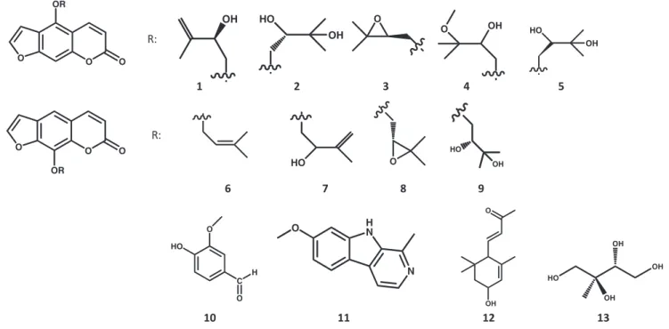
1 2 3 4 5
R:
6 7 8 9
O O O
OR
O
O O O
OR
O HO
C O
H
10 11 12 13 H
N
N O
HO
OH OH
OH HO
OH
OH O
OH OH HO
HO
HO
O OH OH
O
Figure 1. Secondary metabolites isolated fromDucrosia anethifolia.
(thiazolyl blue tetrazolium bromide, Sigma, St. Louis, MO) (from a 5 mg/mL stock) was added to each well. After incubation at 37C for 4 h, 100lL of sodium dodecyl sulphate (SDS, Sigma, St. Louis, MO) solution (10% in 0.01 M HCl) was added to each well and the plates were further incubated at 37C overnight.
The cell growth was determined by measuring the OD at 540 nm (ref. 630 nm) with a Multiscan EX ELISA reader (Thermo Labsystems, Waltham, MA). IC50 values were calculated via the following equation:
IC50 ¼ 100 ODsampleODmediumcontrol
ODcellcontrolODmediumcontrol
100
Assay for cytotoxic effect
The effects of increasing concentrations of compounds on cell growth were tested in 96-well flat-bottomed microtiter plates (Poljarevic et al.2018). The compounds were diluted in a volume of 100lL medium. Then, 1104 cells in 100lL of medium were added to each well, with the exception of the medium con- trol wells. In case of NIH/3T3 cells, the compounds were added after seeding the cells at 37C overnight. The culture plates were incubated at 37C for 24 h; at the end of the incubation period, 20lL of MTT solution (from a 5 mg/mL stock) was added to each well. After incubation at 37C for 4 h, 100lL of SDS solu- tion (10% in 0.01 M HCl) was added to each well and the plates were further incubated at 37C overnight. Cell growth was determined by measuring the optical density (OD) at 540 nm (ref. 630 nm) with a Multiscan EX ELISA reader. Inhibition of the cell growth was determined according to the formula:
IC50 ¼ 100 ODsampleODmediumcontrol
ODcellcontrolODmediumcontrol
100 Results are expressed in terms of IC50, defined as the inhibi- tory dose that reduces by a 50% the growth of the cells exposed to the tested compound.
Assay for multidrug resistance reversing activity
The inhibition of the cancer MDR efflux pump ABCB1 by the tested compounds was evaluated using flow cytometry measuring the retention of rhodamine 123 by ABCB1 (P-glycoprotein) in MDR mouse T-lymphoma cells, as the L5178Y human ABCB1- gene transfected mouse T-lymphoma cell line (MDR) overex- presses P-glycoprotein (Domınguez-Alvarez et al. 2016). This method is a fluorescence-based detection system which uses ver- apamil as reference inhibitor. Briefly, cell number of L5178Y MDR and PAR cell lines was adjusted to 2106 cells/mL, re-suspended in serum-free McCoy’s 5A medium and distributed in 0.5 mL aliquots into Eppendorf centrifuge tubes. The tested compounds were added at different concentrations and the sam- ples were incubated for 10 min at room temperature. Verapamil (Sigma, St. Louis, MO) and tariquidar (Sigma, St. Louis, MO) were applied as positive controls. Next, 10mL (5.2mM final con- centration) of the fluorochrome and ABCB1 substrate rhodamine 123 (Sigma, St. Louis, MO) were added to the samples and the cells were incubated for 20 min at 37C, washed twice and re- suspended in 0.5 mL PBS for analysis. The fluorescence of the cell population was measured with a Partec CyFlowVR flow cytometer (Partec, G€orlitz, Germany). The percentage of mean fluorescence intensity was calculated for the treated MDR cells as compared with the untreated cells. A fluorescence activity ratio
(FAR) was calculated based on the following equation which relates the measured fluorescence values:
FAR ¼ MDRtreated=MDRcontrol
parentaltreated=parentalcontrol
The results obtained from a representative flow cytometry experiment in which 20,000 individual cells of the population were evaluated for amount of rhodamine 123 retained with the aid of the Partec CyFlowVR flow cytometer, are first presented by the histograms and these data converted to FAR units that define fluorescence intensity, standard deviation, peak channel in the total- and in the gated-populations. Parameters calculated are:
forward scatter (FSC, forward scatter count of cells in the sam- ples or cell size ratio); side scatter (SSC, side scatter count of cells in the samples); FL-1 (mean fluorescence intensity of the cells) and FAR, whose values were calculated using the equation given above.
Checkerboard combination assay
A checkerboard microplate method was applied to study the effect of drug interactions between furocoumarins and the che- motherapeutic drug doxorubicin (Takacs et al. 2015). This assay was carried out using multidrug resistant mouse T-lymphoma cells overexpressing the ABCB1 transporter. Doxorubicin is in the class of anthracycline antitumor agents, and it exerts anti- cancer activity as a topoisomerase-II (TI-2) inhibitor. The dilu- tions of doxorubicin (Teva, Debrecen, Hungary, stock solution:
2 mg/mL) were made in a horizontal direction in 100lL (final concentration: 17.242lM), and the dilutions of the test com- pounds vertically in the microtiter plate in 50lL volume. The cells were re-suspended in McCoy’s 5A culture medium and dis- tributed into each well in 50lL containing 6103 cells each.
The plates were incubated for 72 h at 37C in 5% CO2 atmos- phere. The cell growth rate was determined after MTT staining.
At the end of the incubation period, 20lL of MTT solution (from a stock solution of 5 mg/mL) was added to each well.
After incubation at 37C for 4 h, 100lL of SDS solution (10% in 0.01 M HCl) was added to each well and the plates were further incubated at 37C overnight. Optical density was measured at 540/630 nm with Multiscan EX ELISA reader (Thermo Labsystems, Waltham, MA) as described elsewhere (Takacs et al.
2015). Combination index (CI) values at 50% of the growth inhibition dose (ED50) were determined using CompuSyn soft- ware (ComboSyn, Inc., Paramus, NJ) to plot four to five data points to each ratio. CI values were calculated by means of the median-effect equation, where CI <1, CI ¼1 and CI >1 repre- sent synergism, additive effect (or no interaction) and antagon- ism, respectively (Chou and Martin2005; Chou2010).
Results
Isolated compounds
Repeated column chromatography of the bioactive fractions resulted in the isolation of 13 compounds. The compounds were identified by careful interpretation of NMR data and comparison of 1H and 13C chemical shifts with those reported in literature.
Nine linear furocoumarin derivatives, namely pabulenol(1) (Sbai et al. 2016), (þ)-oxypeucedanin hydrate (aviprin) (2) (Sbai et al.
2016), oxypeucedanin (3) (Sbai et al. 2016), oxypeucedanin methanolate (4) (Fujioka et al.1999), (–)-oxypeucedanin hydrate (prangol) (5) (Rahimifard et al. 2018), imperatorin (6) (Lv et al.
PHARMACEUTICAL BIOLOGY 661
2013), isogospherol (7) (Macias et al. 1990), heraclenin (8) (Poonkodi2016), heraclenol (9) (Harkar et al.1984); along with vanillic aldehyde (10) (Chung et al. 2011), harmine (11), 3-hydroxy-a-ionone (12) and 2-C-methyl-erythrytol (13) were identified (1H NMR spectra see inSupporting Information). The diastereomers (þ)-oxypeucedanin hydrate and ()-oxypeuceda- nin hydrate were distinguished by determining their optical rota- tions and comparing with literature (Atkinson et al.1974).
The 1H and 13C NMR spectral data of 11, 12 and 13 in CD3OD are reported here for the first time. Compound11(har- mine): 1H NMR (500 MHz, CD3OD) d¼8.11 (H-3, d, J¼ 5.5 Hz), 8.02 (H-5, d,J¼ 8.7 Hz), 7.86 (H-4, d, J¼ 5.5 Hz), 7.06 (H-8, d,J¼ 1.9 Hz), 6.89 (H-6, dd,J¼ 8.7 Hz, 1.9 Hz), 3.92 (s, 7-OCH3), 2.80 (s, 1-CH3); 13C NMR (125 MHz, CD3OD) d¼162.9, 144.6, 141.6, 137.0, 136.2, 130.7, 123.7, 116.3, 113.5, 111.4, 95.4, 56.0, 19.1. Compound 12 (3-hydroxy-a-ionone): 1H NMR (500 MHz, CD3OD) d¼6.67 (H-7, dd, J¼ 15.8 Hz, 10.3 Hz), 6.13 (H-8, d,J¼ 15.8 Hz), 5.60 (H-4, br s), 4.22 (H-3, br s), 2.58 (H-6, d,J¼ 10.3 Hz), 2.27 (H3-10, s), 1.80 (H-2b, dd, J¼ 13.2 Hz, 5.9 Hz), 1.63 (H3-13, s), 1.38 (H-2a, dd, J¼ 13.2 Hz, 7.2 Hz), 1.01 (H3-11, s), 0.90 (H3-12, s); 13C NMR (125 MHz, CD3OD) d¼200.8, 149.8, 135.9, 134.7, 127.3, 65.9,
55.6, 45.0, 35.0, 29.8, 27.1, 24.5, 22.8. Compound 13 (2-C- methyl-erythrytol):1H NMR (500 MHz, CD3OD)d¼3.80 (H-4a, dd,J¼ 10.4 Hz, 2.5 Hz), 3.61 (H-3, m), 3.59 (H-4b, m), 3.52 (H- 1a, d,J¼ 11.1 Hz), 3.44 (H-1b, d,J¼ 11.1 Hz), 1.11 (2-CH3, s);
13C NMR (125 MHz, CD3OD)d¼76.2, 75.0, 68.5, 63.8, 19.7.
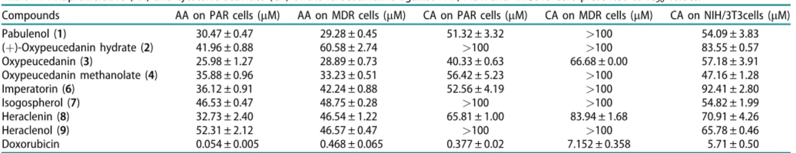
Antiproliferative and cytotoxic activities on cancer cell lines Furocoumarins isolated from D. anethifolia were subjected to bioassay for cytotoxic and antiproliferative activity against cancer cell lines. All compounds exerted potent antiproliferative effect on sensitive and resistant mouse T-lymphoma cells (Table 1).
However, they did not show any selectivity towards the resistant cell line. The most potent compound was oxypeucedanin on both cell lines. Some compounds had no toxic effects ((þ)-oxy- peucedanin hydrate (2), heraclenol (9), isogospherol (7)); fur- thermore, pabulenol (1), oxypeucedanin methanolate (4) and imperatorin (6) were more toxic on the sensitive PAR cell line (IC50 between 52 and 57mM) without any toxicity on MDR cells (Table 1). Oxypeucedanin (3) and heraclenin (8) exhibited cyto- toxic activity; however, they were more potent on the sensitive PAR cell line (Table 1). The cytotoxic activity of furocoumarins was assessed using NIH/3T3 normal murine fibroblast cells.
Some compounds showed slight toxic effect on normal fibro- blasts, namely (þ)-oxypeucedanin hydrate (2), heraclenol (4) and isogospherol (8) with IC50 values of 83.55, 65.78 and 54.82 mM, respectively. Pabulenol (1) possessed similar activity on fibroblast and parental mouse lymphoma cells. In addition, oxypeucedanin (3), oxypeucedin methanolate (5) and heraclenin (9) exhibited mild toxicity on fibroblasts and parental lymphoma cells.
Imperatorin (7) had no toxic activity on fibroblasts.
Multidrug resistance reversing activity
Regarding the efflux pump inhibiting activity of the compounds on ABCB1 overexpressing MDR mouse T-lymphoma cells, only oxypeucedanin (3) showed moderate ABCB1 inhibiting effect (FAR: 2.22); however, this inhibition was lower than in case of the positive controls tariquidar (FAR: 100) and verapamil (FAR:
8.2) (Table 2, figures see inSupporting Information).
Combination assay results on MDR cells
The two most promising compounds in the previous assays were investigated in combination with the standard chemotherapeutic drug doxorubicin. The compounds oxypeucedanin (3) and hera- clenin (8) showed slight synergistic effect with doxorubicin, for this reason, they might be potential adjuvants in combined chemotherapy applying standard anticancer drugs with com- pounds that can act synergistically (Table 3).
Table 1. Antiproliferative (AA) and cytotoxic activities (CA) of the furocoumarins against PAR, MDR and NIH/3T3 cells presented as IC50values.
Compounds AA on PAR cells (mM) AA on MDR cells (mM) CA on PAR cells (mM) CA on MDR cells (mM) CA on NIH/3T3cells (mM)
Pabulenol (1) 30.47 ± 0.47 29.28 ± 0.45 51.32 ± 3.32 >100 54.09 ± 3.83
(þ)-Oxypeucedanin hydrate (2) 41.96 ± 0.88 60.58 ± 2.74 >100 >100 83.55 ± 0.57
Oxypeucedanin (3) 25.98 ± 1.27 28.89 ± 0.73 40.33 ± 0.63 66.68 ± 0.00 57.18 ± 3.91
Oxypeucedanin methanolate (4) 35.88 ± 0.96 33.23 ± 0.51 56.42 ± 5.23 >100 47.16 ± 1.28
Imperatorin (6) 36.12 ± 0.91 42.24 ± 0.88 52.56 ± 4.19 >100 92.41 ± 2.80
Isogospherol (7) 46.53 ± 0.47 48.75 ± 0.28 >100 >100 54.82 ± 1.99
Heraclenin (8) 32.73 ± 2.40 46.54 ± 1.22 65.81 ± 1.00 83.94 ± 1.68 70.91 ± 4.26
Heraclenol (9) 52.31 ± 2.12 46.57 ± 0.47 >100 >100 65.78 ± 0.46
Doxorubicin 0.054 ± 0.005 0.468 ± 0.065 0.377 ± 0.02 7.152 ± 0.358 5.71 ± 0.50
Data were expressed as mean ± standard deviation (n¼3). Different letters represent significant differences (p<0.05).
Table 2. Efflux pump inhibiting activities of furocoumarins.
Samples Conc.lM FSC SSC FL-1 FAR
PAR – 2069 658 98.20 –
MDR – 2152 725 1.79 –
MDR mean 1.182 –
Tariquidar 0.02 2156 719 119 100.68
Verapamil 20 2143 740 9.69 8.20
Pabulenol (1) 2 2324 728 0.596 0.82
20 2323 750 0.544 0.75
(þ)-Oxypeucedanin hydrate (2) 2 2325 725 0.715 0.98
20 2305 769 0.583 0.80
Oxypeucedanin (3) 2 2190 749 0.9 0.76
20 2164 763 2.62 2.22
Oxypeucedanin methanolate (4) 2 2310 737 0.58 0.80
20 2290 766 0.499 0.68
Imperatorin (6) 2 2165 749 0.727 0.62
20 2161 750 0.783 0.66
Isogospherol (7) 2 2305 741 0.531 0.73
20 2326 741 0.548 0.75
Heraclenin (8) 2 2318 723 0.942 1.29
20 2294 764 0.57 0.78
Heraclenol (9) 2 2300 742 0.995 1.37
20 2317 740 0.348 1.30
DMSO 2% (V/V) 2308 762 0.497 0.68
MDR – 2301 746 0.535 –
Table 3.Checkerboard combination assay of selected compounds with doxorubicin.
Compound Best ratio CI at ED50 Interaction SD (±) Oxypeucedanin 1:50 0.85537 Slight synergism 0.07800
Heraclenin 4:100 0.88955 Slight synergism 0.06334
Discussion
Chromatographic separation of the extract ofD. anethifoliaherbs resulted in the isolation of 13 compounds, among them were nine furocoumarins. Compounds 2, 3, 5, 7, 10–13 were identi- fied for the first time fromDucrosiagenus.
The tested furocoumarins exerted antiproliferative effects on sensitive and resistant mouse T-lymphoma cells with no selectiv- ity towards the resistant cell line. This is the first comprehensive analysis of this plant and its furocoumarins on these cells.
Oxypeucedanin (3) had the most remarkable activity on both cell lines. The most effective furocoumarins, oxypeucedanin (3) and heraclenin (9) exhibited marginal toxicity on normal fibroblast cells and sensitive parental mouse lymphoma cells; furthermore, they were less toxic on multidrug resistant lymphoma cells.
From the tested compounds, only oxypeucedanin showed moder- ate MDR reversing activity. In the checkerboard assay, oxypeuce- danin and heraclenin showed slight synergistic effect with doxorubicin. These compounds might improve the cytotoxic effect of the standard chemotherapeutic drug doxorubicin.
Disclosure statement
The authors declare no conflict of interest.
Funding
This work was supported by the National Research, Development and Innovation Office (OTKA K115796), Economic Development and Innovation Operative Programme GINOP-2.3.2-15-2016-00012, EFOP 3.6.3-VEKOP-16-2017-00009 and Janos Bolyai Research Scholarship of the Hungarian Academy of Sciences.
References
Abbasnejad M, Mostafavi A, Kooshki R, Hamzenejad P, Esmaeili-Mahani S.
2017. Effect of Ducrosia anethifolia (DC.) Boiss essential oil on spatial learning and memory in rats. J Gorgan Univ Med Sci. 18:9–15.
Amiri MS, Joharchi MR. 2016. Ethnobotanical knowledge of Apiaceae family in Iran: a review. Avicenna J Phytomed. 6:621–635.
Appendino G, Bianchi F, Bader A, Campagnuolo C, Fattorusso E, Taglialatela-Scafati O, Blanco-Molina M, Macho A, Fiebich BL, Bremner P. 2004. Coumarins fromOpopanax chironium. New dihydrofuranocou- marins and differential induction of apoptosis by imperatorin and heracle- nin. J Nat Prod. 67:532–536.
Asgari Nematian M, Yaghmaei P, Mohammadi S. 2017. Assessment of the antinociceptive, antiinflammatory and acute toxicity effects of Ducrosia anethifoliaessential oil in mice. Sci J Kurdistan Univ Med Sci. 22:74–84.
Ashraf M, Karim A, Bushra B. 1979. Studies on the essential oils of the Pakistani species of the family Umblliferae. Pak J Sci Ind Res. 22:252–254.
Atkinson E, Boyd DR, Grundon MF. 1974. Coumarins ofSkimmia japonica.
Phytochemistry. 13:853–855.
Aynehchi Y. 1991. Materia medica and Iranian medicinal plants. Tehran:
Tehran University Publications.
Bartnik M, Sławinska-Brych A,Zurek A, Kandefer-Szersze_ n M, Zdzisinska B.
2017. 8-methoxypsoralen reduces AKT phosphorylation, induces intrinsic and extrinsic apoptotic pathways, and suppresses cell growth of SK-N-AS neuroblastoma and SW620 metastatic colon cancer cells. J Ethnopharmacol. 207:19–29.
Chou T, Martin N. 2005. A computer program for quantitation of synergism and antagonism in drug combinations, and the determination of IC50and ED50 and LD50 values. CompuSyn for drug combinations: PC software and user’s guide. Paramus: ComboSynInc.
Chou TC. 2010. Drug combination studies and their synergy quantification using the Chou-Talalay method. Cancer Res. 70:440–446.
Chung CP, Hsia SM, Lee MY, Chen HJ, Cheng F, Chan LC, Kuo YH, Lin YL, Chiang W. 2011. Gastroprotective activities of adlay (Coix lachryma- jobiL. var.ma-yuenStapf) on the growth of the stomach cancer AGS cell
line and indomethacin-induced gastric ulcers. J Agric Food Chem. 59:
6025–6033.
Domınguez-Alvarez E, Gajdacs M, Spengler G, Palop JA, Marc MA, Kiec- Kononowicz K, Amaral L, Molnar J, Jacob C, Handzlik J, et al. 2016.
Identification of selenocompounds with promising properties to reverse cancer multidrug resistance. Bioorg Med Chem Lett. 26:2821–2824.
Fujioka T, Furumi K, Fujii H, Okabe H, Mihashi K, Nakano Y, Matsunaga H, Katano M, Mori M. 1999. Antiproliferative constituents from Umbelliferae plants. V. A new furanocoumarin and falcarindiol furano- coumarin ethers from the root of Angelica japonica. Chem Pharm Bull.
47:96–100.
Ghahreman A. 1993. Flora of Iran/Flore de l’Iran, vol. 12. Tehran: Research Institute of Forests and Rangelands.
Habibi H, Ghahtan N, Kohanmoo MA, Eskandari F. 2017. Research in molecular medicine chemical composition and antibacterial effect of medi- cinal plants against some food-borne pathogens. Res Mol Med. 5:14–21.
Haghi G, Safaei A, Safari J. 2004. Extraction and determination of the main components of the essential oil ofDucrosia anethifolia by GC and GC/
MS. Iran J Pharm Res. 3:90–99.
Hajhashemi V, Rabbani M, Ghanadi A, Davari E. 2010. Evaluation of antian- xiety and sedative effects of essential oil ofDucrosia anethifoliain mice.
Clinics (Sao Paulo, Brazil). 65:1037–1042.
Harkar S, Razdan TK, Waight ES. 1984. Steroids, chromone and coumarins fromAngelica officinalis. Phytochemistry. 23:419–426.
Janssen AM, Scheffer JJ, Baerheim Svendsen A, Aynehchi Y. 1984. The essen- tial oil of Ducrosia anethifolia (DC.) Boiss. Chemical composition and antimicrobial activity. Pharm Weekblad Sci Ed. 6:157–160.
Javidnia K, Miri R, Assadollahi M, Gholami M, Ghaderi M. 2009. Screening of selected plants growing in Iran for antimicrobial activity. Iran J Sci Technol Trans A. 33:329–333.
Kaewpiboon C, Surapinit S, Malilas W, Moon J, Phuwapraisirisan P, Tip- Pyang S, Johnston RN, Koh SS, Assavalapsakul W, Chung YH. 2014.
Feroniellin A-induced autophagy causes apoptosis in multidrug-resistant human A549 lung cancer cells. Int J Oncol. 44:1233–1242.
Karami A, Bohlooli A. 2017. Essential oil chemical diversity of Ducrosia anethifolia (DC.) Boiss. accessions from Iran. J Essent Oil Bear Pl. 20:
1342–1348.
Khalid B, Hamid S, Liaqat L, Khan JI. 2009. Seed oils of Pakistani wild spe- cies of Umbelliferae family: Ducrosia anethifolia, Bunium persicum, Bunium cylindricumandAmmi majus; as potential industrial raw material.
Pak J Sci Ind Res. 52:260–263.
Kim Y-K, Kim YS, Ryu SY. 2007. Antiproliferative effect of furanocoumarins from the root ofAngelica dahurica on cultured human tumor cell lines.
Phytother Res. 21:288–290.
Kubrak T, Bogucka-kocka A, Komsta Ł, Za D, Bogucki J, Galkowski D, Kaczmarczyk R, Feldo M, Cioch M, Kocki J. 2017. Modulation of multi- drug resistance gene expression by coumarin derivatives in human Leukemic cells. Oxid Med Cell Longev. 2017:1–13.
Luo KW, Sun JG, Chan JYW, Yang L, Wu SH, Fung KP, Liu FY. 2011.
Anticancer effects of imperatorin isolated fromAngelica dahurica: induc- tion of apoptosis in HepG2 cells through both death-receptor- and mito- chondria-mediated pathways. Chemotherapy. 57:449–459.
Lv X, Liu D, Hou J, Dong P, Zhan L, Wang L, Deng S, Wang C, Yao J, Shu X, et al. 2013. Biotransformation of imperatorin byPenicillium janthinel- lum. Anti-osteoporosis activities of its metabolites. Food Chem. 138:
2260–2266.
Macias FA, Massanet GM, Rodriguez-Luis F, Salva J. 1990.13C NMR of cou- marins. Magn Reson Chem. 28:219–222.
Mahboubi M, Feizabadi MM. 2009. Antimicrobial activity of Ducrosia anethifoliaessential oil and main component, decanal against methicillin- resistant and methicillin-susceptible Staphylococcus aureus. J Essent Oil Bear Pl. 12:574–579.
Mazloomifar A, Valian M. 2015. GC–MS analysis of the leaves essential oil of Ducrosia anethifolia (DC.) Boiss. obtained with three extractions.
J Essent Oil Bear Pl. 18:904–907.
Mirzaei SA, Gholamian Dehkordi N, Ghamghami M, Amiri AH, Dalir Abdolahinia E, Elahian F. 2017. ABC-transporter blockage mediated by xanthotoxin and bergapten is the major pathway for chemosensitization of multidrug-resistant cancer cells. Toxicol Appl Pharmacol. 337:22–29.
Morgan AMA, Kim JH, Lee HW, Lee SH, Lim CH, Jang HD, Kim YH. 2015.
Phytochemical constituents from the aerial part ofDucrosia ismaelisAsch.
Nat Prod Sci. 21:6–13.
Mostafavi A, Afzali D, Mirtadzadini SM. 2008. Chemical composition of the essential oil ofDucrosia anethifolia(DC.) Boiss. from Kerman province in Iran. J Essent Oil Res. 20:509–512.
Mottaghipisheh J, Maghsoudlou MT, Valizadeh J, Arjomandi R. 2014.
Antioxidant activity and chemical composition of the essential oil of PHARMACEUTICAL BIOLOGY 663
Ducrosia anethifolia(DC.) Boiss. from Neyriz. J Med Plants By-Prod. 3:
215–218.
Mozaffarian V. 1996. A dictionary of Iranian plant names. Tehran: Farhang Moaser.
Nyasty F, Oryan S, Sofiabadi M, Eslimi Esfahani D. 2017. Effect of intraperi- toneal injection of hydroalcoholic extract ofDucrosia anethifoliaon penty- lenetetrazol-induced anticonvulsion in male Wistar rats. Horizon Med Sci.
23:49–53.
Poljarevic JM, Tamas Gal G, May NV, Spengler G, D€om€ot€or O, Savic AR, Grguric-Sipka S, EnyedyEA. 2018. Comparative solution equilibrium and structural studies of half-sandwich ruthenium(II)(g6-toluene) complexes of picolinate derivatives. J Inorg Biochem. 181:74–85.
Poonkodi K. 2016. Phytoconstituents fromRichardia scabraL. and its bio- logical activities. Asian J Pharm Clin Res. 9:1–4.
Queslati MH, Bouajila J, Belkacem MA, Harrath AH, Alwasel SH, Ben Jannet H. 2017. Cytotoxicity of new secondary metabolites, fatty acids and tocols composition of seeds ofDucrosia anethifolia(DC.) Boiss. Nat Prod Res.
6419:1–7.
Rahimi N, Samani Jahromi E, Zolghadri Jahromi S. 2016. The effect of the hydro-alcoholic extract of Ducrosia anethifolia on testosterone hormone and the histological changes of the testicle in male adult rats. Armaghane- danesh. 21:682–693.
Rahimifard M, Manayi A, Baeeri M, Gholami M, Saeidnia S, Abdollahi M.
2018. Investigation of b-sitosterol and prangol extracted from Achillea tenoifoliaalong with whole root extract on isolated rat pancreatic islets.
Iran J Pharm Res. 17:317–325.
Sbai H, Saad I, Ghezal N, Greca M, Della, Haouala R. 2016. Bioactive com- pounds isolated fromPetroselinum crispumL. leaves using bioguided frac- tionation. Ind Crops Prod. 89:207–214.
Sefidkon F, Javidtash I. 2002. Essential oil composition ofDucrosia anethifo- lia(DC.) Boiss. from Iran. J Essent Oil Res. 14:278–279.
Shahabipour S, Firuzi O, Asadollahi M, Faghihmirzaei E, Javidnia K. 2013.
Essential oil composition and cytotoxic activity of Ducrosia anethifolia andDucrosia flabellifoliafrom Iran. J Essent Oil Res. 25:160–163.
Shahat AA, Ibrahim AY, Alsaid MS. 2015. Antioxidant capacity and poly- phenolic content of seven Saudi Arabian medicinal herbs traditionally used in Saudi Arabia. Indian J Tradit Know. 14:28–35.
Shalaby NMM, Abd-Alla HI, Aly HF, Albalawy MA, Shaker KH, Bouajila J.
2014. Preliminaryin vitroandin vivoevaluation of antidiabetic activity of Ducrosia anethifolia Boiss. and its linear furanocoumarins. Biomed Res Int. 2014:1–13.
Shokri H, Hekmatpou D, Ebrahimi Fakhar HR, Nyazi A, Azadi M, Taghizadeh M. 2013. Effect of Ducrosia anethifolia (Barilax) on anxiety after acute myocardial infarction. Arak Med Univ J. 16:28–34.
Stavri M, Mathew KT, Bucar F, Gibbons S. 2003. Pangelin, an antimycobac- terial coumarin fromDucrosia anethifolia. Planta Med. 69:956–959.
Syed M, Iqbal MJ, Chaudhary FM, Bhatty MK. 1987. Antimicrobial activity of essential oils of Umbelliferae family. Part VI.Stewartiella baluchistan- ica,Penstemon canescensandDucrosia anethifollia. Pak J Sci Ind Res. 30:
595–598.
Takacs D, CsonkaA, Horv ath A, Windt T, Gajd acs M, Riedl Z, Hajos G, Amaral L, Molnar J, Spengler G. 2015. Reversal of ABCB1-related multi- drug resistance of colonic adenocarcinoma cells by phenothiazines.
Anticancer Res. 35:3245–3251.
Vazirzadeh A, Dehghan F, Kazemeini R. 2017. Changes in growth, blood immune parameters and expression of immune related genes in rainbow trout (Oncorhynchus mykiss) in response to diet supplemented with Ducrosia anethifoliaessential oil. Fish Shellfish Immunol. 69:164–172.
Zamyad M, Abasnejad M, Esmaeili-Mahani S, Mostafavi A. 2016. Alpha- pinene as the main component ofDucrosia anethifolia(Boiss) essential oil is responsible for its effect on locomotor activity in rats. Avicenna J Neuro Psych Physio. 3:e38787.