Review
Effects of Cardiovascular Risk Factors on Cardiac STAT3
Márton Pipicz, Virág Demján, Márta Sárközy and Tamás Csont *
Metabolic Diseases and Cell Signaling (MEDICS) Research Group, Department of Biochemistry, Interdisciplinary Excellence Centre, University of Szeged, Dóm tér. 9., H-6720 Szeged, Hungary;
pipicz.marton@med.u-szeged.hu (M.P.); demjan.virag@med.u-szeged.hu (V.D.);
sarkozy.marta@med.u-szeged.hu (M.S.)
* Correspondence: csont.tamas@med.u-szeged.hu; Tel.: +36-62-545-096
Received: 7 September 2018; Accepted: 8 November 2018; Published: 12 November 2018
Abstract:Nuclear, mitochondrial and cytoplasmic signal transducer and activator of transcription 3 (STAT3) regulates many cellular processes, e.g., the transcription or opening of mitochondrial permeability transition pore, and its activity depends on the phosphorylation of Tyr705 and/or Ser727 sites. In the heterogeneous network of cardiac cells, STAT3 promotes cardiac muscle differentiation, vascular element formation and extracellular matrix homeostasis. Overwhelming evidence suggests that STAT3 is beneficial for the heart, plays a role in the prevention of age-related and postpartum heart failure, protects the heart against cardiotoxic doxorubicin or ischaemia/reperfusion injury, and is involved in many cardioprotective strategies (e.g., ischaemic preconditioning, perconditioning, postconditioning, remote or pharmacological conditioning). Ischaemic heart disease is still the leading cause of death worldwide, and many cardiovascular risk factors contribute to the development of the disease. This review focuses on the effects of various cardiovascular risk factors (diabetes, aging, obesity, smoking, alcohol, depression, gender, comedications) on cardiac STAT3 under non-ischaemic baseline conditions, and in settings of ischaemia/reperfusion injury with or without cardioprotective strategies.
Keywords:STAT family; comorbidity; myocardial infarction; coronary artery disease; cardioprotection;
mPTP; JAK
1. Introduction
Signal transducer and activator of transcription 3 (STAT3) has a central role in transmitting extracellular signals from the plasma membrane to the nucleus and mitochondria, where it influences transcription and mitochondrial function, thereby regulating diverse biological processes.
1.1. Structure of STAT3
The protein encoded by mammalian STAT3 gene contains six functional domains: N-terminal, coiled-coil, DNA-binding, linker, SH2 and C-terminal transactivation domains [1]. There are two phosphorylation sites to activate STAT3: a tyrosine residue on the SH2 domain (Tyr705) and a serine residue on the transactivation domain (Ser727). As a result of alternative splicing two major isoforms exist: the abundant STAT3alpha (770 amino acids) and less abundant STAT3beta (722 amino acids) lacking C-terminal transactivation domain and Ser727 phosphorylation site [1].
1.2. Signalling
A wide variety of extracellular polypeptide ligands like interleukin-(IL)-6-family, leukaemia inhibitory factor, oncostatin M, ciliary neurotrophic factor, and cardiotrophin acts on plasma membrane
Int. J. Mol. Sci.2018,19, 3572; doi:10.3390/ijms19113572 www.mdpi.com/journal/ijms
Int. J. Mol. Sci.2018,19, 3572 2 of 29
receptors and activate STAT3, predominantly via the glycoprotein-130-Janus kinase (gp-130-JAK) axis [2]. Ligand binding by the receptor complex leads to activation of receptor-associated JAKs, thereby activating STAT3 directly and/or non-directly via the extracellular signal-regulated kinase 1 and 2 (ERK1/2) pathway (Figure1).
Int. J. Mol. Sci. 2018, 19, x FOR PEER REVIEW 2 of 29
1.2. Signalling
A wide variety of extracellular polypeptide ligands like interleukin-(IL)-6-family, leukaemia inhibitory factor, oncostatin M, ciliary neurotrophic factor, and cardiotrophin acts on plasma membrane receptors and activate STAT3, predominantly via the glycoprotein-130-Janus kinase (gp-130-JAK) axis [2]. Ligand binding by the receptor complex leads to activation of receptor-associated JAKs, thereby activating STAT3 directly and/or non-directly via the extracellular signal-regulated kinase 1 and 2 (ERK1/2) pathway (Figure 1).
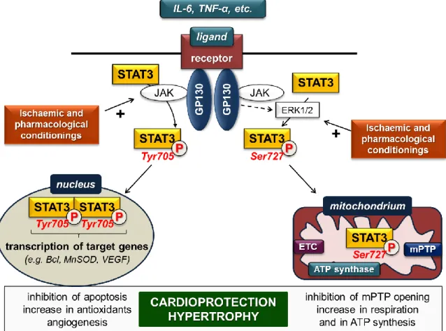
Figure 1. STAT3 signalling in cardiac myocytes in settings of ischaemia/reperfusion. ETC: electron transport chain; GP: glycoprotein; JAK: Janus kinase; mPTP: mitochondrial permeability transition pore; MnSOD: manganese-dependent superoxide dismutase; VEGF: vascular endothelial growth factor. (P in circle represents phosphorylated STAT3 forms; dashed arrow indicates several steps;
plus (+) sign represents activation).
Direct activation occurs once gp-130 is phosphorylated by JAK and provides binding sites for the SH2 domain of STAT3. After recruitment of STAT3, it is phosphorylated and activated by JAKs on Tyr705. Subsequently, STAT3 dissociates from the receptor complex, forms homo- or heterodimers with other STAT proteins, and translocates into the nucleus, where it binds to consensus DNA sequences (so-called gamma-interferon-activated sites (GAS)) and finally initiates transcription of target genes [2]. STAT3 enhances expression of several genes encoding, for instance, anti-apoptotic (e.g., Bcl-xl, MCL-1), anti-oxidant (e.g., MnSOD, metallothionein) and pro-angiogenic (e.g., VEGF) proteins.
The gp-130-JAK axis simultaneously activates ERK1/2, which in turn phosphorylates the Ser727 site of STAT3 monomers and/or Tyr705-phosphorylated STAT3 dimers to influence the dimer’s transcriptional activity. Phosphorylation of STAT3 monomers on Ser727 leads to translocation into the mitochondria without dimerization [3,4]. In the mitochondria, Ser727-STAT3 interacts with complex I of the electron transport chain, the mediator of mitochondrial permeability transition pore
Figure 1.STAT3 signalling in cardiac myocytes in settings of ischaemia/reperfusion. ETC: electron transport chain; GP: glycoprotein; JAK: Janus kinase; mPTP: mitochondrial permeability transition pore; MnSOD: manganese-dependent superoxide dismutase; VEGF: vascular endothelial growth factor.
(P in circle represents phosphorylated STAT3 forms; dashed arrow indicates several steps; plus (+) sign represents activation).
Direct activation occurs once gp-130 is phosphorylated by JAK and provides binding sites for the SH2 domain of STAT3. After recruitment of STAT3, it is phosphorylated and activated by JAKs on Tyr705. Subsequently, STAT3 dissociates from the receptor complex, forms homo- or heterodimers with other STAT proteins, and translocates into the nucleus, where it binds to consensus DNA sequences (so-called gamma-interferon-activated sites (GAS)) and finally initiates transcription of target genes [2].
STAT3 enhances expression of several genes encoding, for instance, anti-apoptotic (e.g., Bcl-xl, MCL-1), anti-oxidant (e.g., MnSOD, metallothionein) and pro-angiogenic (e.g., VEGF) proteins.
The gp-130-JAK axis simultaneously activates ERK1/2, which in turn phosphorylates the Ser727 site of STAT3 monomers and/or Tyr705-phosphorylated STAT3 dimers to influence the dimer’s transcriptional activity. Phosphorylation of STAT3 monomers on Ser727 leads to translocation into the mitochondria without dimerization [3,4]. In the mitochondria, Ser727-STAT3 interacts with complex I of the electron transport chain, the mediator of mitochondrial permeability transition pore (mPTP) cyclophilin D, mitochondrial DNA and may have an impact on complex II and complex V (ATP synthase). Therefore, mitochondrial STAT3 influences ATP synthesis, the opening of mPTP, production of reactive oxygen species and mitochondrial transcription [3,4].
Besides classic ligands, STAT3 signalling is activated or modulated by other endogenous or exogenous peptides including insulin, leptin, angiotensin II, erythropoietin, granulocyte stimulating factor, etc.
1.3. STAT3 in the Heart
STAT3 is expressed in different cell types of the heart such as cardiomyocytes, cardiac fibroblasts, endothelial cells, smooth muscle cells, inflammatory cells and cardiac neurons. In the heterogeneous network of cardiac cells STAT3 regulates cell-to-cell communication (for review, see [5]). For instance, STAT3 promotes cardiac muscle differentiation [6] and vascular element formation in the heart [7], regulatesβ-adrenergic functions [8] and extracellular matrix homeostasis [9]. Overwhelming evidence suggests that STAT3 is beneficial for the heart [10]. STAT3 transduces hypertrophic signals under physiological (e.g., pregnancy) and pathophysiological (e.g., pressure overload) remodelling [11], plays a role in the prevention of age-related and postpartum heart failure, and protects the heart against cardiotoxic doxorubicin and ischaemic injury [2].
Ischaemic heart disease is still a major cause of death and disability worldwide; therefore, cardioprotective strategies and revealing of signalling are needed. Ischaemic conditioning is a manoeuvre for protecting the heart against the detrimental effects of ischaemia/reperfusion injury by means of the application of brief non-harmful ischaemia/reperfusion cycles to elicit endogenous cardioprotective mechanisms. When the conditioning method is applied before, under or after the prolonged lethal ischaemia, it is called ischaemic preconditioning, perconditioning or postconditioning, respectively. Research into the underlying molecular mechanisms of cardioprotection results in investigation of pharmacological agents that mimic the cardioprotective effect of ischaemic conditioning (pharmacological conditioning).
Several studies have reported that ischaemia enhances STAT3 phosphorylation [12–14], and activation of STAT3 protects the myocardium against ischaemia/reperfusion injury (Figure1, for review, see [15]). Moreover, STAT3 is involved in many cardioprotective ischaemic and pharmacological conditioning methods, for instance in ischaemic preconditioning [16], perconditioning [17] or postconditioning [18] (for review, see [19]).
Many cardiovascular risk factors contribute to the development of ischaemic heart disease and interfere with the cardioprotective effect of conditioning manoeuvres (for review, see [20]). Therefore, it is feasible to speculate that risk factors may unfavourably alter protective signalling mechanisms in the heart (e.g., reperfusion injury salvage kinase signalling or survivor activating factor enhancement pathways), thereby resulting in unwanted cardiac consequences. This review focuses on the effects of various cardiovascular risk factors (diabetes, aging, obesity, smoking, alcohol, depression, gender, comedications) on cardiac STAT3, a well-known cardioprotective signal molecule, under non-ischaemic baseline conditions and in settings of ischaemia/reperfusion injury with or without cardioprotective strategies. We systemically analysed phosphorylation (p-STAT3, at Tyr705 and Ser727 sites) and expression (total-STAT3) of cardiac STAT3. Furthermore, the ratio of p-STAT3 and total-STAT3 was considered as STAT3 activation.
2. Effect of Cardiovascular Risk Factors on Cardiac STAT3 under Non-Ischaemic Baseline Conditions
2.1. Diabetes
Diabetes mellitus is a condition characterized by an increased blood glucose level. It is one of the most prevalent metabolic diseases worldwide and is also associated with increased mortality. In 2017, an estimated 425 million people suffered from diabetes, a number that is expected to increase by 48%
within three decades according to the International Diabetes Federation [21]. Diabetic patients have a greater chance of developing cardiovascular complications including ischaemic heart disease such as the life-threatening acute myocardial infarction [22], since diabetes exerts harmful effects on the cardiovascular system, resulting in cardiomyopathy [23] and vasculopathy [24]. An explanation for
Int. J. Mol. Sci.2018,19, 3572 4 of 29
this may be that diabetes leads to molecular changes in the heart, e.g., marked alteration of cardiac gene expression profile in various experimental models [25–27].
Based on a literature review, the effect of diabetes on non-ischaemic baseline expression, phosphorylation or activation of cardiac STAT3 protein seems to be rather controversial. Several publications demonstrated a significant decrease in cardiac STAT3 phosphorylation and/or activation in various experimental models of diabetes (Table1). The first study to demonstrate this phenomenon was written by Wang and colleagues [28]. They induced type I diabetes in male Sprague-Dawley rats by streptozotocin (STZ) injection. After five weeks of diabetes induction, they found that myocardial STAT3 activation (at the Ser727 site) was lower in the diabetic group. Later the same research group confirmed these results in STZ-induced type I diabetes models [29,30] and demonstrated that phosphorylation and activation of STAT3 at Tyr705 was also decreased in diabetes [29–31]. Besides type I diabetes models, reduction of cardiac STAT3 expression was also described in a type II diabetes model, i.e., STAT3 mRNA was downregulated in non-obese Goto-Kakizaki rats [25]. Moreover, similar trends were observed in in vitro models of diabetes. In H9c2 cells subjected to high glucose conditions (25 mM glucose added to the medium), the non-ischaemic baseline STAT3 phosphorylation (Tyr705 and/or Ser727) and activation was significantly decreased [29,32]. Similarly, exposure of isolated adult rat ventricular cardiomyocytes to high glucose conditions also resulted in reduced non-ischaemic baseline phosphorylation and activation of STAT3 (Tyr705) [33].
In contrast to the results above, a few studies found that the non-ischaemic baseline level of phosphorylated STAT3 significantly increased in diabetic hearts (Table1). For instance, the activation of cardiac STAT3 was increased four weeks after the induction of diabetes with STZ injection in C57BL/6 mice [34]. In a longer, 21-week study, a similar increase in phosphorylated STAT3 was observed in Wistar rats, which were first fed a high-glucose and high-fat diet, then injected with STZ [35]. Furthermore, in a Sprague-Dawley rat model of STZ-induced diabetes, a significant increase in non-ischaemic baseline STAT3 activation has been reported [36]. In addition, increases in STAT3 expression was also demonstrated in some studies [36,37]. Baseline phosphorylation and/or activation STAT3 level also increased in some in vitro studies, such as in H9c2 cells [36] or in primary rat cardiac fibroblasts [38] subjected to a high glucose condition. Additionally, increased non-ischaemic baseline STAT3 expression was observed in H9c2 cells [37].
Recently some potential cardioprotective agents have been suggested to attenuate STAT3 dysregulation in diabetes (Table2). The impaired activation or phosphorylation of STAT3 due to STZ-induced diabetes was shown to be restored by N-acetylcysteine [28,30]. In a rat model of diabetes induced by a high-glucose and -fat diet + STZ injection, enhanced phosphorylation of STAT3 due to diabetes was attenuated by losartan treatment [35]. In another study using an STZ-induced rat diabetes model, telmisartan attenuated STAT3 expression, which was increased by diabetes [37].
In summary, the findings regarding the effect of diabetes on cardiac STAT3 phosphorylation, expression and activation are inconsistent. The reasons for these controversies are unclear and may include substantial differences in the method of induction, type, severity, and duration of diabetes as well as differences in the method of detection of STAT3 phosphorylation and expression.
Table 1.Effect of diabetes on cardiac STAT3 under non-ischaemic baseline conditions.
Animal or
Cell Test Group Control Group Tissue Sample
p-STAT3/t-STAT3
Activation p-STAT3 Phosphorylation t-STAT3
Expression Conclusions Ref.
ventricles ↓ ↓(NC) Ser727 – (NC) diabetes decreases STAT3 activation [28]
LV ↓ ↓ Tyr705 Ser727 – (NC) diabetes decreases STAT3 activation and phosphorylation [29]
whole heart
↓(NC) ↓ Tyr705 Ser727 – diabetes decreases STAT3 phosphorylation [30]
↓ ↓ Tyr705 – (NC) diabetes decreases STAT3 activation and phosphorylation [31]
↑ ↑ ? ↑ diabetes increases STAT3 activation, phosphorylation and expression [36]
Rat Sprague Dawley Male
STZ-induced
diabetes nondiabetic
N.D. N.D. N.A. ↑ diabetes increases STAT3 expression [37]
Rat prague Dawley Male
isolated adult diabetic rat cardiomyocytes
isolated adult nondiabetic rat cardiomyocytes
cells ↓ ↓ Tyr705 – (NC) diabetes decreases STAT3 activation and phosphorylation [33]
↓ ↓ Tyr705 – (NC) high glucose condition decreases STAT3 activation and
phosphorylation [32]
↓ ↓ Tyr705 Ser727 – (NC) high glucose condition decreases STAT3 activation and
phosphorylation [29]
↑ ↑(NC) ? ↑(NC) high glucose condition increases STAT3 activation [36]
H9c2 cells high glucose conditions
normal glucose
condition cells
N.D. N.D. N.A. ↑ diabetes increases STAT3 expression [37]
Rat Wistar N.A.
isolated cardiac fibroblasts in high glucose conditions
normal glucose condition
neonatal
cells ↑ ↑ ? – (NC) high glucose condition increases STAT3 activation and
phosphorylation [38]
Rat Wistar Male
high-glucose and -fat diet + STZ-induced
diabetes
nondiabetic whole heart
↑(NC) ↑ ? – (NC) diabetes increases STAT3 phosphorylation [35]
Mouse C57BL/6Male
STZ-induced
diabetes nondiabetic ↑ ↑(NC) ? – (NC) diabetes increases STAT3 activation [34]
↓in green cells: decrease;↑in red cells: increase; – in blue cells: no change; NC: not confirmed; N.D.: not detected; N.A.: not applicable; ?: phosphorylation site was not specified; LV: left ventricle; STZ: streptozotocin; p-STAT3: phosphorylated STAT3; t-STAT3: total STAT3.
Table 2.Effect of pharmacological treatments on non-ischaemic baseline cardiac STAT3 in diabetes.
Animal or
Cell Test Group Control Group Tissue
Sample
p-STAT3/t-STAT3
Activation P-STAT3 Phosphorylation t-STAT3
Expression Conclusions Ref.
ventricles ↑ ↑(NC) Ser727 – (NC) N-acetylcysteine restores impaired
activation of STAT3 in diabetes [28]
RatSprague DawleyMale
STZ-induced diabetes
+ N-acetylcysteine STZ-induced diabetes
whole heart ↑(NC) ↑ Tyr705 Ser727 – N-acetylcysteine restores impaired
phosphorylation of STAT3 in diabetes [30]
RatWistar Male
high-glucose and -fat diet + STZ-induced diabetes + losartan
high-glucose and -fat diet + STZ-induced
diabetes
whole heart ↓(NC) ↓ ? – (NC) losartan attenuates enhanced
phosphorylation of STAT3 in diabetes [35]
RatSprague DawleyMale
STZ-induced diabetes
+ telmisartan STZ-induced diabetes whole heart N.D. N.D. N.A. ↓ telmisartan attenuates enhanced
expression of STAT3 in diabetes [37]
↓in green cells: decrease;↑in red cells: increase; – in blue cells: no change; NC: not confirmed; N.D.: not detected; N.A.: not applicable; ?: phosphorylation site was not specified; LV: left ventricle; STZ: streptozotocin; p-STAT3: phosphorylated STAT3; t-STAT3: total STAT3.
Int. J. Mol. Sci.2018,19, 3572 6 of 29
2.2. Obesity
The definition of obesity among adults is based on a standard cutoff point of body mass index (i.e., BMI≥30 kg/m2) [39]. Obesity is one of the top health concerns worldwide. According to a global survey in 195 countries, 604 million adults and 108 million children were obese [39]. Obesity is a well-known risk factor for cardiovascular diseases [39–41]. In obesity, the adipose tissue is unable to store more extra fat, which results in lipid overflow to other organs, such as the liver, pancreas skeletal muscle and heart [42]. Obese individuals are typically predisposed to increased heart rate and stroke volume, progress to ischaemic cardiomyopathy, compensatory left ventricular hypertrophy and remodelling, and later dilated cardiomyopathy with cardiac fibrosis and apoptosis [43]. Obesity is often accompanied by dyslipidemias, hypertension, and insulin resistance, leading to metabolic syndrome.
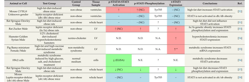
The adipose tissue produces various adipokines such as leptin, which is involved in the regulation of appetite. Obesity is associated with elevated circulating leptin levels and hypothalamic leptin resistance [44]. Clinical studies demonstrated a positive correlation between serum leptin levels and left ventricular hypertrophy independent of blood pressure values [45,46]. Leptin and its receptor are expressed in the heart, and leptin has been shown to promote left ventricular hypertrophy [47–49]. The leptin receptor belongs to cytokine type I receptors, which are known to signal via activation of the JAK2/STAT3 pathway [50]. The activation of the leptin-STAT3 signalling by high-fat diet was reported to be associated with hypertrophy and increased expression and activation of cardiac STAT3 in C57BL/6 mice, while STAT3 activation remained unchanged in leptin-receptor-deficient db/db mice [51,52] (Table3). Another study demonstrated increased STAT3 phosphorylation and expression in the hearts of Zucker rats, a genetic model of obesity [53]. Moreover, increased STAT3 expression has been shown to contribute to the development of left ventricular hypertrophy in hypercholesterolemic hamsters [54] and pigs suffering from metabolic syndrome [55].
In contrast, another study demonstrated that metabolic syndrome mimicked by high glucose, salt, and cholesterol treatment in cardiomyocyte-like H9c2 cells reduced viability and STAT3 activation [56].
In Sprague-Dawley rats, a high-fat diet resulted in decreased phosphorylation and expression of cardiac STAT3, along with an increased sensitivity to doxorubicin-induced cardiotoxicity [57]. Two additional studies showed no alteration of cardiac STAT3 activation and phosphorylation and expression in high-fat-diet-induced obese rats [51] or leptin-receptor-deficient obese mice (ob/ob) [58], respectively.
A preclinical study proved that cardiac ciliary neurotrophic factor (CNTF) ameliorated left ventricular hypertrophy in leptin-deficient ob/ob and leptin-resistant db/db mice via the STAT3 and ERK1/2 signalling pathway by activation of CNTF receptor that is structurally similar to the leptin receptor [59].
Altogether, the majority (but not all) of studies showed no change or increase in cardiac STAT3 phosphorylation, expression and/or activation in animal models of experimental obesity and/or hyperlipidemia. Based on the literature data, it seems that facilitated cardiac STAT3 signalling might contribute to activation of hypertrophic and surviving pathways in obesity.
Table 3.Effect of obesity on cardiac STAT3 under non-ischaemic baseline conditions.
Animal or Cell Test Group Control
Group
Tissue Sample
p-STAT3/t-STAT3
Activation p-STAT3 Phosphorylation t-STAT3
Expression Conclusions Ref.
high-fat-diet-induced
obese mice non-obese ventricles ↑ ↑(NC) Tyr705 – (NC) high-fat diet increases STAT3 activation
[52]
MouseC57BL/6
Unknown gender leptin-receptor-deficient
(db/db) obese mice non-obese ventricles – – (NC) Tyr705 – (NC) STAT3 is not activated in db/db obesity
Rat Sprague-Dawley Male
high-fat-diet-induced
obese rats non-obese whole heart – – (NC) ? – (NC) high-fat diet did not influence
STAT3 activation [51]
Rat Zucker Male leptin receptor deficient
(fa/fa) obese rats non-obese LV ↑(NC) ↑ ? ↑ fa/fa genetic obesity increases STAT3
phosphorylation and expression [53]
Hamster Golden Syrian Male
0.2% cholesterol diet-induced hypercholesterolemic
hamsters
normo-cholesterolemia LV N.D. N.D. N.A. ↑ hypercholesterolemia increases
STAT3 expression [54]
Pig Bama miniature Female/Male
high-fat and high-sucrose diet-induced metabolic
syndrome
non-metabolic
syndrome LV N.D. N.D. N.A. ↑* metabolic syndrome increases STAT3
mRNA expression [55]
H9c2 cells
metabolic syndrome induced by high glucose,
salt, and cholesterol treatment
normal
medium cells ↓(ELISA) N.E. ? N.E. metabolic syndrome decreases
STAT3 activation [56]
Rat Sprague-Dawley Male
high-fat-diet-induced
obese rats non-obese whole heart ↓(NC) ↓ ? ↓ high-fat diet decreases STAT3
phosphorylation and expression [57]
Mouse Leptin-receptor-deficient
(ob/ob) obese Male
leptin-receptor-deficient
(ob/ob) obese mice non-obese whole heart – (NC) – Tyr705 – STAT3 is not activated in ob/ob obesity [58]
↓in green cells: decrease;↑in red cells: increase; – in blue cells: no change; NC: not confirmed; N.D.: not detected; N.A.: not applicable; ?: phosphorylation site was not specified; LV: left ventricle; STZ: streptozotocin; p-STAT3: phosphorylated STAT3; t-STAT3: total STAT3; *: mRNA expression; ELISA: enzyme-linked immunosorbent assay.
Int. J. Mol. Sci.2018,19, 3572 8 of 29
2.3. Hypertension
According to the 2017 High Blood Pressure Clinical Practice Guideline, systolic blood pressure
≥130 and/or diastolic blood pressure≥80 mmHg is considered as hypertension [60]. Observational studies have demonstrated graded associations between hypertension and increased cardiovascular risk to, e.g., myocardial infarction, heart failure, stroke, peripheral artery disease, etc. [61,62]. Moreover, patients with hypertension often have other cardiovascular risk factors such as hypercholesterolemia, obesity, diabetes mellitus, chronic kidney disease and smoking [63].
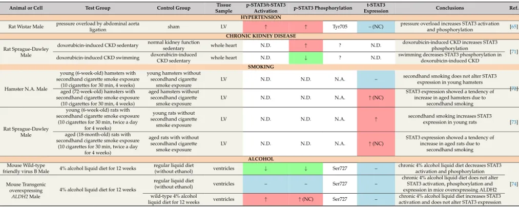
Left ventricular hypertrophy is a secondary consequence of hypertension and independently predicts future cardiovascular events [60,64]. Therefore, it is very difficult to separate the effects of hypertension, left ventricular hypertrophy and later systolic heart failure on cardiac STAT3 in preclinical and clinical studies. Nevertheless, cardiac phosphorylation of STAT3 has been reported to increase shortly after (peaking at 60 min) induction of pressure overload by constriction of the abdominal aorta [65] (Table4). Among the signalling pathways that mediate cardiac hypertrophy and heart failure, the activation of the JAK/STAT pathway is thought to play a pivotal role in the response to various stimuli such as pressure overload, cytokines, neurohormones, growth factors, ischaemia, etc. [11]. Detailed mechanisms by which STAT3 interacts with a broad range of cellular and molecular mechanisms to induce left ventricular hypertrophy and heart failure have been discussed in recent review articles, so here we just refer to these excellent reviews [11,15,66–68].
2.4. Chronic Kidney Disease
Chronic kidney disease (CKD) is a clinical syndrome defined as persistent deterioration of kidney function [41]. The prevalence of all stages of CKD varies between 7% and 12% worldwide [69].
In CKD patients, cardiovascular diseases are the leading cause of death [70]. The high incidence of cardiovascular diseases in CKD can be attributed to different systemic complications of CKD [41];
for instance, hypocalcemia and hyperkalemia often lead to life-threatening arrhythmias [70]. Increased oxidative stress, systemic inflammation, accelerated atherosclerotic process and deteriorating arterial hypertension in CKD often results in cardiac hypertrophy, later progressing to heart failure [70].
Cardiac STAT3 phosphorylation increased in a rat model of doxorubicin-induced CKD and cardiac hypertrophy [71] (Table4). This study also reported that 60 min daily of swimming or running for 11 weeks could attenuate cardiac hypertrophy through the cardiotrophin-1-LIFR-gp130-JAK/STAT3 pathway [71]. Swimming reduced cardiac STAT3 phosphorylation both in the control and CKD group, which might lead to the attenuation of cardiac hypertrophy in CKD animals [71].
Although alteration of cardiac STAT3 has been reported in CKD, whether this is due to direct effects or due to secondary effects via cardiovascular complications of CKD (e.g., cardiac hypertrophy and fibrosis) is very difficult to distinguish.
Table 4.Effect of hypertension, chronic kidney disease, smoking and alcohol on cardiac STAT3 under non-ischaemic baseline conditions.
Animal or Cell Test Group Control Group Tissue
Sample
p-STAT3/t-STAT3
Activation p-STAT3 Phosphorylation t-STAT3
Expression Conclusions Ref.
HYPERTENSION Rat Wistar Male pressure overload by abdominal aorta
ligation sham LV ↑ ↑ Tyr705 – (NC) pressure overload increases STAT3 activation
and phosphorylation [65]
CHRONIC KIDNEY DISEASE doxorubicin-induced CKD sedentary normal kidney function
sedentary whole heart N.D. ↑ ? N.D. doxorubicin-induced CKD increases STAT3
phosphorylation Rat Sprague-Dawley
Male doxorubicin-induced CKD swimming doxorubicin-induced
CKD sedentary whole heart N.D. ↓ ? N.D. swimming decreases STAT3 phosphorylation in
doxorubicin-induced CKD
[71]
SMOKING young (6-week-old) hamsters with
secondhand cigarette smoke exposure (10 cigarettes for 30 min, 4 weeks)
young hamsters without secondhand cigarette
smoke exposure
LV N.D. N.D. N.A. – secondhand smoking does not alter STAT3
expression in young hamsters Hamster N.A. Male aged (72-week-old) hamsters with
secondhand cigarette smoke exposure (10 cigarettes for 30 min, 4 weeks)
aged hamsters without secondhand cigarette
smoke exposure
LV N.D. N.D. N.A. ↑(NC)
STAT3 expression showed a tendency of increase in aged hamsters due to
secondhand smoking
[72]
young (6-week-old) rats with secondhand cigarette smoke exposure
(10 cigarettes for 30 min, twice a day for 4 weeks)
young rats without secondhand cigarette
smoke exposure
LV N.D. N.D. N.A. ↑ secondhand smoking increases STAT3
expression in young rats [73]
Rat Sprague-Dawley
Male aged (18-month-old) rats with secondhand cigarette smoke exposure
(10 cigarettes for 30 min, twice a day for 4 weeks)
aged rats with without secondhand cigarette
smoke exposure
LV N.D. N.D. N.A. ↑(NC)
STAT3 expression showed a tendency of increase in aged rats due to
secondhand smoking ALCOHOL
Mouse Wild-type
friendly virus B Male 4% alcohol liquid diet for 12 weeks regular liquid diet
(without ethanol) ventricles ↓ ↓ Ser727 – chronic 4% alcohol liquid diet decreases STAT3
activation and phosphorylation
4% alcohol liquid diet for 12 weeks
regular liquid diet
(without ethanol) ventricles – – Ser727 –
chronic 4% alcohol liquid diet does not alter STAT3 activation, phosphorylation and expression in mice overexpressing ALDH2 Mouse Transgenic
overexpressing
ALDH2Male wild-type 4% alcohol
liquid diet for 12 weeks ventricles ↑ ↑(NC) Ser727 – chronic 4% alcohol liquid diet increases STAT3
activation and does not alter STAT3 expression [74]
↓in green cells: decrease;↑in red cells: increase; – in blue cells: no change; NC: not confirmed; N.D.: not detected; N.A.: not applicable; ?: phosphorylation site was not specified; LV: left ventricle; STZ: streptozotocin; p-STAT3: phosphorylated STAT3; t-STAT3: total STAT3; CKD: chronic kidney disease; ALDH2: aldehyde dehydrogenase 2.
Int. J. Mol. Sci.2018,19, 3572 10 of 29
2.5. Aging
The aging human population is an epidemiological burden. It is estimated that more than 2 billion individuals will be over the age of 60 by 2050 worldwide [75]. The prevalence of cardiovascular diseases increases with age [76,77], and age might be a dominant risk factor in the elderly since the impact of many traditional risks (e.g., obesity or hypertension) decline with age [78].
The first evidence demonstrating a crucial role for cardiac STAT3 in aging was reported by Jacoby et al., showing the development of cardiac dysfunction and fibrosis with advancing age in mice with a cardiomyocyte-restricted deletion of STAT3 [79]. Later, reductions in STAT3 phosphorylation and expression in the right ventricle were shown in 13-month-old mice compared to three-month-old mice [18] (Table5). Two years later, the same research group demonstrated that the expression of STAT3 in subsarcolemmal mitochondria is reduced in the left ventricles of 21-month-old mice [80].
In accordance with these findings, STAT3 activation was reduced in 12-month-old and 24-month-old rats versus six-month-old controls [81].
In contrast, some studies described no alteration or increase of cardiac STAT3 in association with advanced age. Phosphorylation was unchanged in 14-month-old mice [82] or 20-24-month-old rats [83], and p-STAT3beta was not altered in 24-month-old mice [84] (Table5). Three studies showed that the level of STAT3 expression was increased in response to aging in old mice [82], hamsters [72] and rats [73];
however, these findings are limited since the activation (i.e., phosphorylation) was not examined.
Although age-related STAT3 dysregulation in human hearts has not been reported, research on blood samples from two independent cohort studies showed that STAT3 was positively associated with age [85].
Regarding possible modulation of STAT3 dysregulation in aging, Castello et al. have reported that alternate-day fasting restored the decline in STAT3 activation in elderly rats to young values and protected the heart against age-related hypertrophy [81].
Taken together, it is controversial whether STAT3 expression or activation is affected by aging or not, and further research is needed to elucidate the phenomenon. Nevertheless, fasting seems to be effective for the restoration of aging-associated STAT3 dysregulation.
2.6. Smoking
Smoking has well-known detrimental effects on health. Both active and passive (secondhand) smoking are predominant risk factors for coronary heart disease [86]. We et al. reported that passive smoking increases cardiac STAT3 expression in young rats [73], but does not alter STAT3 expression in hamsters [72] (Table4). In aged rats and hamsters exposed to passive smoking, cardiac STAT3 expression showed a tendency to increase [72,73].
2.7. Alcohol
Alcohol consumption is a widespread social habit. Although several studies showed potential benefits of moderate alcohol consumption on coronary heart diseases [87], heavy alcohol drinking may lead to the development of cardiomyopathy. In mice, chronic 4% alcohol liquid diet for 12 weeks induced cardiomyopathy and was associated with decreased STAT3 phosphorylation, which was reconciled in mice overexpressing aldehyde dehydrogenase 2 [74] (Table4). This finding indicates a potential effect of alcohol on STAT3 signalling.
2.8. Comedications
Patients at risk of cardiovascular disease are often treated with various medications that may also contribute to alteration of cardiac molecular signalling. Administration of the lipid-lowering drug simvastatin (10 mg/kg single daily dose) for five days in male Wistar rats increased cardiac p-STAT3 without affecting t-STAT3 [88]. Controversially, simvastatin gavage (10 mg/kg single daily dose) for 30 days and then for seven days intraperitoneally did not affect p-STAT3 [89].
Table 5.Effect of aging on cardiac STAT3 under non-ischaemic baseline conditions.
Animal or Cell Test Group Control Group Tissue Sample p-STAT3/t-STAT3
Activation p-STAT3 Phosphorylation t-STAT3
Expression Conclusions Ref.
MouseC57Bl6/J
Female aged >13 months young (<3 months) RV – (NC) ↓ Ser727 ↓ age decreases STAT3 phosphorylation and
expression [18]
MouseC57Bl6/J
Female/Male aged 21 months young (8 weeks) LVmitochondrial
fraction N.D. N.D. N.A. ↓ age decreases STAT3 expression [80]
aged 24 months ↓ ↓(NC) Tyr705 – (NC)
Rat Sprague-Dawley
Male adult 12 months young (6 months) LV ↓ ↓(NC) Tyr705 – (NC)
age decreases STAT3 activation and reduced STAT3 activation may contribute to
age-associated hypertrophy
[81]
MouseC57BL/6JMale aged 14 months young (2 months) whole heart – – Tyr705 ↑
age does not alter STAT3 activation and phosphorylation, but increases
STAT3 expression
[82]
Rat Sprague-Dawley Male
aged 20–24
months young (3–4 months) whole heart – – (NC) Ser727 – (NC) age does not influence STAT3 activation [83]
MouseC57BL/6JMale aged 24 months young (3 months) whole heart – (NC) – Tyr705 – age does not influence STAT3
phosphorylation and expression [84]
Hamster N.A. Male aged 72 weeks young (6 weeks) LV N.D. N.D. N.A. ↑ age increases STAT3 expression [72]
Rat Sprague-Dawley
Male aged 18 months young (6 weeks) LV N.D. N.D. N.A. ↑ age increases STAT3 expression [73]
↓in green cells: decrease;↑in red cells: increase; – in blue cells: no change; NC: not confirmed; N.D.: not detected; N.A.: not applicable; RV: right ventricle; LV: left ventricle; p-STAT3:
phosphorylated STAT3; t-STAT3: total STAT3.
Int. J. Mol. Sci.2018,19, 3572 12 of 29
2.9. Summary
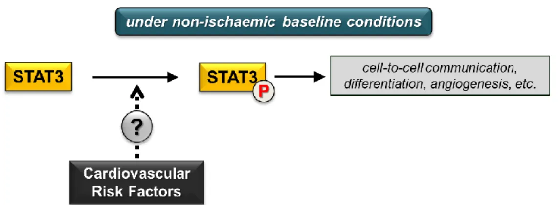
The effect of various cardiovascular risk factors on cardiac STAT3 signalling under non-ischaemic baseline conditions remains inconclusive (Figure2) due to limited literature data or conflicting findings.
Future experimental studies focusing on this area may help us to draw adequate conclusions.
Int. J. Mol. Sci. 2018, 19, x FOR PEER REVIEW 13 of 29
2.9. Summary
The effect of various cardiovascular risk factors on cardiac STAT3 signalling under non-ischaemic baseline conditions remains inconclusive (Figure 2) due to limited literature data or conflicting findings. Future experimental studies focusing on this area may help us to draw adequate conclusions.
Figure 2. Cardiovascular risk factors and cardiac STAT3 activation under non-ischaemic baseline conditions. (P in circle represents phosphorylated STAT3 form; dotted arrow and question mark indicate inconclusive effects of risk factors).
3. Effect of Cardiovascular Risk Factors on Cardiac STAT3 Activation in Settings of Ischaemia/Reperfusion
3.1. Diabetes
Diabetes is a well-known risk factor for the development of ischaemic heart disease. Moreover, clinical studies showed that diabetes mellitus increased the susceptibility of the myocardium to ischaemia/reperfusion injury and that the long-term outcome of ischaemic heart disease is worsened by diabetes. However, the effect of diabetes on the susceptibility of the myocardium to acute ischaemia/reperfusion injury is controversial in animal models. In experimental models of both type I or type II diabetes, infarct size was demonstrated to be significantly larger, unchanged, or significantly smaller in diabetic rats compared to nondiabetic controls [29,90–92].
In contrast to discrepancies regarding the effect of diabetes on infarct size, post-ischaemic phosphorylation/activation of cardiac STAT3 was clearly downregulated in experimental models of diabetes in all investigations (Table 6). In an animal model of STZ-induced type I diabetes, after 30 min ischaemia and 2 h reperfusion there was a significant reduction in phosphorylated STAT3 (Tyr705) levels in the diabetic group compared to the nondiabetic control group (tissue samples were collected from the ischaemic zone of the myocardium) [93]. This reduction of post-ischaemic STAT3 phosphorylation and/or activation due to diabetes was confirmed by several studies of another research group in the ischaemic tissue as well as in whole heart or ventricular tissue samples [29,31,33,92,94]. In addition, the phosphorylated STAT3 (Tyr705) and total STAT3 levels were significantly reduced in a type II diabetes model, i.e., in isolated perfused hearts of leptin receptor null, homozygous db/db mice subjected to ischaemia/reperfusion compared to wild-type hearts subjected to ischaemia/reperfusion [95].
The same trend was observed in in vitro models of diabetes or acute hyperglycaemia. In H9c2 cells subjected to high glucose conditions (25 mM glucose), the post-ischaemic STAT3 phosphorylation and activation (at Ser727 and Tyr705 sites) were significantly lower [29,32].
Similarly, exposure of isolated adult rat ventricular cardiomyocytes to high glucose conditions also resulted in reduced post-ischaemic STAT3 activation (at Tyr705 site) [33,92].
In summary, post-ischaemic STAT3 phosphorylation and/or activation are significantly decreased due to diabetes in all studies irrespective of the applied models, which may contribute to increased susceptibility to myocardial ischaemia/reperfusion injury in diabetes.
Figure 2. Cardiovascular risk factors and cardiac STAT3 activation under non-ischaemic baseline conditions. (P in circle represents phosphorylated STAT3 form; dotted arrow and question mark indicate inconclusive effects of risk factors).
3. Effect of Cardiovascular Risk Factors on Cardiac STAT3 Activation in Settings of Ischaemia/Reperfusion
3.1. Diabetes
Diabetes is a well-known risk factor for the development of ischaemic heart disease. Moreover, clinical studies showed that diabetes mellitus increased the susceptibility of the myocardium to ischaemia/reperfusion injury and that the long-term outcome of ischaemic heart disease is worsened by diabetes. However, the effect of diabetes on the susceptibility of the myocardium to acute ischaemia/reperfusion injury is controversial in animal models. In experimental models of both type I or type II diabetes, infarct size was demonstrated to be significantly larger, unchanged, or significantly smaller in diabetic rats compared to nondiabetic controls [29,90–92].
In contrast to discrepancies regarding the effect of diabetes on infarct size, post-ischaemic phosphorylation/activation of cardiac STAT3 was clearly downregulated in experimental models of diabetes in all investigations (Table6). In an animal model of STZ-induced type I diabetes, after 30 min ischaemia and 2 h reperfusion there was a significant reduction in phosphorylated STAT3 (Tyr705) levels in the diabetic group compared to the nondiabetic control group (tissue samples were collected from the ischaemic zone of the myocardium) [93]. This reduction of post-ischaemic STAT3 phosphorylation and/or activation due to diabetes was confirmed by several studies of another research group in the ischaemic tissue as well as in whole heart or ventricular tissue samples [29,31,33,92,94]. In addition, the phosphorylated STAT3 (Tyr705) and total STAT3 levels were significantly reduced in a type II diabetes model, i.e., in isolated perfused hearts of leptin receptor null, homozygous db/db mice subjected to ischaemia/reperfusion compared to wild-type hearts subjected to ischaemia/reperfusion [95].
The same trend was observed in in vitro models of diabetes or acute hyperglycaemia. In H9c2 cells subjected to high glucose conditions (25 mM glucose), the post-ischaemic STAT3 phosphorylation and activation (at Ser727 and Tyr705 sites) were significantly lower [29,32]. Similarly, exposure of isolated adult rat ventricular cardiomyocytes to high glucose conditions also resulted in reduced post-ischaemic STAT3 activation (at Tyr705 site) [33,92].
In summary, post-ischaemic STAT3 phosphorylation and/or activation are significantly decreased due to diabetes in all studies irrespective of the applied models, which may contribute to increased susceptibility to myocardial ischaemia/reperfusion injury in diabetes.
Table 6.Effect of diabetes on cardiac STAT3 in settings of ischaemia/reperfusion.
Animal or Cell Test Group Control Group Tissue
Sample
p-STAT3/t-STAT3
Activation p-STAT3 Phosphorylation t-STAT3
Expression Conclusions Ref.
ischaemic
zone ↓(NC) ↓ Tyr705 – diabetes decreases post-ischaemic
STAT3 phosphorylation [93]
LV ↓ ↓ Tyr705
Ser727 – (NC) diabetes decreases post-ischaemic STAT3 activation and phosphorylation [29]
whole heart ↓ ↓ Tyr705 – (NC) diabetes decreases post-ischaemic
STAT3 activation and phosphorylation [31]
Rat Sprague-Dawley Male
in vivo regional (LAD) 30 min/120 min I/R in STZ-induced diabetes
I/R nondiabetic
ventricles ↓ ↓ Tyr705
Ser727 – diabetes decreases post-ischaemic STAT3 activation and phosphorylation [92]
Rat Sprague-Dawley Male
in vivo regional (LAD) 30 min/90 min I/R in STZ-induced diabetes
I/R nondiabetic ischaemic
zone ↓ ↓ Tyr705 – (NC) diabetes decreases post-ischaemic
STAT3 activation and phosphorylation [94]
Rat Sprague-Dawley Male
ex vivo global 30 min/120 min
I/R in STZ-induced diabetes I/R nondiabetic LV ↓ ↓(NC) Tyr705 – (NC) diabetes decreases post-ischaemic
STAT3 activation [33]
Mouse leptin receptor null, homozygous db/db
Male
ex vivo global 30 min/60 min I/R in high-fat-diet-induced
diabetes
I/R C57BL/6J wild-type
mouse whole heart ↓ ↓ Tyr705 ↓
diabetes decreases post-ischaemic STAT3 activation, phosphorylation
and expression
[95]
Rat Sprague-Dawley Male
isolated adult diabetic rat cardiomyocytes subjected to
SI/R
isolated adult nondiabetic rat cardiomyocytes subjected
to SI/R
cells ↓ ↓ Tyr705 – (NC) diabetes decreases post-ischaemic
STAT3 activation and phosphorylation [33]
normal glucose conditions + 6 h/12 h
SI/R
cells ↓ ↓ Tyr705
Ser727 – (NC)
high glucose condition decreases post-ischaemic STAT3 activation and
phosphorylation
[29]
H9c2 cells high glucose conditions + 6 h/12 h SI/R
high glucose conditions cells ↓ ↓ Tyr705 – (NC)
high glucose condition decreases post-ischaemic STAT3 activation and
phosphorylation
[32]
Rat Sprague-Dawley Male
isolated adult rat cardiomyocytes subjected to high glucose conditions + 45
min/2 h SI/R
normal glucose conditions + 45 min/2 h
SI/R
cells ↓ ↓(NC) Tyr705 – (NC)
H9c2 cells high glucose conditions + 45 min/2 h SI/R
normal glucose conditions + 45 min/2 h
SI/R
cells ↓ ↓(NC) Tyr705 – (NC)
high glucose condition decreases post-ischaemic STAT3 activation [92]
↓in green cells: decrease;↑in red cells: increase; – in blue cells: no change; NC: not confirmed; LV: left ventricle; STZ: streptozotocin; LAD: left anterior descending coronary artery; I/R:
ischaemia/reperfusion; SI/R: simulated ischaemia/reoxygenation; p-STAT3: phosphorylated STAT3; t-STAT3: total STAT3.
Int. J. Mol. Sci.2018,19, 3572 14 of 29
3.2. Obesity
Obese people are more prone to developing coronary artery disease [40]. Leptin signalling has been shown to ameliorate cardiac dysfunction and remodelling four weeks after myocardial infarction by increasing STAT3 phosphorylation in calorie-restricted lean and obese ob/ob mice [58] as well as in tamoxifen-inducible leptin receptor knockout mice [96] (Table7). The obesity-associated hormone leptin has been shown to exert an infarct size-limiting effect after 35 min global ischaemia and 35 min reperfusion in non-obese C57Bl/6 mice [97]; however, leptin treatment was associated with reduced levels of phosphorylated and total STAT3 after myocardial infarction in this study [97]. Another study demonstrated that cardiac STAT3 activation was not altered due to diet-induced hypercholesterolemia in rabbit hearts subjected to 30 min ischaemia and 10 min reperfusion [98].
3.3. Chronic Kidney Disease
The hypertrophic and fibrotic myocardium is more sensitive to ischaemia; therefore, acute myocardial infarction is a common cause of cardiovascular morbidity and mortality in CKD patients [70]. To date, only one study has reported data on cardiac STAT3 in experimental CKD.
Neither expression nor activation of cardiac STAT3 was affected by CKD in a rat model of in vivo ischaemia/reperfusion [99] (Table7).
3.4. Aging
Aging aggravates myocardial ischaemia/reperfusion injury in humans [100] and rodents [100–103]. An experimental study showed reduced STAT3 activation in the left ventricle at reperfusion after regional ischaemia in 13-month-old mice compared to three-month-old mice [18]
(Table7). Another study found unaltered activation of cardiac STAT3 in aged rats subjected to 30 min ischaemia and 15 min reperfusion compared to young controls undergoing ischaemia/reperfusion [83].
3.5. Gender
It is well known that the risk of cardiovascular diseases is higher in males compared to females. The molecular and cellular basis of the cardiovascular gender difference has been reviewed elsewhere [104]. Scientific evidence suggests that the female sex hormone oestrogen exerts a cardioprotective effect, which also explains why postmenopausal women have a higher cardiovascular risk compared to younger females [105].
Wang et al. demonstrated that in hearts isolated from male wild-type C57BL/6J mice and subjected to ex vivo global ischaemia/reperfusion, the STAT activation was lower compared to hearts from female mice [106] (Table7). This difference was also associated with better functional recovery after ischaemia/reperfusion in female mice [106]. Myocardial STAT3 activation after ex vivo ischaemia/reperfusion was also attenuated in the hearts of male Sprague-Dawley rats compared to females [107]. However, in the same study, in hearts of castrated male rats subjected to ischaemia/reperfusion, the myocardial STAT3 activation was higher compared to hearts of male controls, possibly due to lower levels of endogenous testosterone. Moreover, exogenous testosterone administration decreased activation of STAT3 in hearts of castrated males as well as in females compared to males; therefore, it was concluded that testosterone has a negative effect on myocardial STAT3 activation after ischaemia/reperfusion [107].
3.6. Depression
Depression is common in patients with coronary heart disease and is associated with increased cardiovascular mortality [108]. Depression was not shown to influence STAT3 activation in an ex vivo model of regional ischaemia/reperfusion in hearts isolated from Sprague-Dawley rats exposed to experimental depression induced by chronic mild stress [109] (Table7).
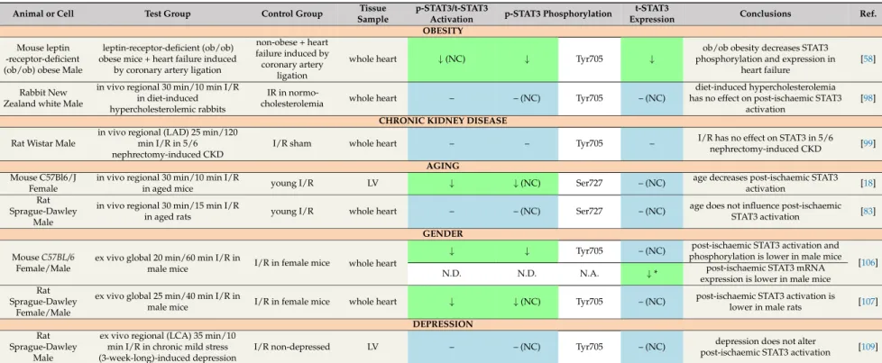
Table 7.Effect of obesity, chronic kidney disease, aging, gender and depression on cardiac STAT3 in settings of ischaemia/reperfusion.
Animal or Cell Test Group Control Group Tissue
Sample
p-STAT3/t-STAT3
Activation p-STAT3 Phosphorylation t-STAT3
Expression Conclusions Ref.
OBESITY Mouse leptin
-receptor-deficient (ob/ob) obese Male
leptin-receptor-deficient (ob/ob) obese mice + heart failure induced
by coronary artery ligation
non-obese + heart failure induced by coronary artery
ligation
whole heart ↓(NC) ↓ Tyr705 ↓
ob/ob obesity decreases STAT3 phosphorylation and expression in
heart failure
[58]
Rabbit New Zealand white Male
in vivo regional 30 min/10 min I/R in diet-induced hypercholesterolemic rabbits
IR in normo-
cholesterolemia whole heart – – (NC) Tyr705 – (NC)
diet-induced hypercholesterolemia has no effect on post-ischaemic STAT3
activation
[98]
CHRONIC KIDNEY DISEASE Rat Wistar Male
in vivo regional (LAD) 25 min/120 min I/R in 5/6 nephrectomy-induced CKD
I/R sham whole heart – – Tyr705 – I/R has no effect on STAT3 in 5/6
nephrectomy-induced CKD [99]
AGING Mouse C57Bl6/J
Female
in vivo regional 30 min/10 min I/R
in aged mice young I/R LV ↓ ↓(NC) Ser727 – (NC) age decreases post-ischaemic STAT3
activation [18]
Rat Sprague-Dawley
Male
in vivo regional 30 min/15 min I/R
in aged rats young I/R whole heart – – (NC) Ser727 – (NC) age does not influence post-ischaemic
STAT3 activation [83]
GENDER
↓ ↓ Tyr705 – (NC) post-ischaemic STAT3 activation and
phosphorylation is lower in male mice MouseC57BL/6
Female/Male
ex vivo global 20 min/60 min I/R in
male mice I/R in female mice whole heart
N.D. N.D. N.A. ↓* post-ischaemic STAT3 mRNA
expression is lower in male mice
[106]
Rat Sprague-Dawley
Female/Male
ex vivo global 25 min/40 min I/R in
male mice I/R in female mice whole heart ↓ ↓(NC) Tyr705 – (NC) post-ischaemic STAT3 activation is
lower in male rats [107]
DEPRESSION Rat
Sprague-Dawley Male
ex vivo regional (LCA) 35 min/10 min I/R in chronic mild stress (3-week-long)-induced depression
I/R non-depressed LV – – (NC) Tyr705 – (NC) depression does not alter
post-ischaemic STAT3 activation [109]
↓in green cells: decrease;↑in red cells: increase; – in blue cells: no change; NC: not confirmed; N.D.: not detected; N.A.: not applicable; LV: left ventricle; LAD: left anterior descending coronary artery; LCA: left coronary artery; I/R: ischaemia/reperfusion; SI/R: simulated ischaemia/reoxygenation; CKD: chronic kidney disease; *: mRNA expression; p-STAT3:
phosphorylated STAT3; t-STAT3: total STAT3.
Int. J. Mol. Sci.2018,19, 3572 16 of 29
3.7. Comedications
Administration of the lipid-lowering drug simvastatin (10 mg/kg single daily dose) for five days in male Wistar rats did not affect ischaemia/reperfusion-induced STAT3 phosphorylation [88].
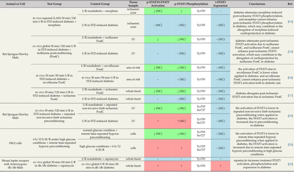
4. Effect of Cardioprotective Strategies against Ischaemia/Reperfusion on Cardiac STAT3 Activation in the Presence of Cardiovascular Risk Factors
4.1. Diabetes
In the literature, studies exist reporting both preserved as well as impaired cardioprotection by ischaemic or pharmacological conditionings [91]. Nevertheless, ischaemic pre- and postconditioning mechanisms, which aim to attenuate ischaemia/reperfusion injury, were inefficient or required extra ischaemia/reperfusion cycles to induce cardioprotection in the majority of animal models of chronic diabetes [90,110,111]. The efficacy of pharmacological preconditioning was also impaired in diabetes, for instance in the case of isoflurane [112] orL-glutamate [113]. In contrast, the infarct-size-limiting effect of remote preconditioning induced by repeated non-invasive limb ischaemia was preserved in STZ-induced diabetes [29].
Several studies have demonstrated that the presence of diabetes attenuated phosphorylation and/or activation of STAT3 in hearts undergoing ischaemia/reperfusion with ischaemic [31], pharmacological [29,33,93,94], or remote [29] conditioning when compared to nondiabetic controls subjected to ischaemia/reperfusion with corresponding conditioning (Table8). In these studies various ischaemic or pharmacological conditioning either increased or did not change STAT3 activation in the diabetic state. These studies suggest that diabetes aggravates the stimulatory effect of various conditioning on the phosphorylation/activation of cardiac STAT3. Similar pattern was shown in an in vitro model of hyperglycaemia in H9c2 cells [29].
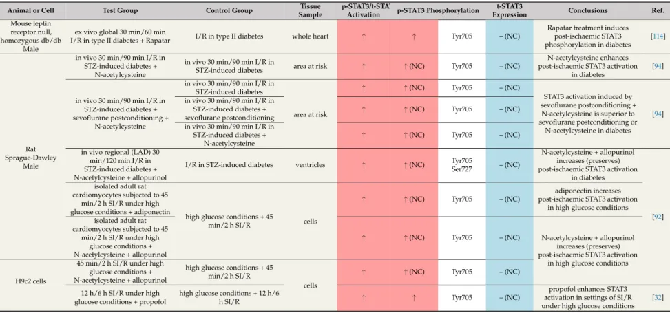
Recently, some promising agents have been suggested to prevent STAT3 dysregulation in diabetes.
In diabetic mice, rapamycin [95] (Table 8) and its nanoformulated form, Rapatar [114] (Table9), increased the phosphorylation and activation of STAT3 (Tyr705), which could contribute to the infarct-size-reducing effect of these treatments. The combination of antioxidant N-acetylcysteine and allopurinol restored the decreased levels of p-STAT3 (Ser727, Tyr705) after ischaemia/reperfusion and so contributed to smaller infarct size in STZ-induced diabetes [92]. N-acetylcysteine treatment together with sevoflurane postconditioning has the same beneficial effects [94] (Table 9). In some studies, the phosphorylation/activation of STAT3 was also increased in response to various cardioprotective agents in diabetes models [32,92,94,114] (Table9).
In summary, the majority of articles revealed that diabetes attenuates ischaemic or pharmacological conditioning-induced cardiac STAT3 activation after ischaemia/reperfusion.
This could be related to the loss of cardioprotection and increased infarct size in diabetes. Interestingly, there are some potential therapeutic agents like the antioxidant N-acetylcysteine and allopurinol or rapamycin that can restore the STAT3 phosphorylation and so can contribute to attenuated myocardial damage.