Research Report
Glucose-monitoring neurons in the mediodorsal prefrontal cortex
Bernadett Nagy ⁎ , István Szabó, Szilárd Papp, Gábor Takács, Csaba Szalay, Zoltán Karádi
Pécs University, Medical School, Institute of Physiology and Neurophysiology Research Group of the Hungarian Academy of Sciences, Pécs, Hungary
A R T I C L E I N F O A B S T R A C T
Article history:
Accepted 11 January 2012 Available online 20 January 2012
The mediodorsal prefrontal cortex (mdPFC), a key structure of the limbic neural circuitry, plays important roles in the central regulation of feeding. As an integrant part of the forebrain dopamine (DA) system, it performs complex roles via interconnections with various brain areas where glucose-monitoring (GM) neurons have been identified. The main goal of the present experiments was to examine whether similar GM neurons exist in the mediodorsal prefrontal cortex. To search for such chemosensory cells here, and to es- timate their involvement in the DA circuitry, extracellular single neuron activity of the mediodorsal prefrontal cortex of anesthetized Wistar and Sprague–Dawley rats was recorded by means of tungsten wire multibarreled glass microelectrodes during microelec- trophoretic administration ofD-glucose and DA. One fourth of the neurons tested changed in firing rate in response to glucose, thus, proved to be elements of the forebrain GM neural network. DA responsive neurons in the mdPFC were found to represent similar proportion of all cells; the glucose-excited units were shown to display excitatory whereas the glucose- inhibited neurons were demonstrated to exert mainly inhibitory responses to dopamine.
The glucose-monitoring neurons of the mdPFC and their distinct DA sensitivity are sug- gested to be of particular significance in adaptive processes of the central feeding control.
© 2012 Elsevier B.V. All rights reserved.
Keywords:
Glucose-monitoring neurons Multibarreled microelectrophoretic technique
Mediodorsal prefrontal cortex Dopamine
1. Introduction
The prefrontal cortex (PFC) is defined as the cortex of the an- terior pole of the mammalian brain, predominantly receiving projections from the mediodorsal thalamic nucleus (Lacroix et al., 2000; Rose and Woolsey, 1948). It has been demonstrat- ed that the prefrontal cortex is implicated in many regulatory processes, including cognitive functions, decision making, working memory, and the control of motivated behaviors
such as the food and fluid intake (Baldwin et al., 2002;
Cardinal et al., 2002; Heidbreder and Groenewegen, 2003;
Kolb, 1984, 1990; Kolb and Nonneman, 1975; Morgane et al., 2005).
The prefrontal cortex is considered to perform its complex roles via multiple interrelationships with forebrain and brain- stem areas. Anatomical studies have shown that the medial– mediodorsal prefrontal cortex (mdPFC) has direct connections with limbic structures, such as the amygdala (AMY), the lateral
⁎Corresponding author at:Pécs University, Medical School, Institute of Physiology, Pécs, Szigeti str. 12., H-7624, Hungary. Fax: +36 72536424.
E-mail address:bernadett.nagy@aok.pte.hu(B. Nagy).
Abbreviations:AMY, amygdala; DA, dopamine; GM, glucose-monitoring; GR, glucose-receptor; GS, glucose-sensitive; LHA, lateral hypo- thalamic area; MB, methylene-blue; mdPFC, mediodorsal prefrontal cortex; NAcc, nucleus accumbens; NTS, nucleus of the solitary tract;
OBF, orbitofrontal cortex; PFC, prefrontal cortex
0006-8993/$–see front matter © 2012 Elsevier B.V. All rights reserved.
doi:10.1016/j.brainres.2012.01.025
A v a i l a b l e o n l i n e a t w w w . s c i e n c e d i r e c t . c o m
w w w . e l s e v i e r . c o m / l o c a t e / b r a i n r e s
hypothalamic area (LHA), the nucleus accumbens (NAcc) and the adjacent orbitofrontal cortex (OBF) (Kita and Oomura, 1981;
Kolb, 1984; Lacroix et al., 2000), all known to be important in the central feeding control. The rat mdPFC also directly projects to the nucleus of the solitary tract (NTS), a brainstem region which integrates a number of autonomic reflexes (Terreberry and Neafsey, 1987) and is well-known as a key structure of the central taste information processing (Norgren and Leonard, 1971; Rolls, 1989) as well.
In previous investigations, particular types of chemosen- sory cells, the so-called glucose-monitoring (GM) neurons– displaying firing rate changes in response to elevation of blood glucose level or to local microelectrophoretic administration of
D-glucose–have been discovered in the above interconnected brain areas. Specific glucose-inhibited (glucose-sensitive, GS) neurons were identified in the LHA of rats (Oomura, 1980;
Oomura et al., 1969) and later in the LHA and in the AMY of rhe- sus monkeys (Aou et al., 1984; Karadi et al., 1992; Nakano et al., 1986), and in the NTS, too (Adachi et al., 1984; Mizuno and Oomura, 1984). By contrast, the NAcc and the OBF were proven to contain not only GS cells but also glucose-excited (glucose- receptor, GR) neurons that are facilitated by increase of the extracellular glucose concentration (Karadi et al., 2004; Papp et al., 2007).
The GM cells were demonstrated to be influenced by cate- cholamines (Karadi et al., 1992, 2004; Lenard et al., 1995), and with respect to this it is especially important to note that the PFC is the major cortical target area of the ascending dopamine (DA) projections (Berger et al., 1976; Björklund and Lindvall, 1984; Descarries et al., 1987; Ungerstedt, 1971). In addition to responding to endogenous chemical stimuli, these chemosen- sory neurons, forming a hierarchically organized neural net- work, are also known to integrate multiple, homeostatically relevant information, such as exogenous chemical and other signals, sensory-motor, perceptual, motivational mechanisms, as well as reinforcement, learning and memory processes, to control feeding and metabolic functions (Aou et al., 1984;
Karadi et al., 1992, 1995, 2004; Oomura and Yoshimatsu, 1984).
Considering the above, it is supposed that the mediodorsal prefrontal cortex accomplishes its complex roles as integrant part of the forebrain glucose-monitoring neural network. In the present experiments, therefore, we aimed to identify GM neurons in the mdPFC, and to examine their responsiveness to DA. To do so, extracellular single neuron activity was recorded in the mdPFC of anesthetized male Wistar and Spra- gue–Dawley rats, by means of tungsten wire multibarreled glass microelectrodes during microelectrophoretic application ofD-glucose and dopamine.
2. Results
Activity changes of altogether 272 neurons have been recorded in the Wistar and Sprague–Dawley rat mdPFC. The mean spon- taneous firing rates were 2.2± 0.2 and 2.4± 0.3 spikes/s, respec- tively, and did not vary significantly between the two preparations. To examine direct neuronal effect of glucose, sin- gle neuron activity was recorded during microelectrophoretic administration ofD-glucose. Results of the neurochemical stim- ulations are summarized inTable 1. Sixty-two (24.3%) of 255
mdPFC neurons showed responsiveness to glucose, thus, these cells were found to be elements of the forebrain GM neu- ral network. The predominant response to glucose was inhibi- tion (43 of the 62 GM neurons, 69.4%), however, definite facilitatory activity changes were also detected (19 /30.6%/ of the 62 neurons). The other 193 neurons (75.7%) did not change in firing rate to glucose and thus, were classified as glucose- insensitive (GIS) cells.
DA responsiveness of 235 cells was examined in the rodent mdPFC. Microiontophoretic application of DA resulted in ac- tivity changes of 55 neurons (23.4%). AsTable 1shows, in the case of DA administration, the proportion of excitatory (28, 11.9%) and inhibitory (27, 11.5%) responses was almost the same.
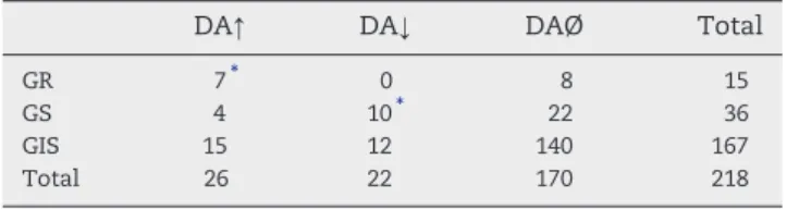
Table 2 demonstrates distinct DA responsiveness of glucose-monitoring and glucose-insensitive neurons in the mdPFC. Twenty-one (41.2%) of the 51 GM units, whereas only 27 (16.2%) of the 167 GIS neurons displayed discharge rate changes to this neurotransmitter, so that DA responsiveness of the GM cells was found to be significantly higher than that of the glucose-insensitive units (p < 0.001;χ2test). DA eli- cited only excitatory response in the GR cells (7 of 15 neurons, 46.7%), whereas both inhibitory (10 of 36 units, 27.8%) and ex- citatory (4 of 36 cells, 11.1%) firing rate changes were observed in the GS neurons. Consequently, direction of the DA induced activity changes of the two types of GM cells differed signifi- cantly (p < 0.01;χ2test). Discharge rate changes of two charac- teristic DA responsive GM cells in the mdPFC are shown in Fig. 1.
The magnitude of the response to microelectrophoretically administered glucose and DA was also examined.Fig. 2dem- onstrates the size of the ejection current-dependent re- sponses of mdPFC neurons. Both in case of glucose and Table 1–Effect of microelectrophoretically applied glucose and dopamine on rat mdPFC neurons.
Glucose DA
↑ 19 28
↓ 43 27
Ø 193 180
Total 255 235
↑: Excitatory response;↓: inhibitory response; Ø: no response.
Table 2–DA responsiveness of GM and GIS neurons in the rat mdPFC.
DA↑ DA↓ DAØ Total
GR 7⁎ 0 8 15
GS 4 10⁎ 22 36
GIS 15 12 140 167
Total 26 22 170 218
GIS: glucose-insensitive neuron; GR: glucose-receptor neuron (excited byD-glucose); GS: glucose-sensitive neuron (inhibited byD-glucose);
DAØ: DA-nonresponsive neurons; DA↑: neurons facilitated by DA;
DA↓: neurons inhibited by DA.
⁎P< 0.001 (χ2test).
dopamine administrations, higher current intensities resulted in significantly bigger firing rate changes of cells of the re- sponsive groups (p < 0.05, Wilcoxon test).
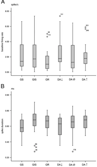
Analysis of other electrophysiological characteristics revealed additional functional attributes of the neurons. As Fig. 3represents, the baseline firing rates and spike durations Fig. 1–Responses of two mdPFC neurons to microelectrophoretically appliedD-glucose and dopamine. A, a glucose-sensitive cell inhibited byD-glucose and DA; B, a glucose-receptor neuron facilitated byD-glucose and dopamine. Spike insets, characteristic action potentials. Horizontal lines, numbers, duration of drug application and ejection current intensities in nA, respectively. Abscissa, time in s; ordinate, firing rate in spikes/s and potential in mV, respectively.
of the cells with various glucose and DA responsiveness did not show significant difference (p = 0.248 and p = 0.30, respec- tively; Kruskal–Wallis test). In addition, neither baseline firing rates nor spike durations were found to correlate with glucose or dopamine responses (p≥0.213).
Burst firing characteristic of the neurons was also examined.
We have found the most burst firing cells among the neurons inhibited by DA (64.3%), and the fewest ones (17.6% and 15%, respectively) among the cells facilitated by this catecholamine as well as among those excited by glucose (p< 0.05;χ2test).
As far as the location of the cells characterized by distinct neurochemical sensitivities is concerned, their topography appeared to be similar, the various neurons were found to be quite homogenously and overlappingly distributed within the mdPFC.
3. Discussion
Results of these experiments provided evidence for the presence of glucose-monitoring neurons in the mediodorsal prefrontal cortex. Although such feeding-associated chemosensory neu- rons have already been identified in several other brain regions (Adachi et al., 1984; Aou et al., 1984; Karadi et al., 1992, 2004;
Mizuno and Oomura, 1984; Nakano et al., 1986; Oomura, 1980;
Oomura et al., 1969; Papp et al., 2007), this is the first systematic study to demonstrate their existence in this cortical division of the forebrain limbic circuitry. The mdPFC is known to be in- volved in a broad variety of regulatory processes, and its impor- tant role in the central feeding control has been demonstrated as well (Cardinal et al., 2002; Kolb, 1984; Kolb and Nonneman, 1975; Mogensen and Divac, 1993). Since GM neurons of several brain areas have already been shown to be indispensable constituents of integration of endogenous and exogenous chemical information, sensory-motor integration, perceptual and motivational processes, as well as reinforcement, learning and memory mechanisms of the regulation of food and fluid in- take behaviors (Aou et al., 1984; Karadi et al., 1992, 1995, 2004;
Lenard et al., 1995; Oomura and Yoshimatsu, 1984; Oomura et al., 1969; Papp et al., 2007), it is reasonable to suppose that these chemosensory cells of the mdPFC possess similar complex functional attributes in the organization of adaptive feeding ac- tions. It is important to note that the former results have been obtained predominantly in the macaques whereas the present Fig. 2–Current response curves showing the magnitude of the
response (mean ± SEM) in percentage of baseline firing for 3 representative current intensities (30 nA, 60 nA, 90 nA, respectively). GR: glucose-receptor cells, GS: glucose-sensitive cells, GIS: glucose-insensitive cells; DA↑: neurons facilitated by DA; DA↓: neurons inhibited by DA; DA Ø: DA-insensitive neurons. *p< 0.05, Wilcoxon test.
Fig. 3–Box diagrams of distribution of baseline firing rates (A) and spike durations (B) of the various cell groups examined.
The duration of the spikes was measured between the negative trough and the positive peak of the spike waveforms.
Numbered open circles refer to neurons out of the continuous data range. Abbreviations are identical with those inFig. 2.
study was performed in the rodent. Nevertheless, findings of the latter gain more general significance in the light of our most re- cent microelectrophysiological experiments in the alert rhesus monkey revealing that GR and GS neurons also exist in the pri- mate mdPFC (unpublished data).
As the other major finding of the present experiments, distinct dopamine sensitivity of the mdPFC neurons has been elucidated:
the feeding-associated GM units were shown to be more likely to change in activity in response to microiontophoretically adminis- tered DA than the glucose-insensitive cells. Furthermore, the GR neurons were found to get facilitated whereas the GS units mainly inhibited by this catecholamine. These data are in concordance with previous results demonstrating higher dopamine respon- siveness of the lateral hypothalamic and pallidal GM neurons compared to that of the GIS cells, as well as the predominance of DA induced inhibitory firing rate changes of the GS neurons in the LHA (Karadi et al., 1992; Lenard et al., 1995).
The dense dopaminergic innervation of the PFC (Berger et al., 1976; Björklund and Lindvall, 1984; Descarries et al., 1987;
Ungerstedt, 1971) has already been indicated to play important roles in a variety of regulatory processes (Dalley et al., 2004;
Goeders et al., 1986; Granon et al., 2000; Hedou et al., 1999;
Ikemoto, 2010; Richardson and Gratton, 1998; Tzschentke, 2001), including feeding-associated and taste mediated learning
and memory mechanisms as well (Baldwin et al., 2002;
Gambarana et al., 2003; Hernadi et al., 2000; Touzani et al., 2010). It is especially worth noting here that food intake itself or stimuli associated with the food have been demonstrated to increase the extracellular DA concentration in the prefrontal cortex (Bassareo and Di Chiara, 1997; Hernandez and Hoebel, 1990). These and our present data are also in agreement with the notion that multiple regulatory functions of the mdPFC are perfectuated via interrelated complex neurochemical mecha- nisms (Morgane et al., 2005; Tzschentke, 2001).
The prefrontal cortical GM neurons are suggested to partic- ipate in the integration of several homeostatically relevant endogenous and exogenous signals. The chemosensory neu- rons at this high decision making level of the neuraxis, by uti- lizing their differential dopamine sensitivity, are supposed to play significant role in the control of adaptive behavioral ac- tions for the well being of the organism.
Previous recording studies have suggested that cortical in- terneurons have briefer spikes than those of pyramidal neu- rons, though cortical pyramidal neurons may exhibit a wide variety of spike durations (Bartho et al., 2004; Contreras, 2004;
Vigneswaran et al., 2011). In our study, examination of spike du- rations revealed no significant difference among the various groups of neurons, and spike durations also did not correlate
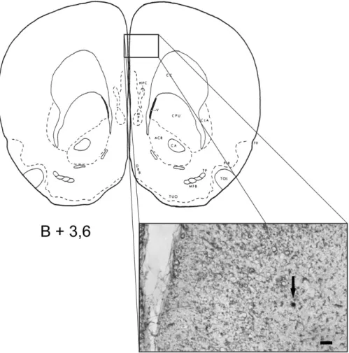
Fig. 4–Drawing of a brain section from the stereotaxic rat atlas (Pellegrino et al., 1979) at the level of the mediodorsal prefrontal cortex (the number refers to the anteroposterior coordinate with reference to bregma). Inset, photomicrograph of a native brain section with the microelectrophoretic methylene blue labeling spot (pointed by arrow) of a representative glucose-monitoring neuron; scale bar, 100μm.
with glucose and dopamine responses. Relationship appears to exist, however, between neurochemical sensitivity of neurons and their burst firing characteristics: the most burst firing cells were found among the neurons inhibited by DA, whereas the fewest burst firing cells were observed among the cells facilitat- ed by glucose and/or the catecholamine.
To understand the significance of these above findings, and to elucidate details of complex functional attributes of the med- iodorsal prefrontal cortical glucose-monitoring neurons, includ- ing their DA receptor mechanisms, further studies are required.
4. Experimental procedures
Thirty-seven Wistar, and fifteen Sprague–Dawley male labora- tory rats (weighing 305–380 g) were used in these experiments.
Individually caged animals were kept and cared for in accor- dance with institutional, national and international regulations (BA02/2000-1/2006, Pécs University, Medical School; Law XXVIII, 1998, Hungary; European Community Council Directive 86/609/
EEC, 1986, 2006; NIH Guidelines, 1997). Anesthesia was induced with a single injection of urethane (0.6 ml/100 g body weight, 25% fresh solution, Sigma, Hungary). Rats were operated on ste- reotaxically, their scalp was incised, and a small hole was drilled through the skull. The microelectrode was led to the mdPFC under microscopic control through the incised dura by means of a hydraulic microdrive (Narishige MO-10, Japan). The stereotaxic coordinates for electrode placements in the mdPFC were chosen according to the rat brain atlas (Pellegrino et al., 1979): anteroposterior, bregma + 3.2–4.0 mm; mediolateral, 0.7– 1.6 mm; vertical, 0.6–2.8 mm. Extracellular recording and micro- electrophoretic application of neurochemicals were accom- plished by means of nine-barreled glass microelectrodes.
Single-neuron activity was recorded via the central barrel con- taining a tungsten wire (10μm in diameter, impedance 1.5–8 M Ωat 50 Hz). Neurochemicals were applied electrophoretically through the capillaries surrounding the central recording elec- trode. Each barrel was filled with one of the following solutions:
D-glucose (0.5 M, pH 7.0), dopamine hydrochloride (0.5 M, pH 6), and monosodiumL-glutamate (0.5 M, pH 7–8; to test the elec- trode tip's vicinity to the recorded neuron). In addition to the above, one barrel was filled with physiological saline used as a current balancing channel, and another one with methylene- blue (MB, Reanal Ltd., Hungary) for labeling the position of the electrode's tip. Constant current source (NeuroPhore BH-2 System, USA), producing constant currents (in the 5–95 nA range) of appropriate polarity, was applied to eject the neuro- chemicals from their respective barrels. Extracellular action po- tentials were passed into a preamplifier, a high gain amplifier including low and high cut filters and a window discriminator to form standard pulses (Supertech Ltd., Hungary), and then into a microprocessor controlled A/D converter device (CED 1401 plus). The Spike 2 software package (Cambridge Electronic Design Ltd., United Kingdom) was used to construct frequency histograms and for real-time and off-line analyses. Neuronal spikes and formed pulses were continuously observed on oscil- loscopes (HAMEG HM-2037, Germany). Only the action poten- tials of spontaneously active, well-isolated cells were recorded.
Neurons that showed non-specific current effects (response to Na+or Cl−ions) were excluded from the analysis.
Similar to our previous studies, a neuron was considered to be responsive to a certain neurochemical if its firing rate changed by at least ±30% or by ±2 SD from the mean baseline level, and if the reactions were dose dependent (by using dif- ferent current intensities), and replicable.
For statistical analysis of the data, the Wilcoxon test, Kruskal– Wallis test, linear regression test, andχ2test were used.
In addition to the stereotaxic determination of the elec- trode position, recording sites were marked by methylene- blue (MB) labeling and confirmed by subsequent histological examination. Accordingly, anodal labeling current (50 nA, 10–15 min) was delivered through the MB containing barrel at the end of the recording sessions (Kovacs et al., 2005).
After completion of the marking procedure, rats were per- fused transcardially with physiological saline followed by paraformaldehyde (4%) and the brains were postfixed over- night in paraformaldehyde. After PBS rinsing, native sections were cut for light microscopic identification of the recording sites (see representative photomicrograph inFig. 4).
5. Conclusion
Our results provided evidence for the existence of special che- mosensory neurons in the mediodorsal prefrontal cortex. The differential dopamine responsiveness of these GS and GR cells is suggested to be of distinguished importance with respect to their complex roles in the central regulation of food and fluid intake behaviors.
Acknowledgments
The authors thank Ms. Ildikó Fuchs, Dr. Dóra Keresztes, Mr. Bence Faragó, Mr. Barnabás Hideg and Dr. Dimitrios Fotakos for their valuable technical help. We also express our gratitude to László Pótó for his help in the statistical analyses. This work was supported by the Health Care Scientific Council (ETT 315/2006), Na- tional Research Fund of Hungary (OTKA K 68431), Ajinomoto 51064/2009, SROP-4.2.1.B-10/2/KONV-2010-0002, SROP-4.2.2/B-10/
1-2010-0029 and Hungarian Academy of Sciences.
R E F E R E N C E S
Adachi, A., Shimizu, N., Oomura, Y., Kobashi, M., 1984.
Convergence of hepatoportal glucose-sensitive afferent signals to glucose-sensitive units within the nucleus of the solitary tract. Neurosci. Lett. 46, 215–218.
Aou, S., Oomura, Y., Lenard, L., Nishino, H., Inokuchi, A., Minami, T., Misaki, H., 1984. Behavioral significance of monkey hypothalamic glucose-sensitive neurons. Brain Res.
302, 69–74.
Baldwin, A.E., Sadeghian, K., Kelley, A.E., 2002. Appetitive instrumental learning requires coincident activation of NMDA and dopamine D1 receptors within the medial prefrontal cortex. J. Neurosci. 22, 1063–1071.
Bartho, P., Hirase, H., Monconduit, L., Zugaro, M., Harris, K.D., Buzsaki, G., 2004. Characterization of neocortical principal cells and interneurons by network interactions and extracellular features. J. Neurophysiol. 92, 600–608.
Bassareo, V., Di Chiara, G., 1997. Differential influence of associative and nonassociative learning mechanisms on the responsiveness of prefrontal and accumbal dopamine transmission to food stimuli in rats fed ad libitum. J. Neurosci. 17, 851–861. Berger, B., Thierry, A.M., Tassin, J.P., Moyne, M.A., 1976.
Dopaminergic innervation of the rat prefrontal cortex: a fluorescence histochemical study. Brain Res. 106, 133–145. Björklund, A., Lindvall, O., 1984. Dopamine-containing systems in
the CNS. In: Björklund, A., Hökfelt, T. (Eds.), Handbook of Chemical Neuroanatomy. : Classical Transmitters in the CNS.
Part I., Vol. 2. Elsevier Science Publishers B.V, Amsterdam-New York-Oxford, pp. 55–122.
Cardinal, R.N., Parkinson, J.A., Hall, J., Everitt, B.J., 2002. Emotion and motivation: the role of the amygdala, ventral striatum, and prefrontal cortex. Neurosci. Biobehav. Rev. 26, 321–352. Contreras, D., 2004. Electrophysiological classes of neocortical
neurons. Neural Netw. 17, 633–646.
Dalley, J.W., Cardinal, R.N., Robbins, T.W., 2004. Prefrontal executive and cognitive functions in rodents: neural and neurochemical substrates. Neurosci. Biobehav. Rev. 28, 771–784.
Descarries, L., Lemay, B., Doucet, G., Berger, B., 1987. Regional and laminar density of the dopamine innervation in adult rat cerebral cortex. Neuroscience 21, 807–824.
Gambarana, C., Masi, F., Leggio, B., Grappi, S., Nanni, G., Scheggi, S., De Montis, M.G., Tagliamonte, A., 2003. Acquisition of a palatable-food-sustained appetitive behavior in satiated rats is dependent on the dopaminergic response to this food in limbic areas. Neuroscience 121, 179–187.
Goeders, N.E., Dworkin, S.I., Smith, J.E., 1986. Neuropharmacological assessment of cocaine self-administration into the medial prefrontal cortex. Pharmacol. Biochem. Behav. 24, 1429–1440. Granon, S., Passetti, F., Thomas, K.L., Dalley, J.W., Everitt, B.J.,
Robbins, T.W., 2000. Enhanced and impaired attentional performance after infusion of D1 dopaminergic receptor agents into rat prefrontal cortex. J. Neurosci. 20, 1208–1215. Hedou, G., Feldon, J., Heidbreder, C.A., 1999. Effects of cocaine on
dopamine in subregions of the rat prefrontal cortex and their efferents to subterritories of the nucleus accumbens. Eur. J.
Pharmacol. 372, 143–155.
Heidbreder, C.A., Groenewegen, H.J., 2003. The medial prefrontal cortex in the rat: evidence for a dorso-ventral distinction based upon functional and anatomical characteristics. Neurosci.
Biobehav. Rev. 27, 555–579.
Hernadi, I., Karadi, Z., Vigh, J., Petyko, Z., Egyed, R., Berta, B., Lenard, L., 2000. Alterations of conditioned taste aversion after microiontophoretically applied neurotoxins in the medial prefrontal cortex of the rat. Brain Res. Bull. 53, 751–758. Hernandez, L., Hoebel, B.G., 1990. Feeding can enhance dopamine
turnover in the prefrontal cortex. Brain Res. Bull. 25, 975–979. Ikemoto, S., 2010. Brain reward circuitry beyond the mesolimbic
dopamine system: a neurobiological theory. Neurosci.
Biobehav. Rev. 35, 129–150.
Karadi, Z., Oomura, Y., Nishino, H., Scott, T.R., Lenard, L., Aou, S., 1992. Responses of lateral hypothalamic glucose-sensitive and glucose-insensitive neurons to chemical stimuli in behaving rhesus monkeys. J. Neurophysiol. 67, 389–400.
Karadi, Z., Faludi, B., Lenard, L., Czurko, A., Niedetzky, C., Vida, I., Nishino, H., 1995. Glucose-sensitive neurons of the globus pallidus:
II. Complex functional attributes. Brain Res. Bull. 37, 157–162. Karadi, Z., Lukats, B., Papp, S., Takacs, G., Egyed, R., Lenard, L.,
2004. The central glucose-monitoring neural network: major protector of the adaptive homeostatic balance for well being of the organism. Int. Congress Ser. 1269, 30–33.
Kita, H., Oomura, Y., 1981. Reciprocal connections between the lateral hypothalamus and the frontal complex in the rat:
electrophysiological and anatomical observations. Brain Res.
213, 1–16.
Kolb, B., 1984. Functions of the frontal cortex of the rat: a comparative review. Brain Res. 320, 65–98.
Kolb, B., 1990. Animal models for human PFC-related disorders.
Prog. Brain Res. 85, 501–519.
Kolb, B., Nonneman, A.J., 1975. Prefrontal cortex and the regulation of food intake in the rat. J. Comp. Physiol. Psychol. 88, 806–815. Kovacs, P., Denes, V., Kellenyi, L., Hernadi, I., 2005.
Microiontophoresis electrode location by neurohistological marking: comparison of four native dyes applied from current balancing electrode channels. J. Pharmacol. Toxicol. Methods 51, 147–151.
Lacroix, L., Spinelli, S., Heidbreder, C.A., Feldon, J., 2000.
Differential role of the medial and lateral prefrontal cortices in fear and anxiety. Behav. Neurosci. 114, 1119–1130.
Lenard, L., Karadi, Z., Faludi, B., Czurko, A., Niedetzky, C., Vida, I., Nishino, H., 1995. Glucose-sensitive neurons of the globus pallidus:
I. Neurochemical characteristics. Brain Res. Bull. 37, 149–155. Mizuno, Y., Oomura, Y., 1984. Glucose responding neurons in the
nucleus tractus solitarius of the rat: in vitro study. Brain Res.
307, 109–116.
Mogensen, J., Divac, I., 1993. Behavioural changes after ablation of subdivisions of the rat prefrontal cortex. Acta Neurobiol. Exp.
(Wars) 53, 439–449.
Morgane, P.J., Galler, J.R., Mokler, D.J., 2005. A review of systems and networks of the limbic forebrain/limbic midbrain. Prog.
Neurobiol. 75, 143–160.
Nakano, Y., Oomura, Y., Lenard, L., Nishino, H., Aou, S., Yamamoto, T., Aoyagi, K., 1986. Feeding-related activity of glucose- and morphine-sensitive neurons in the monkey amygdala. Brain Res. 399, 167–172.
Norgren, R., Leonard, C.M., 1971. Taste pathways in rat brainstem.
Science 173, 1136–1139.
Oomura, Y., 1980. Input–output organisation in the hypothalamus relating to food intake behaviour. In: Morgane, P.J., Panksepp, J.
(Eds.), Handbook of the Hypothalamus II. Marcel Dekker Inc, New York, pp. 557–620. Vol.
Oomura, Y., Yoshimatsu, H., 1984. Neural network of glucose monitoring system. J. Auton. Nerv. Syst. 10, 359–372.
Oomura, Y., Ono, T., Ooyama, H., Wayner, M.J., 1969. Glucose and osmosensitive neurones of the rat hypothalamus. Nature 222, 282–284.
Papp, S., Lukats, B., Takacs, G., Szalay, C., Karadi, Z., 2007.
Glucose-monitoring neurons in the nucleus accumbens.
Neuroreport 18, 1561–1565.
Pellegrino, L.J., Pellegrino, A.S., Cushman, A.J., 1979. A stereotaxic atlas of the rat brain. Plenum Press, New York.
Richardson, N.R., Gratton, A., 1998. Changes in medial prefrontal cortical dopamine levels associated with response-contingent food reward: an electrochemical study in rat. J. Neurosci. 18, 9130–9138.
Rolls, E.T., 1989. Information processing in the taste system of primates. J. Exp. Biol. 146, 141–164.
Rose, J.E., Woolsey, C.N., 1948. The orbitofrontal cortex and its connections with the mediodorsal nucleus in rabbit, sheep and cat. Res. Publ. Assoc. Res. Nerv. Ment. Dis. 27, 210–232 (1 vol.). Terreberry, R.R., Neafsey, E.J., 1987. The rat medial frontal cortex projects directly to autonomic regions of the brainstem. Brain Res. Bull. 19, 639–649.
Touzani, K., Bodnar, R.J., Sclafani, A., 2010. Acquisition of glucose-conditioned flavor preference requires the activation of dopamine D1-like receptors within the medial prefrontal cortex in rats. Neurobiol. Learn. Mem. 94, 214–219.
Tzschentke, T.M., 2001. Pharmacology and behavioral pharmacology of the mesocortical dopamine system. Prog.
Neurobiol. 63, 241–320.
Ungerstedt, U., 1971. Stereotaxic mapping of the monoamine pathways in the rat brain. Acta Physiol. Scand. Suppl. 367, 1–48. Vigneswaran, G., Kraskov, A., Lemon, R.N., 2011. Large identified
pyramidal cells in macaque motor and premotor cortex exhibit
“thin spikes”: implications for cell type classification.
J. Neurosci. 31, 14235–14242.