Aim To study the effect of resveratrol on survival and cas- pase 3 activation in non-transformed cells after serum de- privation.
Methods Apoptosis was induced by serum deprivation in primary mouse embryonic fibroblasts. Caspase 3 activa- tion and lactate dehydrogenase release were assayed as cell viability measure by using their fluorogenic substrates.
The involvement of PI3K, ERK, JNK, p38, and SIRT1 signaling pathways was also examined.
Results Serum deprivation of primary fibroblasts induced significant activation of caspase 3 within 3 hours and re- duced cell viability after 24 hours. Resveratrol dose-depend- ently prevented caspase activation and improved cell via- bility with 50% inhibitory concentration (IC50) = 66.3 ± 13.81 µM. It also reduced the already up-regulated caspase 3 ac- tivity when it was added to the cell culture medium after 3 hour serum deprivation, suggesting its rescue effect.
Among the major signaling pathways, p38 kinase was criti- cal for the protective effect of resveratrol which was abol- ished completely in the presence of p38 inhibitor.
Conclusion Resveratrol showed protective effect against cell death in a rather high dose. Involvement of p38 ki- nase in this effect suggests the role of mild stress in its cy- toprotective action. Furthermore due to its rescue effect, resveratrol may be used not only for prevention, but also treatment of age-related degenerative diseases, but in the higher dose than consumed in conventional diet.
Received: January 15, 2015 Accepted: March 20, 2015 Correspondence to:
Tamás Tábi Nagyvárad tér 4
Budapest, H-1089, Hungary tabi.tamas@pharma.semmelweis- univ.hu
Zsófia Ulakcsai, Fruzsina Bagaméry, István Vincze, Éva Szökő, Tamás Tábi
Department of Pharmacodynamics, Semmelweis University, Budapest, Hungary
Protective effect of resveratrol
against caspase 3 activation in
primary mouse fibroblasts
Age-related degenerative diseases pose enormous chal- lenges both for individuals and society in terms of life quali- ty and economic burden. Since age-related neurodegenera- tive and cardiovascular diseases develop mainly as a result of cell impairments, it is crucial to find agents that prevent and abolish cell damage and death. Resveratrol (3,5,4’-tihydroxy- trans-stilbene) is a widely investigated phytoalexin com- pound, which can be found in numerous plants, mainly in the skin and seeds of red grapes (1). It was reported to pos- sess multiple pharmacological properties including antiag- ing (2), antioxidative, anti-inflammatory (3), anticarcinogen- ic (4), and neuro- and cardioprotective effects (5). However, in the literature its rather contradictory properties, ie, cyto- protective and proapoptotic, were reported (6). The cause of opposite effects may lie in different cell types, cell states, and the duration or dosage of treatment used in the various models (7). Characteristically, resveratrol has an opposite im- pact on apoptosis in non-transformed and transformed cells (8,9). The targets of resveratrol and the mechanisms govern- ing its effects are currently unclear. It was reported to affect different metabolic and signaling pathways, exhibit pro- or antioxidative activities, and modify the functions of several transcription factors and cofactors (10).
Since resveratrol might differently affect apoptotic pro- cess of tumorigenic and non-transformed normal cells, the aim of this study was to investigate its effect on the death of non-transformed cells as a potential lead compound for research of cytoprotective medications. We used pri- mary mouse embryonic fibroblasts as an easily available non-transformed cell culture model. In order to evaluate its cytoprotective effect, caspase 3 activation was exam- ined following serum deprivation as a model of insufficient availability of trophic factors. The specific background mechanisms, involvement of the PI3K, ERK, JNK, p38, and SIRT1 signaling pathways were also determined.
MaTerIalS and MeThodS reagents and animals
Resveratrol, the inhibitors of kinases (SB202190 for p38 MAPK, SP600125 for JNK, PD184352 for ERK, wortman- nin for PI3K) and SIRT1 (EX-527), caspase 3 activity assay kit using fluorogenic caspase 3 substrate (Ac-DEVD-AMC), buffer components and N-acetylcysteine were purchased from Sigma-Aldrich (St. Louis, MO, USA). Non-selective cas- pase inhibitor (Ac-VAD-CMK) was obtained from AnaSpec (Fermont, CA, USA) and CytoxOne lactate dehydrogenase release kit from Promega (Fitchburg, WI, USA). Cell cul-
ture mediums and fetal bovine serum were supplied by GE Healthcare (Little Chalfont, UK) and Life Technologies (Carlsbad, CA, USA), respectively. Test compounds were dissolved in DMSO and used in cell culture medium to provide 0.5% final DMSO concentration. Control cells were treated with the same concentration of DMSO.
Pregnant NMRI mice were supplied by Toxicoop, Gödöllő, Hungary. All animal procedures were approved by the ethics committee of the Semmelweis University (22.1/606/001/2010, February 5, 2010) and were in accor- dance with the EU Council directives on laboratory animals (86/609/EEC).
Cell culture conditions and assay for caspase 3 activity and lactate dehydrogenase release
Mouse embryonic fibroblast culture was established ac- cording to CSH protocol (11). Cells were maintained in DMEM supplemented with 10% fetal bovine serum and used between passage 3 and 7. One day before the experi- ment cells were seeded to 6 cm Petri dishes (3 × 10⁵ cells/
dish). Twenty-four hours later fetal bovine serum was with- drawn from the cell culture medium to induce cell death.
Resveratrol treatment was initiated simultaneously with serum deprivation. When the rescue effect of resveratrol was investigated, resveratrol was added to the cell culture medium after 3-hour serum deprivation. Inhibitors of vari- ous signaling pathways were applied simultaneously with serum deprivation and/or resveratrol treatment.
For caspase activity assay after specified treatment periods (3, 4.5, 6 hours), cells were rinsed with PBS and harvested by trypsin-EDTA, and cytosol extract was prepared by hy- potonic lysis with 0.6% Nonidet P40 according to Andrews and Faller (12). In order to evaluate direct caspase inhibi- tory effect of resveratrol, resveratrol was added directly to cytosol extract of serum-deprived fibroblasts immediately before measuring caspase 3 activity. Ac-VAD-CMK, a non- selective direct caspase inhibitor, was used in 20 µM con- centration as positive control. Caspase 3 activity and lactate dehydrogenase release were measured by commercially available kits according to the manufacturer instructions.
Caspase 3 activity is shown as nanomol substrate cleaved by miligram protein in 3 hours.
Statistical analysis
Data were expressed as mean ± standard deviation.
Comparisons were made by paired t test. P < 0.05
was considered statistically significant. Data were analyzed by Microsoft Excel 2010 (Redmond, WA, USA).
reSUlTS
resveratrol dose-dependently prevented serum deprivation-induced caspase 3 activation in primary mouse embryonic fibroblasts
Primary mouse fibroblasts were exposed to serum depri- vation, which after 3-6 hours induced significant caspase
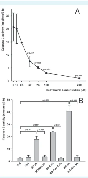
3 activation (P < 0.001). In order to evaluate the protec- tive effect of resveratrol, the cells were treated with several concentrations (10, 25, 50, 75, 100, 200 µM) of resveratrol simultaneously with serum deprivation. Resveratrol pre- vented caspase 3 activation in a dose-dependent manner, with 50% inhibitory concentration (IC50) = 66.3 ± 13.81 µM.
Caspase 3 activation following 3 hour serum deprivation was completely inhibited at 200 µM resveratrol concentra- tion (Figure 1A), and thus this level was used in the further experiments. This protective effect was also obtained after up to 6 hours of serum deprivation (Figure 1B). To verify whether resveratrol regulates the cellular response or di- rectly interacts with caspase 3, resveratrol was added di- rectly to the cytosol extract rather than to cell culture me- dium. Resveratrol showed no direct caspase inhibitory effect, although the known direct inhibitor Ac-VAD-CMK, used as positive control, completely blocked caspase 3 ac- tivity (Figure 2).
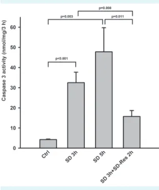
resveratrol exhibited rescue effect on serum deprivation-induced caspase 3 activation
We further investigated whether resveratrol reduced the already up-regulated caspase 3 activity. Primary fibroblasts were exposed to serum deprivation for 3 hours, after which
FIgUre 1. resveratrol dose-dependently prevented caspase 3 activation after 3 h serum deprivation. Control value of caspase 3 activity in serum supported cells: 1.76 ± 0.097 nmol/
mg/3 h (A). 200 µM of resveratrol prevented caspase 3 activa- tion after 3, 4.5, and 6 h serum deprivation (B).
FIgUre 2. resveratrol showed no direct caspase 3 inhibi- tory effect. When 200 µM of resveratrol was added to cytosol extract of serum-deprived fibroblast during caspase 3 activity measurement, it did not significantly reduce caspase 3 activity.
a known direct caspase inhibitor, ac-Vad-CMK, was used as positive control in 20 µM concentration.
the culture medium was supplemented with 200 µM res- veratrol for an additional 2 hours. Resveratrol significantly reduced the already activated caspase 3. It prevented not only its further increase but also reduced it to a level be- low that observed after 3-hour serum deprivation. These experiments indicate that resveratrol may have both pro- tective and rescue effect on cells (Figure 3).
resveratrol reduced lactate dehydrogenase release induced by serum deprivation
Lactate dehydrogenase release was measured to evaluate whether the inhibition of caspase 3 activation by resveratrol was accompanied by increased cell viability. Cell viability decreased by 24 hour serum deprivation was significantly improved by 200 µM resveratrol treatment (Figure 4).
The effect of resveratrol on caspase 3 activity involves p38 kinase pathway
In order to investigate the signaling cascades involved in the protective effects of resveratrol, we carried out experi- ments in the presence of specific inhibitors of p38 (50 µM SB202190), JNK (50 µM SP600125), ERK (50 µM PD184352), PI3K (10 µM wortmannin) kinase pathways, and SIRT1 (5
µM EX-527). Among them, only p38 MAPK inhibitor SB 202190 decreased the protective effect of resveratrol on caspase 3 activation (Figure 5).
FIgUre 3. resveratrol showed rescue effect on caspase 3 acti- vation. Following 3 h of serum deprivation, 200 µM resveratrol was supplemented for an additional 2 h.
FIgUre 4. 200 µM of resveratrol reduced lactate dehydroge- nase release after 24 h serum deprivation.
FIgUre 5. The effect of 50 µM SB202190 (p38 MaPK inhibitor), 50 µM SP600125 (JnK inhibitor), 50 µM Pd184352 (erK inhibi- tor), 10 µM wortmannin (PI3 kinase inhibitor), and 5 µM eX-527 (SIrT-1 inhibitor) on 3-h serum deprivation-induced caspase 3 activation and the protective action of 200 µM resveratrol.
only p38 MaPK inhibitor SB202190 abolished the effect of resveratrol on caspase activation.
The role of oxidative stress in the effect of resveratrol Considering that p38 kinase pathway is activated by mild intracellular stress (13) and pro- and antioxidant properties of resveratrol had been previously described (14), we hy- pothesized that reactive oxygen species generation could be involved in caspase 3 activation induced by serum de- privation and/or the protective effect of resveratrol. To clar- ify if the antioxidant property of resveratrol may play a key role in its cytoprotective effect, we investigated the effect of 5 mM N-acetylcysteine, a well-known antioxidant agent, on caspase 3 activation. Contrary to our expectations, it did not prevent caspase 3 activation but exacerbated it. How- ever, 200 µM resveratrol abolished the combined effect of serum deprivation and N-acetylcysteine on caspase 3 ac- tivation (Figure 6).
dISCUSSIon
Cytoprotective effect of resveratrol
Resveratrol prevented serum deprivation-induced caspase 3 activation in primary fibroblasts and increased their via- bility. These results are in line with those of previous studies
performed on non-transformed cells using various toxic in- sults (15,16). In this study, cytoprotective effect of resvera- trol was considerable, in 100-200 µM concentration range, which is similar to another study (17). However, some re- cent studies observed lower concentrations, in the 10-20 µM range to be efficient as well (9,16). The effective dose probably depends on the cell type and the intensity of the damaging insult used. The concentration found to be ef- fective in the present study is considerably higher than the concentration that can be obtained from dietary sources, suggesting the need for resveratrol supplementation. Fur- thermore, resveratrol can serve as a lead compound for re- search of more potent cytoprotective medications.
To the best of our knowledge this is the first report demon- strating that resveratrol abolishes the already elevated cas- pase 3 activity induced by serum deprivation, suggesting its rescue effect. Resveratrol was found to prevent and im- prove cardiac function in cardiac fibroblasts (18,19) and to play a neuroprotective role in neurotoxic injury (20). How- ever, our results showed that it is a promising cytoprotec- tive agent which should be explored not only for preven- tion of age-related degenerative disorders, but also in the early treatment of degeneration following an acute insult.
Probable mechanism of resveratrol action
It has already been suggested that several kinase pathways have a role in the cytoprotective effects of resveratrol. Cy- toprotective functions of resveratrol were associated with the activation of PI3-kinase/Akt (21,22), p38 MAPK/JNK/ERK (23,24) signaling, and molecular pathways involving SIRT1 (9), an NAD dependent histone deacetylase. Our present findings indicate that the most critical signaling pathway in the protective effect of resveratrol against serum de- privation-induced caspase 3 activation is the activation of p38. The reports about the effects of resveratrol on p38 ki- nase pathway are rather contradictory. It was shown that through inhibition of p38 pathway resveratrol suppresses macrophage and vascular smooth muscle cell apoptosis (17,23). On the other hand, it exerted protective effect in H9c2 embryonic rat heart derived cells by up-regulating the p38 MAPK signaling (25). It was also shown to inhib- it the proliferation of human primary fibroblasts and en- hance their entry to senescence in p38 dependent man- ner (26). Therefore, p38 kinase seems to have a dual role as a regulator of cell fate, mediating either survival or death.
Adams et al (27) reported that the specific function of p38 MAPKs in apoptosis depended on the cell type, stimuli, and/or p38 isoform. In accordance with their findings, we FIgUre 6. Five mM of n-acetylcysteine (naC) exacerbated
serum deprivation-induced caspase 3 activation, but 200 µM of resveratrol prevented their combined effect.
showed that p38 MAPK had a cytoprotective rather than proapoptotic role.
Several articles discuss antioxidant properties of resvera- trol as the cause of its cytoprotective effect (14,16). Con- sidering that N-acetylcysteine exacerbated rather than prevented serum deprivation-induced caspase activation and resveratrol abolished their combined effect, antioxi- dant properties cannot explain its protective action. Pre- vious articles reported similar effect of N-acetylcysteine, concluding that the elevated glutathione level can inhibit NF-κB induced transcription of inhibitor of apoptosis pro- tein, which can explain its potentiating effect on caspase activation (28,29). Since several previous reports demon- strated not only antioxidant but prooxidant characteristics of resveratrol (14,30), the latter might be involved in the activation of p38 MAPK and reduction of caspase 3 acti- vation. A previous study suggested that the prooxidant activity of resveratrol was responsible for its inhibitory ef- fect on apoptosis by creating an intracellular milieu non- permissive for caspase activation (30). These findings are in line with our results, which also indicate the role of p38 kinase in the protective effect of resveratrol and activation of this pathway by mild intracellular stress (13). However, the effect of resveratrol on oxidative state of cells requires further research.
Activation of p38 was also connected to increase in au- tophagic flux. This process is involved in the degradation of misfolded proteins or damaged organelles, such as de- polarized mitochondria, which can prevent the release of proapoptotic mediators and the consequent caspase acti- vation (25). Accordingly, two recent papers reported that resveratrol improved autophagic flux and prevented cas- pase cleavage in H9c2 rat cardiomyoblast cells (25,31). Sim- ilarly to our results, the protective effect of resveratrol de- pended on p38 MAPK activity (25). Based on these data, we can hypothesize that the effect of resveratrol on caspase 3 activation and cell survival might be connected with its prooxidant property, which may enhance autophagic flux via p38 activation.
A major limitation of this study is its in vitro nature, which is why further translational experiments are required to analyze the cytoprotective effect of resveratrol. In conclu- sion, we demonstrated the p38 MAPK signaling pathway- dependent cytoprotective effect of resveratrol against se- rum deprivation induced caspase 3 activation in primary fibroblasts. Also, resveratrol exhibited a rescue effect and reduced the already up-regulated caspase 3 activity. This
finding may contribute to the research of drugs used for prevention and treatment of age-related disorders.
Funding The research was supported by the RECOOP HST Association.
ethical approval received from the ethics committee of the Semmelweis University (22.1/606/001/2010, February 5, 2010)
declaration of authorship ZU, FB, and IV performed the experiments and evaluated the results, and ZU, ES, and TT participated in the planning of ex- periments, evaluation of results, and manuscript preparation.
Competing interests All authors have completed the Unified Competing Interest form at www.icmje.org/coi_disclosure.pdf (available on request from the corresponding author) and declare: no support from any organi- zation for the submitted work; no financial relationships with any organiza- tions that might have an interest in the submitted work in the previous 3 years; no other relationships or activities that could appear to have influ- enced the submitted work.
references
1 Fonseca-Kelly Z, nassrallah M, Uribe J, Khan rS, dine K, dutt M, et al. resveratrol neuroprotection in a chronic mouse model of multiple sclerosis. Frontiers in neurology. 2012;3:84.
Medline:22654783 doi:10.3389/fneur.2012.00084
2 darzynkiewicz Z, Zhao h, halicka hd, li J, lee YS, hsieh TC, et al. In search of antiaging modalities: evaluation of mTor- and roS/dna damage-signaling by cytometry. Cytometry a. 2014;85:386-99.
Medline:24677687 doi:10.1002/cyto.a.22452 3 Jang Jh, Surh YJ. Protective effect of resveratrol on beta-
amyloid-induced oxidative PC12 cell death. Free radic Biol Med. 2003;34:1100-10. Medline:12684095 doi:10.1016/S0891- 5849(03)00062-5
4 Jang M, Cai l, Udeani go, Slowing KV, Thomas CF, Beecher CW, et al. Cancer chemopreventive activity of resveratrol, a natural product derived from grapes. Science. 1997;275:218-20.
Medline:8985016 doi:10.1126/science.275.5297.218 5 rege Sd, geetha T, griffin gd, Broderick Tl, Babu Jr.
neuroprotective effects of resveratrol in alzheimer disease pathology. Frontiers in aging neuroscience. 2014;6:218.
Medline:25309423 doi:10.3389/fnagi.2014.00218 6 li Q, huyan T, Ye lJ, li J, Shi Jl, huang QS. Concentration-
dependent biphasic effects of resveratrol on human natural killer cells in vitro. J agric Food Chem. 2014;62:10928-35.
Medline:25360711 doi:10.1021/jf502950u
7 Barger Jl, Kayo T, Vann JM, arias eB, Wang J, hacker Ta, et al. a low dose of dietary resveratrol partially mimics caloric restriction and retards aging parameters in mice. PloS one. 2008;3:e2264.
Medline:18523577 doi:10.1371/journal.pone.0002264 8 Trincheri nF, nicotra g, Follo C, Castino r, Isidoro C. resveratrol
induces cell death in colorectal cancer cells by a novel pathway involving lysosomal cathepsin d. Carcinogenesis. 2007;28:922-31.
Medline:17116725 doi:10.1093/carcin/bgl223
9 Zhang l, guo X, Xie W, li Y, Ma M, Yuan T, et al. resveratrol exerts an anti-apoptotic effect on human bronchial epithelial cells undergoing cigarette smoke exposure. Molecular Medicine
reports. 2015;11:1752-8. Medline:25385506
10 Khan rS, Fonseca-Kelly Z, Callinan C, Zuo l, Sachdeva MM, Shindler KS. SIrT1 activating compounds reduce oxidative stress and prevent cell death in neuronal cells. Front Cell neurosci. 2012;6:63.
Medline:23293585 doi:10.3389/fncel.2012.00063
11 nagy a, gertsenstein M, Vintersten K, Behringer r. Preparing mouse embryo fibroblasts. CSh Protocols. 2006;2006(1).
12 andrews nC, Faller dV. a rapid micropreparation technique for extraction of dna-binding proteins from limiting numbers of mammalian cells. nucleic acids res. 1991;19:2499.
Medline:2041787 doi:10.1093/nar/19.9.2499
13 Zarubin T, han J. activation and signaling of the p38 MaP kinase pathway. Cell res. 2005;15:11-8. Medline:15686620 doi:10.1038/
sj.cr.7290257
14 de la lastra Ca, Villegas I. resveratrol as an antioxidant and pro- oxidant agent: mechanisms and clinical implications. Biochem Soc Trans. 2007;35:1156-60. Medline:17956300 doi:10.1042/
BST0351156
15 liang Q, Wang XP, Chen TS. resveratrol protects rabbit articular chondrocyte against sodium nitroprusside-induced apoptosis via scavenging roS. apoptosis. 2014;19:1354-63. Medline:25001340 doi:10.1007/s10495-014-1012-1
16 Zhou X, Chen M, Zeng X, Yang J, deng h, Yi l, et al. resveratrol regulates mitochondrial reactive oxygen species homeostasis through Sirt3 signaling pathway in human vascular endothelial cells. Cell death and disease. 2014;5:e1576. Medline:25522270 doi:10.1038/cddis.2014.530
17 guo r, Su Y, liu B, li S, Zhou S, Xu Y. resveratrol suppresses oxidised low-density lipoprotein-induced macrophage apoptosis through inhibition of intracellular reactive oxygen species generation, loX-1, and the p38 MaPK pathway.
Cell Physiol Biochem. 2014;34:603-16. Medline:25116358 doi:10.1159/000363026
18 liu J, Zhuo X, liu W, Wan Z, liang X, gao S, et al. resveratrol inhibits high glucose induced collagen upregulation in cardiac fibroblasts through regulating TgF-beta1-Smad3 signaling pathway. Chem Biol Interact. 2015;227:45-52. Medline:25559857 doi:10.1016/j.
cbi.2014.12.031
19 Chen T, li J, liu J, li n, Wang S, liu h, et al. activation of SIrT3 by resveratrol ameliorates cardiac fibrosis and improves cardiac function via the TgF-beta/Smad3 pathway. am J Physiol heart Circ Physiol. 2015;308:h424-34. Medline:25527776 doi:10.1152/
ajpheart.00454.2014
20 ai Z, li C, li l, he g. resveratrol inhibits beta-amyloid-induced neuronal apoptosis via regulation of p53 acetylation in PC12 cells.
Molecular Medicine reports. 2015;11:2429-34. Medline:25483559
21 liu Mh, Yuan C, he J, Tan TP, Wu SJ, Fu hY, et al. resveratrol protects PC12 cells from high glucose-induced neurotoxicity via PI3K/akt/
Foxo3a pathway. Cell Mol neurobiol. 2015. Medline:25471227 doi:10.1007/s10571-014-0147-5
22 Simao F, Matte a, Pagnussat aS, netto Ca, Salbego Cg. resveratrol prevents Ca1 neurons against ischemic injury by parallel modulation of both gSK-3beta and CreB through PI3-K/akt pathways. eur J neurosci. 2012;36:2899-905. Medline:22817531 doi:10.1111/j.1460-9568.2012.08229.x
23 guo r, li W, liu B, li S, Zhang B, Xu Y. resveratrol protects vascular smooth muscle cells against high glucose-induced oxidative stress and cell proliferation in vitro. Medical Science Monitor Basic research. 2014;20:82-92. Medline:24971582 doi:10.12659/
MSMBr.890858
24 Kutuk o, Poli g, Basaga h. resveratrol protects against 4-hydroxynonenal-induced apoptosis by blocking JnK and c-JUn/aP-1 signaling. Toxicological Sciences. 2006;90:120-32.
Medline:16322078 doi:10.1093/toxsci/kfj055
25 lv XC, Zhou hY. resveratrol protects h9c2 embryonic rat heart derived cells from oxidative stress by inducing autophagy: role of p38 mitogen-activated protein kinase. Can J Physiol Pharmacol.
2012;90:655-62. Medline:22537597 doi:10.1139/y2012-051 26 Faragher rg, Burton dg, Majecha P, Fong nS, davis T, Sheerin
a, et al. resveratrol, but not dihydroresveratrol, induces premature senescence in primary human fibroblasts. age (dordr).
2011;33:555-64. Medline:21318333 doi:10.1007/s11357-010- 9201-5
27 adams rh, Porras a, alonso g, Jones M, Vintersten K, Panelli S, et al. essential role of p38alpha MaP kinase in placental but not embryonic cardiovascular development. Mol Cell. 2000;6:109-16.
Medline:10949032 doi:10.1016/S1097-2765(05)00014-6 28 Zhang h, limphong P, Pieper J, liu Q, rodesch CK, Christians e, et
al. glutathione-dependent reductive stress triggers mitochondrial oxidation and cytotoxicity. FaSeB J. 2012;26:1442-51.
29 Qanungo S, Wang M, nieminen al. n-acetyl-l-cysteine enhances apoptosis through inhibition of nuclear factor-kappaB in hypoxic murine embryonic fibroblasts. J Biol Chem. 2004;279:50455-64.
Medline:15375156 doi:10.1074/jbc.M406749200 30 ahmad Ka, Clement MV, Pervaiz S. Pro-oxidant activity of
low doses of resveratrol inhibits hydrogen peroxide-induced apoptosis. ann n Y acad Sci. 2003;1010:365-73. Medline:15033754 doi:10.1196/annals.1299.067
31 Wang B, Yang Q, Sun YY, Xing YF, Wang YB, lu XT, et al. resveratrol- enhanced autophagic flux ameliorates myocardial oxidative stress injury in diabetic mice. J Cell Mol Med. 2014;18:1599-611.
Medline:24889822 doi:10.1111/jcmm.12312