Molecular Background of Leak K
⫹Currents: Two-Pore Domain Potassium Channels
PE´ TER ENYEDI AND GA´BOR CZIRJA´K
Department of Physiology, Semmelweis University, Budapest, Hungary
I. Introduction 559
II. TWIK (Tandem of Pore Domains in a Weak Inward Rectifying K⫹Channel) 562 A. (Non-?)functional properties of the members of TWIK subfamily 562 B. Possible mechanisms restricting the functional expression of TWIK channels 562
C. TWIK channels at the gene, mRNA, and protein levels 563
III. TREK (TWIK-Related K⫹Channel) 564
A. Electrophysiological diversity in the TREK subfamily 564
B. Multiplex regulation of TREK/TRAAK channels 565
C. Physiological function and pathophysiology of TREK/TRAAK channels 571
IV. TASK (TWIK-Related Acid-Sensitive K⫹Channel) 579
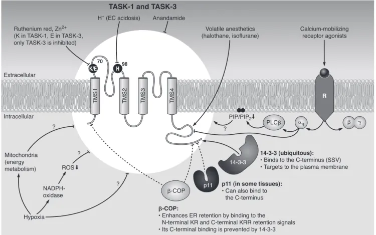
A. Biophysical properties and the molecular mechanisms of acid sensitivity 579 B. Regulation by receptors, endocannabinoids, and interacting proteins 581
C. pH-dependent and other functions mediated by TASK channels 583
V. TALK (TWIK-Related Alkaline pH-Activated K⫹Channel) 590
A. Expression patterns, electrophysiology, and regulation by alkaline pH 590
B. Alkaline pH-related functions of TALK channels 592
VI. THIK (Tandem Pore Domain Halothane-Inhibited K⫹Channel) 593
VII. TRESK (TWIK-Related Spinal Cord K⫹Channel) 593
A. Unique single-channel behavior 593
B. Regulation by Ca2⫹and protein interactions 594
C. Physiological significance of TRESK 595
VIII. Conclusions 596
Enyedi P, Czirja´ k G.Molecular Background of Leak K⫹Currents: Two-Pore Domain Potassium Channels.Physiol Rev90: 559 – 605, 2010; doi:10.1152/physrev.00029.2009.—Two-pore domain K⫹(K2P) channels give rise to leak (also called background) K⫹currents. The well-known role of background K⫹currents is to stabilize the negative resting membrane potential and counterbalance depolarization. However, it has become apparent in the past decade (during the detailed examination of the cloned and corresponding native K2Pchannel types) that this primary hyperpolar- izing action is not performed passively. The K2Pchannels are regulated by a wide variety of voltage-independent factors. Basic physicochemical parameters (e.g., pH, temperature, membrane stretch) and also several intracellular signaling pathways substantially and specifically modulate the different members of the six K2Pchannel subfamilies (TWIK, TREK, TASK, TALK, THIK, and TRESK). The deep implication in diverse physiological processes, the circumscribed expression pattern of the different channels, and the interesting pharmacological profile brought the K2Pchannel family into the spotlight. In this review, we focus on the physiological roles of K2Pchannels in the most extensively investigated cell types, with special emphasis on the molecular mechanisms of channel regulation.
I. INTRODUCTION
The high resting potassium conductance of the plasma membrane was recognized in the era when the principal concepts of electrophysiology about ion chan- nels and the generation of membrane potential were de- veloped (135, 136). The high K⫹ conductance could be most simply explained by the presence of (unregulated)
potassium-selective pores in the plasma membrane and a background (leak) K⫹ current conducted through these pores. On the basis of the constant field theory of Gold- man, Hodgkin and Katz (114, 137), the current-voltage relationship of these hypothetical background K⫹ chan- nels could be predicted. However, the molecular entities responsible for the background K⫹ currents have not been found for a half century. The first representative
(and several further) coding DNAs of all the other major (voltage-gated, inwardly rectifying, and calcium-depen- dent) K⫹ channel families had already been cloned (8, 106, 179), when finally in 1996, the identification of TWIK (tandem of pore domains in a weak inward rectifying K⫹ channel, now called TWIK-1) gave birth to the last, rapidly emerging family of mammalian background potassium channels (190). Actually, cloning and functional expres- sion of the first K⫹ channel with two pore domains per subunit was reported from Saccharomyces cerevisiae, and a new family of “potassium channels with two pore domains in tandem” was predicted in 1995 (159). How- ever, this yeast channel, TOK1 (YORK) (159, 189), pos- sessed eight putative transmembrane segments, and it was strongly outwardly rectifying. As TOK1 differs from the mammalian K2P channels both structurally and func- tionally, we will not discuss it further in this review.
Between 1996 and 2003, the family of cloned mam- malian background K⫹ channels embraced 14 further members encoded by different genes (7, 11, 47, 84, 99, 112, 165, 170, 192, 194, 285, 286, 289, 294, 299). The members were divided into six subfamilies (TWIK, TREK, TASK, TALK, THIK, and TRESK) on the basis of sequence simi- larity and functional resemblance (Fig. 1). The categori- zation into subfamilies was reasonable despite the rela- tively low numbers of members, since the sequence vari- ation between the different background K⫹ channel subfamilies proved to be almost as high as that between
the families of K⫹ channels. Although the members of different subfamilies show relatively low sequence simi- larity (e.g., 28% identity between TWIK-1 and TREK-1 at the protein level; Ref. 99), all members of the background potassium channel family are characterized by the same general molecular architecture. The name of the family,
“two-pore domain” potassium (K2P) channels, was given on the basis of the distinguishing topology of the K⫹ channel subunits. Each subunit contains two K⫹channel pore loop forming (P) domains, in contrast to the other K⫹ channel families characterized by one P domain per one subunit stoichiometry. (Accordingly, K2P subunits dimerize to constitute the functional K⫹selectivity filter containing four pore loop domains, a structure character- istic for all known K⫹ channels). It is important to note that all mammalian K2P channel subunits possess four transmembrane segments; the 4TM/2P structure defines the membership in the K2P channel family (see Fig. 1). It is also worth mentioning that the K2P channels are not restricted to mammals; their genes can also be found in lower organisms. ORK1 (later renamed by the authors to KCNKO), a Drosophila melanogaster background K⫹ channel with 4TM/2P structure, was also reported in 1996 (116). An especially high number (⬎40 genes) of K2P channels were found inCaenorhabditis elegans(76, 182).
Although they had significantly different domain compo- sition from most of the animal channels outside the 4TM/2P core, two-pore domain K⫹ channels have also
FIG. 1. Schematic transmembrane topology (left) and dendrogram of human two-pore domain potassium (K2P) channels (right). Transmem- brane segments are indicated by blue cylinders (TMS1-4). Note that the length of the COOH-terminal tail and the intracellular loop between TMS2 and -3 is highly variable in the different channels. The topology is not drawn to scale, and only one subunit of the functional dimer is depicted. On the dendrogram, both the conventional and systematic (HUGO) names are indicated. Note that KCNK8, KCNK11, and KCNK14 do not exist.
been cloned from different plants (62, 242), indicating that channels with the 4TM/2P architecture are widespread both in the animal and plant kingdoms.
In addition to the 4TM/2P topology, the electro- physiological features of K2Pchannels are also similar.
It is well-accepted in the literature that K2P channels give rise to background K⫹ currents. Therefore, it is practical to keep in mind what kind of properties an ideal background K⫹current should have if it followed the Goldman-Hodgkin-Katz (GHK) current equation. An ideal background K⫹current is not voltage dependent, meaning that the probability of opening (Po) of the channels is the same at all membrane potential values.
(Pois also independent from the K⫹concentrations on the two sides of the plasma membrane.) Another im- portant feature of the hypothetical background K⫹cur- rent is that the amplitude of the current instanta- neously follows the changes of the membrane potential.
[It is also said that the channel is “time independent”
(190) or the channel does not have activation, deacti- vation, and inactivation kinetics (84).] In sharp contrast to the K2Pchannels, thePoof the voltage-gated channel types is regulated by the membrane potential. This is achieved via a relatively slow rearrangement of protein conformation. Therefore, the alteration of the voltage- gated K⫹ current is delayed after the change of the membrane potential at the millisecond timescale (Fig. 2A). Since the membrane potential does not influ- ence the Po of the ideal background K⫹ channel, and the driving force of K⫹is determined by the membrane potential at the submillisecond timescale, a rapid change of the membrane potential results in an “imme- diate” alteration of the background K⫹ current; thus a voltage step in a voltage-clamp experiment induces a square wave-like K⫹ current (Fig. 2B). Moreover, an ideal background channel is not rectifying; opposite driving forces (electrochemical gradients) of equal am- plitudes induce opposite currents of equal amplitudes.
This means that the apparent rectification in physiolog- ical solutions (mentioned as “open rectification” by some authors, Ref. 188) is caused exclusively by the unequal K⫹ concentrations on the two sides of the plasma membrane (Fig. 3). (If [K⫹] was equal on both sides, then the current-voltage relationship would be a line passing through the origin.) If a K⫹current approx- imately meets the above criteria, then it can be consid- ered as a background (leak) K⫹ current. Although real K2Pchannels do not perfectly fulfill these criteria (due to weak voltage dependence, interactions of multiple ions in the channel pore, rectification in symmetrical [K⫹], etc.), they provide the best approximation by far, compared with the other K⫹channel families. In the follow- ing sections we are going to highlight how the currents of the different K2P channels deviate from the above princi- ples, and also discuss the (possible or established) phys-
iological roles of the given channel with special emphasis on the regulation of channel activity.
In this review, where it is possible, we use the conventional names of K2P channels (given by the re- searchers, generally beginning with the letter T), as these names can most often be found in the scientific literature. A systematic nomenclature was accepted by the Human Genome Organization (HUGO) for the K2P channels genes (KCNK1-18) and another for the protein products (K2P1-18) (115). (This latter nomenclature also takes the splice variants into account by extending the name with a dot and a further number, e.g., K2P1.1.) In parentheses, we also give the systematic names of gene and protein product once for each channel.
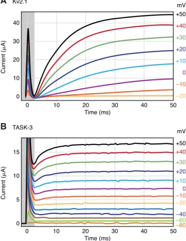
FIG. 2. Representative current traces of voltage-gated and back- ground potassium channels. Kv2.1 voltage-gated (A) or TASK-3 back- ground K⫹channels (B) were expressed inXenopusoocytes. The cur- rents were elicited by voltage steps from ⫺80 to ⫹50 mV in 10-mV increments in a solution containing 2 mM [K⫹]. After the capacitative transient (indicated by the gray-shaded area), Kv2.1 is not yet active at 2.5 ms (A), whereas TASK-3 current is close to its maximum (B). TASK-3 recordings also contain a voltage-dependent component mainly reflect- ing the increasedPoof the channel at depolarized potentials (286). This whole oocyte recording is compatible with the instantaneous nature of the major fraction of TASK-3 current. (Kv2.1 was not activated by voltage steps from⫺80 to⫺30 mV.) The voltage labels of these curves on therightofA(and those of every second TASK-3 curves below⫺20 mV inB) were omitted. The holding potential was⫺80 mV for both cells.
II. TWIK (TANDEM OF PORE DOMAINS IN A WEAK INWARD RECTIFYING Kⴙ CHANNEL)
A. (Non-?)functional Properties of the Members of TWIK Subfamily
Electrophysiological characterization and understand- ing of the functional significance of TWIK channels [TWIK-1 (KCNK1, K2P1.1), TWIK-2 (KCNK6, K2P6.1), and KCNK7 (K2P7.1)] has been impeded by the low or absent functional expression in heterologous expression sys- tems. TWIK-1 has been reported to give rise to a weakly inwardly rectifying K⫹current inXenopus laevisoocytes in the first study of K2P channels (190). In this original report, despite the high amount of injected cRNA, only small currents were induced (190), the amplitudes of which were close to those of endogenous currents of Xenopusoocytes (190). In subsequent independent exper- iments, TWIK-1 (also mentioned as HOHO1) could not be expressed in the same expression system (67, 117, 279, 283). This suggests that presently unidentified (perhaps oocyte preparation-specific) factors may have enabled the moderate functional expression in the first study. In sym- metrical [K⫹], TWIK-1 single-channel conductance was 34
pS (at ⫺80 mV in symmetrical 140 mM [K⫹]) in the first study (190); this value was also confirmed by others with a mutant form (K274E, see sect. IIB) of human TWIK-1, producing higher current density than the wild-type chan- nel (283). TWIK-1 was inhibited by intracellular acidifica- tion and activated by the application of phorbol 12-myris- tate 13-acetate (PMA, the pharmacological activator of protein kinase C), although the latter effect was suggested to be indirect (190, 191). These functional properties were also found to be characteristic for TWIK-2 current (47).
Expression of human TWIK-2 in Xenopus oocytes also resulted in small current amplitudes (⬍0.5–1A, in 115 mM [K⫹] at⫺100 mV; Ref. 47), and as in the case of TWIK-1, others could not detect human or mouse TWIK-2 (also called TOSS) current inXenopusoocytes (67, 279).
Coexpression of TWIK-1 and TWIK-2 subunits also failed to induce K⫹current (279). Interestingly, rat TWIK-2 pro- duced 15-fold higher currents than the human channel in COS-7 cells; thus rat TWIK-2 was more readily character- ized (270). According to these (sometimes contradictory) reports, the expression of TWIK-2 may also be limited in the different expression systems (with the possible ex- ception of rat TWIK-2).
TWIK-2 (like TWIK-1) induced a weakly inwardly rectifying current in symmetrical [K⫹] (47, 270). The cur- rent deviated from that of an ideal background channel as the activity was influenced by the membrane potential.
About 50% of TWIK-2 inactivated slowly (⬃2 s) at strong depolarization (steady-state half inactivation at ⫹65 mV) (270), while the remaining fraction showed background K⫹ current properties (270). The inactivation was also modified by the extracellular K⫹concentration. TWIK-2 inactivated at physiological [K⫹] distribution, whereas no inactivation was observed in symmetrical high [K⫹] (47, 270).
The third member of the TWIK subfamily, K2P7 (KCNK7), could not be heterologously expressed in Xe- nopusoocytes or COS-7 cells (27, 294). (KNCK7 of differ- ent species was also called KCNK6 or KCNK8 in the first studies. Later KCNK6 was assigned to TWIK-2, and the KCNK8 name was omitted.) A structural trait of mouse KCNK7 is its COOH-terminal EF-hand (potential Ca2⫹ binding) domain (294), which is unique in the mammalian K2P channels. Human KCNK7 has an unconventional se- quence (GLE) in the second K⫹channel pore (P2) domain (294), whereas in other K⫹ channels in homologous posi- tions, the glycine residues are strictly conserved (GYG, GLG, or GFG) and considered as indispensable in forming the selectivity filter. This raises the question whether the human KCNK7 protein may function as a K⫹channel at all.
B. Possible Mechanisms Restricting the Functional Expression of TWIK Channels
Two different hypotheses were suggested to explain the low/absent functional expression of TWIK-1. Accord-
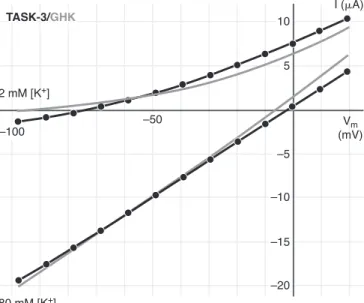
FIG. 3. Approximation of the current of TASK-3 with Goldman- Hodgkin-Katz (GHK) current equation. K⫹currents of aXenopusoocyte expressing TASK-3 were evoked by depolarizing voltage steps from
⫺100 to⫹20 mV in 2 or 80 mM extracellular [K⫹]. The instantaneous components of TASK-3 current, measured at 2.4 ms after the initiation of the voltage steps, were plotted (circles connected with black lines). The data in 2 and 80 mM [K⫹] were approximated by the GHK current equation (gray curves) in a single fitting process applying the least- squares method with the permeability constant and the intracellular [K⫹] as variables. Note that the GHK curves only approximate the instantaneous component of the current, and the steady-state current (e.g., at 50 ms, see Fig. 2B, also including the voltage-dependent com- ponent of TASK-3 current) would be even less well fitted (not shown).
ing to the first one, TWIK-1 is mainly located in intracel- lular compartments, and its transfer to the plasma mem- brane is tightly regulated. Expressed TWIK-1 was present primarily in the pericentriolar recycling endosome in non- polarized cell types (HeLa, CHO, and COS cells) and in the subapical recycling endosome in polarized (MDCK) cells (78). The native channel was also abundant in the subapical region of renal proximal tubule cells (78). An ARF6-depen- dent interaction was demonstrated between TWIK-1 and EFA6 (78). EFA6 is a GDP/GTP exchange factor for ARF6 small G protein, which is known to regulate membrane recycling, actin cytoskeleton organization, and vesicle trafficking (102). It has been hypothesized that sequestra- tion of the channel in intracellular compartments may be the consequence of this ternary interaction, and the im- portance of TWIK-1 in the modulation of endocytosis and intracellular vesicle trafficking has also been suggested (78). Nevertheless, functional TWIK-1 expression in the plasma membrane has not been achieved by manipulation of the ARF6/EFA6 system (78).
According to the other hypothesis, TWIK-1 is inserted into the plasma membrane, but it is silenced by a special posttranslational modification: sumoylation (283). SUMO (small ubiquitin-related modifier) proteins are reversibly and covalently linked to the ⑀-amino group of a lysine residue located in consensus sumoylation sequences of target proteins (246). K274E mutation of the putative sumoylation site of TWIK-1 rendered the channel func- tional in Xenopus oocytes and in COS-7 cells (283).
Sumoylation of wild-type TWIK-1 was biochemically ver- ified; desumoylation of the channel with SENP1 enzyme resulted in an appropriate molecular weight shift of the protein. SENP1-treated wild-type and sumoylation-resis- tant K274E mutants were indistinguishable on Western blots. Coexpression of SENP1 increased TWIK-1 whole cell current in Xenopus oocytes, and application of the desumoylating enzyme to excised patches also increased the activity of the channel (283).
However, the theory of TWIK-1 sumoylation was soon challenged (96). It was shown that the current den- sities of wild-type and K274R-mutant TWIK-1 were iden- tical, although this mutant could not be sumoylated. (In these experiments the HcRed-K2P1 fusion construct was used, which resulted in measurable “wild-type” TWIK-1 current.) Therefore, it has been suggested that the higher current density of K274E mutant could be the conse- quence of a charge effect unrelated to sumoylation. Fur- thermore, the previous results on the wild-type channel regarding the molecular weight shift and functional con- sequences of desumoylation with SENP1 could not be reproduced (96). Because both sets of conflicting exper- iments (96, 283) seem to be convincing, further investiga- tion is required to decide the principal question whether TWIK-1 is sumoylated or not.
Despite the small (47, 270) or absent (279) human TWIK-2 currents inXenopus oocytes and the 15-fold dif- ference between human and rat TWIK-2 currents in COS-7 cells (270), the mechanism restricting functional expres- sion of TWIK-2 has not been examined so far. In contrast, the third member of the TWIK subfamily, KCNK7, clearly failed to induce K⫹current in heterologous systems, and therefore, the question was immediately raised whether the protein was unable to reach the plasma membrane or whether it was inactive on the cell surface. Immunofluo- rescence localization of KCNK7 in permeabilized COS-7 cells indicated that the channel protein was sequestered in the endoplasmic reticulum (294). This result suggests that KCNK7 may reside in the endoplasmic reticulum under physiological conditions or a presently unknown factor, absent from COS-7 cells, is required for its surface expression.
C. TWIK Channels at the Gene, mRNA, and Protein Levels
While the electrophysiology of TWIK channels re- mains enigmatic, an exciting direct interaction of KCNK1 gene has been recently revealed (20). A member of the p53 tumor suppressor family, TAp73, has been shown to transactivate the KCNK1 gene. TAp73 inhibits anchorage- independent growth of tumor cells, and it has been sug- gested that TWIK-1 might mediate its effect. In fact, si- lencing of TWIK-1 expression promoted growth of tumor cells similarly to the knockdown of TAp73. As TWIK-1 is downregulated in different tumors, including glioblas- toma, melanoma, and advanced stages of ovarian cancer, it was plausible to assume that the channel modulated tumor aggressiveness and metastasis (20).
Expression of TWIK-1 has been detected in several tissues, including the kidney, brain, and lung (5, 190). In the kidney, TWIK-1 immunoreactivity has been found in different parts of the nephron; however, the expression pattern showed considerable differences in various spe- cies (59, 195, 233, 251, 261). Unexpectedly, the principal cells of cortical collecting duct of TWIK-1⫺/⫺ knockout mice were hyperpolarized (233), and the increased uri- nary phosphate and free water clearance reflected re- duced expression of the sodium-phosphate cotransporter in the proximal tubule and inappropriate internalization of aquaporin-2 in the collecting duct (251). Thus the ab- sence of TWIK-1 at these locations appeared to interfere with proper targeting and/or regulation of other transport- ers. TWIK-1 was consistently expressed in the thick as- cending limb of the loop of Henle in all examined species, raising the possibility that the function of TWIK-1 is re- lated to the K⫹recycling in these cells, the mechanism of which is primarily attributed to the inwardly rectifying K⫹ channel, ROMK (Kir1.1) (59).
TWIK-1 was also detected in the heart (86, 329), and its expression was altered in atrial fibrillation. However, these results are inconsistent as both increased (108) and reduced (86) TWIK-1 mRNA levels have been reported.
Thus further work is required to elucidate whether TWIK-1 participates in the electrophysiological maladap- tation characteristic of chronic atrial fibrillation (249, 335).
TWIK-1, like many other K2P channel subunits, is expressed along the auditory and vestibular sensory sys- tem, including neurons and also nonsensory epithelia (53, 61, 138, 250). After bilateral cochlear ablation, TWIK-1 mRNA levels were reduced significantly (although not as dramatically as TASK-5 mRNA) in both the cochlear nu- cleus and colliculus inferior (61, 138), suggesting that TWIK-1 might also contribute to the deafness-associated sensitization of the neural auditory pathway.
TWIK-1 expression has been documented in several other tissues: in the brain (29, 175, 282), in airway epithe- lial cells (75, 143), and in the umbilical cord vein (266).
Different theories were proposed for the physiological functions of TWIK channels at these locations. We will not discuss these theories further, as they remain clearly hypothetical until TWIK K⫹current is analyzed directly in the corresponding cell types.
In the absence of apparent functional expression, another successful line of investigation was the biochem- ical analysis of TWIK-1 protein (193). It was demonstrated that TWIK-1 subunits formed homodimers in vitro, which were dissociated by treatment with-mercaptoethanol. A disulfide bridge, formed between the cysteines at residue 69 in the first extracellular loops (193), linked the two subunits. The experimental data suggested that TWIK-1 constitutes functional homodimers also in vivo, and both pore domains of TWIK-1 subunits contribute to the for- mation of K⫹-selective channel pore (193). Although the same cysteine (and thus the disulfide bridge) is not con- served in all the other K2Pchannels, a similar conclusion, regarding the dimeric structure, was derived in the case of the functional TASK-1 channel (213).
III. TREK (TWIK-RELATED Kⴙ CHANNEL)
A. Electrophysiological Diversity in the TREK Subfamily
The first member of the subfamily, TREK-1 (TWIK- related K⫹ channel) (KCNK2, K2P2), was discovered in 1996, as the second mammalian K2Pchannel (99). Subse- quent cloning of the closely related TRAAK (TWIK-related arachidonic acid activated K⫹ channel, KCNK4, K2P4) (100) and TREK-2 (KCNK10, K2P10) (11, 194) completed this group. Functionally active alternative splice variants of both TREK-1 (340), TREK-2 (122), and TRAAK (263)
also contribute to the diversity of the subfamily. [In addi- tion, significant expression of a nonfunctional, truncated form (consisting of only 67 amino acids) of TRAAK (TRAAKt) has also been detected (100).] The structural difference of the functional splice variants is restricted to the extreme NH2-terminal intracellular part. To our present knowledge, these minor variations influence nei- ther the basic biophysical characteristics of the currents nor the regulatory processes of the channels (171, 269), although, intriguingly, the expression pattern of the splice variants shows significant tissue specificity (122, 340). It has been recently reported that the heterogeneity of both TREK-1 and TREK-2 is further increased by alternative translation initiation. In these alternatively translated forms of the channel, a longer segment of the intracellular NH2terminus is missing than in the splice variants. This structural alteration has significant consequences on the biophysical characteristics of the channels (see below).
TREK-1 has been originally characterized as an out- wardly rectifying potassium channel (99). Two indepen- dent mechanisms underlie the deviation from the ideal leak conductance. In the absence of extracellular divalent cations, the single-channel current-voltage (I-V) relation- ship of TREK-1 in symmetrical K⫹ solution is linear or slightly inwardly rectifying (194, 340). However, in the presence of physiological concentrations of extracellular Mg2⫹ or Ca2⫹, the unitary conductance of TREK-1 is progressively reduced in the negative membrane potential range and has a tendency to saturate at very negative values (168, 269). This results in a marked outward rec- tification. The sensitivity to extracellular divalent cations (a mirror image of the mechanism, by which intracellular Mg2⫹or spermine regulate the inwardly rectifying potas- sium channels) is unique for TREK-1 in the subfamily. The amplitude of TREK-2 and TRAAK single-channel currents is not influenced significantly by the presence of extracel- lular Mg2⫹(11, 168, 194).
Voltage-dependent gating of TREK-1 is the other ma- jor factor responsible for outward rectification. TREK-1 single-channel activity has a characteristic, pronounced bursting behavior (99, 269). At highly positive membrane potentials, the Po of TREK-1 can be as high as 0.6 (also depending on the mechanical activation). In turn, at neg- ative membrane potentials, thePoof the channel is greatly reduced (218), and the bursting activity completely disap- pears (269). Early reports described TREK-1 as an almost instantly activating channel (99). Closer analysis revealed that the instantaneous current change during depolariza- tion (reflecting the alteration of the electrochemical driv- ing force) is in fact accompanied by a fast (⫽4 – 6 ms), time-dependent component, representing the voltage-de- pendent activation of TREK-1 (28). TREK-1 does not in- activate, and the channel has a low deactivation time constant ( ⫽ 5–7 ms) (218). The rapid activation and deactivation kinetics may hide the voltage dependence of
TREK-1, when the voltage clamp is not efficient enough during whole cell measurements, and may manifest as an apparent rectification of the K⫹current.
Progressive deletion of the intracellular tail of TREK-1 gradually diminished the transient component of the current (evoked by depolarizing voltage steps), thus simultaneously reducing the apparent rectification of the channel (218). Studying hybrid constructs of TREK-1 and a less voltage-dependent K2P channel, TASK-1, it was demonstrated that the COOH-terminal tail of TREK-1 was not only necessary for the voltage dependence, but its transposition to the core of TASK-1 conferred voltage sensitivity to the chimera (218). However, it should also be noted that the same domain, required for the effect of the membrane potential, is also prominent in a wide variety of other regulatory mechanisms. It is a salient feature of TREK-1 regulation that the highly divergent regulatory factors affect either directly or indirectly the proximal intracellular tail region, and many of these were also shown to modulate the voltage dependence (28, 218).
In symmetrical high (⬃140 mM) K⫹ solution, the single-channel conductance of TREK-1 is 95–130 pS at positive membrane potentials (140, 173, 269, 340). At neg- ative potentials, in the absence of inhibitory extracellular (EC) Mg2⫹, the slope conductance is somewhat higher (28). In addition to this high conductance mode, another lower conductance level of TREK-1 was characterized more recently. Heterologous expression of either the longer or the shorter splice variants produced two popu- lations of channels with the high and low single-channel conductance values (⬃100 and 40 pS, respectively), and the activity of the channels in both conductance modes was changed similarly by the different regulatory factors of TREK-1. Transition between the two conductance val- ues was observed very rarely. Accordingly, it has been proposed that the different conductance levels might arise because of the interaction of the channel with un- known accessory intracellular proteins (340).
TREK-1 gene has a weak natural Kozak sequence, which may be skipped, and translation can initiate from another, internal start codon (AUG site 57 amino acids downstream of the expected NH2terminus). A truncated form of the TREK protein was detected in heterologous expression systems as well as in native tissues. The dele- tion construct (⌬1–56) was shown to have⬃30% larger unitary current but lower Po than the M57I mutant, in which the mutation prevents translation to the shorter deleted form. This mechanism clearly explains the previ- ously observed heterogeneity of the unitary current of TREK-1 expressed from the same mRNA species. A quite unexpected property of the truncated channel was also observed, which may have striking consequences; the ion selectivity of the truncated channel was impaired. Al- though the missing part of the NH2terminus is located far from the selectivity filter, it has been found that TREK-
1(⌬1–56) is also permeable to Na⫹. Thus, considering the physiological ion concentrations, the high relative expres- sion of the⌬1–56 channel form may induce depolarization toward the firing threshold (316).
Alternative translation initiation is not restricted to TREK-1. Three different TREK-2 channels are produced from the same mRNA as a result of skipping the first or the first two initiation site(s). It has long been known that expressed TREK-2 channels have low (⬃50 pS) and high (⬃220 pS) conductance levels and that these levels do not represent different subconductance states, as they are never converted from one to the other. The long TREK-2 variant turned out to be responsible for the lower, while the two shorter forms for the higher conductance chan- nel. The previously reported channels of intermediate conductance may have reflected the heteromer assembly of the short and long variants. Unlike the shorter TREK-1 forms, all the TREK-2 variants preserve their selectivity for K⫹. They were also identical in their response to different regulatory manipulations, which had previously been assigned to the proximal intracellular tail region (303).
TREK-2 shows high sequence similarity to TREK-1 in the proximal region of the intracellular COOH-terminal tail (in addition to the transmembrane segments). As expected on the basis of this resemblance, the membrane potential also affects the gating of TREK-2. ThePoof the channel at ⫹40 mV was determined to be about twice (194) or four times (11) as high as at⫺40 mV. In contrast to TREK-1, however, depolarization decreases the unitary conductance of TREK-2 (11, 122, 168, 194). At the macro- scopic (whole cell) current level, this inward rectification of the unitary current may balance the effect of the volt- age dependence of the gating, resulting in an almost linear (11) or only slightly outwardly rectifying current (194) in symmetrical [K⫹] solutions.
TRAAK current is not influenced by EC Mg2⫹, and its single-channel I-V relationship is only slightly inwardly rectifying in symmetrical high [K⫹] solutions (168). The slope conductance, in physiological [K⫹] solutions, is 45 pS measured in the positive membrane potential range (between 0 and ⫹60 mV) (100). Although it has been reported that the single-channel activity (NPo) of TRAAK in isolated membrane patches was increased three- to eightfold by depolarization (168, 217), at the whole cell level TRAAK behaves as a classical nonrectifying leak channel (100).
B. Multiplex Regulation of TREK/TRAAK Channels
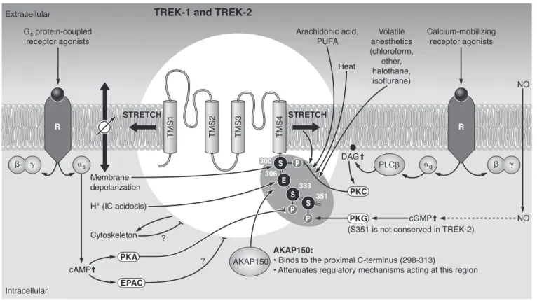
The channels belonging to the TREK subfamily share complex regulatory properties. The first 30 amino acids of the cytoplasmic tail adjacent to the fourth transmembrane segment (TMS) turned out to be absolutely critical for
such diverse regulatory processes as the voltage depen- dence, mechano-, lipid-, and thermosensitivity as well as the modulation by G protein-coupled receptors. On the basis of the analogy with other potassium channels of known crystal structure (238), the second and fourth transmembrane segments of K2Pchannels are envisioned to form the inner pore helices. Secondary structure pre- dictions suggest that the fourth transmembrane ␣-helix extends intracellularly, and this rodlike extension appar- ently has a pivotal role in the regulation of channel activ- ity. TREK-1 and TREK-2 are highly conserved in this region, explaining the very similar regulatory mechanisms of the two channels (see Fig. 4). Analysis of the response of TREK mutants to various stimuli pinpointed single amino acid residues in this ␣-helix, which are targeted during the different types of regulation. Aligning the se- quences of the proximal COOH terminus in the three members of the family clearly shows that TRAAK differs from TREK-1 and TREK-2 at some critical positions. Ac- cordingly, its regulation also deviates from that of the two TREK channels. (A homologous part plays an important role in the receptor-mediated regulation of TASK chan-
nels, suggesting that the intracellular helical structure following the fourth TMS is also a regulatory focal point in the other extensively examined K2P channel subfamily.)
1. Mechanosensitivity
Under basal conditions, the activity of the TREK/
TRAAK channels is rather low, while applying subatmo- spheric (negative) pressure to the patch pipette in cell- attached configuration reversibly activates TREK-1 (269), TREK-2 (194), and TRAAK (168, 217). Laminar shear stress stimulates TREK-1 (269), whereas the shrinkage of the cell induced by extracellular hyperosmolarity reduces the amplitude of TREK-1 and TRAAK currents. This indi- cates that the tension of the cell membrane efficiently regulates channel activity (222, 269). Mechanosensitivity does not depend on cellular integrity; rather, it is clearly membrane delimited. The response to negative pressure in the pipette is not only maintained after patch excision, but the physical disruption of cytoskeletal interactions by the isolation of the membrane patch even increases the basal activity and augments the susceptibility to mechan-
FIG. 4. Multiplex control of TREK-1 and TREK-2. The channels are activated by physicochemical changes, such as stretch or convex deformation of the plasma membrane, depolarization, heat, and intracellular acidosis. Polyunsaturated fatty acids (PUFA) including arachidonic acid (AA) and a wide variety of volatile anesthetics also open TREK channels. Activation of the Gs/cAMP/PKA and the Gq/PLC/DAG/PKC signaling pathways inhibit TREK channels by phosphorylating defined serine residues on the COOH-terminal tail. In some tissues, the effect of the elevated cAMP concentration is mediated by the small G protein EPAC. The interaction of the cytoskeleton with TREK channels has bidirectional consequences; the channels shape the cytoskeleton while being inhibited by it. Another established partner of TREK-1 is AKAP 150, which binds to the key regulatory domain of the channel and modifies its sensitivity towards various modulating factors. TREK-1 is activated via the NO/cGMP/PKG pathway, but the PKG phosphorylation consensus site is missing in TREK-2. (Arrows indicate stimulation; lines withⳕending represent inhibition.)
ical stimuli (168, 186, 217). Pharmacological disintegra- tion of the cytoskeletal network (pretreatment with col- chicine or cytochalasin D) also activates these channels, suggesting that the cytoskeleton exerts a tonic inhibitory effect on them (168, 186). The action of the cytoskeleton might be transmitted by membrane shape or tension, which in turn directly regulates channel activity. Interest- ingly, the regulatory effect seems to be bidirectional; it has been reported that TREK channels influence the cy- toskeletal organization (see more details in sect.IIIC1).
In general, the opening of all TREK/TRAAK channels is facilitated by convex membrane deformation. However, detailed characterization of their response to physical stress revealed delicate variations. TREK-1 (269) and TREK-2 (194) are activated by negative, and to a much smaller degree by positive, pressure applied to the extra- cellular side of the plasma membrane. On the other hand, TRAAK can be activated only by suction from the extra- cellular direction (positive pressure activates only from the intracellular side) (168, 217). Amphipathic molecules, which asymmetrically insert into the lipid bilayer, are known to modify the shape of the membrane character- istically. The anionic crenator trinitrophenol (TNP) causes convex curvature (like the negative pressure does) and activates TREK-1 (269) and TRAAK (217). The oppo- site (concave) change of shape created by the cationic cup former antipsychotic drug chlorpromazine inhibits TREK-1 but does not affect TRAAK (222).
Activation of the channel by the applied mechanical stimulus is graded and reversible. The negative pressure that activated half-maximally TREK-1 and TRAAK was
⫺36 and⫺46 mmHg, respectively (221); however, these values significantly depended on the experimental system and on other regulating factors, such as the intracellular pH, membrane potential, and arachidonic acid (217).
Truncation of the COOH terminus results in the re- duction of mechanosensitivity of TREK channels, if the region next to the fourth TMS is also truncated. A more distal deletion (after T322 in TREK-1), leaving the first 30 amino acids of the COOH-terminal tail intact, does not influence mechanosensitivity, whereas progressive trun- cation before this position gradually decreases mechano- (and other forms of) stimulation and also the amplitude of the basal current (269). The basal channel activity was rescued by fusing the COOH terminus of TASK-1 to the TREK-1 channel core (retaining only the first six amino acids of the COOH-terminal cytoplasmic part of TREK-1);
however, this chimera remained insensitive to shearing forces (269). [The reverse chimera, in which the tail of TREK-1 was transferred to TASK-1, was also entirely insensitive to shear stress (269).] In contrast to the above results, a similar TREK-2/TASK-3C chimera (in which the two channel parts were joined exactly at the transmem- brane/cytoplasmic border) was responsive to the negative pressure, applied directly to the membrane (168). The
discrepancy between the two studies might have arisen because different members of the TREK and TASK sub- families were used, or an alternative explanation may be that the⫺60 mmHg suction, applied directly to the mem- brane exerted a stronger mechanical stimulus than the shear stress (168). It is also possible that the important
␣-helix (the rodlike structure extending the fourth TMS in the cytoplasm) was better preserved in the TREK-2/
TASK-3C chimera despite the shorter sequence retained from the mechanosensitive TREK channel. In addition to the above truncation and chimera experiments, the sig- nificance of the fourth TMS and its COOH-terminal cyto- plasmic ␣-helical appendage in the determination of mechanosensitivity has been further supported by site- directed mutagenesis studies (140) (see further details in sect.IIIB4). These results together suggest that the trans- membrane segments (primarily the fourth one) play a pivotal role in the mechanosensitivity, while an appropri- ate (but not necessarily TREK subfamily specific) COOH- terminal tail fragment is also necessary for proper regu- lation.
2. Arachidonic acid, phospho-, lysophospho-, and other lipids
Both TREK channels and TRAAK are activated by arachidonic acid (AA). In fact, the name TRAAK refers to this sensitivity. AA need not be metabolized to act, and other polyunsaturated fatty acids (PUFA), docosahexae- noic acid, linolenic acid, linoleic acid, etc., also directly stimulate TREK/TRAAK channels. On the contrary, satu- rated fatty acids are not effective (11, 73, 100, 194). In addition to the activation, PUFA sensitize the channels to mechanical stimulation; the channels open in response to less negative pressure. PUFA also interfere with the cAMP/protein kinase A (PKA)-induced inhibition of TREK-1 (269). Stimulation by PUFA does not rely on cellular integrity; it also develops in excised patches.
PUFA are effective from either side of the membrane (100, 168); however, their effect is evoked more rapidly if they are administered to the intracellular compartment (11). The first 28 –30 amino acids in the proximal COOH- terminal tail of TREK-1 (269) and TREK-2 (171) mediate the effect of PUFA; however, in the case of TRAAK, it has been suggested that this region is not absolutely critical and can be substituted by a homologous region of TASK-3 without losing the regulation (168).
Different lysophospholipids (independently of their charge) open TREK and TRAAK channels even more rap- idly than AA (194, 222). In contrast to the fatty acids, however, the effect of lysophosphatidylcholine (LPC) needs cellular integrity. If LPC is applied to the intracel- lular side of excised membrane patches, then it becomes even inhibitory (222). Native TREK-1 in bovine adrenal fasciculata cells was activated by LPC and lysophosphati-
dylinositol (LPI) similarly to the heterologously ex- pressed channel; however, the activation rapidly desensi- tized and subsequent stimuli were ineffective. These re- sults suggest a more complex mode of action with the possible involvement of signaling, metabolism, or auxil- iary channel subunit(s) (73). Unlike the lysophospholip- ids, lysophosphatidic acid (LPA) does not act from out- side (neither on whole cells, nor on right-side-out vesi- cles) (50, 222). However, it activates TREK and TRAAK channels on inside-out vesicles, suggesting that LPA, formed intracellularly during the activation of phospho- lipase C (PLC) and phospholipase A2(PLA2), may have a regulatory potential. LPA also shifts the mechanogat- ing of the channels toward less negative pressure val- ues and converts them to voltage-independent leak con- ductance (50).
Acidic inner leaflet membrane phospholipids, phos- phatidylinositol-4,5-bisphosphate (PIP2) and to a lesser degree phosphatidylserine (PS), also activate TREK-1 from the intracellular side (51, 212). It has been suggested that these phospholipids interact with positively charged amino acid residues in the proximal COOH-terminal do- main (51). Quenching the charge of endogenous phospho- lipids by the polycationic agent polylysine significantly reduces the channel activity in excised patches. In re- sponse to the application of PIP2to the intracellular sur- face, the characteristics of TREK-1 current were also modified, e.g., the channel became less voltage dependent (51, 212). The PIP2 level of the plasma membrane is reduced substantially by the activation of PLC, when the cells are stimulated via Gq protein-coupled receptors.
Therefore, the PIP2-mediated activation of K⫹ current may also change dynamically, also depending on the strength of channel-PIP2interaction (212).
PA and its precursor, diacylglycerol (DAG), were shown to reduce the AA-activated TREK-1 and TREK-2 currents in patches excised from HEK293 cells expressing the channels heterologously (48). In a similar experiment (147), however, inhibition of TREK-2 by DAG was incon- sistent; it was apparent only in ⬃25% of the examined patches. A potentially significant difference between the two experiments was that in the latter, the channels were not preactivated by AA (147). This suggests that the inhi- bition by PA and DAG may be either indirect even in the excised membrane patch, or (more likely) it depends on the simultaneous presence of other local regulating fac- tors (e.g., on the lipid composition of the surrounding membrane).
3. Thermosensitivity
TREK-1 is activated gradually and reversibly, in re- sponse to elevation of the ambient temperature from 14 to 42°C (219). TREK-2 and TRAAK channels share this sen- sitivity in the same temperature range (146). The activity
of TREK-1 increased most steeply between 22 and 42°C.
In this range, the amplitude of TREK-1 (whole cell) cur- rent was enhanced about sevenfold (which is a much larger increase than the activation of most other K⫹chan- nels under identical conditions, e.g.,⬃2-fold for TASK-1).
Thermal activation was also apparent in cell-attached configuration in addition to whole cell measurements.
However, interestingly, after patch excision, the thermo- sensitivity of TREK/TRAAK channels was completely lost (146, 219), under conditions when mechano-, AA, and pH regulation was maintained. Therefore, it has been hypoth- esized that thermosensitivity is not an inherent property of the channels and may depend on an auxiliary (sensor) protein (219). If this theory is correct, then the auxiliary protein must be widespread and present not only in spe- cialized temperature sensor cells but also in a wide vari- ety of other cell types (even in amphibian oocytes), since thermosensitivity was very similar in different expression systems (COS-7 cells, Xenopus oocytes) and in the cell types expressing the channels endogenously (146).
Single-channel conductance of TREK-2 is not influ- enced by temperature, and activation is exclusively due to the elevatedPo(146). Thus temperature affects the gating of these channels, similarly to the (above discussed) other regulatory factors. In the case of TREK-1, deletion of the COOH-terminal tail or its replacement with the COOH terminus of TASK-1 abolished the thermosensitivity of the construct (219). However, the similar chimera composed of the core of TREK-2 and the COOH terminus of TASK-3 remained thermosensitive, suggesting that, in this con- text, the TREK-specific COOH terminus was not abso- lutely critical (146). The apparent contradiction of the results obtained with the two different chimeras is quite similar to the discrepancy discussed more extensively in section IIIB1 and suggests that mechano- and ther- mosensitivity may work through related mechanisms, which are structurally associated with the fourth TMS and the following COOH-terminal tail region.
4. Regulation by pH
A) TREK/TRAAK CHANNELS ARE EFFICIENTLY REGULATED BY PH FROM THE INTRACELLULAR SIDE. In contrast to the canonic acid- and alkaline-sensitive (TASK and TALK) K2Pchannels, the activity of the members of TREK subfamily is modified predominantly by intracellular pH changes. Protons acti- vate both TREK-1 and TREK-2 (171, 194, 221), and also sensitize these channel to mechanical stimuli. TRAAK is not influenced by the acidification, but it is stimulated by the alkalinization of the intracellular solution (from an initial value of pH 7.3) (168). The effect of pH on all TREK/TRAAK channels is direct and apparent not only in cell-attached and whole cell configurations but also in excised patches.
Truncation experiments indicated that the proton sensor function of TREK channels resides in a 30-amino acid stretch of their proximal COOH-terminal intracellu- lar tail. Progressive truncation of this COOH-terminal re- gion of TREK-1 (221) and TREK-2 (171) gradually de- creased the proton sensitivity, and the effect of pH com- pletely disappeared when the truncation approached E306 in TREK-1 or the homologous E333 in TREK-2. The chimera, assembled from the core of TREK-2 and the COOH terminus of TASK-3 (a K2P channel which is not influenced by IC pH changes), was insensitive to acidifi- cation (171), indicating that the COOH terminus of TREK-2 was indispensable for this type of regulation.
Glutamate-306 was identified as the cornerstone of the intracellular proton sensor in TREK-1 by alanine scanning mutagenesis. Its substitution with neutral or positively charged residues rendered the channel constitutively ac- tive, independent of the membrane stretch, and even re- stored the mechanosensitivity of a mutant, truncated more distantly at the COOH terminus. This latter result suggests that the mechanosensitivity is determined by the complicated interaction of the COOH terminus and the channel core, and the protonation of E306 has a pivotal role in the regulation of this interaction. At the same time, the robust activity of the E306A channel was resistant to cAMP-mediated inhibition (characteristic for wild-type TREK-1, see sect. IIIB5). This indicates that E306 inter- twines the regulatory action of the intracellular pH not only with mechanosensitivity but also with the suscepti- bility to the stimulation of Gs protein-coupled receptors (140).
TRAAK also has a glutamate in the position homolo- gous to E306 of TREK-1, but this channel is not influenced by intracellular (IC) acidification (below pH 7.2). Further- more, the mutation of this glutamate to alanine does not affect channel activity (140). In addition, transposition of the COOH-terminal tail of TREK-1 confers acid sensitivity to TRAAK (221), indicating that the transmembrane seg- ments and pore domains of TRAAK can be modulated (and the channel activated by IC protons), if an appropri- ate COOH-terminal proton sensor is installed artificially.
Without this engineered proton sensor, wild-type TRAAK can be modulated by intracellular pH changes only in the alkaline range. Alkalinization activates the channel sug- gesting that the effect is mediated by deprotonation of a residue (or residues), which is protonated at neutral pH.
Although the contributing amino acid(s) have not been unequivocally determined, it is clear that they are not located in the intracellular tail, as the whole cytoplasmic COOH terminus of TRAAK could be replaced by that of TASK-3 without losing alkaline activation. These results indicate that the elements involved in the pH regulation of TRAAK are entirely different from those of TREK-1 and TREK-2 channels (168).
B) EXTRACELLULAR PH MODERATELY INHIBITS HUMAN TREK-1.
Although the investigation of the effects of intracellular pH changes on TREK/TRAAK channels has dominated the literature for almost a decade, the regulation by extracel- lular acidification has also been demonstrated and its mechanism recently analyzed in detail (60). Two histidine residues (H87 and H141) in the first EC turret loop (lo- cated before the P1 pore domain) of human TREK-1 pro- vide significant pH sensitivity in the physiological pH range. (Human TREK-1 is inhibited by ⬃35% at pH 6.5, compared with the value measured at pH 7.5. The inhibi- tion by acidification was characterized by a pKvalue of
⬃7.5 and a Hill coefficient of 0.51, suggesting that the H⫹-binding sites interact with negative cooperativity.) However, the sensitivity of human TREK-1 to EC pH in the physiological range seems to be the exception rather than the rule in the subfamily. In murine TREK-1, a glutamine is located in the position corresponding to H87 of the human channel, and that explains the much lower acid sensitivity of the mouse ortholog. An analog of H87 is also absent in human TREK-2, in accordance with the low sensitivity of this channel to EC pH alterations.
Extracellular acidification reduces the Po of human TREK-1, while it does not influence the single-channel conductance (60). It has been proposed that the proton- ated histidine(s) attracted glutamate-84, thereby displac- ing the residue from its normal pore structure-stabilizing location. The evoked collapse of the selectivity filter and reduction of the Po resembled in many respects to the C-type inactivation of voltage-dependent K⫹ channels:
attenuation of the pH effect by high extracellular K⫹ concentration, reduction of ion selectivity (K⫹/Na⫹) si- multaneously with the proton induced destabilization of the channel pore, and sensitization of the channel to acidification by mutating the pore-proximal serine-164 to tyrosine. These characteristics suggested that the inhibi- tory effect of acidification is related to the conformational change of the external pore gate.
In conclusion, human TREK-1 should be considered as a channel regulated by pH from both sides of the plasma membrane; its activity is modified in the opposite direction depending on the intra- or extracellular domi- nance of acidification. These data also point to the impor- tance of careful evaluation of the results, where the iden- tification of a natural leak K⫹ current as TASK or TALK relied predominantly on the sensitivity to pH.
5. G protein-coupled receptors and phosphorylation
The first publication about TREK-1 already de- scribed its G protein-mediated regulation (99). InXeno- pusoocytes expressing TREK-1, intracellular microinjec- tion of the nonhydrolyzable GTP analog GTP␥S inhibited the background K⫹current. The inhibition was explained by the stimulation of PKA and protein kinase C (PKC), as
pharmacological activation of these kinases by forskolin plus 3-isobutyl-1-methylxanthine (IBMX) or PMA, respec- tively, also decreased the K⫹ current. The effects of the maximal pharmacological activation of PKA and PKC were additive, suggesting that separate sites were phos- phorylated (99).
The COOH-terminal region of TREK-1 possesses two putative PKA consensus sites (S333 and S351). Mutational analysis revealed that serine-333 is the primary target of PKA; the S333A mutant lost cAMP sensitivity (99, 219). In excised patch experiments, phosphorylation by PKA de- creased the Po of TREK-1 only at negative membrane potentials, and thus enhanced the degree of rectification.
The S348D mutant (in the long variant of TREK-1, which corresponds to S333D mutation in the short ortholog), mimicking the phosphorylated state at the PKA consen- sus site, also showed reduced Poat negative membrane potentials leading to the characteristic outward rectifica- tion (28). The opposite modification, treatment with alka- line phosphatase or mutation of the PKA target serine to alanine, converted TREK-1 (in the absence of Mg2⫹) into a leak channel with high Po. These results led to the conclusion that phosphorylation by PKA converted TREK-1 into a voltage-dependent channel (28). However, this explanation for the mechanism of cAMP inhibition has been challenged, as under whole cell conditions the current of the wild-type and the S333A mutant channel showed equal voltage dependence, i.e., the same propor- tion of their currents was activated instantly and time dependently (218).
Recent results suggested that cAMP may also influ- ence TREK-1 by a PKA-independent mechanism. In bo- vine adrenal zona fasciculata cells, cAMP inhibited TREK-1 by activating the Epac2 pathway (208). As the distribution of Epac2 is not uniform, this inhibitory mech- anism is restricted to a population of cells or tissues, which express this cAMP-regulated small G protein.
The first argument in favor of the PKC-mediated reg- ulation of TREK-1 was the inhibition of the channel by PMA (99). Serine-300, located in a highly conserved re- gion, at the border between the fourth TMS and the intracellular tail, turned out to be essential for this regu- lation. An interesting relationship between PKA- and PKC- induced phosphorylations was also revealed in elegant experiments using a series of mutant channels (247). The protein kinase target serines (S300 and S333) were re- placed (either alone or in different combinations) by amino acids that mimic the phosphorylated or dephos- phorylated state (aspartate or alanine, respectively). Ac- cording to this study, PKA- and PKC-induced phosphory- lations at S333 and S300, respectively, are intimately re- lated. The basal current densities and the inhibition of these mutants by PKA and/or PKC suggested that the phosphorylation of TREK-1 by PKA at S333 predisposes
the channel for subsequent phosphorylation by PKC at S300 (247).
In different native cells and expression systems, TREK-1 activity at negative membrane potentials is low and the channel shows marked outward rectification and voltage-dependent activation (28, 93, 99, 211, 247). Stim- ulation of the Gi-coupled mGluR2 (194), mGluR4 (43), or
␣2A-adrenergic receptors (343) activated the expressed and native TREK currents, respectively. In accordance, application of protein kinase inhibitors activated wild- type TREK current while the current of the S333A/S300A mutant was unaltered, suggesting that a significant frac- tion of the wild-type channels was phosphorylated under basal conditions (43, 194). Moreover, the phosphorylation state may change during the course of patch-clamp ex- periments. A slow increase of the background K⫹current after establishing whole cell configuration was apparent in cells expressing native (93) or cloned TREK-1 (247).
This gradual run-up of the current was attributed to de- phosphorylation of the channel, since the phenomenon was absent in mutant channels, lacking the S300 or S333 phosphorylation sites (247).
TREK-2 possesses the conserved serines of regula- tory importance (S326 and S359, corresponding to S300 and S333 of TREK-1). Accordingly, TREK-2 current was inhibited by a membrane-permeable cAMP analog (i.e., the channel was regulated by PKA identically to TREK-1) (194). Different (NH2-terminal) splice variants of TREK-2 were also inhibited by PMA similarly to TREK-1, indicat- ing that the PKC-mediated regulation of TREK-2 was also operational (122). TREK-2 was inhibited by the Gq pro- tein-coupled M3 muscarinic receptor, and this effect was prevented by the PKC inhibitor bisindolylmaleimide (147). Alanine substitution of the regulatory serines re- sulted in high basal channel activity, whereas the replace- ment with negatively charged aspartate caused low basal channel activity. All of these TREK-2 mutants were also insensitive to acetylcholine acting on the coexpressed M3 receptor (147). Thus, to our present knowledge, inhibition of TREK-2 by PKA or PKC is indistinguishable from that of TREK-1. In addition to the kinases, regulated by G protein-coupled receptors, TREK channels turned out to be targets of the AMP-activated protein kinase (AMPK).
The inhibitory effect of AMPK relies on phosphorylation of the same serine residues, which are the substrate sites of PKA and/or PKC. AMPK may influence the activity of TREK channels depending on the metabolic state of the cell (178).
Interestingly, serine-351 of TREK-1, originally pre- dicted as a PKA phosphorylation site, was also found to contribute to regulation, although its mutation to alanine failed to interfere with the inhibition evoked by PKA.
Closer analysis of the PKA-mediated regulation of TREK-1 revealed that robust activation of the kinase may result in a biphasic response. The initial inhibition was followed by
activation of the current after long-term (15 min) stimu- lation. The S351A mutant was not activated by PKA (173), indicating that slow phosphorylation of S351 was respon- sible for the second (activatory) phase of regulation.
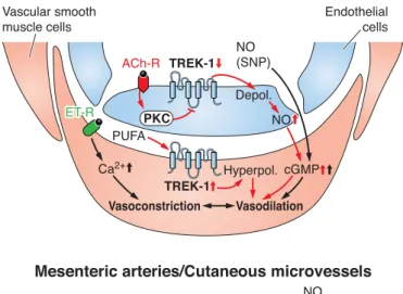
While S351 is a poor substrate for PKA, its phosphoryla- tion is central in TREK-1 current activation by the nitric oxide/cGMP/protein kinase G pathway (173). As serine- 351 is not conserved in TREK-2, this channel cannot be the target of nitrergic stimulation. From the regulatory point of view, this is an important difference between the two, otherwise nearly identical, TREK channels.
On the basis of the above studies, it is clear that the phosphorylation of TREK channels by PKA or PKC plays a pivotal role in the channel inhibition during Gs or Gq protein-coupled receptor stimulation, respectively. In- deed, phosphorylation by PKC proved to be indispensable for the inhibition of TREK-1 (or TREK-2) by the Gq-cou- pled thyrotropin releasing hormone (TRH)-, orexin-, and M3 muscarinic receptors (147, 247). However, during the Gq-coupled receptor activation, complex signal transduc- tion pathways are activated, and several metabolites are formed with potential regulatory function. Considering the multiplex and hierarchical regulation of TREK chan- nels, the direct effect of the generated DAG, phosphatidic acid (PA), or lysophospholipides and the breakdown of polyphosphoinositides must also be considered. While most (48, 147, 211), but not all (207), studies accept that PLC activation is necessary for TREK-1 inhibition, the relative importance of the complementary or alternative pathways activated simultaneously with PKC is less well defined and may be cell specific.
In contrast to the PKC dependence of TREK inhibition during the stimulation of TRH, orexin-B, or M3 receptors, the inhibition of TREK-1 and TREK-2 via mGluR1 glutamate receptor was not influenced by a PKC inhibitory peptide and staurosporine. In this case, the liberated DAG and PA were suggested to be the most important regulators, since a DAG lipase inhibitor slowed down the recovery of the current after glutamate application, and DAG and PA reduced the current of the channels preactivated by AA (48). Direct activation of TREK by phosphoinositides was clearly shown by different groups (52, 211, 212). Thus it has been hypoth- esized that the breakdown of PIP2in response to Gqactiva- tion contributed to the inhibition of TREK current. However, direct experimental demonstration of this mechanism has not been presented.
Neither PKA nor PKC influences TRAAK activity (100).
The inefficiency of PKA can be predicted from the amino acid sequence, since TRAAK does not contain a serine in homologous position to S333 of TREK-1. In contrast, S261 of TRAAK corresponds to the PKC phosphorylation site (S300) of TREK-1, and the amino acids in its neighborhood are also highly conserved between the two channels. On the basis of the sequential phosphorylation theory of TREK channels (247), it may be speculated that S261 of TRAAK cannot be
efficiently phosphorylated by PKC in the absence of the PKA phosphorylation site. In fact, TRAAK proved to be insensi- tive to the stimulation of M1 muscarinic receptor, under the same conditions when TASK and TREK channels were effi- ciently inhibited (212).
6. Interaction with partner proteins
The A-kinase anchoring protein AKAP150 is known as a scaffolding protein, which binds to several partners [includ- ing PKA, PDZ binding proteins (PSD95 and SAP97), and G protein-coupled receptors]. A proteomic approach revealed interaction between TREK-1 and AKAP150. The proximal tail of TREK-1 between V298 and R313, the region having the primary importance in the regulation of channel activity, was identified as the AKAP150-interacting domain (298).
Coexpression of AKAP150 profoundly changed the func- tional properties of TREK-1 (and TREK-2 but not TRAAK).
The channel was transformed into a voltage-independent leak conductance. The gained high basal activity could not be stimulated further by AA, stretch, or intracellular acidifi- cation. AKAP150 rendered TREK-1 resistant to PKC, which inhibited the channel by phosphorylating S300 within the AKAP-binding domain in the absence of the scaffolding pro- tein. On the contrary, the inhibition by PKA, which phos- phorylates distal to the AKAP-interacting site, was retained.
The effect of2-adrenergic receptor was even accelerated, probably reflecting that the channel and the receptor were assembled to a protein complex by AKAP150 (298).
Microtubule-associated protein, Mtap2, increases the surface expression of TREK-1 and TREK-2 without influ- encing their I-Vrelationship. The enhanced targeting de- pends on the simultaneous interaction of Mtap2 with the microtubular system. Mtap2 attaches to the amino acid stretch between E335 and Q360 on the COOH-terminal tail of TREK-1, which is clearly distinct from the AKAP150 binding site. This allows simultaneous binding of AKAP150 and Mtap2 to the channel with an additive in- tensifying effect on TREK current (297).
Interaction between TREK-1 and a cellular prion pro- tein (PrPC) was detected in a bacterial two-hybrid system.
The interaction was verified in transfected eukaryotic cells by coimmunoprecipitation and by confocal micro- scopic visualization of the transfected proteins (9). Al- though the colocalization and binding of PrPCto TREK-1 is interesting, demonstration of the functional conse- quences of the interaction would highly increase the sig- nificance of the finding.
C. Physiological Function and Pathophysiology of TREK/TRAAK Channels
Long before the discovery of K2P channels, unusual outwardly rectifying and noninactivating potassium cur- rents were described in different tissues, including neu-