Short Article
MICU1 Interacts with the D-Ring of the MCU Pore to Control Its Ca 2+ Flux and Sensitivity to Ru360
Graphical Abstract
Highlights
d
MICU1 controls the RuRed/Ru360 sensitivity of the MCU complex
d
MICU1’s R440/443 supports a direct interaction with MCU
d
MICU1 R440/443-dependently competes with RuRed for interacting with the D-ring of MCU
d
MICU1-MCU interaction is central for MCU gatekeeping and cell survival
Authors
Melanie Paillard, Gyo¨rgy Csorda´s, Kai-Ting Huang, Peter Va´rnai,
Suresh K. Joseph, Gyo¨rgy Hajno´czky
Correspondence
gyorgy.hajnoczky@jefferson.edu
In Brief
Paillard et al. report that mitochondrial Ca
2+uptake 1 (MICU1) interacts with the D-ring of MCU, the pore-forming protein of the mitochondrial Ca
2+uniporter, through a DIME interacting domain involving the arginines 440 and 443 to control both the Ca
2+flux and the ruthenium red sensitivity of the MCU complex.
Paillard et al., 2018, Molecular Cell72, 778–785 November 15, 2018ª2018 Elsevier Inc.
https://doi.org/10.1016/j.molcel.2018.09.008
Molecular Cell
Short Article
MICU1 Interacts with the D-Ring of the MCU Pore to Control Its Ca 2+ Flux and Sensitivity to Ru360
Melanie Paillard,1Gyo¨rgy Csorda´s,1Kai-Ting Huang,1Peter Va´rnai,2Suresh K. Joseph,1and Gyo¨rgy Hajno´czky1,3,*
1MitoCare Center, Department of Pathology, Anatomy and Cell Biology, Thomas Jefferson University, Philadelphia, PA 19107, USA
2Department of Physiology, Semmelweis University, Budapest, 1094 Hungary
3Lead Contact
*Correspondence:gyorgy.hajnoczky@jefferson.edu https://doi.org/10.1016/j.molcel.2018.09.008
SUMMARY
Proper control of the mitochondrial Ca
2+uniporter’s pore (MCU) is required to allow Ca
2+-dependent acti- vation of oxidative metabolism and to avoid mito- chondrial Ca
2+overload and cell death. The MCU’s gatekeeping and cooperative activation is mediated by the Ca
2+-sensing MICU1 protein, which has been proposed to form dimeric complexes anchored to the EMRE scaffold of MCU. We unexpectedly find that MICU1 suppresses inhibition of MCU by ruthe- nium red/Ru360, which bind to MCU’s DIME motif, the selectivity filter. This led us to recognize in MICU1’s sequence a putative DIME interacting domain (DID), which is required for both gatekeeping and cooperative activation of MCU and for cell sur- vival. Thus, we propose that MICU1 has to interact with the D-ring formed by the DIME domains in MCU to control the uniporter.
INTRODUCTION
The mitochondrial Ca2+ uniporter (mtCU) or MCU complex needs to be tightly regulated to avoid cell death by Ca2+overload and to allow the Ca2+-dependent physiological stimulation of oxidative metabolism. Among the different components of the MCU complex, the Ca2+-sensing protein MICU1 has been characterized as a key mediator of gatekeeping and cooperative activation of MCU, the pore-forming unit of the MCU complex (Csorda´s et al., 2013; Kamer and Mootha, 2014; Mallilankaraman et al., 2012; Patron et al., 2014). MICU1 has been linked to hu- man neurological and muscular diseases (Lewis-Smith et al., 2016; Logan et al., 2014), as well as to liver regeneration (Antony et al., 2016), cardiac ischemia-reperfusion injury (Xue et al., 2017), diabetic cardiomyopathy (Ji et al., 2017) and ovarian can- cer (Chakraborty et al., 2017).
In addition to MCU and MICU1, other MCU complex com- ponents have also been identified: two paralogs of MICU1, MICU2 and MICU3 (Plovanich et al., 2013), a dominant-nega- tive paralog of MCU, MCUb (Raffaello et al., 2013), and the indispensable transmembrane protein EMRE (Sancak et al., 2013). MICU1 interacts with MICU2 through a disulfide bond
involving C465 in MICU1 and C410 in MICU2 to form homo/
heterodimers that control the MCU complex activity (Patron et al., 2014; Petrungaro et al., 2015). On the other hand, MICU1 binding to the polyaspartate tail of EMRE has been shown to anchor MICU1 to the MCU complex in the mitochon- drial intermembrane space and thus confer gatekeeping of the pore (Tsai et al., 2016). However, in the same study, MICU1’s interaction with MCU without EMRE was also shown by coimmunoprecipitation (Tsai et al., 2016), which is also sup- ported by surface plasmon resonance data (Vecellio Reane et al., 2016).
Topology studies showed that most of MCU’s amino acid residues are in the two transmembrane domains and the N and C termini that are exposed to the mitochondrial matrix and identified a short sequence between the two transmem- brane domains, which is exposed to the intermembrane space (IMS) and might be accessible for direct interaction with IMS proteins like MICU1. This motif was termed as DIME (or DXXE) based on its amino acid sequence conserved across species (D261 and E264 in hsMCU) (Baughman et al., 2011).
A recent study characterized the DIME domain as the ion selectivity filter of the MCU with ion binding to the two carbox- ylate rings (Cao et al., 2017). Notably, mutation of DIME domain also impaired the MICU1-MCU interaction (Patron et al., 2014). Interestingly, the inhibitory effect of the most potent inhibitors of the MCU complex, ruthenium red (RuRed) and ruthenium 360 (Ru360), requires S259 (Baughman et al., 2011). Two recent NMR studies further indicated that Ru360/
RuRed binds the D-ring of MCU’s selectivity filter, which is sol- vent-exposed compared to the E-ring located deeper in the pore (Arduino et al., 2017; Cao et al., 2017). Thus, we reasoned that MICU1 might affect the RuRed sensitivity of the MCU complex through a potential interaction with the DIME domain of MCU.
We here find that MICU1 deletion increases the MCU complex sensitivity to RuRed/Ru360, suggesting that MICU1 interferes with the binding site of these inhibitors in the DIME motif of MCU. We then identify a complementing sequence in MICU1 to the DIME motif, which we refer to as DIME interacting domain (DID) and predict that two arginines (R440 and R443) engage in potential salt bridges with the D-ring of MCU. Using an imaging approach to assess both threshold and cooperativity of the MCU complex in MICU1-KO mouse embryonic fibroblasts (MEFs) and HEK293 cells (HEKs), we show that the DID in MICU1 is required to control the MCU complex-mediated Ca2+influx and, in turn,
cell survival. Finally, we show that the DID of MICU1 alters the RuRed/Ru360 sensitivity by competing for the interaction with the DIME domain of MCU, thus opening a new avenue for ther- apeutic targeting of the MCU complex.
RESULTS
MICU1 Controls the RuRed/Ru360 Sensitivity of the MCU Complex
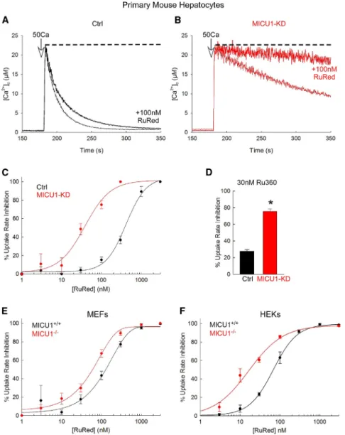
While recording mitochondrial clearance of Ca2+added to the cytoplasmic buffer of permeabilized hepatocytes isolated from wild-type (Ctrl) and hepatocyte-specific MICU1/ mouse (MICU1-KD), we noticed that deletion of MICU1 caused sensiti- zation to RuRed (Figures 1A and 1B). Specifically, MICU1-defi- cient mitochondria showed less-effective uptake when a large Ca2+bolus was applied (50mM CaCl2) as we reported earlier (Antony et al., 2016), and it was almost completely suppressed by a submaximal dose of RuRed (100 nM), which caused only a small inhibition in wild-type (WT) mitochondria. When dose
Figure 1. MICU1 Controls the RuRed/Ru360 Sensitivity of MCU Complex
(A and B) Time courses of the mitochondrial clearance of the [Ca2+]crise upon addition of a 50mM CaCl2bolus (50Ca) in permeabilized Ctrl (A) and MICU1-KD hepatocytes (B), with and without 100 nM RuRed. Dashed line indicates complete inhibition of the mitochondrial Ca2+uptake.
(C) [RuRed] dose response for the percentage of inhibition of the initial mitochondrial uptake rate for a 50Ca bolus in Ctrl (black) and MICU1-KD (red) hepatocytes. A sigmoidal fit is displayed for each.
(D) Percentage of uptake rate inhibition by 30 nM Ru360 in Ctrl (black) and MICU1-KD (red) hepatocytes.
(E and F) [RuRed] dose response for the percent- age of uptake rate inhibition for a 20Ca bolus in MICU1+/+and MICU1/MEFs (E) and HEKs (F). A sigmoidal fit is displayed for each.
Mean ± SEM, n = 3–4, *p < 0.05 versus Ctrl, t test.
See alsoFigure S1.
response curves were compiled, an in- crease in sensitivity for RuRed of almost an order of magnitude was observed in MICU1-deficient hepatocytes (Figure 1C).
Higher sensitivity to RuRed was also observed in MICU1-deficient hepatocytes in response to a smaller bolus of Ca2+
(7mM; raising [Ca2+]cto 1.5–2 mM) (Fig- ure S1), for which the mitochondrial Ca2+
uptake is similar to the control (Antony et al., 2016). The sensitivity was also increased toward Ru360, which is a more specific inhibitor of MCU complex (Figure 1D). Furthermore, sensitization to RuRed by MICU1 deletion was also found in MEFs (Figure 1E) and in HEKs (Fig- ure 1F). Thus, these results provide evidence that MICU1 decreases the RuRed/Ru360 sensitivity of the MCU complex, probably by competing with RuRed/Ru360 for binding to MCU.
MICU1 Has a Complementing Sequence to the Region of MCU Determining RuRed Sensitivity and Ca2+Selectivity RuRed/Ru360 was recently shown to bind the DIME domain of MCU (Arduino et al., 2017; Baughman et al., 2011; Cao et al., 2017). The results shown inFigure 1led us to postulate that there might be a domain in MICU1 that interacts with the DIME domain of MCU. It has been previously suggested that MICU1 interacts indirectly with MCU through EMRE, involving the transmem- brane domains of MCU and EMRE, and EMRE binding to MICU1 via EMRE’s C-terminal polyaspartate tail (Sancak et al., 2013; Tsai et al., 2016). Nevertheless, two studies using different techniques have indicated that MICU1 can interact with MCU without EMRE (Tsai et al., 2016; Vecellio Reane et al., 2016), sup- porting the possibility of an additional protein-protein interaction component. Moreover, along with the DIME domain of MCU (Pa- tron et al., 2014), the C-terminal structure of MICU1 has been
shown to be necessary for co-immunoprecipitation with MCU (Kamer and Mootha, 2014; Wang et al., 2014). However, the exact sequence in MICU1 involved in the binding to MCU is still unknown. Alignment of MICU1, MICU2, and MICU3 revealed a potential DIME interacting domain in the C-terminal sequence of all three MICUs, involving lysine (K438) and two arginines (R440 and R443), which could form potential salt bridges with the DIME domain in MCU (Figure 2A). Among the marked poten- tial interactions, the one between K438 and the corresponding serine is more likely a dipole-dipole interaction than an actual salt bridge between the positively charged amino group of lysine and the OH of serine. The crystal structure of MICU1 shows that the two arginine residues are orientated in the same direction in a helix (Wang et al., 2014), thus suggesting that the side chains of R440 and R443 could act like RuRed to bind to the outer D-ring
of the pore of the MCU pentamer (Cao et al., 2017). To validate our hypothesis, we mutated in hsMICU1 these 3 residues to alanine (MICU1-DDID: 440KQRLMR445 > 440AQALMA445) and analyzed its binding with MCU. MICU1-DDID (tagged with HA) was pulled down 60% less than WT MICU1 by MCU (tagged with FLAG), and MCU was pulled down to a lesser extent by MICU1-DDID than by WT MICU1 (Figure 2B). These results sug- gest that the KQRLMR sequence in MICU1, which we now named DID (DIME interaction domain), interacts with MCU, probably through its DIME motif.
Next, using MICU1-KO MEFs, in which MICU2 is maintained (Antony et al., 2016), we aimed at better characterizing the func- tional role of the DID in MICU1 in the control of the MCU complex Ca2+flux. To assess MCU complex activity in individual cells, [Ca2+]mwas followed via loading the mitochondria in WT and Figure 2. Mutation of the Complementing Sequence Interferes with MICU1 Binding to and Control of MCU
(A) Alignment of MICU1, MICU2 and MICU3 showing previously identified sites in MICU1 involved in interactions with EMRE and MICU1/MICU2, and proposing a potential MCU binding domain.
(B) Upper: representative immunoblotting of coimmunoprecipitation (coIP) between MCU-FLAG and HA-tagged WT MICU1 or MICU1-DDID mutant expressed in MCU-FLAG HEKs. Arrow indicates specific MICU1-HA band, while asterisk is an unspecific band. Lower: quantification of the relative MICU1 amount pulled down by MCU-FLAG in HEKs expressing WT MICU1 or MICU1-DDID. Mean ± SEM, n = 3, *p < 0.05 versus WT, t test.
(C) [Ca2+]mmeasurements in permeabilized WT MEFs and MICU1-KO MEFs expressing empty vector, MICU1 or MICU1-DDID and challenged with a bolus of 20mM CaCl2.
(D) Quantification of resting [Ca2+]mas an index of thresholding of the MCU complex.
(E) Calculations of the difference (D[Ca2+]m) between the [Ca2+]m30 s post-20Ca addition and the resting [Ca2+]mas an index of cooperativity of the MCU complex.
Mean ± SEM, n = 6, *p < 0.05 versus WT, one-way ANOVA.
See alsoFigure S2.
MICU1-KO MEF cells with the ratiometric probe furaFF/AM; cells were then permeabilized and subjected to a two-step Ca2+addi- tion protocol (3 and 20mM CaCl2, 3 and 20Ca). Mitochondrial localization of the probe was confirmed by complete inhibition of the Ca2+ response by RuRed (Figure S2) (Paillard et al., 2017). As expected, MICU1-KO MEFs displayed an increased resting [Ca2+]m, which could be prevented by RuRed (Figure S2).
The resting [Ca2+]mwas used then as a measure of gatekeeping activity of various MICU1 mutants. Rescue was performed with WT MICU1 (MICU1-HA) or MICU1-DDID in the MICU1-KO cells.
While MICU1-HA restored the low resting [Ca2+]m, MICU1-DDID failed to lower it (Figures 2C and 2D). As a simple measure of the Ca2+-induced highly cooperative activation of MCU, in a single- addition protocol, the initial response to 20Ca (difference be- tween the [Ca2+]m 30 s post-20Ca addition and the resting [Ca2+]m) was used. MICU1-DDID expression decreased D [Ca2+]mas in MICU1-KO MEFs (with empty vector), compared to WT MEFs (Figures 2C and 2E). Thus, our data support that mu- tation of the complementing sequence with MCU DIME motif in- terferes with MICU1 binding to and control of the Ca2+fluxes of MCU.
Role of DID and the Other Motifs of MICU1 in Intracellular Ca2+Homeostasis and Cell Survival
MICU1 is thus engaged in at least three interactions with compo- nents of the MCU complex: the previously documented dimer- ization with MICU2/MICU1, an interaction with EMRE, and our newly demonstrated interaction with MCU via the DID. To eval- uate the MCU complex regulation by the interactions of MICU1 with MICU2, EMRE and MCU, respectively, we interfered with each separately or in combination and performed simultaneous [Ca2+]cand [Ca2+]mmeasurements during store-operated Ca2+
entry (SOCE) in intact WT and MICU1-KO HEKs. We opted for SOCE as the Ca2+source because it stimulates mitochondrial Ca2+ uptake mostly via the global [Ca2+]c increase that we measured, so we could plot [Ca2+]mas both a function of time and [Ca2+]c. We generated three additional HA-tagged mutants of MICU1: the MICU1-Ddimer (C465A), which is incompetent for dimerization, MICU1-DEMRE (99KKKKR > 99AAAAA), which lacks the EMRE-binding motif, and a combination of MICU1- DDID+DEMRE+Ddimer. Co-transfection of MICU1-KO cells with MICU1 mutants and the mitochondrial matrix-targeted Ca2+indicator, mtCepia, allowed us to evaluate in each single cell [Ca2+]m and [Ca2+]c by fura2 (Figures 3A and 3B). The SOCE-mediated [Ca2+]mincrease appeared earlier in MICU1- KO HEKs rescued by either the empty vector, the MICU1- DDID, or the MICU1-DDID+DEMRE+Ddimer mutant, compared to WT HEKs or MICU1-KO cells rescued by the other MICU1 mu- tants (Figure 3B). The [Ca2+]mresponse 60 s post-Ca2+addition revealed that both MICU1-DDID and MICU1-DDID+DEMRE+D dimer mutant rescues took up Ca2+similarly to the MICU1-KO cells, while the MICU1-DEMRE and MICU1-Ddimer rescued cells took up initially less Ca2+, like the WT MICU1 rescue (Fig- ure 3C). Plotting [Ca2+]magainst [Ca2+]cfurther supported that MICU1-KO cells rescued by either the empty vector or the MICU1-DDID or the MICU1-DDID+DEMRE+Ddimer mutant showed a significant [Ca2+]mincrease at lower [Ca2+]cthan WT cells or the ones rescued by WT MICU1 and the other MICU1
mutants in MICU1-KO HEKs (Figure 3D). Indeed, to reach a similar level of [Ca2+]m(arbitrarily set at 1.2 ratio units), around 0.2mM of [Ca2+]cwas sufficient in MICU1-KO and the MICU1- DDID or MICU1-DDID+DEMRE+Ddimer rescues, whereas WT HEKs and MICU1-KO cells rescued by WT MICU1, MICU1- DEMRE, or MICU1-Ddimer constructs required a significantly higher [Ca2+]c(Figure 3E). We further assessed if the MICU1- DDID mutant operated as a dominant-negative MCU complex component in these intact cell experiments. Similarly to MICU1-KO HEKs, co-expression of WT MICU1 and MICU1- DDID led to a significant [Ca2+]mincrease at lower [Ca2+]cthan MICU1-KO HEKs rescued by WT MICU1 itself or along with a point mutant in EF1 of MICU1 (MICU1-EF1-D9E), thus unraveling a dominant-negative effect of MICU1-DDID over WT MICU1 (Fig- ures S3A and S3B). MICU1-EF1-D9E was created to cause a specific perturbation in D239 of the EF hand, but it was found to unalter the resting [Ca2+]mor the rescue of cooperativity and Ca2+-dependent activation of MCU complex (Figures S3C–
S3E). Therefore, we used this construct as an alternative example of full rescue. Together, these data suggest that the MICU1-MCU interaction through the MICU1’s DID and MCU’s DIME motif is the most critical for the gatekeeping of the MCU complex.
Because the gatekeeping of MCU complex by MICU1 is cen- tral to avoiding mitochondrial Ca2+overload, oxidative stress, and cell death (Antony et al., 2016; Csorda´s et al., 2013; Liu et al., 2016; Mallilankaraman et al., 2012), we then proceeded to study the so-called delayed Ca2+dysregulation by following the [Ca2+]cin intact HEKs subjected to prolonged Ca2+entry (Fig- ure 3F). After the initial [Ca2+]crise, a large delayed increase was observed only in mock- and MICU1-DDID-rescued cells, whereas [Ca2+]c was maintained at a low level in MICU1-KO HEKs rescued by WT MICU1 or with the MICU1-EF1-D9E (Figure 3G).
The mitochondrial oxidative stress generated during pro- longed SOCE conditions was measured using mtGrx1-RoGFP2, the fluorescence of which shows the ratio of oxidized and reduced glutathione (GSSG/GSH) and is unaffected by pH (Gutscher et al., 2008). The oxidative stress recorded in mock rescued cells was duplicated in the cells rescued by the MICU1-DDID mutant, whereas oxidative stress was avoided in the cells rescued with WT MICU1 or MICU1-EF1-D9E constructs (Figure 3H). Importantly, all the MICU1 constructs were effec- tively expressed in the MICU1-KO cells, with no difference between the MICU1-DDID and MICU1-EF1-D9E mutants (Fig- ure S4A). To conclude, these results indicate that MICU1 interac- tion with the D-ring of the DIME domain of MCU is required for gatekeeping of the MCU complex and, in turn, for cell survival.
DID Mutant Fails to Rescue RuRed Sensitivity
Finally, we investigated how RuRed sensitivity will be altered with expression of DID mutant of MICU1. While in WT and in MICU1-KO HEKs rescued by WT MICU1, or MICU1-EF1-D9E constructs, 30 nM RuRed elicited a partial inhibition of the mito- chondrial Ca2+ uptake rate, MICU1-KO HEKs rescued with empty vector or MICU1-DDID displayed an almost complete in- hibition of mitochondrial Ca2+uptake, which could only be at- tained with 3mM RuRed in WT HEKs (Figures 4A and 4B). Indeed,
a significantly higher inhibition of mitochondrial Ca2+uptake by 30 nM RuRed was obtained in MICU1-KO HEKs rescued with empty vector or MICU1-DDID compared to either WT MICU1 or MICU1-EF1-D9E rescue (Figure 4B). Importantly, rescuing MICU1-KO HEKs by the MICU1-DDID or MICU1-EF1-D9E con- structs both lead to a higher MICU1 protein expression than that in the WT HEKs (Figure S4B). Because no release of any of the MICU1 constructs was noticed upon selective permeabilization of the plasma membrane and in the immunofluorescence exper- iments, the expressed MICU1 constructs co-localized with the mitochondria (Figure S4C), they seem to be correctly targeted to the mitochondria. To test the localization to the IMS and proper interaction with EMRE for both MICU1-HA and MICU1- DDID-HA, we performed for the revision an immunoprecipitation experiment.Figure S4D shows that EMRE (tagged with Myc) pulled down a comparable amount of WT MICU1 and MICU1- DDID mutant (tagged with HA), thus confirming the proper local-
ization of the MICU1-DDID construct in the IMS and also that alteration of the MICU1-MCU interaction does not interfere with the MICU1-EMRE binding. Furthermore, the dominant- negative effect of MICU1-DDID (Figures S3A and S3B) also provides functional evidence for the correct localization of MICU1-DDID. Thus, the failure of the DID mutant to restore the lower RuRed sensitivity confirms that the interaction between the DID of MICU1 and MCU, likely through its DIME domain, con- trols the Ca2+ flux and the RuRed sensitivity of the MCU complex.
DISCUSSION
The main finding of this study is that MICU1 interacts with the DIME motif of MCU and this interaction is required for keeping MCU complex’s pore both closed at low [Ca2+]cand optimally activated at high [Ca2+]c. Furthermore, this interaction also Figure 3. Critical Role of the DID of MICU1 in Ca2+Homeostasis and Cell Survival
(A and B) SOCE-induced [Ca2+]c(A: Fura2 after calibration inmM) and [Ca2+]m(B: mt-Cepia) time courses in WT and MICU1-KO HEKs expressing the indicated MICU1 constructs, after addition of 3 mM CaCl2.
(C) Bar graph shows [Ca2+]mresponse at 60 s, normalized to MICU1-KO HEK response, from data in (B).
(D) [Ca2+]mplotted against [Ca2+]cin individual cells.
(E) Measurements of [Ca2+]crequired to reach 1.2 [Ca2+]mbased on data from (D). Mean ± SEM, n = 4–7, *p < 0.05 versus WT, one-way ANOVA.
(F) Representative traces of [Ca2+]cmeasured by fura-ff-AM in intact HEKs after an addition of 10 mM CaCl2and in the presence of 100mM H2O2. (G) Quantification of the increase in [Ca2+]cfrom data in (F) (50 min–10 min).
(H) Quantification of the mitochondrial GSSG/GSH ratio using the mtGrx1-RoGFP2 sensor in the same conditions as in (F).
Mean ± SEM, n = 3 independent experiments, *p < 0.05 versus MICU1-KO, one-way ANOVA.
See alsoFigures S3andS4.
controls the MCU complex’s accessibility for its most-character- ized inhibitors. Thus, our findings are central to understanding the mechanisms that allow MICU1 to support cell survival by pre- venting mitochondrial Ca2+overload and cause human disease in MICU1 deficiency, and provide important clues for mitochon- drial drug development.
It has been broadly recognized that the Ca2+-sensing MICU1 is required for the control of MCU complex activity. As to the underlying mechanism, MICU1’s interactions with some compo- nents of the MCU complex have been dissected recently: disul- fide bonds between the cysteines of MICU1 and MICU2 to form dimers (Patron et al., 2014; Petrungaro et al., 2015) and electro- static interaction between the polyaspartate tail of EMRE and the polybasic sequence of MICU1 (Tsai et al., 2016). While Hoffman et al. had suggested a potential interaction between the N-termi- nal polybasic domain of MICU1 and the two interacting coiled- coil domains of MCU (Hoffman et al., 2013), this was later demonstrated to be indirect and mediated through EMRE (Tsai et al., 2016). MICU1’s interaction with MCU without EMRE was also indicated (Kova´cs-Bogda´n et al., 2014; Tsai et al., 2016), but until now no direct interaction sites between MCU and MICU1 have been established. Given the involvement of the
MCU complex, and specifically MICU1, in cell death and in dis- eases (Liu et al., 2017; Mammucari et al., 2017), a better under- standing of the MICU1 interactions in the MCU complex was clearly needed.
We here report a structural and functional interaction be- tween the DID of MICU1 and the region identified as the selec- tivity filter domain of MCU, which controls the Ca2+flux and the RuRed sensitivity of the MCU complex. Using sequence align- ment, we identified a putative DIME interacting domain, DID, in the C terminus of MICU1, engaging R440 and R443 in potential salt bridges with the D-ring of MCU. Decreased co-immunopre- cipitation between MCU and a MICU1 mutant of these two ar- ginines (MICU1-DDID: 440KQRLMR445 > 440AQALMA445) supported a structural role for the DID in MICU1-MCU interac- tion. Using Ca2+ imaging approaches in genetically rescued MICU1-KO MEFs and HEKs, we showed that the DID in MICU1 is required for both the threshold and cooperative activation of the MCU complex-mediated Ca2+uptake. Further- more, the DID was required to avoid mitochondrial Ca2+over- load, ROS dysregulation, and the ensuing cell injury. Addition- ally, unlike MICU1, MICU1-DDID expression in MICU1-KO HEKs did not decrease the RuRed sensitivity. Based on the Figure 4. MICU1-MCU Interaction through the DID of MICU1 Is Required to Maintain RuRed Sensitivity
(A) Time courses of the mitochondrial clearance of the [Ca2+]crise upon addition of a 10mM CaCl2bolus (10Ca) in permeabilized WT and MICU1-KO HEKs with different MICU1 constructs, with or without RuRed (3mM or 30 nM).
(B) Bar graph shows percentage of uptake rate inhibition by 30 nM RuRed in data from (A). Mean ± SEM, n = 3, *p < 0.05 versus WT,yp < 0.05 versus MICU1-EF1- D9E, one-way ANOVA.
(C) Proposed model for MICU1 interaction with the MCU complex. In addition to the previously identified sites of MICU1 interaction with MICU1/2 through a disulfide bridge and with EMRE via electrostatic binding, we have identified a new functional domain in MICU1, the DID, for interaction with the DIME domain of MCU. More precisely, the two arginines 440 and 443 in MICU1 would interact via salt bridges with the accessible D-ring of the filter selectivity domain of MCU to control the Ca2+flux and RuRed sensitivity of the MCU complex.
See alsoFigure S4.
proposedin vitroarchitecture of MCU as a pentamer (Oxenoid et al., 2016) and on our identification of the MICU1 DID interac- tion site with MCU, we here propose an updated model of the interactions of MICU1 within the MCU complex (Figure 4C): the two arginines R440 and R443 from one MICU1 would interact with the exposed D-ring (D261) of two MCU units from the pen- tamer to assure the physiological gatekeeping of the pore. In such a model, however, it would be predicted that only one MICU1 dimer is required to block a pentameric MCU complex.
Nevertheless, the pentameric assembly was establishedin vitro using C. elegansMCU that lacked the N-terminal 165 amino acids (Oxenoid et al., 2016), and thein vivoMCU stoichiometry is yet to be determined. Some earlier studies, based on biochemical data, proposed tetrameric stoichiometry (Raffaello et al., 2013), in which case our model would be compatible with a pair of DID domains (in 2 MICU1 dimers) being able to fully engage with the D-ring of the MCU selectivity filter. Thus, eluci- dation of thein vivoarchitecture of MCU will be crucial to better appreciate the stoichiometry between MICU1 and MCU per MCU complex.
As to the relevance of the interactions with the different MCU complex components, our results identify the MICU1 interaction with MCU via its DID to be critical for the gatekeeping. In our ex- periments, relevance of EMRE and dimer formation is also confirmed, but the MICU1-EMRE interaction rather appears as an additional stabilizing interaction to support the tight regulation of MCU by MICU1, which also fits with the only partial loss of threshold observed by Tsai et al. when disrupting the MICU1- EMRE interaction (Tsai et al., 2016). Notably, direct control of MCU by MICU1 as an evolutionarily conserved critical factor in gatekeeping is supported by the finding that the functional MCU complex of Dictyostelium discoideum consists of only MCU and one MICU isoform (Kova´cs-Bogda´n et al., 2014).
Our alignment of MICUs suggests that the DID, comprising the two arginines, is conserved among the three MICUs. However, MICU2 fails to act as a gatekeeper in the absence of MICU1 (Kamer and Mootha, 2014). Because some interactions re- mained between MICU1-DDID and MCU (Figure 2B), we specu- late that those interactions might be different for MICU1 and MICU2. Thus, further studies will be required to test whether the DID of MICU2 (or MICU3) can support an interaction with MCU.
One of the most unexpected findings of this work was that removal of MICU1 greatly sensitized mitochondria toward RuRed/Ru360, and this seems to be because of the DID limiting access of RuRed to its target site in the DIME of MCU. In the light of recent demonstration of tissue-specific differences in the MICU1 abundance relative to MCU (Paillard et al., 2017), the pre- sent finding might predict different RuRed/Ru360 sensitivity of MCU complex in various tissues, such as the particularly high sensitivity in the little MICU1-containing cardiac muscle (Matlib et al., 1998). Furthermore, pharmacological targeting of the MCU complex by new and potentially therapeutically useful mol- ecules has become an area of concerted research efforts (Ardu- ino et al., 2017; Arduino and Perocchi, 2018). Our findings might aid drug design and call attention to possible organ-specific dif- ferences in the sensitivity to future drugs sharing the target of RuRed/Ru360.
STAR+METHODS
Detailed methods are provided in the online version of this paper and include the following:
d KEY RESOURCES TABLE
d CONTACT FOR REAGENT AND RESOURCE SHARING
d EXPERIMENTAL MODEL AND SUBJECT DETAILS B Cell lines
B Primary mouse hepatocytes
d METHOD DETAILS
B Measurements of mitochondrial Ca2+ uptake and membrane potential
B Construction of the HA-tagged MICU1 mutants B Co-immunoprecipitation
B Live cell Ca2+imaging
d QUANTIFICATION AND STATISTICAL ANALYSIS SUPPLEMENTAL INFORMATION
Supplemental Information includes four figures and can be found with this article online athttps://doi.org/10.1016/j.molcel.2018.09.008.
ACKNOWLEDGMENTS
M.P. was a recipient of postdoctoral fellowships from La Fondation pour la Re- cherche Me´dicale (FRM SPE20130326561) and American Heart Association (14POST19830021) and a grant ‘‘Aide a` la mobilite´’’ from the Institut Servier (France). The study was funded by an NIH grant (RO1 GM102724) to G.H.
AUTHOR CONTRIBUTIONS
Conceptualization: G.H., G.C., S.K.J., and M.P.; Investigation: G.H., G.C., S.K.J., M.P., K.-T.H., and P.V.; Writing: G.H., G.C., S.K.J., and M.P.; Funding Acquisition: G.H., M.P.
DECLARATION OF INTERESTS
The authors declare no competing interests.
Received: February 28, 2018 Revised: July 20, 2018 Accepted: September 6, 2018 Published: October 25, 2018 REFERENCES
Antony, A.N., Paillard, M., Moffat, C., Juskeviciute, E., Correnti, J., Bolon, B., Rubin, E., Csorda´s, G., Seifert, E.L., Hoek, J.B., and Hajno´czky, G. (2016).
MICU1 regulation of mitochondrial Ca(2+) uptake dictates survival and tissue regeneration. Nat. Commun.7, 10955.
Arduino, D.M., and Perocchi, F. (2018). Pharmacological modulation of mito- chondrial calcium homeostasis. J. Physiol.596, 2717–2733.
Arduino, D.M., Wettmarshausen, J., Vais, H., Navas-Navarro, P., Cheng, Y., Leimpek, A., Ma, Z., Delrio-Lorenzo, A., Giordano, A., Garcia-Perez, C., et al. (2017). Systematic Identification of MCU Modulators by Orthogonal Interspecies Chemical Screening. Mol. Cell67, 711–723.e7, e717.
Baughman, J.M., Perocchi, F., Girgis, H.S., Plovanich, M., Belcher-Timme, C.A., Sancak, Y., Bao, X.R., Strittmatter, L., Goldberger, O., Bogorad, R.L., et al. (2011). Integrative genomics identifies MCU as an essential component of the mitochondrial calcium uniporter. Nature476, 341–345.
Cao, C., Wang, S., Cui, T., Su, X.C., and Chou, J.J. (2017). Ion and inhibitor binding of the double-ring ion selectivity filter of the mitochondrial calcium uni- porter. Proc. Natl. Acad. Sci. USA114, E2846–E2851.
Chakraborty, P.K., Mustafi, S.B., Xiong, X., Dwivedi, S.K.D., Nesin, V., Saha, S., Zhang, M., Dhanasekaran, D., Jayaraman, M., Mannel, R., et al. (2017).
MICU1 drives glycolysis and chemoresistance in ovarian cancer. Nat.
Commun.8, 14634.
Csorda´s, G., Golena´r, T., Seifert, E.L., Kamer, K.J., Sancak, Y., Perocchi, F., Moffat, C., Weaver, D., de la Fuente Perez, S., Bogorad, R., et al. (2013).
MICU1 controls both the threshold and cooperative activation of the mito- chondrial Ca2+uniporter. Cell Metab.17, 976–987.
Gutscher, M., Pauleau, A.L., Marty, L., Brach, T., Wabnitz, G.H., Samstag, Y., Meyer, A.J., and Dick, T.P. (2008). Real-time imaging of the intracellular gluta- thione redox potential. Nat. Methods5, 553–559.
Hoffman, N.E., Chandramoorthy, H.C., Shamugapriya, S., Zhang, X., Rajan, S., Mallilankaraman, K., Gandhirajan, R.K., Vagnozzi, R.J., Ferrer, L.M., Sreekrishnanilayam, K., et al. (2013). MICU1 motifs define mitochondrial cal- cium uniporter binding and activity. Cell Rep.5, 1576–1588.
Ji, L., Liu, F., Jing, Z., Huang, Q., Zhao, Y., Cao, H., Li, J., Yin, C., Xing, J., and Li, F. (2017). MICU1 Alleviates Diabetic Cardiomyopathy Through Mitochondrial Ca2+-Dependent Antioxidant Response. Diabetes 66, 1586–1600.
Kamer, K.J., and Mootha, V.K. (2014). MICU1 and MICU2 play nonredundant roles in the regulation of the mitochondrial calcium uniporter. EMBO Rep.15, 299–307.
Kamer, K.J., Grabarek, Z., and Mootha, V.K. (2017). High-affinity cooperative Ca2+binding by MICU1-MICU2 serves as an on-off switch for the uniporter.
EMBO Rep.18, 1397–1411.
Kova´cs-Bogda´n, E., Sancak, Y., Kamer, K.J., Plovanich, M., Jambhekar, A., Huber, R.J., Myre, M.A., Blower, M.D., and Mootha, V.K. (2014).
Reconstitution of the mitochondrial calcium uniporter in yeast. Proc. Natl.
Acad. Sci. USA111, 8985–8990.
Lewis-Smith, D., Kamer, K.J., Griffin, H., Childs, A.M., Pysden, K., Titov, D., Duff, J., Pyle, A., Taylor, R.W., Yu-Wai-Man, P., et al. (2016). Homozygous deletion in MICU1 presenting with fatigue and lethargy in childhood. Neurol Genet2, e59.
Liu, J.C., Liu, J., Holmstro¨m, K.M., Menazza, S., Parks, R.J., Fergusson, M.M., Yu, Z.X., Springer, D.A., Halsey, C., Liu, C., et al. (2016). MICU1 Serves as a Molecular Gatekeeper to Prevent In Vivo Mitochondrial Calcium Overload.
Cell Rep.16, 1561–1573.
Liu, J.C., Parks, R.J., Liu, J., Stares, J., Rovira, I.I., Murphy, E., and Finkel, T.
(2017). The In Vivo Biology of the Mitochondrial Calcium Uniporter. Adv.
Exp. Med. Biol.982, 49–63.
Logan, C.V., Szabadkai, G., Sharpe, J.A., Parry, D.A., Torelli, S., Childs, A.M., Kriek, M., Phadke, R., Johnson, C.A., Roberts, N.Y., et al.; UK10K Consortium (2014). Loss-of-function mutations in MICU1 cause a brain and muscle disor- der linked to primary alterations in mitochondrial calcium signaling. Nat. Genet.
46, 188–193.
Mallilankaraman, K., Doonan, P., Ca´rdenas, C., Chandramoorthy, H.C., M€uller, M., Miller, R., Hoffman, N.E., Gandhirajan, R.K., Molgo´, J., Birnbaum, M.J., et al. (2012). MICU1 is an essential gatekeeper for MCU-mediated mitochon- drial Ca(2+) uptake that regulates cell survival. Cell151, 630–644.
Mammucari, C., Gherardi, G., and Rizzuto, R. (2017). Structure, Activity Regulation, and Role of the Mitochondrial Calcium Uniporter in Health and Disease. Front. Oncol.7, 139.
Matlib, M.A., Zhou, Z., Knight, S., Ahmed, S., Choi, K.M., Krause-Bauer, J., Phillips, R., Altschuld, R., Katsube, Y., Sperelakis, N., and Bers, D.M. (1998).
Oxygen-bridged dinuclear ruthenium amine complex specifically inhibits Ca2+ uptake into mitochondria in vitro and in situ in single cardiac myocytes.
J. Biol. Chem.273, 10223–10231.
O-Uchi, J., Jhun, B.S., Xu, S., Hurst, S., Raffaello, A., Liu, X., Yi, B., Zhang, H., Gross, P., Mishra, J., et al. (2014). Adrenergic signaling regulates mitochon- drial Ca2+ uptake through Pyk2-dependent tyrosine phosphorylation of the mitochondrial Ca2+ uniporter. Antioxid. Redox Signal.21, 863–879.
Oxenoid, K., Dong, Y., Cao, C., Cui, T., Sancak, Y., Markhard, A.L., Grabarek, Z., Kong, L., Liu, Z., Ouyang, B., et al. (2016). Architecture of the mitochondrial calcium uniporter. Nature533, 269–273.
Paillard, M., Csorda´s, G., Szanda, G., Golena´r, T., Debattisti, V., Bartok, A., Wang, N., Moffat, C., Seifert, E.L., Sp€at, A., and Hajno´czky, G. (2017).
Tissue-Specific Mitochondrial Decoding of Cytoplasmic Ca2+ Signals Is Controlled by the Stoichiometry of MICU1/2 and MCU. Cell Rep. 18, 2291–2300.
Patron, M., Checchetto, V., Raffaello, A., Teardo, E., Vecellio Reane, D., Mantoan, M., Granatiero, V., Szabo`, I., De Stefani, D., and Rizzuto, R.
(2014). MICU1 and MICU2 finely tune the mitochondrial Ca2+ uniporter by ex- erting opposite effects on MCU activity. Mol. Cell53, 726–737.
Petrungaro, C., Zimmermann, K.M., K€uttner, V., Fischer, M., Dengjel, J., Bogeski, I., and Riemer, J. (2015). The Ca(2+)-Dependent Release of the Mia40-Induced MICU1-MICU2 Dimer from MCU Regulates Mitochondrial Ca(2+) Uptake. Cell Metab.22, 721–733.
Plovanich, M., Bogorad, R.L., Sancak, Y., Kamer, K.J., Strittmatter, L., Li, A.A., Girgis, H.S., Kuchimanchi, S., De Groot, J., Speciner, L., et al. (2013). MICU2, a paralog of MICU1, resides within the mitochondrial uniporter complex to regu- late calcium handling. PLoS ONE8, e55785.
Raffaello, A., De Stefani, D., Sabbadin, D., Teardo, E., Merli, G., Picard, A., Checchetto, V., Moro, S., Szabo`, I., and Rizzuto, R. (2013). The mitochondrial calcium uniporter is a multimer that can include a dominant-negative pore- forming subunit. EMBO J.32, 2362–2376.
Sancak, Y., Markhard, A.L., Kitami, T., Kova´cs-Bogda´n, E., Kamer, K.J., Udeshi, N.D., Carr, S.A., Chaudhuri, D., Clapham, D.E., Li, A.A., et al. (2013).
EMRE is an essential component of the mitochondrial calcium uniporter com- plex. Science342, 1379–1382.
Tsai, M.F., Phillips, C.B., Ranaghan, M., Tsai, C.W., Wu, Y., Willliams, C., and Miller, C. (2016). Dual functions of a small regulatory subunit in the mitochon- drial calcium uniporter complex. eLife5, 5.
Vecellio Reane, D., Vallese, F., Checchetto, V., Acquasaliente, L., Butera, G., De Filippis, V., Szabo`, I., Zanotti, G., Rizzuto, R., and Raffaello, A. (2016). A MICU1 Splice Variant Confers High Sensitivity to the Mitochondrial Ca2+
Uptake Machinery of Skeletal Muscle. Mol. Cell64, 760–773.
Wang, L., Yang, X., Li, S., Wang, Z., Liu, Y., Feng, J., Zhu, Y., and Shen, Y.
(2014). Structural and mechanistic insights into MICU1 regulation of mitochon- drial calcium uptake. EMBO J.33, 594–604.
Xue, Q., Pei, H., Liu, Q., Zhao, M., Sun, J., Gao, E., Ma, X., and Tao, L. (2017).
MICU1 protects against myocardial ischemia/reperfusion injury and its control by the importer receptor Tom70. Cell Death Dis.8, e2923.
STAR + METHODS
KEY RESOURCES TABLE
REAGENT or RESOURCE SOURCE IDENTIFIER
Antibodies
Rabbit anti-MICU1 Sigma-Aldrich HPA037480; RRID: AB_10696934
Rabbit anti-MCU Sigma-Aldrich HPA016480; RRID: AB_2071893
Rabbit anti-MICU2 Abcam ab101465; RRID: AB_10711219
Mouse anti-mtHsp70 Thermo Fisher MA3-028; RRID: AB_325474
Rabbit anti-prohibitin Abcam ab28172; RRID: AB_777457
Anti-HA polyclonal Invitrogen 71-5500; RRID: AB_2533988
Anti-FLAG M2 Sigma-Aldrich F1804; RRID: AB_262044
Anti-Myc monoclonal Precision AG10004
Chemicals, Peptides, and Recombinant Proteins
Anti-DYKDDDDK (FLAG) beads Genscript A00187-200
Ruthenium Red Sigma R2751
Fura-2 (AM) Teflabs 0-103
Fura-2 (salt) Teflabs 0-104
Fura-2 low affinity (AM) Teflabs 0-136
Fura-2 low affinity (salt) Teflabs 0-137
Thapsigargin Enzo Life Sciences BML-PE180-0005
CGP-37157 Enzo Life Sciences BML-CM119-0005
Lipofectamine 3000 Life Technologies L3000008
Complete protease inhibitor cocktail (EDTA free) Roche 11873580001
Protein A Sepharose Abcam ab193256
Critical Commercial Assays
DC Protein Assay Biorad 5000112
Experimental Models: Cell Lines
MICU1loxp/loxpMEF Antony et al., 2016 N/A
MICU1KO/KOMEF Antony et al., 2016 N/A
MICU1-KO HEK293T Sancak et al., 2013 N/A
EMRE-KO HEK293T Sancak et al., 2013 N/A
HEK293T stably overexpressing mouse MCU-FLAG O-Uchi et al., 2014 N/A
WT HEK293T Sancak et al., 2013 N/A
Recombinant DNA
pcDNA-dest40-MICU1-HA Kamer et al., 2017 N/A
pcDNA3.1-MICU1-C465A-HA Patron et al., 2014 N/A
pcDNA-dest40-M1-EF1-D9E-HA, M1-DDID-HA and M1-DEMRE-HA This paper N/A
EMRE-Myc This paper N/A
Mt-Cepia This paper N/A
mtGrx1-RoGFP This paper N/A
Software and Algorithms
Canvas X N/A N/A
Endnote N/A N/A
SigmaPlot 12.5 N/A N/A
CONTACT FOR REAGENT AND RESOURCE SHARING
Further information and requests for resources and reagents should be directed to and will be fulfilled by the Lead Contact, Gyorgy Hajnoczky (gyorgy.hajnoczky@jefferson.edu).
EXPERIMENTAL MODEL AND SUBJECT DETAILS Cell lines
Mouse embryonic fibroblasts (MEFs) were isolated by trypsin digestion from e14.5 embryos, usingMICU1KO/loxPmice (for generation see (Antony et al., 2016)), and then immortalized. MEFs were cultured in DMEM (ATCC 30-2002) supplemented with penicillin, strep- tomycin at 37C/5%O2. MICU1-KO, and EMRE-KO HEKs were kindly provided by Dr Vamsi Mootha and grown as previously described (Sancak et al., 2013) and the stable MCU-Flag HEK by Dr Shey-Shing Sheu. Co-transfection of the control plasmid (pcDNA3-dest40) or the HA-tagged MICU1 constructs with the mitochondrial Ca2+sensor mtCepia or the redox sensor mtGrx1- RoGFP2, was performed in MEFs and HEKs using Lipofectamine 3000 according to manufacturers’ protocol. Co-transection effi- ciency was confirmed by immunofluorescence via the HA tag (data not shown). Since a 4-fold higher expression was observed for the WT MICU1-HA construct compared to the other MICU1-HA mutants, we used 1/4 of the DNA amount for the WT MICU1- HA plasmid.
Primary mouse hepatocytes
Primary hepatocytes were isolated byin situretrograde perfusion with collagenase as previously described (Csorda´s et al., 2013), fromMICU1loxP/loxPmale mice 3 weeks after tail vein-injection with an AAV8-TBG-Cre or an AAV8-TBG Null (1.3x10¹¹pfu/mouse) (Penn Vector Core). Only preparations with greater than 90% viability were used for subsequent experiments. Studies were done in accordance with the Thomas Jefferson University institutional review board guidelines.
METHOD DETAILS
Measurements of mitochondrial Ca2+uptake and membrane potential
Saponin-permeabilized hepatocytes (2 millions), MEFs or HEKs (2.4 mg) were resuspended in 1.5 mL of intracellular medium (ICM) containing 120 mM KCl, 10 mM NaCl, 1 mM KH2PO4, 20 mM Tris-HEPES at pH 7.2, and supplemented with proteases inhibitors (leupeptin, antipain, pepstatin, 1mg/ml each), 2 mM MgATP, 2mM thapsigargin and maintained in a stirred thermostated cuvette at 35C. Assays were performed in presence of 20mM CGP-37157 and 1 mM succinate using a multiwavelength-excitation dual- wavelength-emission fluorimeter (DeltaRAM, PTI). The extramitochondrial Ca2+concentration [Ca2+]cwas assessed using the ratio- metric Ca2+probe Fura2-FA (1.5mM, Teflabs) or Fura-loAff (formerly Fura-FF) (1mM, Teflabs).Dcm was measured with 1.5mM TMRM (Invitrogen). Fura and TMRM fluorescence were recorded simultaneously using 340-380 nm excitation and 500 nm emission, and 545 nm excitation and 580 nm emission, respectively. Complete depolarization (maximum de-quench of TMRM fluorescence) was elicited using of the protonophore FCCP (2mM). Calibration of the Fura signal was carried out at the end of each measurement, adding 1 mM CaCl2, followed by 10 mM EGTA/Tris, pH 8.5.
Construction of the HA-tagged MICU1 mutants
MICU1-EF1-D9E and MICU1-DEMRE were generated by site-directed mutagenesis in the cDNA sequence pcDNA-DEST40 contain- ing the wild-type human MICU1 (provided by Vamsi K. Mootha). MICU1-DDID was created by inserting the synthetized sequence coding the appropriate mutations (Blue Heron) between the HindII/EcoNI restriction sites. Finally, the sequence resulting the C-ter- minal HA tag was inserted into each construct between the EcoNI/AgeI restriction sites. All constructs have been sequenced.
Co-immunoprecipitation
For coIP experiments, HEK293 cells were grown in 10cm plates. After 48h post-transfection the plates were lysed in 1ml of a buffer containing 120mM NaCl, 50mM MOPS (pH 7.2), 0.5mM EGTA, 1% 3-[(3-Cholamidopropyl)dimethylammonio]-1-propanesulfonate hydrate (CHAPS) and protease inhibitor cocktail. Two equal aliquots (approximately 0.5mg protein) were immunoprecipitated with either 15ml FLAG-Ab beads or 1mg of HA or Myc antibody with 50ml of Protein A Sepharose (50% slurry).
Live cell Ca2+imaging
First, the cells were pre-incubated in a serum-free extracellular medium (ECM, 120 mM NaCl, 5 mM NaHCO3, 10 mM Na-HEPES, 4.7 mM KCl, 1.2 mM KH2PO4, 1.2 mM MgSO4, 2 mM CaCl2, 10 mM glucose, pH7.4) containing 2% BSA. For SOCE experiments, ER stores were depleted by 10 min pretreatment with 2mM thapsigarginin in Ca2+-free ECM. For permeabilized cells imaging, mito- chondrial loading with 4mM Fura-FF AM was performed for 1h at 37C, in in a serum-free extracellular medium (ECM, 120 mM NaCl, 5 mM NaHCO3, 10 mM Na-HEPES, 4.7 mM KCl, 1.2 mM KH2PO4, 1.2 mM MgSO4, 2 mM CaCl2, 10 mM glucose, pH7.4) containing 2% BSA and 0.012% Pluronic Acid. After plasma membrane permeabilization for 5 min at 37C with 40mg/ml saponin, cells were
washed once with ICM and then incubated with ICM supplemented with 2 mM MgATP, 2mM thapsigargin, 2 mM Succinate, 20mM CGP-37157 and 1mM Rhod2-FA. Fluorescence wide field imaging of [Ca2+]cand [Ca2+]mwas carried out using a ProEMICU1024 EM- CCD (Princeton Instruments), fitted to Leica DMI 6000B inverted epifluorescence microscopes as previously described (Paillard et al., 2017).
QUANTIFICATION AND STATISTICAL ANALYSIS
Data are expressed as mean ± SEM. Experiments were performed at least 3 times, in duplicates or more. Statistical analysis was performed using ANOVA-1 followed by a Dunn’s post hoc test for comparisons between multiple groups.